
94% of researchers rate our articles as excellent or good
Learn more about the work of our research integrity team to safeguard the quality of each article we publish.
Find out more
REVIEW article
Front. Environ. Sci. , 07 September 2022
Sec. Toxicology, Pollution and the Environment
Volume 10 - 2022 | https://doi.org/10.3389/fenvs.2022.948041
This article is part of the Research Topic Further Rare Earth Elements Environmental Dissemination: Observation, Analysis and Impacts View all 8 articles
 Antonios Apostolos Brouziotis1,2*
 Antonella Giarra2
 Giovanni Libralato1,3
 Giovanni Pagano2,3
 Marco Guida1,3
 Marco Trifuoggi2,3*
Rare earth elements (REEs) are metals including the 15 lanthanides together with Yttrium and Scandium. China is the leading country in their exploitation and production (∼90%). REEs are necessary for the production of several technological devices. This extended use of REEs has raised concerns about human health safety. In this review, we investigated the hazard of REEs to human health and the main gaps into the knowledge like as the need to develop further focused research activity. We categorized the research papers collected into eight main sections: environmental exposure, association of REEs with health problems, exposure to REEs due to lifestyle, REE exposure through the food chain, Gd contrast agents causing health problems, occupational REE exposure, and cytotoxicity studies of REEs. This review provided information about the exposome of REEs (the exposure of REEs to the human body), the existing research data, and the gaps that require attention and must be further investigated. More than one third of the literature about REE toxicity to human health concerns their cytotoxicity to human cell lines, while hair, blood serum and blood are the most studied matrices. The main results evidenced that REEs can enter human body via several routes, are associated with numerous diseases, can cause ROS production, DNA damage and cell death, and are more toxic to cancer cells than normal cells.
The term rare earth elements (REEs) refers to a group of metals that consists of the 15 lanthanides [Lanthanum (La), Cerium (Ce), Praseodymium (Pr), Neodymium (Nd), Promethium (Pm), Samarium (Sm), Europium (Eu), Gadolinium (Gd), Terbium (Tb), Dysprosium (Dy), Holmium (Ho), Erbium (Er), Thulium (Tm), Ytterbium (Yb), and Lutetium (Lu)] generally along with Scandium (Sc) and Yttrium (Y)] (Wall, 2014). These metals have similar ionic radii and physicochemical properties and are sub-divided into three groups according to their atomic number and masses (Brown et al., 1990): La, Ce, Pr, Nd, and Pm are called light rare earth elements (LREEs); Sm, Eu, Gd, Tb, Dy, and Ho are called middle rare earth elements (MREEs); Er, Tm, Yb, and Lu are called heavy rare earth elements (HREEs).
China is the leader country in the production of REEs, having provided since recently more than approximately 90% of the global REE supply (Long, 2011).However, China’s development in REE production has been decreased during the last years, resulting in production of only 60% of the global REE supply in 2021 (USGS, 2022). REEs are used in several technological applications since the manufacturing of many devices relies on their utilization (Du and Graedel, 2011; US Environmental Protection Agency, 2012; Du and Graedel., 2013; Pagano et al., 2019), with the glass industry being the number one consumer of REE raw materials (Van Gosen et al., 2017).
Human exposures to REEs occur due to anthropogenic activity in many ways. Atmospheric particulates undergo an REE-enrichment by anthropogenic sources and are influenced by strong winds (Wang and Liang 2014). Workers in recycling facilities are exposed to significant amounts of REEs through inhalation, ingestion, and skin contact (Shin et al., 2019). REEs can be transferred to the human body also through the food chain since they are used as feed additives to improve animal health and production without affecting their safety (He et al., 2001; He et al., 2010; Abdelnour et al., 2019), and in pesticides, herbicides, and fertilizers to improve crop yield and quality (Pang et al., 2002). However, unlike animals, an REE accumulation occurs not only in soil (Naccarato et al., 2020) but also in the roots and tops of plants (Xu et al., 2002). As a result, many concerns about human health due to REE exposure have been raised.
Some review papers have investigated the hazards of REEs to human health based on research of REE toxicity both to humans and generally to several biota (Hirano and Suzuki, 1996; Cassee et al., 2011; Pagano et al., 2015a; Pagano et al., 2015b; Rim, 2016; Gwenzi et al., 2018). In a recent review paper, we investigated the environmental species sensitivity distribution of REEs and calculated the environmental hazard concentration of each REE for the 5% (HC5) and the 50% (HC50) of the species, including only research concerning acute toxicity and bioaccumulation of REEs to animals, plants and microorganisms (Albarano et al., 2022).
In the present review, we attempt to investigate the impacts of REEs on human health and find out the main knowledge gaps. Through this literature review, we wish to accomplish three critical issues: i) to gather all the studies that investigated REE toxicity to the human body; ii) to find out the gaps concerning the information about the exposure and toxicity of REEs directly to human health, and iii) to investigate the exposome of REEs directly to human health, i.e. to understand by which routes REEs can enter the human body, its tropism, and the level of toxicity they can exert.
We used the following main keywords for searching activities: rare earth elements, human, toxicity, accumulation. The literature search was made up to 28th July 2022 using SCOPUS, PubMed and Google Scholar as the main search engines. We found 135 articles mainly about accumulation due to REE mining, risk assessment to human health, cytotoxicity to human cell lines. They were categorized into seven sections: environmental exposure, association of REEs with health problems, exposure to REEs due to daily habits, REE exposure through the food chain, Gd contrast agents causing health problems, occupational exposure to REEs, cytotoxicity studies of REEs. Table 1 is demonstrating a schematic summary of this review paper. The relative abundance (%) of papers for each section studied in response to REEs are shown in Figure 1.
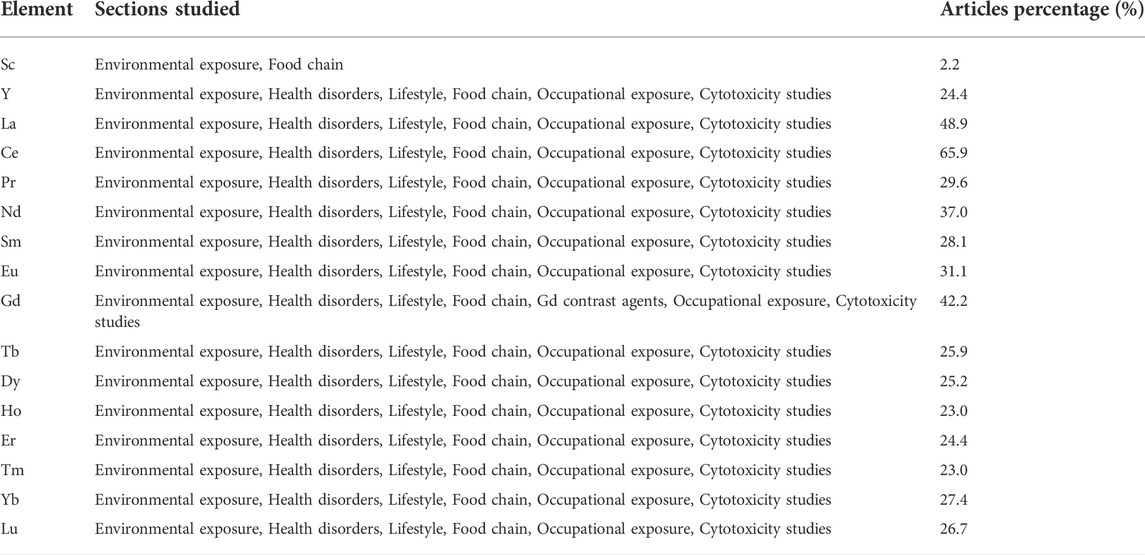
TABLE 1. Schematic summary table demonstrating the different sections that each REE has been studied about at least once, and the relative abundance (%) of papers including information for the toxicity of each REE to human health.
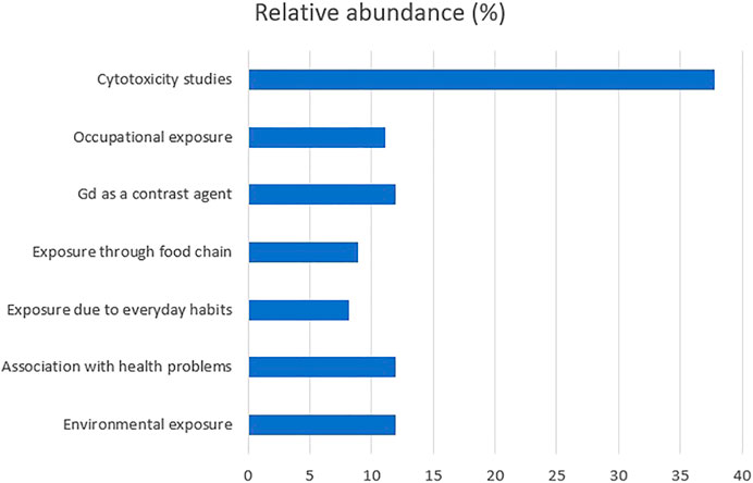
FIGURE 1. Comparison of the relative abundance (%) of papers for each section studied in response to REEs.
Comparing the sections studied about REE toxicity, 37.8% of papers included cytotoxicity studies, followed by a 11.9% for environmental exposure, association of REEs with health problems and Gd-based contrast agents. The comparison among the analyzed biological matrices is shown in Figure 2. Hair was the most analyzed matrix (19%), followed by blood serum (16%) and blood (13%). Comparing the percentage of the articles considering the investigated REEs, Ce was present in 65.9% of the papers, La in 48.9%, Gd in 42.2%, while Nd in 37%. The complete data is shown in Figure 3. Comparing the different types of Ce for which we found information, more than half of them (52%) were reports in which Ce levels were traced in biological matrices, and studies about the cytotoxicity of CeO2 nanoparticles (NPs) were 24%. Comparison of the relative abundance (%) of each type of Ce studied is shown in Figure 4.
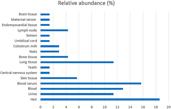
FIGURE 2. Comparison of number of available articles in which each biological matrices was analyzed for the toxicity of REEs.
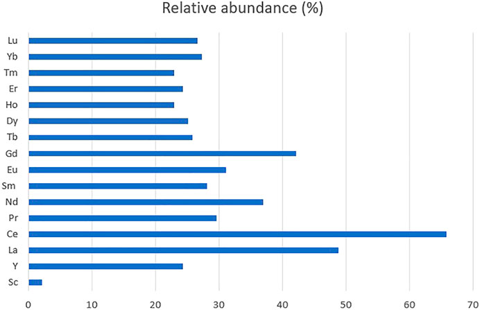
FIGURE 3. Comparison of the relative abundance (%) of papers including information for the toxicity of each REE to human health.
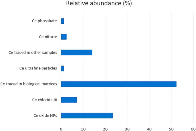
FIGURE 4. Comparison of the relative abundance (%) of each type of Ce studied about its toxicity to human health.
Some studies showed that REE mining affected the residents in the areas near the mining sites by analyzing the REE levels in human samples. Urine REE concentrations of people living near Baiyun Obo mining area were significantly higher than in residents outside the mining area (Hao et al., 2015; Liang et al., 2018). Other studies have investigated the REE-pollution through analyses of hair samples. In the study of Wei et al. (2013), scalp hair of people living near a mining site in Inner Mongolia contained significantly higher REE levels compared to people from the control area. In a similar study, the hair of children living in Shang and Liao villages, near a mining site, contained higher REE levels versus children from reference areas (Tong et al., 2004). As reported by Yu et al. (2007), REE blood levels were higher in people living near the mining site of Xunwu. They had some effects on the peripheral blood mononuclear cells, such as increasing telomerase activity, promoting the diploid DNA replication, and increasing the percentage of cells in the S-phase and the G2/M phase. The blood and hair of people living near a large-scale mining site in southwest Fujian Province, China, were found enriched with REEs but much more with LREEs (Li et al., 2014). A correlation between the concentration of REEs in soil and human samples was found in Hezhang County, China, where REEs levels (mostly La, Ce, and Nd) in the soil near a smelting area were higher than those in the soil near a mining area, and the same applied for urine and hair of residents near these two areas (Meryem et al., 2016). Tian et al. (2019) found an enrichment of Ce in dust from mining areas and the potential of causing health problems through inhalation. According to the Life Cycle Assessment of Wang C. et al. (2020), the most significant impact of Sc2O3 production from Bayan Obo mine is the non-cancerous human toxicity. All these studies indicated that the REE mining process influenced the environment in mining areas and the people living nearby.
However, REEs environmental exposure can occur, not only due to REE mining but also to other factors. Ugandan children presented significantly higher REE concentrations in their primary teeth than the children from the United Kingdom (Brown et al., 2004) due to an environmental exposure. The levels of REEs were significantly higher in the blood of Moroccans than in Canarians, possibly related to the improper management of e-waste (Henríquez-Hernández et al., 2018). Lanthanum and Ce levels were higher in resident serum from an e-waste area and were associated with higher thyroid-stimulating hormone (TSH) (Guo et al., 2020). Hollriegl et al. (2010) associated the increased Ce levels in the serum of women in Madrid with the high traffic volume in this town. The accumulated concentration of REEs in the rib bone tissue was found to be age dependent by Zaichick et al. (2011). Street dust in industrial cities is enriched with REEs mainly due to heavy local soil and less due to anthropogenic pollution, however, the existing REE levels were not found as a threat to cancer occurrence (Sun et al., 2017). Analysis of soil samples from a tropical area and a subsequent health risk assessment from Da Silva Ferreira et al. (2022) showed that REEs represent less than 20% of the metal content and the risk for cancer due to these metals is very low.
Some studies have investigated the association between REEs and specific diseases, with different results. Brown et al. (2004) concluded that the higher Ce concentration in primary teeth of Ugandan children, compared to children from the United Kingdom, failed to be associated with endomyocardial fibrosis. Huo et al. (2017) suggested that although LREEs are higher in the hair of women with neural tube defects (NTDs) affected pregnancies, they are not associated with the risk of NTDs, even if Wei et al. (2020) found that the REEs levels were higher in the maternal serum of pregnant women affected by NTDs and that the risk for NTDs increases with REE concentrations, especially La. Henríquez-Hernández et al. (2017) have pointed out the possibility of REEs playing a role in the development of anemia since they were found to be higher in the blood of sub-Saharan immigrants with anemia and independent of Fe levels. De Vathaire et al. (1998) identified an excess of mortality due to lung and bladder cancer in a French population living near a site where Ce was extracted. Similarly, Kutty et al. (1996) found that deposits of monazite elements, like Ce, increase the rate of occurrence of endomyocardial fibrosis (EMF). In utero exposure to REEs, mainly La and Nd, increase the risk of orofacial clefts to infants (Liu L. et al., 2021). Exposure of pregnant women to Ce and Yb has decreased TSH levels in infants (Liu et al., 2019). REEs may accumulate in the cerebral cortex by long-term environmental intake of a small amount and cause subclinical damage (Zhu et al., 1997). REEs, especially La, Ce, and Gd, are significantly higher in brain-tumor tissues of patients with astrocytoma compared to normal brain tissues (Zhuang et al., 1996). People living near high-REE background areas have illnesses like indigestion, diarrhea, abdominal distension, anorexia, weakness, and fatigue. They have been shown to have lower levels of total protein, globulin, albumin, and serum-glutamic pyruvic transaminase (SGPT) and higher levels of IgM in their blood serum (Zhu et al., 2005). A high amount of REEs in hair has been associated with low levels of Ca in hair and a high risk of hypertension among housewives (Wang B. et al., 2017). Lanthanum levels in the serum of women undergoing in vitro fertilization-embryo transfer have been associated with a 30% increase in clinical pregnancy failure, a 230% increase in preclinical spontaneous abortion, and a negative correlation with the number of good-quality oocytes (Li et al., 2021). Cerium levels in toenails have been associated with an increased risk of acute myocardial infarction (Gómez-Aracena et al., 2006). In the study of Medina-Estévez et al. (2020), Ce levels were higher in control people group than in the subjects, and so, inversely associated with the risk of stroke. Increased REEs in the hair of housewives have been correlated with indoor air pollution and the risk of hypertension by Wang et al. (2018).
Everyday habits can also affect the concentration of metals, including REEs, in the human body. Marzec-Wroblewska et al. (2015) found that the concentrations of La, Ce, Eu, and Gd were significantly lower in the non-drinkers’ and short-term smokers’ semen than in the semen from drinkers and long-term smokers. Moreover, Zhang et al. (2020) evidenced that smoking was positively associated with Dy, Er, and Yb levels, whereas drinking habits showed no significant effect on REE levels, while La, Ce, Pr, and Nd were higher in hair and nails of smokers (Rodushkin and Axelsson, 2000). Poniedziałek et al. (2017) found that Nd concentration was significantly higher in the colostrum milk of smoking women compared to those who had never smoked. Zhang et al. (2020) confirmed also that REE levels are higher in biological matrices of smokers, but not in drinkers. Housewives exposed to REEs due to passive smoking presented elevated levels of REEs in their hair according to Na et al. (2022).
The rolling paper, especially a flavored paper, is the part of the cigarette that contains the highest amount of hazardous metals, including REEs, while black tobacco contains more REEs compared to the other tobacco types, and so, smokers are exposed to REEs mainly due to the rolling paper, as well as to the tobacco. (Zumbado et al., 2019). LREEs are contained in the flint of lighters, and even short-term contact with the flint during the light-up could result in exposure to LREEs (Rodushkin and Axelsson, 2000; Kastury et al., 2020).
Tobacco smoking has been rated as one of the most significant sources of REEs in indoor inhalable particles. Cerium and La levels significantly increased in the households of smokers and hospitality pubs like restaurants, pubs, dance clubs, offices, and cafeterias due to tobacco smoke, and imply a threat to the human respiratory system (Slezakova et al., 2009; Böhlandt et al., 2012). Increased indoor Ce and La have also been associated with indoor smoking and respiratory symptoms in children (Drago et al., 2018). Although REEs in PM10 are higher in smokers’ households, their dissolution in PM10 and inhalation bioaccessibility are higher in non-smokers’ households (Kastury et al., 2020).
E-cigarettes can be considered also as a source of an increased REE intake. Badea et al. (2018) found that REE levels are higher in serum of e-cigarette users than cigarette users, and the longer someone uses e-cigarettes, the higher the levels of Ce and Er in their blood serum. Ytterbium levels were found higher in teeth of people wearing porcelain bridges, and so the latter can be considered as well as a another potential source of REEs intake (Rodushkin and Axelsson, 2000).
REEs can be transferred in the human body also via the food chain. In the study by Zhang et al. (2020), centenarians who consumed smoked and pickled food, egg, milk, and high amounts of salt had higher REE levels in their hair. REE oxides (LREOs) in Oolong tea consider a negligible threat for human health as its consumption does not exceed the accepted daily intake (ADI) of REE levels (Guo et al., 2015). Cereals, especially wheat, and vegetables, especially leaf vegetables, bioaccumulate significantly high amounts of LREEs when growing near mining areas; however, these concentrations are much lower than the ADI (Zhuang et al., 2017a; Zhuang et al., 2017b). Guo et al. (2012) investigated the REE levels in the most consumed foods in China and concluded that REEs are at a shallow concentration and do not exert a hazard for human health. Freshwater and marine fish in Shandong Province, the largest center for fishery production and processing in China, contain a much higher amount of LREEs versus HREEs; however, the estimated daily intake (EDI) of REEs through fish food were significantly lower than the ADI; therefore, its consumption presented little risk to human health (Yang et al., 2016). The health risk assessment of Li et al. (2013) concluded that vegetable consumption would not exceed the EDI values for REEs. The same applies for the health risk assessment of Shi et al. (2022) concerning fruit and vegetable consumption. REE levels are higher in the aquatic than the terrestrial food, get lower at higher trophic levels in the environment and their human intake at present is acceptable (Dai et al., 2022). To the best of our knowledge, the studies, up to now, about the REEs level in food showed that they are present in low concentrations and can hardly exert problems in human health. Squadrone et al. (2019) analyzed REEs level in several terrestrial and marine matrices and organisms. Although the highest REE levels were found at low trophic levels in both environments, they suggested a biomonitoring of these elements for possible cumulative effects to human health, since they can be transferred to human body through the trophic chain.
Zhang et al. (1999) indicated that exposure to REEs through the food chain could result in low total serum protein (TSP), albumin, β-globulin, glutamic pyruvic transaminase, serum triglycerides, and immunoglobulin, but high cholesterol. Cerium concentration was found higher in endomyocardial samples of people dying from endomyocardial fibrosis and has been associated with the Ce levels in leafy vegetables and root tubers like tapioca and yam (Valiathan et al., 1986).
Gadolinium-based contrast agents (GBCAs) have been used for years in magnetic resonance imaging (MRI) and magnetic resonance angiography (MRA). In the first research about the safety of these agents to human health, no side effects were caused to patients. Carr et al. (1984) found that a small intravenous administration of Gd-diethylenetriamine pentaacetic acid (Gd-DTPA) could enhance the contrast of tumors during an MRI operation, causing no side effects to patients. However, subsequent studies reported that these agents are not completely safe for human health. GBCAs can cause acute renal failure in patients with underlying chronic renal insufficiency (Sam et al., 2003). Chien et al. (2011) found out that potential acute kidney injury is after administration of a GBCA, under sepsis condition, at the dose given for MRI and MRA examinations in patients with renal impairment. Ergun et al. (2006) reported that an acute renal failure (ARF) can occur after a GBCA administration in patients with moderate to severe chronic renal failure. Risk factors for ARF after Gd toxicity include diabetic nephropathy and low glomerular filtration rate (GFR). Similarly, Thomsen (2004) reported that GBCAs can induce nephropathy even at doses below 0.2 mmol/kg body weight (a usual dose of GBCA for MRI) in patients with multiple risk factors. Gd-DTPA could play a role in developing nephrogenic fibrosing dermopathy (NFD) in renal disease patients undergoing MRA (Grobner, 2006). There is a high risk of nephrogenic systemic fibrosis in patients with chronic kidney disease at stage 5 due to exposure to gadodiamide during MRI (Rydahl et al., 2008). Gadolinium remained in human bone tissue after administration of a standard clinical dose (0.1 mmol/kg) of Omniscan or Prohance a few days before surgery to patients undergoing hip replacement (Gibby et al., 2004; White et al., 2006). GBCAs can induce central torso and peripheral arm and leg distribution pain in patients (Semelka et al., 2016). Gadovist, a GBCA was reported to cause fibromyalgia in one patient following repeated administrations (Lattanzio and Imbesi, 2020).
A few studies have investigated the potential toxicity of GBCAs to human health by a number of cytotoxicological experiments. Four GBCAs, Gadovist, Magnevist, Multihance, and Omniscan, were not toxic to the embryonic fetal lung fibroblast cell line Hel-299, which rapidly recovered after the initial antiproliferative effects of the agents (Wiesinger et al., 2010). Omniscan increased the proliferation and the levels of the matrix metalloproteinase-1 (MMP-1) and the inhibitor of metalloproteinases-1 (TIMP-1) to monolayer culture of human dermal fibroblasts and to organ culture of human dermal skin, but they did not stimulate the proliferation of epidermal keratinocytes (Bhagavathula et al., 2009; Varani et al., 2009). Magnevist, Multihance, and Prohance (i.e., another GBCAs) increased the proliferation and levels of MMP-1 and TIMP-1 in human dermal fibroblasts like Omniscan did (Varani et al., 2009). Edward et al. (2010) examined the effect of several GBCAs on human skin fibroblasts. Some of them stimulated cell proliferation and hyaluronan production, while no GBCA could affect collagen synthesis. Omniscan increased MMP-1 and TIMP-1 in a human skin organ culture, but reduced the collagenolytic activity (Perone et al., 2010). Gadolinium chloride had similar effects with the GBCAs, having no effect on epidermal keratinocytes, enhancing the proliferation and the levels of MMP-1 and TIMP-1, and having no effect on type I procollagen in fibroblasts via activation of the PDGF receptor signaling pathway (Bhagavathula et al., 2010).
The content of REEs in the hair of miners, particularly light REEs (La, Ce, Pr, and Nd), was much higher than the values in the hair of non-miners from both mining area and control area (Wei et al., 2013). Liu et al. (2015) found higher REE hair levels in miners working at the Baiyun Obo mining area (China), affecting the levels of proteins in serum that participate in various biological processes. Increased La levels were also found in the lung tissue of deceased smelter workers (Gerhardsson and Nordberg, 1993). Elevated REE levels were found in the hair of miners, which were associated with Fe and Ca in hair and a lower bone mineral density at lumbar vertebrae, femoral neck, greater trochanter, and intertrochanter (Liu H. et al., 2021). These studies suggested that the miners can be exposed to increased REE levels in their occupational environment.
Rare earth pneumoconiosis is an uncommon occupational disease caused by the inhalation of REE-containing dust and has been associated with dendriform pulmonary ossification (Yoon et al., 2005). Some early studies showed that workers active in movie projection were exposed to REEs, produced in the fumes and dust of arc carbon lamps that contained a significant REEs amount. A movie projectionist who had approximately 25 years of occupational exposure to carbon arc lamp fumes was found to be exposed to La, Ce, and Nd, with evidence of their redistribution throughout the reticuloendothelial system. However, the exposed did not suffer from pneumoconiosis, as there were no respiratory symptoms or radiographic or histological pulmonary changes attributable to a progressive REEs accumulation (Waring and Watling 1990). Porru et al. (2000) investigated another movie projectionist exposed for 12 years to REE-containing dust from cored arc light carbon electrodes. The exposed presented interstitial lung disease, emphysema, and a severe obstructive impairment with a marked decrease of carbon monoxide diffusion capacity. The biopsy confirmed the diffuse interstitial lung fibrosis and significantly higher concentrations of Ce, La, Nd, Sm, Tb, and Yb compared to the unexposed control subjects. Three other cases of photoengravers, exposed for many years to the fumes and dust from carbon arc lamps, suffered from pneumoconiosis and the clinical analyses showed evident high REEs concentration in the pulmonary and lymph node biopsies, suggesting a relationship between the pneumoconiosis and the occupational exposure to REE dusts, and the need to define air exposure threshold limit values of REEs to prevent workers occupational exposure (Sabbioni et al., 1982; Vocaturo et al., 1983).
Rare earth elements are biopersistent and can remain in the human body many years after occupational exposure (Pairon et al., 1994; Pairon et al., 1995). High Ce, La, and Nd levels were found in the lung tissue of a subject having worked in printing shops and exposed to carbon-arc lamp emissions (Dufresne et al., 1994). REE dust exposure in a lens grinder was reported by Yoon et al. (2005), who found pneumoconiosis associated with dendriform pulmonary ossification. The urines of workers producing ultrafine and nano-sized particles containing Ce and La oxide; especially those at separation and packaging locations presented greater levels of La and Ce (Li et al., 2017). In the study by Li et al. (2016), the REE levels in the urine of workers that manufacture cerium and lanthanum oxide ultrafine and nano-sized particles were more than five times higher versus controls. Their results, however, suggested that only the urinary levels of La, Ce, Nd, and Gd among the exposed workers were significantly higher than the levels measured in the control subjects. High REE levels in the blood have been associated with significant DNA oxidative damage and high blood concentration of Cr in chromate exposed workers (Bai et al., 2019). The urines and blood of e-waste recyclers contain more Eu, but for La, this applies only for urines (Takyi et al., 2021). An uptake of REEs has been found in triple negative breast cancer cells of a woman having worked in the ceramic industry (Roncati et al., 2018).
Nanoparticles (NPs) of Ce can significantly mitigate reactive oxygen species (ROS) production and DNA oxidative damage in the BEAS-2B cell line (Rubio et al., 2016). The co-exposure of CeO2 NPs and diesel exhaust on a sophisticated in vitro 3D co-culture model of the human epithelial airway barrier indicates that a short-term co-exposure filed to result in adverse effects, long-term co-exposure caused undesired effects to human respiratory health (Steiner et al., 2012). CeO2 NPs induced mitochondrial damage and overexpression of apoptosis to human peripheral blood monocytes (CD14+) (Hussain et al., 2012) and human lung adenocarcinoma (A549) (Mittal and Pandey, 2014). CeO2 NPs protected human colon cells (CRL 1541) from radiation by reducing ROS production and increasing the expression of superoxide dismutase 2 (SOD2) (Colon et al., 2010). CeO2 NPs enhanced the antitumor effect of anthracycline doxorubicin, one of the most effective anti-cancer drugs, in human melanoma cells (A375), and even protected human dermal fibroblasts from doxorubicin-induced cytotoxicity (Sack et al., 2014). Nanoparticles of CeO2 did not cause any cytotoxicity to human hepatic carcinoma cell line (HepG2) and human lung carcinoma cell line (A549) during a short-term exposure (24 h) but only during a long-term period (10 days); moreover, they did not cause any cytotoxicity to cells from human colon carcinoma (CaCo2) at any of the investigated times (De Marzi et al., 2013).
Diesel particulate matter containing CeO2 and Fe(C5H5)2 NPs decrease the viability of human-type II cell alveolar epithelial cells (A549) (Zhang and Balasubramanian, 2017). Nanoparticles of CeO2 enhanced the proliferation of human keratinocytes and human microvascular endothelial cells (Chigurupati et al., 2013). Nanoparticles of CeO2 were not found to be genotoxic to eye lens epithelial cells (ATCC-LGC CRL-11421), suggesting a potential safe use in drug delivery for treating cataract (Pierscionek et al., 2010). They were not toxic to the cell lines T98G (from a human glioblastoma multiform tumor) but caused cell death to BEAS-2B cell line (Park et al., 2008) and human bronchoalveolar carcinoma derived cell line (A549) (Lin et al., 2006) by an apoptotic process, mainly by increasing the levels of ROS and decreasing the levels of GSH and α-tocopherol. In the study of Alpaslan et al. (2015), the dextran-coated nanoceria killed much more effectively the osteosarcoma cell line MG-63 (ATCC CRL-1427) than the osteoblast noncancerous cell line (PromoCell, C-12720), suggesting that nanoceria can promisingly be used for treating bone cancer without the appearance of adverse effects to healthy bone cells. CeO2 NPs showed a rapid uptake from human keratinocyte cells (HaCaT) and co-localized with mitochondria, lysosomes, endoplasmic reticulum, cytoplasm, and nucleus, suggesting that they act as cellular antioxidants in multiple compartments (Singh et al., 2010). CeO2 NPs are taken up into caveolin-1 endosomal compartments by BEAS-2B cells without causing inflammation or cytotoxicity (Xia et al., 2008). Tarnuzzer et al. (2005) showed that CeO2 NPs could protect almost 100% of the normal breast epithelial cell line CRL8798 and simultaneously did not protect the breast carcinoma cell line MCF-7. According to the study by Celardo et al. (2011), in two tumor cell lines, the human tumor monocytes U937 and the human tumor T lymphocytes Jurkat, the intracellular antioxidant effect of CeO2 NPs is due to their anti-apoptotic and prosurvival activity. They also found that doping of CeO2 NPs with increasing concentrations of Sm3+ blunted these effects by decreasing Ce3+ and not affecting oxygen vacancies, demonstrating that Ce3+/Ce4+ redox reactions might be responsible for the biological properties of nanoceria.
In Yongxing et al. (2000), La and Gd were indicated as possible mutagenic factors for human primary peripheral lymphocytes. The compounds LaCl3 and CeCl3 inhibited the growth of the leukemic cell lines HL-60 and NB4, respectively, and simultaneously LaCl3 had no inhibitory effect on normal bone marrow hematopoietic progenitor cells (Dai et al., 2002). Lanthanum and Ce nitrates decrease the proliferation of the human osteoblast MG63 cell line when combined with radiation but not alone (Iwahara et al., 2019). Lanthanum oxyiodide nanosheets with low dose of doxorubicin, an anticancer drug, can kill more effectively the A375 cells compared to the doxorubicin alone (Xu et al., 2020). Nd2O3 and La2O3 NPs are cytotoxic, while Gd2O3 and CeO2-Gd NPs enhance ROS production to the U-87 MG tumor cell line (Lu et al., 2019). Lanthanum is cytotoxic through the necrosis pathway and genotoxic by increasing ROS production in Jurkat cells and human peripheral lymphocytes (Paiva et al., 2009). The ionic forms La3+ and Tb3+ caused disruptions on potassium channels on the plasma membrane of HEK293 cells (Wang L. et al., 2017). Lanthanides can decrease the ATP production and the cell viability of the HepG2 cells, and also can decrease the mitochondrial membrane potential most probably by interfering with the calcium regulation due to the similar ionic radius (Kajjumba et al., 2021). In the report by Feyerabend et al. (2010), lanthanides are cytotoxic against the human osteosarcoma cell line MG63, but not to the human umbilical cord perivascular (HUCPV) cells, with some of them increasing the levels of the inflammatory factors TNF-α and IL-1a. Lanthanides did not stimulate the growth of HaCaT cells (keratinocytes), while in human dermal fibroblasts they enhanced the proliferation and the production of matrix metalloproteinase-1 (MMP-1) and did not influence the production of type I procollagen (Jenkins et al., 2011). The “Raman fingerprints” of human sperm DNA exposed to CeCl3 demonstrated that Ce can cause DNA damage through oxidative damage and destruction of the antioxidant defense systems of sperm and even lead to damage or death (Chen et al., 2015). This implies that excess REEs exposure may be a risk factor for human infertility. A cytotoxicity study of CeO2 NPs in six cancers and two normal cell lines was elaborated by Pešic et al. (2015). Only two cancer cell lines were sensitive to CeO2 NPs, melanoma 518A2 and colorectal adenocarcinoma HT-29, showing a median inhibitory concentration (IC50) of 125 μΜ and 183 μΜ, respectively. In this study, human cells also resulted in lower ROS production and higher antioxidant capacity, implying that normal cells are more resistant to CeO2 NPs versus cancer cells. Ultrafine particles of Y2O3 were not cytotoxic against human aortic endothelial cells and resulted in an inflammatory response by increasing the concentration of IL-8, ICAM-1, and MCP-1 (Gojova et al., 2007), but CeO2 ultrafine particles failed to cause any inflammation on the same cell lines (Gojova et al., 2009). Lanthanum and Ce, alone or in combination, inhibited cell proliferation and altered genes involved in oxidative stress pathways in HepG2 and HT-29 cell lines (Benedetto et al., 2018). Particles enriched with REEs, collected from an REE mining area, showed no cytotoxicity to lung epithelial A549 cells; however, they increased ROS production (Tian et al., 2020). Erbium-laser photoablation was found to cure oligoasthenozoospermia and enhance fertilization (Antinori et al., 1994). Europium selenide nanoparticles synthesized in vivo by recombinant Escherichia coli cells were cytotoxic to the cancer cell lines 293T, HeLa and SKOV-3, and thus could be promising drug carrying agents against cancers (Kim et al., 2016). An Eu complex bearing 8-hydroxyquinoline-N-oxide and 1,10-phenanthroline ligands exhibits low toxicity to normal HL-7702 cells, but high antiproliferative activity against cisplatin-resistant A549/DDP cells by upregulating the expression of LC3 and Beclin1 and downregulating p62 to induce apoptosis which is related to its cell autophagy-inducing properties (Yang et al., 2021). Neodymium-iron-boron magnets are cytotoxic against human oral mucosal fibroblasts (Donohue et al., 1995). Although not causing cell death, the powder of Nd2Fe14B magnet can cause a dose-dependent ROS production to A549 cells (Rumbo et al., 2021). Gadolinium oxide nanoparticles were found cytotoxic to human umbilical vein endothelial cells, inducing lipid peroxidation, ROS production, mitochondrial dysfunction and autophagic modulation through apoptosis and necrosis (Akhtar et al., 2020). Nanoparticles of Y2O3 induced apoptosis and necrosis to human embryonic kidney (HEK293) cells, by elevating the cellular ROS levels, increasing the mitochondrial membrane permeability, and causing DNA damage (Selvaraj et al., 2014). Yttrium aluminum borates nanoparticles are cytotoxic to human bronchial epithelial cells (Cieślik et al., 2019). The study by Perry et al. (2020) was the first to provide in vitro data on the efficacy of radiation therapy with Tm nanoparticles for the management of metastatic cutaneous squamous cell carcinoma (cSCC). REE salts are not skin sensitizers and do not exhibit endocrine disruption to epidermal keratinocytes (Rucki et al., 2021). The chlorides of Ce, La, Eu and Yb are cytotoxic against the embryonal kidney HEK-293 cells (Heller et al., 2019). La citrate induces anoikis (a type of programmed cell death) in HeLa cells by reorganizing actin cytoskeleton and increasing the co-localization of F-actin with mitochondria (Su et al., 2009). La citrate is also cytotoxic against SiHa cells causing a mitochondrial dysfunction that induces apoptosis (Shen et al., 2010). EuCl2 and EuCl3 reduced the viability of the human monocyte (U937) and primary human mononuclear cells (PBMNCs) (Bladen et al., 2013). In the study of Wang L. et al. (2020) a REE-doped up conversion nanoparticle (UCNP), sodium yttrium fluoride NP, doped with Yb and Er (NaYF4: Yb3+, Er3+), induced cytotoxicity to HepG2 cells after their internalization by promoting ROS generation, and the induced apoptosis was related to the mitochondria mediated pathway after seeing an increase of cleaved caspase-9, cleaved caspase-3 and Bax and a decrease in the anti-apoptotic protein, Bcl-2. They also compared to the cytotoxicity of the UCNP with that of Y3+ ions and concluded that the properties of NPs did not play any significant role in the cytotoxicity of the UCNP. In contrast to the above mentioned, GdCl3 increased the survival and the proliferation of HeLa cells by promoting S phase and activating FAK and JPK, but also attenuated the serum deprivation-induced cell loss (Zhang et al., 2009).
More than one third of the literature about REE effects on human health regards cytotoxicity. Hair, blood and blood serum are the most biomonitored matrices. In the near future, research should focus on other potential REE targeted tissues and organs. The effects on LREEs are much more studied than for MREEs or HREEs, thus requiring more attention. Cerium is the most investigated among all REEs.
The REEs mining showed to affect human health as they are accumulated in hair, urine or blood, not only of mining workers, who are exposed directly to these elements, but also of residents near the mining areas. Precautionary regulation should be evaluated for their protection—in the perspective of the “prevention is better than cure” paradigm.
Human daily exposure to REEs can occur also due to other factors such as improper management of e-wastes or high traffic volume since they are used for developing technological devices and as additives in diesel fuels. Although REE levels in the environment and food are currently present at low levels and do not possess a huge threat for human health, they should be considered as a potential future risk since REEs concentration is expected to increase along time.
REEs exposure has been associated with several diseases like NTD, anemia or endomyocardial fibrosis. The lifestyle (smoking and drinking) could contribute to increase the exposure to REEs.
A number of studies have shown that Gd used as a contrast media in MRA and MRI can be accumulated in kidney or muscles causing renal failure and fibromyalgia. Through cytotoxicity studies, GBCAs showed to increase the levels of MMP-1 and TIMP-1, possibly interfering in biological processes where collagen is involved.
A regulation should be implemented also in occupational REE exposures since many jobs may lead to adverse effects. To date, most studies have focused on REE miners and workers involved in movie projection. More focus should be given also to other jobs, concerning occupational exposure to REEs, like workers in diesel or technological devices production.
The mechanism of action and the metabolic pathways that REEs activate are still unclear in most cases. GBCAs can activate the pathways related to MPP-1 and TIMP-1. Activation of the PDGF receptor pathway is referred above. REEs can activate the metabolic pathways resulting to ROS production, DNA damage and apoptosis to several cell lines. They seem to be more cytotoxic to cancer cells than normal cells, probably by promoting apoptosis, suggesting their potential protective action against cancer. In some studies, they acted as antioxidants activating pathways related to numerous proteins such as SOD2, GSA, TNF-α, IL-1α, IL-8, ICAM-1, MCP-1, LC3, Beclin1, p62, caspase-9, caspase-3, BAX, Bcl-2, FAK and JPK. They can be uptaken by several cellular compartments like mitochondria, lysosomes, cytoplasm and nucleus. Attention should be given to the case of mitochondria, since they have been mentioned to cause mitochondrial disruption. REEs have similar ionic radius to calcium and so they may interfere with its regulation. Future cytotoxic studies should focus on the mechanisms of action of REEs to elucidate the metabolic pathways they can activate.
As a general conclusion, REEs seem to constitute a potential risk for human health. Specific human biomonitoring programs should take place especially for occupational exposures (i.e., miners, e-waste workers), evidencing, particularly, how tobacco smoke can change the background exposure level in the population. Further investigations are necessary to highlight stronger cause-effect relationships between pathological status and REEs exposure, looking for their presence in the right target matrix.
Conceptualization, AB, GP, GL, MT, MG; Literature gathering, AB, AG; Writing—original draft preparation, AB, GP, AG, MT, GL; Writing—review and editing, AB, GP, AG, MT, GL; Visualization, AB, GP, GL, MT, MG; Supervision, MT, GL, MG; Project administration, MT, GL; Funding acquisition, MT, MG, GL. All authors have read and agreed to the published version of the manuscript.
This project has received funding from European Union’s Horizon 2020 research and innovation program under the Marie Sklodowska-Curie Grant Agreement N°857989.
The authors declare that the research was conducted in the absence of any commercial or financial relationships that could be construed as a potential conflict of interest.
All claims expressed in this article are solely those of the authors and do not necessarily represent those of their affiliated organizations, or those of the publisher, the editors and the reviewers. Any product that may be evaluated in this article, or claim that may be made by its manufacturer, is not guaranteed or endorsed by the publisher.
Abdelnour, S. A., Abd El-Hack, M. E., Khafaga, A. F., Noreldin, A. E., Arif, M., Chaudhry, M. T., et al. (2019). Impacts of rare Earth elements on animal health and production: Highlights of cerium and lanthanum. Sci. Total Environ. 672, 1021–1032. doi:10.1016/j.scitotenv.2019.02.270
Akhtar, M. J., Ahamed, M., and Alhadlaq, H. (2020). Gadolinium oxide nanoparticles induce toxicity in human endothelial HUVECs via lipid peroxidation, mitochondrial dysfunction and autophagy modulation. Nanomaterials 10, 1675. doi:10.3390/nano10091675
Albarano, L., Guida, M., Tommasi, F., Lofrano, G., Padilla Suarez, E., Gjata, I., et al. (2022). Species sensitivity distribution of rare Earth elements: A full overview. Sci. Total Environ.
Alpaslan, E., Yazici, H., Golshan, N. H., Ziemer, K. S., and Webster, T. J. (2015). PH-dependent activity of dextran-coated cerium oxide nanoparticles on prohibiting osteosarcoma cell proliferation. ACS Biomater. Sci. Eng. 1, 1096–1103. doi:10.1021/acsbiomaterials.5b00194
Antinori, S., Versaci, C., Fuhrberg, P., Panci, C., Caff, B., and Gholami, G. H. (1994). Andrology: Seventeen live births after the use of an erbium-yytrium aluminium garnet laser in the treatment of male factor infertility. Hum. Reprod. 9, 1891–1896. doi:10.1093/oxfordjournals.humrep.a138354
Badea, M., Luzardo, O. P., González-Antuña, A., Zumbado, M., Rogozea, L., Floroian, L., et al. (2018). Body burden of toxic metals and rare Earth elements in non-smokers, cigarette smokers and electronic cigarette users. Environ. Res. 166, 269–275. doi:10.1016/j.envres.2018.06.007
Bai, Y., Long, C., Hub, G., Zhou, D., Gao, X., Chen, Z., et al. (2019). Association of blood chromium and rare Earth elements with the risk of DNA damage in chromate exposed population. Environ. Toxicol. Pharmacol. 72, 103237. doi:10.1016/j.etap.2019.103237
Benedetto, A., Bocca, C., Brizio, P., Cannito, S., Abetea, M. C., and Squadrone, S. (2018). Effects of the rare elements lanthanum and cerium on the growth of colorectal and hepatic cancer cell lines. Toxicol. Vitro 46, 918. doi:10.1016/j.tiv.2017.09.024
Bhagavathula, N., Da Silva, M., Aslam, M. N., Dame, M. K., Warner, R. L., Xu, Y., et al. (2009). Regulation of collagen turnover in human skin fibroblasts exposed to a gadolinium-based contrast agent. Investig. Radiol. 44, 433–439. doi:10.1097/RLI.0b013e3181a4d7e9
Bhagavathula, N., Dame, M. K., Da Silva, M., Jenkins, W., Aslam, M. N., Perone, P., et al. (2010). Fibroblast response to gadolinium: Role for platelet-derived growth factor receptor. Investig. Radiol. 45, 769–777. doi:10.1097/RLI.0b013e3181e943d2
BladenTzu-Yin, C. L, L., Fisher, J., and Tipper, J. L. (2013). In vitro analysis of the cytotoxic and anti-inflammatory effects of antioxidant compounds used as additives in ultra high-molecular weight polyethylene in total joint replacement components. J. Biomed. Mat. Res. 101B, 407–413. doi:10.1002/jbm.b.32798
Böhlandt, A., Schierl, R., Diemer, J., Koch, C., Bolte, G., Kiranoglu, M., et al. (2012). High concentrations of cadmium, cerium and lanthanum in indoor air due to environmental tobacco smoke. Sci. Total Environ. 414, 738–741. doi:10.1016/j.scitotenv.2011.11.017
Brown, C. J., Chenery, S. R. N., Smith, B., Mason, C., Tomkins, A., Roberts, G. J., et al. (2004). Environmental influences on the trace element content of teeth-implications for disease and nutritional status. Archives Oral Biol. 49, 705–717. doi:10.1016/j.archoralbio.2004.04.008
Brown, P. H., Rathjen, A. H., Graham, R. D., and Tribe, D. E. (1990). “Chapter 92 rare earth elements in biological systems” in Handbook on the physics and chemistry of rare earths. 13, ed. Amsterdam, Netherlands: Elsevier, 423–452.
Carr, D. H., Brown, J., Bydder, G. M., Steiner, R. E., Weinmann, H. J., Speck, U., et al. (1984). Gadolinium-DTPA as a contrast agent in MRI: Initial clinical experience in 20 patients. Am. J. Roentgenol. 143, 215–224. doi:10.2214/ajr.143.2.215
Cassee, F. R., Van Balen, E. C., Singh, C., Green, D., Muijser, H., Weinstein, J., et al. (2011). Exposure, health and ecological effects review of engineered nanoscale cerium and cerium oxide associated with its use as a fuel additive. Crit. Rev. Toxicol. 41, 213–229. doi:10.3109/10408444.2010.529105
Celardo, I., De Nicola, M., Mandoli, C., Pedersen, J. Z., Traversa, E., and Ghibelli, L. (2011). Ce3+ ions determine redox-dependent anti-apoptotic effect of cerium oxide nanoparticles. ACS Nano 5, 4537–4549. doi:10.1021/nn200126a
Chen, J., Xiao, H. J., Qi, T., Chen, D. L., Long, H. M., and Liu, S. H. (2015). Rare earths exposure and male infertility: The injury mechanism study of rare earths on male mice and human sperm. Environ. Sci. Pollut. Res. 22, 2076–2086. doi:10.1007/s11356-014-3499-y
Chien, C. C., Wang, H. Y., Wang, J. J., Kan, W. C., Chien, T. W., Lin, C. Y., et al. (2011). Risk of acute kidney injury after exposure to gadolinium-based contrast in patients with renal impairment. Ren. Fail. 33, 758–764. doi:10.3109/0886022X.2011.599911
Chigurupati, S., Mughal, M. R., Okun, E., Das, S., Kumar, A., McCaffery, M., et al. (2013). Effects of cerium oxide nanoparticles on the growth of keratinocytes, fibroblasts and vascular endothelial cells in cutaneous wound healing. Biomaterials 34, 2194–2201. doi:10.1016/j.biomaterials.2012.11.061
Cieślik, I., Płocińska, M., Płociński, T., Zdunek, J., Woźniak, M. J., Bil, M., et al. (2019). Influence of polymeric precursors on the viability of human cells of yttrium aluminum borates nanoparticles doped with ytterbium ions. Appl. Surf. Sci. 488, 874–886. doi:10.1016/j.apsusc.2019.05.001
Colon, J., Hsieh, N., Ferguson, A., Kupelian, P., Seal, S., Jenkins, D. W., et al. (2010). Cerium oxide nanoparticles protect gastrointestinal epithelium from radiation-induced damage by reduction of reactive oxygen species and upregulation of superoxide dismutase 2. Nanomedicine Nanotechnol. Biol. Med. 6, 698–705. doi:10.1016/j.nano.2010.01.010
Da Silva Ferreira, M., Ferreira Fontes, M. P., Weitzel Dias Carneiro Lima, M. T., Cordeiro, S. G., Passamani Wyatt, N. L., Lima, H. N., et al. (2022). Human health risk assessment and geochemical mobility of rare Earth elements in Amazon soils. Sci. Total Environ. 806, 151191. doi:10.1016/j.scitotenv.2021.151191
Dai, Y., Li, J., Li, J., Yu, L., Dai, G., Hu, A., et al. (2002). Effects of rare Earth compounds on growth and apoptosis of leukemic cell lines. Vitro Cell. Dev. Biol. – Animal 38, 373–375. doi:10.1290/1071-2690(2002)038<0373:EORECO>2.0.CO;2
Dai, Y., Sun, S., Li, Y., Yang, J., Zhang, C., Cao, R., et al. (2022). Residual levels and health risk assessment of rare Earth elements in Chinese resident diet: A market-based investigation. Sci. Total Environ. 828, 154119. doi:10.1016/j.scitotenv.2022.154119
De Marzi, L., Monaco, A., De Lapuente, J., Ramos, D., Borras, M., Di Gioacchino, M., et al. (2013). Cytotoxicity and genotoxicity of ceria nanoparticles on different cell lines in vitro. Int. J. Mol. Sci. 14, 3065–3077. doi:10.3390/ijms14023065
De Vathaire, F., De Vathaire, C. C., Ropers, J., and Mollie, A. (1998). Cancer mortality in the commune of Pargny sur Saulx in France. J. Radiol. Prot. 18, 23–27. doi:10.1088/0952-4746/18/1/004
Donohue, V. E., McDonald, F., and Evans, R. (1995). In vitro cytotoxicity testing of neodymium-iron-boron magnets. J. Appl. Biomater. 6, 69–74. doi:10.1002/jab.770060110
Drago, G., Perrino, C., Canepari, S., Ruggieri, S., L’Abbate, L., Longo, V., et al. (2018). Relationship between domestic smoking and metals and rare Earth elements concentration in indoor PM2.5. Environ. Res. 165, 71–80. doi:10.1016/j.envres.2018.03.026
Du, X., and Graedel, T. E. (2013). Uncovering the end uses of the rare Earth elements. Sci. Total Environ. 461-462, 781–784. doi:10.1016/j.scitotenv.2013.02.099
Du, X., and Graedel, T. E. (2011). Uncovering the global Life cycles of the rare earth elements. Sci. Rep. 1, 145. doi:10.1038/srep00145
Dufresne, A., Krier, G., Muller, J. F., Case, B., and Perrault, G. (1994). Lanthanide particles in the lung of a printer. Sci. Total Environ. 151, 249–252. doi:10.1016/0048-9697(94)90474-X
Edward, M., Quinn, J. A., Burden, A. D., Newton, B. B., and Jardine, A. G. (2010). Effect of different classes of gadolinium-based contrast agents on control and nephrogenic systemic fibrosis–derived fibroblast proliferation. Radiology 256, 735–743. doi:10.1148/radiol.10091131
Ergun, I., Keven, K., Uruc¸, I., Ekmekci, Y., Canbakan, B., Erden, I., et al. (2006). The safety of gadolinium in patients with stage 3 and 4 renal failure. Nephrol. Dial. Transpl. 21, 697–700. doi:10.1093/ndt/gfi304
Feyerabend, ., Fischer, J., Holtz, J., Witte, F., Willumeit, R., Drücker, H., et al. (2010). Evaluation of short-term effects of rare Earth and other elements used in magnesium alloys on primary cells and cell lines. Acta Biomater. 6, 1834–1842. doi:10.1016/j.actbio.2009.09.024
Gerhardsson, L., and Nordberg, G. F. (1993). Lung cancer in smelter workers–interactions of metals as indicated by tissue levels. Scand. J. Work Environ. Health 19, 90–94. https://www.jstor.org/stable/40966382.
Gibby, W. A., Gibby, K. A., and Gibby, W. A. (2004). Comparison of Gd DTPA-BMA (omniscan) versus Gd HP-DO3A (ProHance) retention in human bone tissue by inductively coupled plasma atomic emission spectroscopy. Invest. Radiol. 39, 138–142. doi:10.1097/01.rli.0000112789.57341.01
Gojova, A., Guo, B., Kota, R. S., Rutledge, J. C., Kennedy, I. M., and Barakat, A. I. (2007). Induction of inflammation in vascular endothelial cells by metal oxide nanoparticles: Effect of particle composition. Environ. Health Perspect. 115, 403–409. doi:10.1289/ehp.8497
Gojova, A., Lee, J. T., Jung, H. S., Guo, B., Barakat, A. I., and Kennedy, I. M. (2009). Effect of cerium oxide nanoparticles on inflammation in vascular endothelial cells. Inhal. Toxicol. 21, 123–130. doi:10.1080/08958370902942582
Gómez-Aracena, J., Riemersma, R. A., Gutiérrez-Bedmar, M., Bode, P., Kark, J. D., Garcia-Rodríguez, A., et al. (2006). Toenail cerium levels and risk of a first acute myocardial infarction: The EURAMIC and heavy metals study. Chemosphere 64, 112–120. doi:10.1016/j.chemosphere.2005.10.062
Grobner, T. (2006). Gadolinium – A specific trigger for the development of nephrogenic fibrosing dermopathy and nephrogenic systemic fibrosis? Nephrol. Dial. Transpl. 21, 1104–1108. doi:10.1093/ndt/gfk062
Guo, C., Wei, Y., Yan, L., Li, Z., Qian, Y., Liu, H., et al. (2020). Rare Earth elements exposure and the alteration of the hormones in the hypothalamic-pituitary-thyroid (hpt) axis of the residents in an e-waste site: A cross-sectional study. Chemosphere 252, 126488. doi:10.1016/j.chemosphere.2020.126488
Guo, J. D., Jie, Y., Shuo, Z., and Jin, Y. D. (2012). A survey of 16 rare earth elements in the major foods in China. Biomed. Environ. Sci. 25, 267–271. doi:10.3967/0895-3988.2012.03.003
Guo, Y., Zhang, S., Lai, L., and Wang, G. (2015). Rare Earth elements in Oolong tea and their human health risks associated with drinking tea. J. Food Compos. Analysis 44, 122–127. doi:10.1016/j.jfca.2015.08.001
Gwenzi, W., Mangori, L., Danha, C., Chaukura, N., Dunjana, N., and Sanganyado, E. (2018). Sources, behaviour, and environmental and human health risks of high-technology rare Earth elements as emerging contaminants. Sci. Total Environ. 636, 299–313. doi:10.1016/j.scitotenv.2018.04.235
Hao, Z., Li, Y., Li, H., Wei, B., Liao, X., Liang, T., et al. (2015). Levels of rare Earth elements, heavy metals and uranium in a population living in Baiyun Obo, inner Mongolia, China: A pilot study. Chemosphere 128, 161–170. doi:10.1016/j.chemosphere.2015.01.057
He, M. L., Ranz, D., and Rambeck, W. A. (2001). Study on the performance enhancing effect of rare Earth elements in growing and fattening pigs. J. Anim. Physiol. Anim. Nutr. Berl. 85, 263–270. doi:10.1046/j.1439-0396.2001.00327.x
He, M. L., Wehr, U., and Rambeck, W. A. (2010). Effect of low doses of dietary rare Earth elements on growth performance of broilers. J. Anim. Physiol. Anim. Nutr. Berl. 94, 86–92. doi:10.1111/j.1439-0396.2008.00884.x
Heller, A., Barkleit, A., Bok, F., and Wober, J. (2019). Effect of four lanthanides onto the viability of two mammalian kidney cell lines. Ecotoxicol. Environ. Saf. 173, 469–481. doi:10.1016/j.ecoenv.2019.02.013
Henríquez-Hernández, L. A., Boada, L. D., Carranza, C., Pérez-Arellano, J. L., González-Antuña, A., Camacho, M., et al. (2017). Blood levels of toxic metals and rare Earth elements commonly found in e-waste may exert subtle effects on hemoglobin concentration in sub-Saharan immigrants. Environ. Int. 109, 20–28. doi:10.1016/j.envint.2017.08.023
Henríquez-Hernández, L. A., González-Antuña, A., Boada, L. D., Carranza, C., Pérez-Arellano, J. L., Almeida-González, M., et al. (2018). Pattern of blood concentrations of 47 elements in two populations from the same geographical area but with different geological origin and lifestyles: Canary Islands (Spain) vs. Morocco. Sci. Total Environ. 636, 709–716. doi:10.1016/j.scitotenv.2018.04.311
Hirano, S., and Suzuki, K. T. (1996). Exposure, metabolism, and toxicity of rare earths and related compounds. Environ. Health Perspect. 104, 85–95. doi:10.1289/ehp.96104s185
Hollriegl, V., Gonzalez-Estecha, M., Trasobares, E. M., Giussani, A., Oeh, U., Herraiz, M. A., et al. (2010). Measurement of cerium in human breast milk and blood samples. J. Trace Elem. Med. Biol. 24, 193–199. doi:10.1016/j.jtemb.2010.03.001
Huo, W., Zhu, Y., Li, Z., Pang, Y., Wang, B., and Li, Z. (2017). A pilot study on the association between rare Earth elements in maternal hair and the risk of neural tube defects in north China. Environ. Pollut. 226, 89–93. doi:10.1016/j.envpol.2017.03.046
Hussain, S., Al-Nsour, F., Rice, A. B., Marshburn, J., Yingling, B., Ji, Z., et al. (2012). Cerium dioxide nanoparticles induce apoptosis and autophagy in human peripheral blood monocytes. ACS Nano 6, 5820–5829. doi:10.1021/nn302235u
Iwahara, L. K. D. F., De Oliveira, M. S., and De Alencar, M. A. V. (2019). Evaluation of the effect of three constituent metals of monazita on the radiosensibility of human osteoblasts. J. Environ. Radioact. 208-209, 106011–106209. doi:10.1016/j.jenvrad.2019.106011
Jenkins, W., Perone, P., Walker, K., Bhagavathula, N., Aslam, M. N., DaSilva, M., et al. (2011). Fibroblast response to lanthanoid metal ion stimulation: Potential contribution to fibrotic tissue injury. Biol. Trace Elem. Res. 144, 621–635. doi:10.1007/s12011-011-9041-x
Kajjumba, G. W., Attene-Ramos, M., and Marti, E. J. (2021). Toxicity of lanthanide coagulants assessed using four in vitro bioassays. Sci. Total Environ. 800, 149556. doi:10.1016/j.scitotenv.2021.149556
Kastury, F., Ritch, S., Rasmussen, P. E., and Juhasz, A. L. (2020). Influence of household smoking habits on inhalation bioaccessibility of trace elements and light rare Earth elements in Canadian house dust. Environ. Pollut. 262, 114132. doi:10.1016/j.envpol.2020.114132
Kim, E. B., Seo, J. M., Kim, G. W., Lee, S. Y., and Park, T. J. (2016). In vivo synthesis of europium selenide nanoparticles and related cytotoxicity evaluation of human cells. Enzyme Microb. Technol. 95, 201–208. doi:10.1016/j.enzmictec.2016.08.012
Kutty, V. R., Abraham, S., and Kartha, C. C. (1996). Geographical distribution of endomyocardial fibrosis in south Kerala. Int. J. Epidemiol. 25, 1202–1207. doi:10.1093/ije/25.6.1202
Lattanzio, S. M., and Imbesi, F. (2020). Fibromyalgia associated with repeated gadolinium contrast-enhanced MRI examinations. Radiol. Case Rep. 15, 534–541. doi:10.1016/j.radcr.2020.02.002
Li, M., Zhuang, L., Zhang, G., Lan, C., Yan, L., Liang, R., et al. (2021). Association between exposure of light rare Earth elements and outcomes of in vitro fertilization-embryo transfer in North China. Sci. Total Environ. 762, 143106. doi:10.1016/j.scitotenv.2020.143106
Li, X., Chen, Z., Chen, Z., and Zhang, Y. (2013). A human health risk assessment of rare Earth elements in soil and vegetables from a mining area in Fujian Province, Southeast China. Chemosphere 93, 1240–1246. doi:10.1016/j.chemosphere.2013.06.085
Li, X. F., Chen, Z. B., and Chen, Z. Q. (2014). Distribution and fractionation of rare Earth elements in soil–water system and human blood and hair from a mining area in southwest Fujian Province, China. Environ. Earth Sci. 72, 3599–3608. doi:10.1007/s12665-014-3271-0
Li, Y., Yu, H., Li, P., and Bian, Y. (2017). Assessment the exposure level of rare earth elements in workers producing cerium, lanthanum oxide ultrafine and nanoparticles. Biol. Trace Elem. Res. 175, 298–305. doi:10.1007/s12011-016-0795-z
Li, Y., Yu, H., Zheng, S., Miao, Y., Yin, S., Li, P., et al. (2016). Direct quantification of rare Earth elements concentrations in urine of workers manufacturing Cerium, Lanthanum oxide ultrafine and nanoparticles by a developed and validated ICP-MS. Int. J. Environ. Res. Public Health 13, 350. doi:10.3390/ijerph13030350
Liang, Q., Yin, H., Li, J., Zhang, L., Hou, R., and Wang, S. (2018). Investigation of rare Earth elements in urine and drinking water of children in mining area. Medicine 97, e12717. doi:10.1097/MD.0000000000012717
Lin, W., Huang, Y. W., Zhou, X. D., and Ma, Y. (2006). Toxicity of cerium oxide nanoparticles in human lung cancer cells. Int. J. Toxicol. 25, 451–457. doi:10.1080/10915810600959543
Liu, H., Liu, H., Yang, Z., and Wang, K. (2021). Bone mineral density in population long-term exposed to rare earth elements from a mining area of China. Biol. Trace Elem. Res. 199, 453–464. doi:10.1007/s12011-020-02165-0
Liu, H., Wang, J., Yang, Z., and Wang, K. (2015). Serum proteomic analysis based on iTRAQ in miners exposed to soil containing rare earth elements. Biol. Trace Elem. Res. 167, 200–208. doi:10.1007/s12011-015-0312-9
Liu, L., Wang, L., Ni, W., Pan, Y., Chen, Y., Xie, Q., et al. (2021). Rare Earth elements in umbilical cord and risk for orofacial clefts. Ecotoxicol. Environ. Saf. 207, 111284. doi:10.1016/j.ecoenv.2020.111284
Liu, Y., Wu, M., Zhang, L., Bi, J., Song, L., Wang, L., et al. (2019). Prenatal exposure of rare Earth elements cerium and ytterbium and neonatal thyroid stimulating hormone levels: Findings from a birth cohort study. Environ. Int. 133, 105222. doi:10.1016/j.envint.2019.105222
Long, K. R. (2011). The future of rare earth elements—will these high-tech industry elements continue in short supply? Reston, VA: U.S. Geological Survey. Open-File Report 2011–1189 http://pubs.usgs.gov/of/2011/1189/.
Lu, V. M., Crawshay-Williams, F., White, B., Elliot, A., Hill, M. A., and Townley, H. E. (2019). Cytotoxicity, dose-enhancement and radiosensitization of glioblastoma cells with rare Earth nanoparticles. Artif. Cells Nanomed. Biotechnol. 47, 132–143. doi:10.1080/21691401.2018.1544564
Marzec-Wroblewska, U., Kaminski, P., Łakota, P., Ludwikowski, G., Szymanski, M., Wasilow, K., et al. (2015). Determination of rare earth elements in human sperm and association with semen quality. Arch. Environ. Contam. Toxicol. 69, 191–201. doi:10.1007/s00244-015-0143-x
Medina-Estévez, F., Zumbado, M., Luzardo, O. P., Rodríguez-Hernández, Á., Boada, L. D., Fernández-Fuertes, F., et al. (2020). Association between heavy metals and rare earth elements with acute ischemic stroke: A case-control study conducted in the canary islands (Spain). Toxics 8, 66. doi:10.3390/toxics8030066
Meryem, B., Hongbing, J. I., Yang, G., Huajian, D., and Cai, L. (2016). Distribution of rare Earth elements in agricultural soil and human body (scalp hair and urine) near smelting and mining areas of Hezhang, China. J. Rare Earths 34, 1156–1167. doi:10.1016/S1002-0721(16)60148-5
Mittal, S., and Pandey, A. K. (2014). Cerium oxide nanoparticles induced toxicity in human lung cells: Role of ROS mediated DNA damage and apoptosis. BioMed Res. Int. 2014, 1–14. doi:10.1155/2014/891934
Na, J., Chen, H., An, H., Li, N., Yan, L., Ye, R., et al. (2022). Association of rare earth elements with passive smoking among housewives in shanxi Province, China. Int. J. Environ. Res. Public Health 19, 559. doi:10.3390/ijerph19010559
Naccarato, A., Tassone, A., Cavaliere, F., Elliani, R., Pirrone, N., Sprovieri, F., et al. (2020). Agrochemical treatments as a source of heavy metals and rare Earth elements in agricultural soils and bioaccumulation in ground beetles. Sci. Total Environ. 749, 141438. doi:10.1016/j.scitotenv.2020.141438
Pagano, G., Aliberti, F., Guida, M., Oral, R., Siciliano, A., Trifuoggi, M., et al. (2015a). Rare Earth elements in human and animal health: State of art and research priorities. Environ. Res. 142, 215–220. doi:10.1016/j.envres.2015.06.039
Pagano, G., Guida, M., Tommasi, F., and Oral, R. (2015b). Health effects and toxicity mechanisms of rare Earth elements—knowledge gaps and research prospects. Ecotoxicol. Environ. Saf. 115, 40–48. doi:10.1016/j.ecoenv.2015.01.030
Pagano, G., Thomas, P. J., Di Nunzio, A., and Trifuoggi, M. (2019). Human exposures to rare Earth elements: Present knowledge and research prospects. Environ. Res. 171, 493–500. doi:10.1016/j.envres.2019.02.004
Pairon, J. C., Roos, F., Iwatsubo, Y., Janson, X., Billon-Galland, M. A., Bignon, J., et al. (1994). Lung retention of cerium in humans. Occup. Environ. Med. 51, 195–199. doi:10.1136/oem.51.3.195
Pairon, J. C., Roos, F., Sebastien, P., Chamak, B., Abd-Alsamad, I., Bernaudin, J. F., et al. (1995). Biopersistence of cerium in the human respiratory tract and ultrastructural findings. Am. J. Ind. Med. 27, 349–358. doi:10.1002/ajim.4700270304
Paiva, A. V., De Oliveira, M. S., Yunes, S. N., De Oliveira, L. G., Cabral-Neto, J. B., and De Almeida, C. E. B. (2009). Effects of lanthanum on human lymphocytes viability and DNA strand break. Bull. Environ. Contam. Toxicol. 82, 423–427. doi:10.1007/s00128-008-9596-1
Pang, X., Li, D., and Peng, A. (2002). Application of rare-Earth elements in the agriculture of China and its environmental behavior in soil. Environ. Sci. Pollut. Res. 9, 143–148. doi:10.1007/BF02987462
Park, E. J., Choi, J., Park, Y. K., and Park, K. (2008). Oxidative stress induced by cerium oxide nanoparticles in cultured BEAS-2B cells. Toxicology 245, 90–100. doi:10.1016/j.tox.2007.12.022
Perone, P. A., Weber, S. L., DaSilva, M., Paruchuri, T., Bhagavathula, N., Aslam, M. N., et al. (2010). Collagenolytic activity is suppressed in organ-cultured human skin exposed to a gadolinium-based MRI contrast agent. Investig. Radiol. 45, 42–48. doi:10.1097/RLI.0b013e3181bf95eb
Perry, J., Minaei, E., Engels, E., Ashford, B. G., McAlary, L., Clark, J. R., et al. (2020). Thulium oxide nanoparticles as radioenhancers for the treatment of metastatic cutaneous squamous cell carcinoma. Phys. Med. Biol. 65, 215018. doi:10.1088/1361-6560/abaa5d
Pešic, M., Podolski-Renic, A., Stojkovic, S., Matovic, B., Zmejkoski, D., Kojic, V., et al. (2015). Anti-cancer effects of cerium oxide nanoparticles and its intracellular redox activity. Chemico-Biological Interact. 232, 85–93. doi:10.1016/j.cbi.2015.03.013
Pierscionek, B. K., Li, Y., Yasseen, A. A., Colhoun, L. M., Schachar, R. A., and Chen, W. (2010). Nanoceria have no genotoxic effect on human lens epithelial cells. Nanotechnology 21, 035102. doi:10.1088/0957-4484/21/3/035102
Poniedziałek, B., Rzymski, P., Pięt, M., Niedzielski, P., Mleczek, M., Wilczak, M., et al. (2017). Rare-Earth elements in human colostrum milk. Environ. Sci. Pollut. Res. 24, 26148–26154. doi:10.1007/s11356-017-0359-6
Porru, S., Placidi, D., Quarta, C., Sabbioni, E., Pietra, R., and Fortaner, S. (2000). The potencial role of rare earths in the pathogenesis of interstitial lung disease: A case report of movie projectionist as investigated by neutron activation analysis. J. Trace Elem. Med. Biol. 14, 232–236. doi:10.1016/S0946-672X(01)80008-0
Rim, K. T. (2016). Effects of rare Earth elements on the environment and human health: A literature review. Toxicol. Environ. Health Sci. 8, 189–200. doi:10.1007/s13530-016-0276-y
Rodushkin, I., and Axelsson, M. D. (2000). Application of double focusing sector field ICP-MS for multielemental characterization of human hair and nails. Part II. A study of the inhabitants of northern Sweden. Sci. Total Environ. 262, 21–36. doi:10.1016/S0048-9697(00)00531-3
Roncati, L., Gatti, A. M., Barbolini, G., Piscioli, F., Pusiol, T., and Maiorana, A. (2018). In vivo uptake of rare earth metals by triple-negative breast cancer cells. Pathology Oncol. Res. 24, 161–165. doi:10.1007/s12253-017-0209-3
Rubio, L., Annangi, B., Vila, L., Hernández, A., and Marcos, R. (2016). Antioxidant and anti-genotoxic properties of cerium oxide nanoparticles in a pulmonary-like cell system. Arch. Toxicol. 90, 269–278. doi:10.1007/s00204-015-1468-y
Rucki, M., Kejlova, K., Vlkova, A., Jirova, D., Dvorakova, M., Svobodova, L., et al. (2021). Evaluation of toxicity profiles of rare Earth elements salts (lanthanides). J. Rare Earths 39, 128343. doi:10.1016/j.jre.2020.02.011
Rumbo, C., Espina, C. C., Popov, V. V., Skokov, K., and Tamayo-Ramos, J. A. (2021). Toxicological evaluation of MnAl based permanent magnets using different in vitro models. Chemosphere 263, 128343. doi:10.1016/j.chemosphere.2020.128343
Rydahl, C., Thomsen, H. S., and Marckmann, P. (2008). High prevalence of nephrogenic systemic fibrosis in chronic renal failure patients exposed to gadodiamide, a gadolinium-containing magnetic resonance contrast agent. Investig. Radiol. 43, 141–144. doi:10.1097/RLI.0b013e31815a3407
Sabbioni, E., Pietra, R., Gaglione, P., Vocaturo, G., Colombo, F., Zanoni, M., et al. (1982). Long-term occupational risk of rare-Earth pneumoconiosis. A case report as investigated by neutron activation analysis. Sci. Total Environ. 26, 19–32. doi:10.1016/0048-9697(82)90093-6
Sack, M., Alili, L., Karaman, E., Das, S., Gupta, A., Seal, S., et al. (2014). Combination of conventional chemotherapeutics with redox-active cerium oxide nanoparticles-A novel aspect in cancer therapy. Mol. Cancer Ther. 13, 1740–1749. doi:10.1158/1535-7163.MCT-13-0950
Sam, A. D., Morasch, M. D., Collins, J., Song, G., Chen, R., and Pereles, F. S. (2003). Safety of gadolinium contrast angiography in patients with chronic renal insufficiency. J. Vasc. Surg. 38, 313–318. doi:10.1016/S0741-5214(03)00315-X
Selvaraj, V., Bodapati, S., Murray, E., Rice, K. M., Winston, N., Shokuhfar, T., et al. (2014). Cytotoxicity and genotoxicity caused by yttrium oxide nanoparticles in HEK293 cells. Int. J. Nanomedicine 9, 1379–1391. doi:10.2147/IJN.S52625
Semelka, R. C., Commander, C. W., Jay, M., Burke, L. M. B., and Ramalho, M. (2016). Presumed gadolinium toxicity in subjects with normal renal function. Invest. Radiol. 51, 661–665. doi:10.1097/rli.0000000000000318
Shen, L., Lan, Z., Sun, X., Shi, L., Liu, Q., and Ni, J. (2010). Proteomic analysis of lanthanum citrate-induced apoptosis in human cervical carcinoma SiHa cells. Biometals 23, 1179–1189. doi:10.1007/s10534-010-9368-3
Shi, Z., Yong, L., Liu, Z., Wang, Y., Sui, H., Mao, W., et al. (2022). Risk assessment of rare Earth elements in fruits and vegetables from mining areas in China. Environ. Sci. Pollut. Res. 29, 48694–48703. doi:10.1007/s11356-022-19080-7
Shin, S. H., Kim, H. Y., and Rim, K. Y. (2019). Worker safety in the rare earth elements recycling process from the review of toxicity and issues. Saf. Health Work 10, 409–419. doi:10.1016/j.shaw.2019.08.005
Singh, S., Kumar, A., Karakoti, A., Seal, S., and Self, W. T. (2010). Unveiling the mechanism of uptake and sub-cellular distribution of cerium oxide nanoparticles. Mol. Biosyst. 6, 1813. doi:10.1039/c0mb00014k
Slezakova, K., Pereira, M. C., and Alvim-Ferraz, M. C. (2009). Influence of tobacco smoke on the elemental composition of indoor particles of different sizes. Atmos. Environ. 43, 486–493. doi:10.1016/j.atmosenv.2008.10.017
Squadrone, S., Brizio, P., Stella, C., Mantia, M., Battuello, M., Nurra, N., et al. (2019). Rare Earth elements in marine and terrestrial matrices of Northwestern Italy: Implications for food safety and human health. Sci. Total Environ. 660, 1383–1391. doi:10.1016/j.scitotenv.2019.01.112
Steiner, S., Mueller, L., bPopovicheva, O. B., Raemy, D. O., Czerwinski, J., Comte, P., et al. (2012). Cerium dioxide nanoparticles can interfere with the associated cellular mechanistic response to diesel exhaust exposure. Toxicol. Lett. 214, 218–225. doi:10.1016/j.toxlet.2012.08.026
Su, X., Zheng, X., and Ni, J. (2009). Lanthanum citrate induces anoikis of Hela cells. Cancer Lett. 285, 200–209. doi:10.1016/j.canlet.2009.05.018
Sun, G., Li, Z., Liu, T., Chen, J., Wu, T., and Feng, X. (2017). Rare Earth elements in street dust and associated health risk in a municipal industrial base of central China. Environ. Geochem. Health 39, 1469–1486. doi:10.1007/s10653-017-9982-x
Takyi, S. A., Basu, N., Arko-Mensah, J., Dwomoh, D., Houessionon, K. G., and Fobil, J. N. (2021). Biomonitoring of metals in blood and urine of electronic waste (E-waste) recyclers at Agbogbloshie, Ghana. Chemosphere 280, 130677. doi:10.1016/j.chemosphere.2021.130677
Tarnuzzer, R. W., Colon, J., Patil, S., and Seal, S. (2005). Vacancy engineered ceria nanostructures for protection from radiation-induced cellular damage. Nano Lett. 5, 2573–2577. doi:10.1021/nl052024f
Thomsen, H. S. (2004). Gadolinium-based contrast media may be nephrotoxic even at approved doses. Eur. Radiol. 14, 1654–1656. doi:10.1007/s00330-004-2379-0
Tian, S., Li, K., Møller, P., Ying, S. C., Wang, L., Li, Z., et al. (2020). Assessment of reactive oxygen species production and genotoxicity of rare Earth mining dust: Implications for public health and mining management. Sci. Total Environ. 740, 139759. doi:10.1016/j.scitotenv.2020.139759
Tian, S., Liang, T., and Li, K. (2019). Fine road dust contamination in a mining area presents a likely air pollution hotspot and threat to human health. Environ. Int. 128, 201–209. doi:10.1016/j.envint.2019.04.050
Tong, S. L., Zhu, W. Z., Gao, Z. H., Meng, Y. X., Peng, R. L., and Lu, G. C. (2004). Distribution characteristics of rare earth elements in children's scalp hair from a rare earths mining area in southern China. J. Environ. Sci. Health Part A 39, 2517–2532. doi:10.1081/ESE-200026332
US Environmental Protection Agency (2012). Rare earth elements: A review of production, processing, recycling, and associated environmental issues. Washington, DC: U.S. Environmental Protection Agency. EPA 600/R-12/572 www.epa.gov/ord.
USGS (2022). “Rare earths,” in U.S. Mineral commodity summaries 2022 (Reston, VA: U.S. Geological Survey), 134–135.
Valiathan, M. S., Kartha, C. C., Panday, V. K., Gang, H. S., and Sunta, C. M. (1986). A geochemical basis for endomyocardial fibrosis. Cardiovasc. Res. 20, 679–682. doi:10.1093/cvr/20.9.679
Van Gosen, B. S., Verplanck, P. L., Seal, R. R., Long, K. R., and Gambogi, J. (2017). “Rare-Earth elements,” in Chapter O” in Critical mineral resources of the United States—economic and environmental geology and prospects for future supply. Editors K. J. Schulz, J. H. DeYoungJr., and R. R. Seal (Bradley, D.C.: U.S. Geological Survey), O1–O31. Professional Paper 1802.
Varani, J., DaSilva, M., Warner, R. L., O’Brien Deming, M., Barron, A. G., Johnson, K. J., et al. (2009). Effects of gadolinium-based magnetic resonance imaging contrast agents on human skin in organ culture and human skin fibroblasts. Investig. Radiol. 44, 74–81. doi:10.1097/RLI.0b013e31818f76b5
Vocaturo, G., Colombo, F., Zanoni, M., Rodi, F., Sabbioni, E., and Pietra, R. (1983). Human exposure to heavy metals: Rare Earth pneumoconiosis in occupational workers. Chest 83, 780–783. doi:10.1378/chest.83.5.780
Wall, F. (2014). “Rare earth elements,” in Critical metals handbook. Editor G. Gunn (Keyworth Nottingham, UK: John Wiley & Sons), 312–339.
Wang, B., Yan, L., Huo, W., Lu, Q., Cheng, Z., Zhang, J., et al. (2017). Rare Earth elements and hypertension risk among housewives: A pilot study in shanxi Province, China. Environ. Pollut. 220, 837–842. doi:10.1016/j.envpol.2016.10.066
Wang, B., Zhu, Y., Pang, Y., Xie, J., Hao, Y., Yan, H., et al. (2018). Indoor air pollution affects hypertension risk in rural women in northern China by interfering with the uptake of metal elements: A preliminary cross-sectional study. Environ. Pollut. 240, 267–272. doi:10.1016/j.envpol.2018.04.097
Wang, C., He, M., Chen, B., and Hu, B. (2020). Study on cytotoxicity, cellular uptake and elimination of rare-Earth-doped upconversion nanoparticles in human hepatocellular carcinoma cells. Ecotoxicol. Environ. Saf. 203, 110951. doi:10.1016/j.ecoenv.2020.110951
Wang, L., He, J., Xia, A., Cheng, M., Yang, Q., Du, C., et al. (2017). Toxic effects of environmental rare Earth elements on delayed outward potassium channels and their mechanisms from a microscopic perspective. Chemosphere 181, 690–698. doi:10.1016/j.chemosphere.2017.04.141
Wang, L., and Liang, T. (2014). Accumulation and fractionation of rare Earth elements in atmospheric particulates around a mine tailing in Baotou, China. Atmos. Environ. 88, 23–29. doi:10.1016/j.atmosenv.2014.01.068
Wang, L., Wang, P., Chen, W. Q., Wang, Q. Q., and Lu, H. S. (2020). Environmental impacts of scandium oxide production from rare earths tailings of Bayan Obo mine. J. Clean. Prod. 270, 122464. doi:10.1016/j.jclepro.2020.122464
Waring, P. M., and Watling, R. J. (1990). Rare Earth deposits in a deceased movie projectionist. A new case of rare Earth pneumoconiosis? Med. J. Aust. 153, 726–730. doi:10.5694/j.1326-5377.1990.tb126334.x
Wei, B., Li, Y., Li, H., Yu, J., Ye, B., and Liang, T. (2013). Rare Earth elements in human hair from a mining area of China. Ecotoxicol. Environ. Saf. 96, 118–123. doi:10.1016/j.ecoenv.2013.05.031
Wei, J., Wang, C., Yin, S., Pia, X., Jin, L., Lia, Z., et al. (2020). Concentrations of rare Earth elements in maternal serum during pregnancy and risk for fetal neural tube defects. Environ. Int. 137, 105542. doi:10.1016/j.envint.2020.105542
White, G. W., Gibby, W. A., and Tweedle, M. F. (2006). Comparison of Gd(DTPA-BMA) (omniscan) versus Gd(HP-DO3A) (ProHance) relative to gadolinium retention in human bone tissue by inductively coupled plasma mass spectroscopy. Investig. Radiol. 41, 272–278. doi:10.1097/01.rli.0000186569.32408.95
Wiesinger, B., Kehlbach, R., Bebin, J., Hemsen, J., Bantleon, R., Schmehl, J., et al. (2010). Effects of MRI contrast agents on human embryonic lung fibroblasts. Investig. Radiol. 45, 513–519. doi:10.1097/RLI.0b013e3181eb2fe7
Xia, T., Kovochich, M., Liong, M., Madler, L., Gilbert, B., Shi, H., et al. (2008). Comparison of the mechanism of toxicity of zinc oxide and cerium oxide nanoparticles based on dissolution and oxidative stress properties. ACS Nano 2, 2121–2134. doi:10.1021/nn800511k
Xu, L., Xue, Y., Xia, J., Qu, X., Lei, B., Yang, T., et al. (2020). Construction of high quality ultrathin lanthanide oxyiodide nanosheets for enhanced CT imaging and anticancer drug delivery to efficient cancer theranostics. Biomaterials 230, 119670. doi:10.1016/j.biomaterials.2019.119670
Xu, X., Zhu, W., Wang, Z., and Witkamp, G. J. (2002). Distributions of rare earths and heavy metals in field-grown maize after application of rare Earth-containing fertilizer. Sci. Total Environ. 293, 97–105. doi:10.1016/S0048-9697(01)01150-0
Yang, L., Wang, X., Nie, H., Shao, L., Wang, G., and Liu, Y. (2016). Residual levels of rare Earth elements in freshwater and marine fish and their health risk assessment from Shandong, China. Mar. Pollut. Bull. 107, 393–397. doi:10.1016/j.marpolbul.2016.03.034
Yang, Y., Zhou, Z., Wei, Z. Z., Qin, Q. P., Yang, L., and Liang, H. (2021). High anticancer activity and apoptosis- and autophagy-inducing properties of novel lanthanide(III) complexes bearing 8-hydroxyquinoline-N-oxide and 1, 10-phenanthroline. Dalton Trans. 50, 5828–5834. doi:10.1039/D1DT00450F
Yongxing, W., Xiaorong, W., and Zichun, H. (2000). Genotoxicity of lanthanum (III) and gadolinium (III) in human peripheral blood lymphocytes. Bull. Environ. Contam. Toxicol. 64, 611–616. doi:10.1007/s001280000047
Yoon, H. K., Moon, H. S., Park, S. H., Song, J. S., Lim, Y., and Kohyama, N. (2005). Dendriform pulmonary ossification in patient with rare Earth pneumoconiosis. Thorax 60, 701–703. doi:10.1136/thx.2003.006270
Yu, L., Dai, Y., Yuan, Z., and Li, J. (2007). Effects of rare earth elements on telomerase activity and apoptosis of human peripheral blood mononuclear cells. Biol. Trace Elem. Res. 116, 53–59. doi:10.1007/BF02685918
Zaichick, S., Zaichick, V., Karandashev, V., and Nosenko, S. (2011). Accumulation of rare Earth elements in human bone within the lifespan. Metallomics 3, 186–194. doi:10.1039/c0mt00069h
Zhang, H., Feng, J., Zhu, W., Liu, C., Xu, S., Shao, P., et al. (1999). Chronic toxicity of rare-earth elements on human beings. Biol. Trace Elem. Res. 73, 1–17. doi:10.1385/BTER:73:1:1
Zhang, R., Wang, L., Li, Y., Li, H., and Xu, Y. (2020). Distribution characteristics of rare earth elements and selenium in hair of centenarians living in China longevity region. Biol. Trace Elem. Res. 197, 15–24. doi:10.1007/s12011-019-01970-6
Zhang, Y., Fu, L. J., Li, J. X., Yang, X. G., Yang, X. D., and Wang, K. (2009). Gadolinium promoted proliferation and enhanced survival in human cervical carcinoma cells. Biometals 22, 511–519. doi:10.1007/s10534-009-9208-5
Zhang, Z. H., and Balasubramanian, R. (2017). Effects of cerium oxide and ferrocene nanoparticles addition as fuel-borne catalysts on diesel engine particulate emissions: Environmental and health implications. Environ. Sci. Technol. 51, 4248–4258. doi:10.1021/acs.est.7b00920
Zhu, W., Xu, S., Shao, P., Zhang, H., Wu, D., Yang, W., et al. (1997). Bioelectrical activity of the central nervous system among populations in a rare Earth element area. Biol. Trace Elem. Res. 57, 71–77. doi:10.1007/BF02803871
Zhu, W., Xu, S., Shao, P., Zhang, H., Wu, D., Yang, W., et al. (2005). Investigation on liver function among population in high background of rare Earth area in South China. Biol. Trace Elem. Res. 104, 001–008. doi:10.1385/BTER:104:1:001
Zhuang, G., Zhou, Y., Lu, H., Lu, W., Zhou, M., Wang, Y., et al. (1996). Concentration of rare Earth elements, As, and Th in human brain and brain tumors, determined by neutron activation analysis. Biol. Trace Elem. Res. 53, 45–49. doi:10.1007/BF02784543
Zhuang, M., Wang, L., Wu, G., Wang, K., Jiang, X., Liu, T., et al. (2017a). Health risk assessment of rare Earth elements in cereals from mining area in Shandong, China. Sci. Rep. 7, 9772. doi:10.1038/s41598-017-10256-7
Zhuang, M., Zhao, J., Li, S., Liu, D., Wang, K., Xiao, P., et al. (2017b). Concentrations and health risk assessment of rare Earth elements in vegetables from mining area in Shandong, China. Chemosphere 168, 578–582. doi:10.1016/j.chemosphere.2016.11.023
Keywords: rare earth elements, human health, toxicity, exposome, accumulation
Citation: Brouziotis AA, Giarra A, Libralato G, Pagano G, Guida M and Trifuoggi M (2022) Toxicity of rare earth elements: An overview on human health impact. Front. Environ. Sci. 10:948041. doi: 10.3389/fenvs.2022.948041
Received: 19 May 2022; Accepted: 16 August 2022;
Published: 07 September 2022.
Edited by:
Nsikak U. Benson, Covenant University, NigeriaReviewed by:
Panpan Xie, Hebei University of Technology, ChinaCopyright © 2022 Brouziotis, Giarra, Libralato, Pagano, Guida and Trifuoggi. This is an open-access article distributed under the terms of the Creative Commons Attribution License (CC BY). The use, distribution or reproduction in other forums is permitted, provided the original author(s) and the copyright owner(s) are credited and that the original publication in this journal is cited, in accordance with accepted academic practice. No use, distribution or reproduction is permitted which does not comply with these terms.
*Correspondence: Antonios Apostolos Brouziotis, YW50b25pcy5icm91emlvdGlzQG91dGxvb2suY29t; Marco Trifuoggi, bWFyY28udHJpZnVvZ2dpQHVuaW5hLml0
Disclaimer: All claims expressed in this article are solely those of the authors and do not necessarily represent those of their affiliated organizations, or those of the publisher, the editors and the reviewers. Any product that may be evaluated in this article or claim that may be made by its manufacturer is not guaranteed or endorsed by the publisher.
Research integrity at Frontiers

Learn more about the work of our research integrity team to safeguard the quality of each article we publish.