
95% of researchers rate our articles as excellent or good
Learn more about the work of our research integrity team to safeguard the quality of each article we publish.
Find out more
SYSTEMATIC REVIEW article
Front. Endocrinol. , 01 July 2024
Sec. Thyroid Endocrinology
Volume 15 - 2024 | https://doi.org/10.3389/fendo.2024.1420540
 Jiani Liu1†
 Min Tian2†Haiyue Qin3Danrong Chen4Sabitina Mrisho Mzava5
 Xu Wang6*Francis Manyori Bigambo6*
Background: Evidence from animal experiments and epidemiological studies has reported controversial results about the effects of prenatal bisphenols (BPs) exposure on childhood thyroid function. This study aims to explore the associations of prenatal exposure to BPs with thyroid-related hormones (THs) in newborns and early childhood, with a particular focus on the sex-dependent and exposure level effects.
Methods: Correlated studies were systematically searched from PubMed, Web of Science, Medline, Cochrane, and Embase until February 21, 2024. The exposures assessed include bisphenol A (BPA), bisphenol F (BPF), bisphenol S (BPS), bisphenol AF (BPAF), and tetrachlorobisphenol A (TCBPA). THs measured were thyroid stimulating hormone (TSH), total tri-iodothyronine (TT3), total thyroxine (TT4), free tri-iothyronine (FT3), and free thyroxine (FT4). Effect estimates were quantified using coefficients from multivariable regression models. Statistical analyses were completed using Stata 16.0. The methodological quality of the included studies was evaluated using the Newcastle-Ottawa Scale (NOS).
Results: Eleven cohort studies comprising 5,363 children were included in our meta-analysis. Prenatal bisphenol concentrations were statistically significant related to alterations in thyroid hormones in children, exclusively in female offspring, including reduced TSH (β = -0.020, 95% CI: -0.036, -0.005) and increased TT3 levels (β = 0.011, 95% CI: 0.001, 0.021), and exposure to high concentration of bisphenols (>1.5 ug/g creatinine) significantly reduced FT3 levels in children (β = -0.011, 95% CI: -0.020, -0.003).
Conclusion: Prenatal bisphenol exposure is linked to alterations in thyroid hormone levels in girls, necessitating enhanced measures to control bisphenol exposure levels during pregnancy for child health protection.
Systematic Review Registration: https://inplasy.com, identifier INPLASY202450129.
Bisphenols (BPs) are integral to plasticizers in the synthesis of polycarbonate plastics and epoxy resins (1–5). They are pervasive in the manufacturing of industrial, agricultural, and personal care products (6), such as toys, beverage containers, the interior coating of food cans, medical tubes, dental sealants, and water supply pipes (1, 3, 7–9). Bisphenols are found in urine samples from nearly all individuals undergoing medical examinations (10–12), as well as in samples of placenta, amniotic fluid, breast milk, and umbilical cords from pregnant women (2, 13–20). This suggests that BP exposure begins prenatally as it crosses the placental barrier, and continues across postnatal life (21). BPA is the most frequently utilized and extensively-studied contaminant. Low doses of BPA estrogenic activity and a high affinity for uterine tissue (22), and exposure of the fetus to BPA in utero might have more pronounced adverse effects, including poor sperm quality, abnormal menstrual cycle, adiposis, obvious neurobehavioral issues in children, and fluctuations in thyroid hormone levels and blood pressure. Considering the reported low metabolism rate of BPA in animal studies (23), the fetus might be particularly sensitive to BPA exposure (24), although this contaminant is present in low concentrations in the human body. Since the European Union, the United States, and China announced bans on the utilization of BPA in certain infant products in 2011, there has been a significant increase in the use of other bisphenol derivatives such as BPF, BPS, BPAF, and TCBPA as substitutes for BPA.
Thyroid hormones play complex roles in body growth and brain development both during prenatal and postnatal stages (1, 3). Thyroid hormones maintain homeostasis in our bodies, help synthesize proteins, maintain cardiovascular function, facilitate the growth and development of both the central nervous system and the skeletal system, as well as regulate hematopoiesis (5). Since BP is structurally similar to thyroid hormones (25), BPs may cause thyroid hormone disturbances (2, 26), which may affect thyroid-stimulating hormone (TSH), free and total triiodothyronine (FT3, TT3), or free and total thyroxine (FT4, TT4), by directly interacting with TH receptors acid. Previous researches have indicated that BPs interfere with the expression of genes related to thyroid function in both FRTL5 cells and zebrafish embryos by modulating the expression of thyroid transcription factors Foxe1, Nkx2-1, and Pax8 (27, 28). In addition, BPs also inhibit sodium/iodine transporters, thereby affecting thyroid hormone signaling and action (27). Multiple in-vitro studies and animal experiments suggested that interference with thyroid hormone action by BPs may adversely affect neurobehavioral outcomes and be related to the elevated potential for cognitive decline and children’s behavioral problems (29–32). BPA has been identified as capable of targeting the thyroid, leading to interference with its functions (33–35). Several in vivo and in vitro investigations, along with animal experiments, have delineated that specific concentrations of BPS and BPF can induce thyroid dysfunction by disrupting thyroid hormone synthesis (36) and perturbing endocrine equilibrium through modulation of gene transcription implicated in the hypothalamic-pituitary-thyroid (HPT) axis (28, 37). 7-day zebrafish embryo toxicity assays conducted by (38) have revealed a notable decrement in thyroid hormone receptor (TR) levels upon exposure to ≥12.5μg/L of BPAF, indicative of its potential as a TR antagonist (39). Furthermore, rodent studies (40, 41) and research involving amphibians (42) have postulated that TBBPA and TCBPA may serve as thyroid hormone disruptors. These findings underscore the plausible hazards posed by these compounds on thyroid function within organisms, warranting further investigation and vigilance within the scientific community.
In animal studies, elevated serum T4 levels were presented in female and male offspring after BPA exposure in pregnant mice (26, 43–45). In another study, FT4 levels increased only in male offspring (postnatal day 7) and then decreased after about two weeks (postnatal day 21) (44). There are also studies showing that prenatal exposure to BPA in pregnant rats did not affect offspring TSH and TT4 (1). In human studies, several prospective cohort studies have found that maternal urinary exposure to BPA is linked to lower TSH and TT4 among children, and a positive correlation with TT3 (1, 46). On the contrary, a cohort study conducted in China demonstrated a positive association between prenatal exposure to BPA or BPS with neonatal TSH levels, particularly pronounced in girls, while no significant correlation was detected between BPF exposure and thyroid hormone levels (47). A Netherlands cohort study showed that antenatal maternal high BPA exposure was only related to lower FT4 in six-year-old children, and also suggested that higher concentrations of BPF were correlated with higher levels of FT3. However, no significant effect of BPS was observed (48). Also, Fen Lin et al. confirmed that there is a negative association between BPA concentrations and FT3 in neonates (4), but American research indicated that there is no such relationship (2). Megan E. Romano et al. demonstrated that prenatal exposure to BPA did not affect any type of thyroid hormones (2). Studies conducted in vivo and in vitro have demonstrated that BPA halogenated derivatives exhibit similar or even greater endocrine toxicity than BPA, due to their structural similarity (49). Human exposure to environmental pollutants often involves simultaneous exposure to multiple substances rather than a single compound. In addition to investigating the relationship between individual bisphenol compound exposures and thyroid function, numerous studies have examined the collective impact of bisphenol mixtures on THs. A cohort study conducted in China unveiled a positive trend in the cumulative impact of six bisphenol compounds, including BPA, bisphenol B (BPB), bisphenol C (BPC), BPF, BPS, and BPAF, on FT4 and FT3 levels (50). Another study found significant correlations between maternal urinary bisphenol mixtures (BPA, BPF, BPS, BPAF, TCBPA) and TT3 concentration in newborn cord blood, with a slight but significant correlation with FT3 concentration (51). However, conflicting results also exist. Research by Arash Derakhshan et al. identified a link between prenatal bisphenols mixture exposure (BPA and BPS) and reduced FT4 levels in children, but found no correlation with TSH levels (48). The Bayesian kernel machine regression (BKMR) and restricted cubic spline (RCS) models revealed a non-linear correlation between bisphenol mixtures and FT3 (52).
Due to controversies in the findings of the previous studies, a meta-analysis was performed to investigate the effect of BPs as single-exposure and mixture exposure during pregnancy on the levels of TSH, TT3, TT4, FT3, and FT4 in children, particularly in the sex-specific manners.
This systematic review and meta-analysis adhered strictly to the standard protocols outlined in the Cochrane Handbook and complied with the Preferred Reporting Items for Systematic Reviews and Meta-Analyses (PRISMA) guidelines (53). This meta-analysis was registered in INPLASY as INPLASY202450129, and no major amendments were made compared to the proposal.
Comprehensive literature searches were performed in PubMed, Web of Science, Medline, Cochrane, and Embase from the earliest available dates stated in each database until February 21, 2024. We employed a broad array of search terms to encompass various aspects of prenatal exposure and thyroid function, including “gestation”, “pregnancy”, “prenatal”, “antepartum”, “maternal”, “BPA”, “BPF”, “BPS” “bisphenol”, child” “kid”, “ped”, “neonatal”, “neonate”, “baby”, “newborn”, “infant”, “boy”, “girl”, “thyroid”, “TSH”, “TT3”, “TT4”, “FT3”, “FT4”, “total triiodothyronine”, “total thyroxine”, “free triiodothyronine”, “free thyroxine”, “thyroid-stimulating hormone”, “thyroid disease”, “hyperthyroidism”, “subclinical hyperthyroidism”, “hypothyroidism”, “subclinical hypothyroidism”. The detailed search terms are shown in Supplementary Table 1. Study search, data extraction, and quality assessment were conducted independently by two researchers, and discrepancies were resolved through group discussions until reach consensus.
Studies were included if they (i) focused on prenatal bisphenols (BPs) exposure like (BPA, BPF, BPS, BPAF, and TCBPA and children’s thyroid function presented by thyroid hormone metric including TSH, TT3, TT4, FT3, and FT4); (ii) detected BPs from maternal blood, urine, amniotic fluid (AF), or umbilical cord blood samples; (iii) quantified measured the correlation between BP concentrations and TH levels in children. Studies were excluded if they were reviews, animal studies, conference abstracts, lectures, literature, or editorial materials, and if full texts were not available. After removing duplicates, the remaining literature was meticulously screened for relevance based on title, abstract, and full text, utilizing Endnote X20 for record management.
Two researchers extracted information from eligible papers using predefined templates independently. Data extracted include first author name, publication year, country/region, research design, number of children, bisphenol types measured, thyroid hormones measured, maternal age, reported effect estimates, the concentrations of bisphenols, and covariates adjusted in the analyses.
The quality of each eligible study was assessed using the Newcastle Ottawa Scale (NOS). NOS consists of 3 categories (topic selection, comparability, and results) and 8 items. NOS scores range from 0 to 9 stars, with 4 stars allocated for selection, 2 stars for comparability, and 3 stars for results. A study is of high quality if the number of stars is greater than or equal to 6; medium quality if the number of stars is between 3 and 5; and low quality if the total number of stars is less than 3.
In our study, we investigated the effects of prenatal exposure to BPs on THs in total children and by sex. Subgroup analyses were performed based on types of thyroid hormones with all BPs included in the model. Furthermore, since BPA was predominantly reported among several eligible studies and highly controversial effects on THs existed, subgroup analyses of BPA and thyroid hormones were also performed. The effect sizes of eligible studies were synthesized using coefficients from multivariable regression models, alongside their 95% confidence intervals (CIs). In instances where studies reported percentage changes in thyroid hormone levels, conversions were performed to align with the common effect size metric (β represents the regression coefficient) as shown in the formula (2):
Heterogeneity among studies was assessed using the I^2 statistic, with I2≥50% indicating considerable heterogeneity and necessitating a random-effects model. Conversely, a fixed-effects model was applied for I2<50%. Forest plots were employed to visually depict the results of the meta-analysis. Results were visually represented through forest plots, and subgroup analyses were conducted to explore the sex-specific and exposure level effects. There is no clear threshold for the concentration of prenatal bisphenols exposure that may cause thyroid dysfunction. We sorted all included studies by bisphenol concentrations and used the 50th percentile as the boundary to classify the included studies into low-exposure and high-exposure groups. To ascertain the stability of the results, sensitivity analyses were performed. Egger’s linear regression tests were used to check publication bias. The data acquisition and analysis processes were conducted utilizing Stata 16.0 software. Statistically significant was at p-value< 0.05.
An initial query across five major databases (PubMed, Web of Science, Medline, Cochrane, and Embase) retrieved 591 potential articles. After removing duplicates, the remaining 450 articles were screened by title and abstract resulting in 97 articles that were assessed in full text. This meticulous evaluation led to the exclusion of 59 articles due to irrelevance to the associations of prenatal BPs exposure with THs levels in children, 16 reviews, 5 animal studies, 1 abstract, and 5 articles lacking necessary data. As a result, 11 articles (1–5, 20, 48, 51, 54–56) were eligible for this meta-analysis (Figure 1).
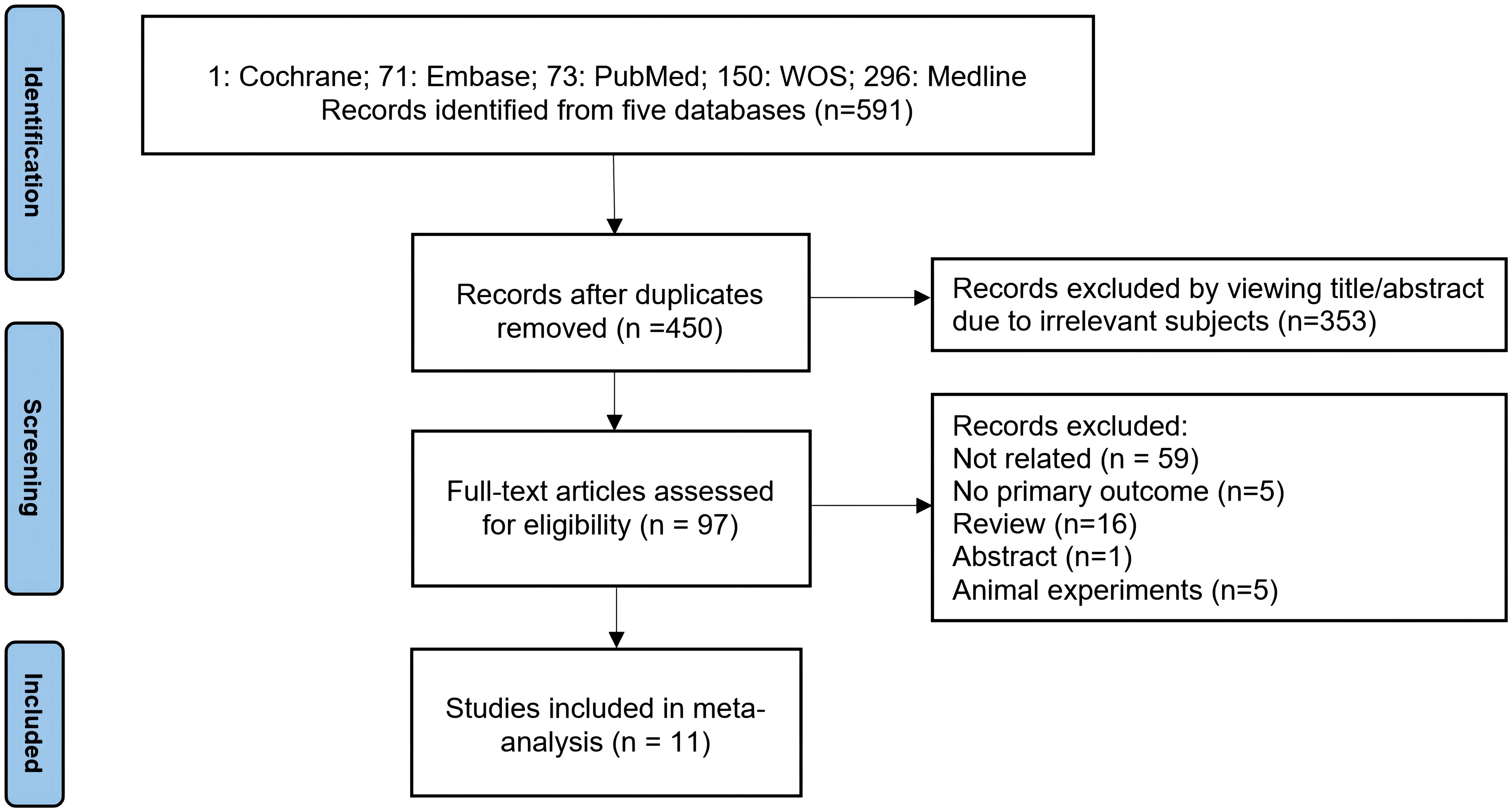
Figure 1 Flowchart of Literature Search.
The comprehensive meta-analysis encompassed 11 cohort studies spanning a geographical range that included the United States, China, Japan, Korea, France, Spain, and the Netherlands, with publication years extending from 2013 to 2023. The number of children in the eligible included studies ranged from 249 to 1,735, and the mean age of pregnant women across studies was between 26.4 and 32.5 years. A detailed examination of TH levels was predominantly focused on neonatal assessments in seven studies, while three studies focused on early childhood, assessing thyroid function at mean ages of 2, 4, 6, and 10 years. One study uniquely investigated both neonatal and childhood stages, around the age of six years. These studies collectively reported on maternal BPA concentrations, with a subset also exploring exposure to other bisphenols. Notably, the investigations in particular highlighted the exploration of sex-specific effects (Table 1).
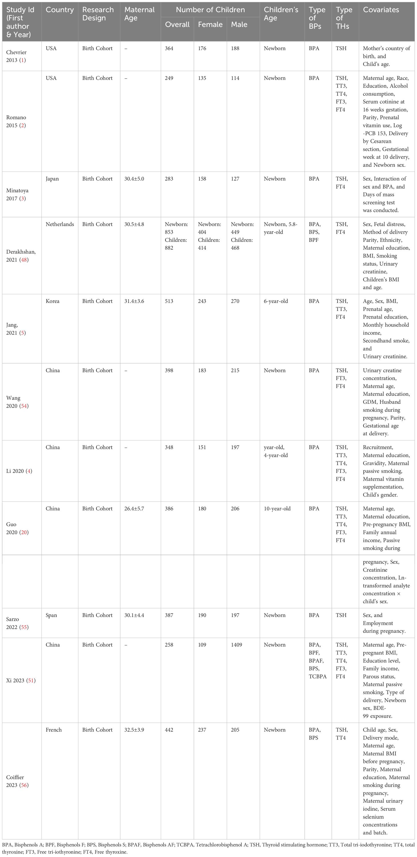
Table 1 Characteristics of Included Studies.
The NOS quality assessment results of the studies included are shown in Table 2. The NOS scores ranged from 6 to 8 stars, affirming the high quality of the included studies in our analysis, and underscoring the robustness and reliability of the synthesized evidence presented (Table 2).
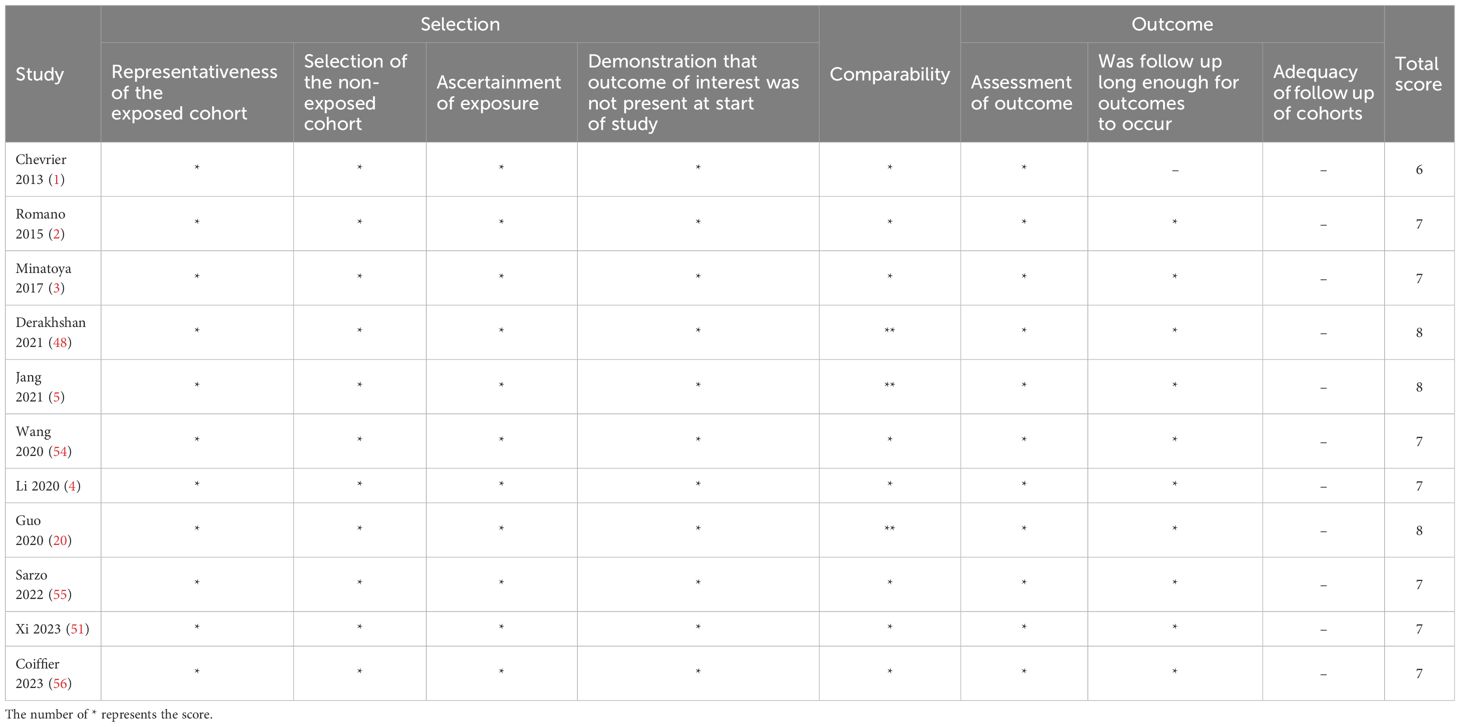
Table 2 Study quality of cohort studies.
Analysis from eleven cohort studies assessing the impact of prenatal BPs exposure on TSH levels indicated a modest, yet statistically significant reduction in TSH levels in total children (β = -0.013, 95% CI: -0.025, -0.001), a fixed-effects model was employed due to low heterogeneity (I2< 50%). Notably, a sex-specific analysis revealed that this reduction was pronounced in female offspring (β = -0.020, 95% CI: -0.036, -0.005), and no significant association was observed in male (β = -0.005, 95%CI: -0.024, 0.015) (Figure 2).
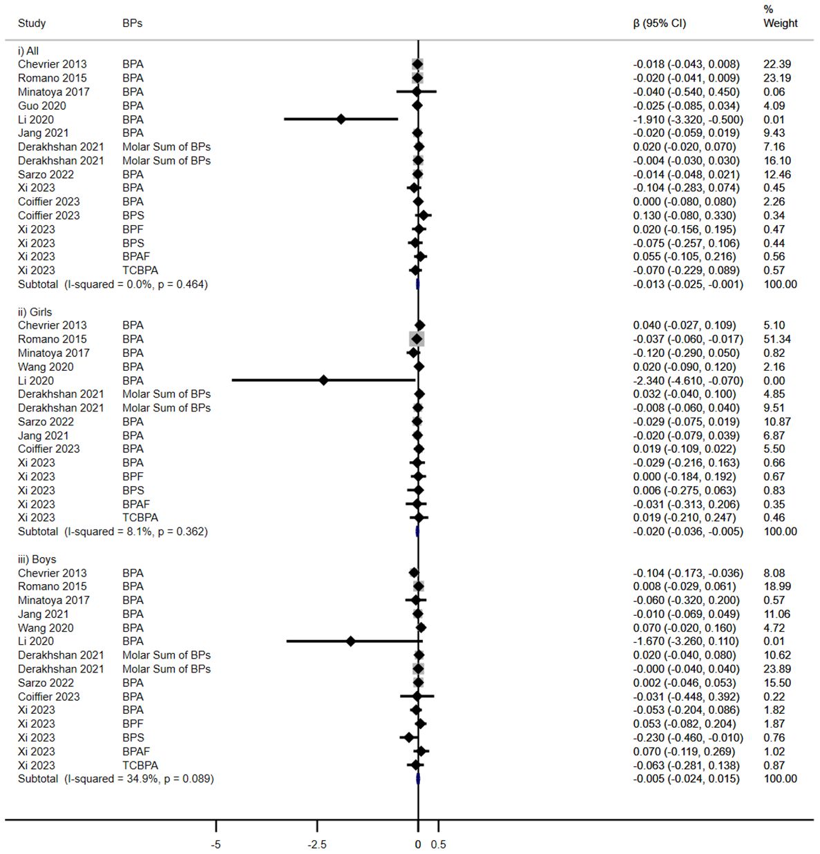
Figure 2 Effect of Prenatal BPs Mixtured Exposure on TSH Levels in Children of Different Sex.
Five cohorts evaluated the impact of prenatal exposure to BPs on TT3 levels in children (Figure 3). In subgroup analyses, a significant increase was noted in females (β = 0.011, 95% CI: 0.001, 0.021), contrary to no discernible effect in overall children (β = 0.000, 95%CI: -0.007, 0.007) or in males (β = -0.009, 95%CI: -0.020, 0.003).
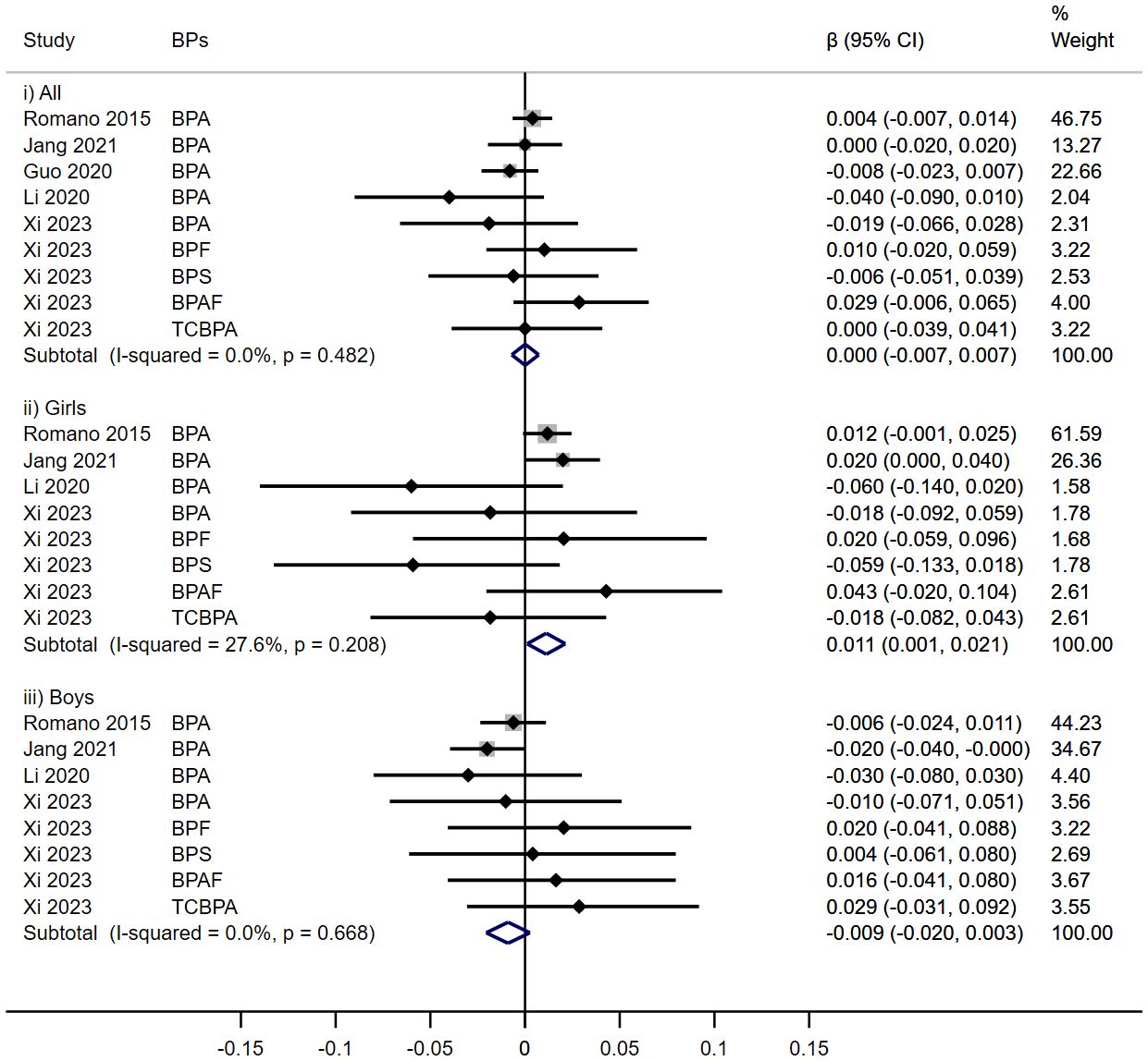
Figure 3 Effect of Prenatal BPs Mixtured Exposure on TT3 Levels in Children of Different Sex.
No significant associations were observed between prenatal BPs exposure and TT4 levels among children across five cohort studies (β = 0.000, 95%CI: -0.001, 0.001), irrespective of sex (Girls: β = 0.001, 95%CI: -0.001, 0.002; Boys: β = -0.000, 95%CI: -0.002, 0.001) (Figure 4).
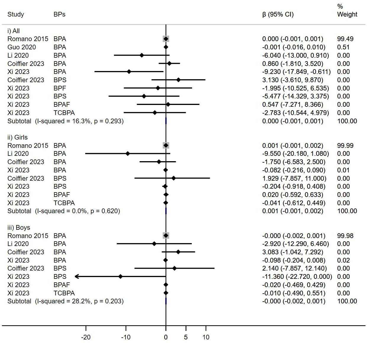
Figure 4 Effect of Prenatal BPs Mixtured Exposure on TT4 Levels in Children of Different Sex.
In this subgroup analysis, prenatal BPs exposure was related to decreased FT3 levels in all children (β = -0.011, 95% CI: -0.019, -0.003). However, this relationship was neither found in females (β = 0.005, 95%CI: -0.012, 0.022) nor males (β = -0.018, 95%CI: -0.040, 0.004) (Figure 5).
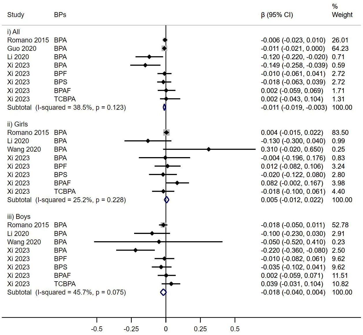
Figure 5 Effect of Prenatal BPs Mixtured Exposure on FT3 Levels in Children of Different Sex.
In an aggregated analysis of eight cohort studies, no significant associations were observed in prenatal BPs exposure and decreased FT4 levels in children (β= -0.00001, 95%CI: -0.0001, 0.0000), irrespective of sex (Girls: β = -0.0001, 95% CI: -0.0002, 0.00001; Boys: β = 0.0000, 95%CI: -0.0001, 0.0001) (Figure 6).
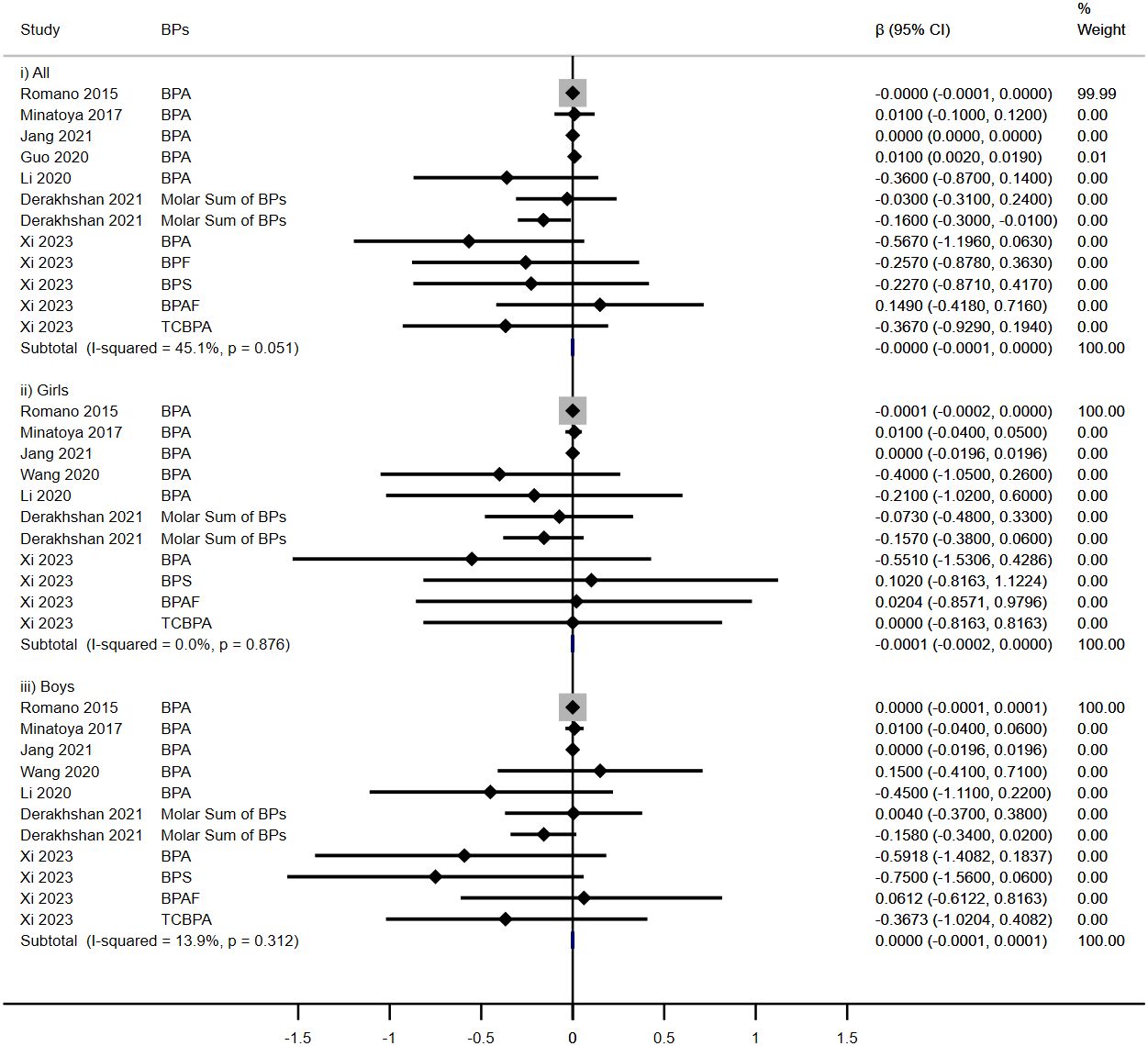
Figure 6 Effect of Prenatal BPs Mixtured Exposure on FT4 Levels in Children of Different Sex.
Subgroup analysis further elucidated that the observed relationships between prenatal BPA concentrations and alterations in thyroid hormone levels mirrored those identified in the broader bisphenol exposure analysis. Intriguingly, increased TT3 was observed in female offspring (β = 0.012, 95% CI: 0.002, 0.023), and a decrease in TT3 levels was also noted in male offspring (β = -0.013, 95% CI: -0.025, -0.001) (Supplementary Figures 1A–E).
We established 1.5 ug/g creatinine (Cr) as the threshold value to categorize the exposure groups into high and low levels. Subgroup analysis showed that high levels of bisphenols exposure significantly reduced FT3 levels (β = -0.011, 95% CI: -0.020, -0.003) in contrast to low exposure groups (β = -0.008, 95% CI: -0.037, -0.020), but no specific effects of exposure concentration were found in relation to other thyroid hormones (Supplementary Figures 2A–E).
Sensitivity analysis was conducted to evaluate the stability of the relationship between prenatal bisphenols mixtured exposure and THs in all children, as shown in Supplementary Figures 3A–E. The pooled β and 95% CI did not show evident differences after excluding each individual article one by one, indicating the credibility of the studies included in our meta-analysis.
Publication bias in the relationship between prenatal bisphenols mixtured exposure and thyroid hormones in all children was assessed with Egger’s test (Supplementary Table 2). Our statistical test showed no evidence of publication bias (Egger’s test p > 0.05).
This meta-analysis investigated the association of maternal bisphenols (BPs) exposure with offspring’s THs levels, and sex differences and exposure levels may change the outcomes. The salient findings are multifaceted: 1) Exposure to BP mixtures notably reduced TSH levels across the pediatric spectrum, and more effects were reported in girls; 2) BPs singularly escalates TT3 levels in female offspring; 3) BPA exposure parallels these effects (reduce TSH levels and increase TT3 levels in girls) while additionally diminishing TT3 levels in boys; 4) High level of BPs mixtured exposure (>1.5 ug/g Cr) could reduce FT3 levels. The detailed results were summarized in Table 3.
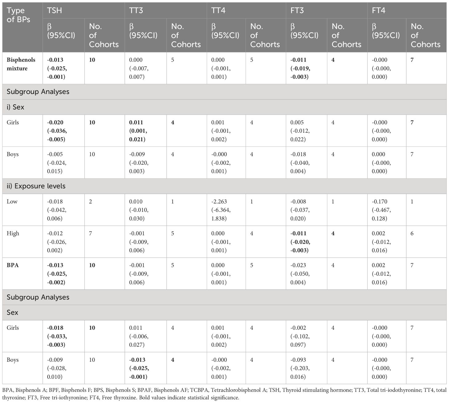
Table 3 Effects of Prenatal BPs on Children by Meta-analyses.
Prenatal exposure to bisphenols may traverse the placental barrier, potentially exerting long-term effects on fetal thyroid development and function, as well as postnatal life outcomes (21). Therefore, comprehending these impacts is pivotal for pediatric health. Prior studies have indicated that maternal exposure to bisphenols may influence maternal thyroid hormone levels. For instance, urinary BPA has been associated with thyroid function, with elevated concentrations of BPA potentially decreasing levels of TSH (54, 57), TT4 (1, 58), TT3 and FT3 (59) in women. Conversely, exposure to BPB, BPF, and TBBPA (60) may elevate FT4 levels (52), while BPS exposure might lower TT3 levels (52) and elevate TT4 levels (48). Moreover, studies have reported a notable positive trend in serum FT3 and FT4 levels with cumulative exposure to bisphenols mixture (50), although some have suggested that BPA exposure, either single-exposure (48) or in combination with other bisphenols (2, 55), may not correlate with maternal thyroid function. Despite inconsistent findings, the significance of this issue cannot be overstated. A meta-analysis focusing on childhood exposure to bisphenols and pediatric thyroid hormone levels indicated that BPA exposure lowers TSH levels (61). The prenatal period is a critical window for fetal organ and system development. Given the potential window of exposure and the cumulative effects over time, the impact of bisphenol exposure on fetal thyroid function may manifest postnatally. Hence, understanding how maternal exposure to bisphenols influences offspring thyroid function holds crucial implications for preventing and managing pediatric thyroid disorders in public health.
The indispensable role of thyroid hormones in supporting fetal growth, neurological maturation, metabolic regulation, and overall physiological harmony underscores the clinical significance of these findings (4, 62). The contrasting outcomes documented in previous literature (conclusions of the included studies were shown in Supplementary Table 3) underscore the intricacy involved in comprehending the association between prenatal exposure to bisphenols and thyroid function. While our study accentuates significant correlations between BPs and THs, disparities with preceding inquiries warrant a meticulous exploration of plausible influencing factors. The diversity within study populations, including demographic characteristics and health status of pregnant women, may significantly influence the outcomes. The study population by Romano et al. was mainly white primiparous women aged 25 - 35 years old from the United States, with higher socioeconomic status and incomes, and usually had regular intake of vitamin pills (2). In contrast, studies by Xi et al. and Li et al., most of the exposed subjects were elderly Asian multipara women who basically did not consume vitamin pills (4, 51). The combined superposition of these factors may be the reason for the differences that Romano et al. observed a relationship between prenatal BPA exposure and reduced TSH levels in female neonates, while the other two studies noted a negative correlation between BPA exposure and TSH levels in male infants. Furthermore, methodological differences, particularly in the sample matrix and the timing and frequency of bisphenols measurement, may contribute to inconsistent results. Minatoy et al. utilized serum samples, with BPA concentrations much lower than those reported in studies using maternal urine samples (3). Furthermore, Sarzo et al. only measured phenolics once in the first trimester (55). Other studies have taken more than one measurement and calculated average concentrations of BPA exposed throughout pregnancy. In addition, the study by Sarzo et al. used a birth cohort in Spain, which gradually restricted or banned the use of BPA as a pollutant (55). Rare relevant literature from Spain shows a decrease in urinary BPA concentrations in pregnant women in 2015-2016 (63) similar to the study by Wang and colleague (54). Phenolics are non-persistent pollutants, and the half-life of BPA is relatively short, and a single measurement may not adequately reflect BPA exposure throughout pregnancy. The lower levels of bisphenols exposure may have contributed to the lack of association between BPA and thyroid parameters observed in studies by Minatoya et al., Sarzo et al., and Wang et al. Another study analyzed BPA concentrations in tertiles indicated that girls in the middle tertile had lower TSH levels, whereas boys in the highest tertile had lower TSH, TT3, and FT3 in cord plasmas. However, BPA exposure did not cause an increase or decrease in TT4 or FT4 (4).
Our study elucidates the relationship between prenatal bisphenol exposure and offspring thyroid hormone levels, particularly highlighting significant differences based on sex, and exposure levels. We found that the mixture of BPs exposure and single exposure to BPA were related to decreased TSH and increased TT3 levels in girls, while only in the case of single BPA exposure, reduced TT3 levels were observed in boys. This observed pattern can be attributed to the structural similarity of BPA to TSH and TT3, as well as its role as an antagonist (43, 64) or agonist of thyroid receptors (65, 66). Specifically, BPA functions as an antagonist to thyroid hormones, disrupting TSH secretion through hormonal feedback mechanisms (67). The sex-specific effects observed in studies on endocrine-disrupting chemicals (EDCs) are reinforced by numerous investigations into the diverse effects of BPA (68, 69). Biological factors for dimorphism include the lower expression levels of uridine diphosphate-glucuronosyltransferase 2B1 (UGT2B1), which is involved in BPA glucuronidation, in males than females (70). Glucuronidation is a crucial step in the metabolism and elimination of phenolic compounds in the body. It involves the conjugation of phenolics with glucuronic acid, rendering them more water-soluble and facilitating their excretion via urine or bile, thereby eliminating phenolic compounds (71, 72). Additionally, the thyroid system exhibits a sexually dimorphic sensitivity to maternal exposure to BPs (70). Gonadal hormones, including estradiol, significantly influence the sex-specific neuroendocrine pathway, impacting the hypothalamic-pituitary-thyroid (HPT) axis, which governs the secretion of thyroid hormones (73). These molecular interactions and physiological responses form a complex network through which bisphenols exert their effects on thyroid hormone levels, revealing the intricacy of endocrine disruption and its varied manifestations between sexes. This meta-analysis also revealed that exposure to a mixture of bisphenols exceeding 1.5 µg/g creatinine is associated with a decrease in FT3 levels. A study found a significant correlation between high-dose bisphenol exposure and thyroid function alterations, whereas exposure at low doses may not have a significant impact (74). Similarly, another study noted that the effects of bisphenols on thyroid function may exhibit dose-dependent effects (35). Furthermore, at low concentrations, bisphenols may not adequately bind to thyroid receptors, leading to receptor saturation. In contrast, high-concentration bisphenol exposure may increase the binding affinity of bisphenols to thyroid hormone receptors, thereby intensifying interference with thyroid function. Additionally, exposure to bisphenols at lower concentrations may be more readily metabolized and excreted by the body, reducing their accumulation and duration in the body.
Further to these pathways, bisphenols may affect the transcriptional expression by altering transcription-related genes (Foxe1, Pax8, Nkx2-1) and thyroid hormone synthesis genes (Tpo, Tg, Slc5a5), with bisphenol analogs BPAF and BPS influencing transcriptional changes at doses lower than BPA (75, 76). BPA has the potential to hinder iodine uptake by interacting with the sodium/iodide symporter (NIS) (77, 78), a crucial mediator of iodine transport in thyroid and extrathyroidal tissues, thereby impacting thyroid function (75, 79). NIS plays a crucial role in transporting iodine from the bloodstream into thyroid cells, a pivotal step in thyroid hormone synthesis (80, 81). NIS is responsible for transporting iodine into thyroid cells, which is essential for the synthesis of thyroid hormones, particularly thyroxine (T4) (82, 83). Additionally, since TSH stimulates the thyroid to release T4, compensatory elevation of TSH levels may occur (84). Another possible mechanism is that their ability to interact with estrogen and androgen receptors (85). Animal studies provide evidence that BPA impacts hormone synthesis in the pituitary gland, affecting TSH production through estrogen receptor signaling (86–89), independently of thyroid hormone feedback loops. These studies have reported varying results, possibly attributable to differences in the chemical structures tested, dosages used, and species studied. Further in vivo controlled experiments are needed to elucidate the extent and mechanisms of BP influence on NIS and thyroid hormone levels. The mechanisms of action of bisphenol compounds were shown in Supplementary Table 4.
This study is the first meta-analysis to investigate the effect of maternal BPs exposure on thyroid hormone levels in children and explore the sex-specific effect on thyroid function, and incorporated studies were high-caliber prospective cohort investigations, defined by explicit search criteria, potentially reinforcing the trustworthiness of our research. However, it has certain limitations. First, studies were interpreted in different subgroup analyses, such as types of exposure and THs, and children’s sex. These subgroup analyses demonstrated the potential directions of further public health concerns, but, unfortunately, the analyses were limited by a relatively small sample size, we were unable to analyze the single effects of other bisphenol analogs on thyroid hormones. Second, studies used different types of outcome measurements, although we conducted sensitivity analyses on individual tests, substantial heterogeneity across the included studies was not fully interpreted. Third, most of the research subjects were neonates, but there were only four studies focused on early childhood, which may lead to imprecise results. Finally, the included cohort studies were mainly from North Americans and Asians, and there is a dearth of relevant studies in other regions, potentially leading to geographic bias.
Prenatal exposure to bisphenols reduced TSH and increased TT3 levels in female offspring. Maternal BPA exposure showed an inverse relationship with TT3 levels in male offspring. Notably, high concentrations of bisphenol exposure have also been found to decrease FT3 levels in offspring. Given these findings, it is critical to strengthen control over prenatal exposure to bisphenols to safeguard early childhood health. To validate our findings, a larger and broader prospective cohort study should be conducted to explore the effects of prenatal bisphenols exposure on thyroid hormone levels in children.
The original contributions presented in the study are included in the article/Supplementary Material. Further inquiries can be directed to the corresponding authors.
JL: Data curation, Investigation, Methodology, Visualization, Writing – original draft, Writing – review & editing. MT: Data curation, Investigation, Writing – review & editing. HQ: Writing – review & editing. DC: Writing – review & editing. SM: Writing – review & editing. XW: Resources, Supervision, Writing – review & editing. FB: Resources, Supervision, Writing – review & editing.
The author(s) declare that financial support was received for the research, authorship, and/or publication of this article.
The authors declare that the research was conducted in the absence of any commercial or financial relationships that could be construed as a potential conflict of interest.
All claims expressed in this article are solely those of the authors and do not necessarily represent those of their affiliated organizations, or those of the publisher, the editors and the reviewers. Any product that may be evaluated in this article, or claim that may be made by its manufacturer, is not guaranteed or endorsed by the publisher.
The Supplementary Material for this article can be found online at: https://www.frontiersin.org/articles/10.3389/fendo.2024.1420540/full#supplementary-material
1. Chevrier J, Gunier RB, Bradman A, Holland NT, Calafat AM, Eskenazi B, et al. Maternal urinary bisphenol a during pregnancy and maternal and neonatal thyroid function in the CHAMACOS study. Environ Health Perspect. (2013) 121:138–44. doi: 10.1289/ehp.1205092
2. Romano ME, Webster GM, Vuong AM, Thomas Zoeller R, Chen A, Hoofnagle AN, et al. Gestational urinary bisphenol A and maternal and newborn thyroid hormone concentrations: the HOME Study. Environ Res. (2015) 138:453–60. doi: 10.1016/j.envres.2015.03.003
3. Minatoya M, Sasaki S, Araki A, Miyashita C, Itoh S, Yamamoto J, et al. Cord blood bisphenol A levels and reproductive and thyroid hormone levels of neonates: the Hokkaido study on environment and children’s health. Epidemiology. (2017) 28 Suppl 1:S3–s9. doi: 10.1097/EDE.0000000000000716
4. Li F, Yang F, Li DK, Tian YP, Miao MH, Zhang Y, et al. Prenatal bisphenol A exposure, fetal thyroid hormones and neurobehavioral development in children at 2 and 4 years: A prospective cohort study. Sci Total Environ. (2020) 722:137887. doi: 10.1016/j.scitotenv.2020.137887
5. Jang Y, Choi Y-J, Lim Y-H, Lee K-S, Kim B-N, Shin CH, et al. Associations between thyroid hormone levels and urinary concentrations of bisphenol A, F, and S in 6-year-old children in Korea. J Prev Med Public Health. (2021) 54:37–45. doi: 10.3961/jpmph.20.310
6. Chou WC, Chen JL, Lin CF, Chen YC, Shih FC, Chuang CY. Biomonitoring of bisphenol A concentrations in maternal and umbilical cord blood in regard to birth outcomes and adipokine expression: a birth cohort study in Taiwan. Environ Health. (2011) 10:94. doi: 10.1186/1476-069X-10-94
7. Liao C, Kannan K. Widespread occurrence of bisphenol A in paper and paper products: implications for human exposure. Environ Sci Technol. (2011) 45:9372–9. doi: 10.1021/es202507f
8. Huang YQ, Wong CK, Zheng JS, Bouwman H, Barra R, Wahlström B, et al. Bisphenol A (BPA) in China: a review of sources, environmental levels, and potential human health impacts. Environ Int. (2012) 42:91–9. doi: 10.1016/j.envint.2011.04.010
9. Wang H, Liu L, Wang J, Tong Z, Yan J, Zhang T, et al. Urinary sexual steroids associated with bisphenol A (BPA) exposure in the early infant stage: Preliminary results from a Daishan birth cohort. Sci Total Environ. (2017) 601-602:1733–42. doi: 10.1016/j.scitotenv.2017.05.257
10. Calafat AM, Ye X, Wong LY, Reidy JA, Needham LL. Exposure of the U.S. population to bisphenol A and 4-tertiary-octylphenol: 2003-2004. Environ Health Perspect. (2008) 116:39–44. doi: 10.1289/ehp.10753
11. Koch HM, Kolossa-Gehring M, Schröter-Kermani C, Angerer J, Brüning T. Bisphenol A in 24 h urine and plasma samples of the German Environmental Specimen Bank from 1995 to 2009: a retrospective exposure evaluation. J Expo Sci Environ Epidemiol. (2012) 22:610–6. doi: 10.1038/jes.2012.39
12. Zhang T, Sun H, Kannan K. Blood and urinary bisphenol A concentrations in children, adults, and pregnant women from China: partitioning between blood and urine and maternal and fetal cord blood. Environ Sci Technol. (2013) 47:4686–94. doi: 10.1021/es303808b
13. Lee YJ, Ryu HY, Kim HK, Min CS, Lee JH, Kim E, et al. Maternal and fetal exposure to bisphenol A in Korea. Reprod Toxicol. (2008) 25:413–9. doi: 10.1016/j.reprotox.2008.05.058
14. Geens T, Aerts D, Berthot C, Bourguignon JP, Goeyens L, Lecomte P, et al. A review of dietary and non-dietary exposure to bisphenol-A. Food Chem Toxicol. (2012) 50:3725–40. doi: 10.1016/j.fct.2012.07.059
15. Philippat C, Wolff MS, Calafat AM, Ye X, Bausell R, Meadows M, et al. Prenatal exposure to environmental phenols: concentrations in amniotic fluid and variability in urinary concentrations during pregnancy. Environ Health Perspect. (2013) 121:1225–31. doi: 10.1289/ehp.1206335
16. Arbuckle TE, Davis K, Marro L, Fisher M, Legrand M, LeBlanc A, et al. Phthalate and bisphenol A exposure among pregnant women in Canada–results from the MIREC study. Environ Int. (2014) 68:55–65. doi: 10.1016/j.envint.2014.02.010
17. Cantonwine DE, Ferguson KK, Mukherjee B, McElrath TF, Meeker JD. Urinary bisphenol A levels during pregnancy and risk of preterm birth. Environ Health Perspect. (2015) 123:895–901. doi: 10.1289/ehp.1408126
18. Lee J, Choi K, Park J, Moon HB, Choi G, Lee JJ, et al. Bisphenol A distribution in serum, urine, placenta, breast milk, and umbilical cord serum in a birth panel of mother-neonate pairs. Sci Total Environ. (2018) 626:1494–501. doi: 10.1016/j.scitotenv.2017.10.042
19. Wiraagni IA, Mohd MA, Bin Abd Rashid R, Haron D. Validation of a simple extraction procedure for bisphenol A identification from human plasma. PloS One. (2019) 14:e0221774. doi: 10.1371/journal.pone.0221774
20. Guo JQ, Wu CH, Zhang JM, Li WT, Lv SL, Lu DS, et al. Maternal and childhood urinary phenol concentrations, neonatal thyroid function, and behavioral problems at 10 years of age: The SMBCS study. Sci Total Environ. (2020) 743:140678. doi: 10.1016/j.scitotenv.2020.140678
21. Li LX, Chen L, Meng XZ, Chen BH, Chen SQ, Zhao Y, et al. Exposure levels of environmental endocrine disruptors in mother-newborn pairs in China and their placental transfer characteristics. PloS One. (2013) 8:e62526. doi: 10.1371/journal.pone.0062526
22. Sugiura-Ogasawara M, Ozaki Y, Sonta S, Makino T, Suzumori K. Exposure to bisphenol A is associated with recurrent miscarriage. Hum Reprod. (2005) 20:2325–9. doi: 10.1093/humrep/deh888
23. Casals-Casas C, Desvergne B. Endocrine disruptors: from endocrine to metabolic disruption. Annu Rev Physiol. (2011) 73:135–62. doi: 10.1146/annurev-physiol-012110-142200
24. Salian S, Doshi T, Vanage G. Perinatal exposure of rats to Bisphenol A affects fertility of male offspring–an overview. Reprod Toxicol. (2011) 31:359–62. doi: 10.1016/j.reprotox.2010.10.008
25. Lassen TH, Frederiksen H, Jensen TK, Petersen JH, Joensen UN, Main KM, et al. Urinary bisphenol A levels in young men: association with reproductive hormones and semen quality. Environ Health Perspect. (2014) 122:478–84. doi: 10.1289/ehp.1307309
26. Sheng ZG, Tang Y, Liu YX, Yuan Y, Zhao BQ, Chao XJ, et al. Low concentrations of bisphenol a suppress thyroid hormone receptor transcription through a nongenomic mechanism. Toxicol Appl Pharmacol. (2012) 259:133–42. doi: 10.1016/j.taap.2011.12.018
27. Gentilcore D, Porreca I, Rizzo F, Ganbaatar E, Carchia E, Mallardo M, et al. Bisphenol A interferes with thyroid specific gene expression. Toxicology. (2013) 304:21–31. doi: 10.1016/j.tox.2012.12.001
28. Zhang DH, Zhou EX, Yang ZL. Waterborne exposure to BPS causes thyroid endocrine disruption in zebrafish larvae. PloS One. (2017) 12:e0176927. doi: 10.1371/journal.pone.0176927
29. Kundakovic M, Champagne FA. Epigenetic perspective on the developmental effects of bisphenol A. Brain Behav Immun. (2011) 25:1084–93. doi: 10.1016/j.bbi.2011.02.005
30. Boas M, Feldt-Rasmussen U, Main KM. Thyroid effects of endocrine disrupting chemicals. Mol Cell Endocrinol. (2012) 355:240–8. doi: 10.1016/j.mce.2011.09.005
31. Henrichs J, Ghassabian A, Peeters RP, Tiemeier H. Maternal hypothyroxinemia and effects on cognitive functioning in childhood: how and why? Clin Endocrinol (Oxf). (2013) 79:152–62. doi: 10.1111/cen.12227
32. Kundakovic M, Gudsnuk K, Franks B, Madrid J, Miller RL, Perera FP, et al. Sex-specific epigenetic disruption and behavioral changes following low-dose in utero bisphenol A exposure. Proc Natl Acad Sci U.S.A. (2013) 110:9956–61. doi: 10.1073/pnas.1214056110
33. Zoeller RT, Brown TR, Doan LL, Gore AC, Skakkebaek NE, Soto AM, et al. Endocrine-disrupting chemicals and public health protection: a statement of principles from The Endocrine Society. Endocrinology. (2012) 153:4097–110. doi: 10.1210/en.2012-1422
34. Liao C, Kannan K. A survey of alkylphenols, bisphenols, and triclosan in personal care products from China and the United States. Arch Environ Contam Toxicol. (2014) 67:50–9. doi: 10.1007/s00244-014-0016-8
35. Rochester JR, Bolden AL. Bisphenol S and F: A systematic review and comparison of the hormonal activity of bisphenol A substitutes. Environ Health Perspect. (2015) 123:643–50. doi: 10.1289/ehp.1408989
36. Hu C, Xu Y, Wang M, Cui S, Zhang H, Lu L. Bisphenol analogues induce thyroid dysfunction via the disruption of the thyroid hormone synthesis pathway. Sci Total Environ. (2023) 900:165711. doi: 10.1016/j.scitotenv.2023.165711
37. Huang GM, Tian XF, Fang XD, Ji FJ. Waterborne exposure to bisphenol F causes thyroid endocrine disruption in zebrafish larvae. Chemosphere. (2016) 147:188–94. doi: 10.1016/j.chemosphere.2015.12.080
38. Chen P, Wang R, Chen G, An B, Liu M, Wang Q, et al. Thyroid endocrine disruption and hepatotoxicity induced by bisphenol AF: Integrated zebrafish embryotoxicity test and deep learning. Sci Total Environ. (2022) 822:153639. doi: 10.1016/j.scitotenv.2022.153639
39. Kwon B, Kho Y, Kim PG, Ji K. Thyroid endocrine disruption in male zebrafish following exposure to binary mixture of bisphenol AF and sulfamethoxazole. Environ Toxicol Pharmacol. (2016) 48:168–74. doi: 10.1016/j.etap.2016.10.018
40. Kitamura S, Jinno N, Ohta S, Kuroki H, Fujimoto N. Thyroid hormonal activity of the flame retardants tetrabromobisphenol A and tetrachlorobisphenol A. Biochem Biophys Res Commun. (2002) 293:554–9. doi: 10.1016/S0006-291X(02)00262-0
41. Kitamura S, Suzuki T, Sanoh S, Kohta R, Jinno N, Sugihara K, et al. Comparative study of the endocrine-disrupting activity of bisphenol A and 19 related compounds. Toxicol Sci. (2005) 84:249–59. doi: 10.1093/toxsci/kfi074
42. Kitamura S, Kato T, Iida M, Jinno N, Suzuki T, Ohta S, et al. Anti-thyroid hormonal activity of tetrabromobisphenol A, a flame retardant, and related compounds: Affinity to the mammalian thyroid hormone receptor, and effect on tadpole metamorphosis. Life Sci. (2005) 76:1589–601. doi: 10.1016/j.lfs.2004.08.030
43. Zoeller RT, Bansal R, Parris C. Bisphenol-A, an environmental contaminant that acts as a thyroid hormone receptor antagonist in vitro, increases serum thyroxine, and alters RC3/neurogranin expression in the developing rat brain. Endocrinology. (2005) 146:607–12. doi: 10.1210/en.2004-1018
44. Xu X, Liu Y, Sadamatsu M, Tsutsumi S, Akaike M, Ushijima H, et al. Perinatal bisphenol A affects the behavior and SRC-1 expression of male pups but does not influence on the thyroid hormone receptors and its responsive gene. Neurosci Res. (2007) 58:149–55. doi: 10.1016/j.neures.2007.02.011
45. Fénichel P, Déchaux H, Harthe C, Gal J, Ferrari P, Pacini P, et al. Unconjugated bisphenol A cord blood levels in boys with descended or undescended testes. Hum Reprod. (2012) 27:983–90. doi: 10.1093/humrep/der451
46. Brucker-Davis F, Ferrari P, Boda-Buccino M, Wagner-Mahler K, Pacini P, Gal J, et al. Cord blood thyroid tests in boys born with and without cryptorchidism: Correlations with birth parameters and in utero xenobiotics exposure. Thyroid. (2011) 21:1133–41. doi: 10.1089/thy.2010.0459
47. Xiong C, Xu LL, Dong XH, Cao ZQ, Wang YJ, Chen K, et al. Trimester-specific associations of maternal exposure to bisphenols with neonatal thyroid stimulating hormone levels: A birth cohort study. Sci Total Environ. (2023) 880:163354. doi: 10.1016/j.scitotenv.2023.163354
48. Derakhshan A, Philips EM, Ghassabian A, Santos S, Asimakopoulos AG, Kannan K, et al. Association of urinary bisphenols during pregnancy with maternal, cord blood and childhood thyroid function. Environ Int. (2021) 146:106160. doi: 10.1016/j.envint.2020.106160
49. Lin J, Deng L, Sun M, Wang Y, Lee S, Choi K, et al. An in vitro investigation of endocrine disrupting potentials of ten bisphenol analogues. Steroids. (2021) 169:108826. doi: 10.1016/j.steroids.2021.108826
50. Lu W, Sun Z, Wang Z, Qu M, Shi Z, Song Q, et al. The joint effects of bisphenols and iodine exposure on thyroid during pregnancy. Nutrients. (2023) 15(15):3422. doi: 10.3390/nu15153422
51. Xi JY, Su XJ, Wang ZL, Ji HL, Chen Y, Liu XF, et al. The associations between concentrations of gestational bisphenol analogues and thyroid related hormones in cord blood: A prospective cohort study. Ecotoxicology Environ Saf. (2023) 256:114838. doi: 10.1016/j.ecoenv.2023.114838
52. Huang H, Liang J, Tang P, Yu C, Fan H, Liao Q, et al. Associations of bisphenol exposure with thyroid hormones in pregnant women: a prospective birth cohort study in China. Environ Sci pollut Res Int. (2022) 29:87170–83. doi: 10.1007/s11356-022-21817-3
53. Moher D, Liberati A, Tetzlaff J, Altman DG, Group P. Preferred reporting items for systematic reviews and meta-analyses: the PRISMA statement. PloS Med. (2009) 6:e1000097. doi: 10.1371/journal.pmed.1000097
54. Wang X, Tang N, Nakayama SF, Fan PP, Liu ZW, Zhang J, et al. Maternal urinary bisphenol A concentration and thyroid hormone levels of Chinese mothers and newborns by maternal body mass index. Environ Sci pollut Res. (2020) 27:10939–49. doi: 10.1007/s11356-020-07705-8
55. Sarzo B, Abumallouh R, Marín N, Llop S, Beneito A, Lopez-Flores I, et al. Association between phenols and thyroid hormones: The role of iodothyronine deiodinase genes. Environ pollut. (2022) 311:119926. doi: 10.1016/j.envpol.2022.119926
56. Coiffier O, Nakiwala D, Rolland M, Malatesta A, Lyon-Caen S, Chovelon B, et al. Exposure to a mixture of non-persistent environmental chemicals and neonatal thyroid function in a cohort with improved exposure assessment. Environ Int. (2023) 173:107840. doi: 10.1016/j.envint.2023.107840
57. Aung MT, Johns LE, Ferguson KK, Mukherjee B, McElrath TF, Meeker JD. Corrigendum to “Thyroid hormone parameters during pregnancy in relation to urinary bisphenol A concentrations: A repeated measures study” [Environment International 104 (2017) 33-40]. Environ Int. (2019) 122:417. doi: 10.1016/j.envint.2018.11.063
58. Derakhshan A, Shu H, Peeters RP, Kortenkamp A, Lindh CH, Demeneix B, et al. Association of urinary bisphenols and triclosan with thyroid function during early pregnancy. Environ Int. (2019) 133:105123. doi: 10.1016/j.envint.2019.105123
59. McGee G, Génard-Walton M, Williams PL, Korevaar TIM, Chavarro JE, Meeker JD, et al. Associations of maternal urinary concentrations of phenols, individually and as a mixture, with serum biomarkers of thyroid function and autoimmunity: results from the EARTH study. Toxics. (2023) 11(6):521. doi: 10.3390/toxics11060521
60. Aker AM, Ferguson KK, Rosario ZY, Mukherjee B, Alshawabkeh AN, Calafat AM, et al. A repeated measures study of phenol, paraben and Triclocarban urinary biomarkers and circulating maternal hormones during gestation in the Puerto Rico PROTECT cohort. Environ Health. (2019) 18:28. doi: 10.1186/s12940-019-0459-5
61. Koutaki D, Paltoglou G, Vourdoumpa A, Charmandari E. The impact of bisphenol A on thyroid function in neonates and children: A systematic review of the literature. Nutrients. (2022) 14(1):168. doi: 10.3390/nu14010168
62. Fukushi M, Honma K, Fujita K. Maternal thyroid deficiency during pregnancy and subsequent neuropsychological development of the child. N Engl J Med. (1999) 341:2016.
63. Martínez M, González N, Martí A, Marquès M, Rovira J, Kumar V, et al. Human biomonitoring of bisphenol A along pregnancy: An exposure reconstruction of the EXHES-Spain cohort. Environ Res. (2021) 196:110941. doi: 10.1016/j.envres.2021.110941
64. Moriyama K, Tagami T, Akamizu T, Usui T, Saijo M, Kanamoto N, et al. Thyroid hormone action is disrupted by bisphenol A as an antagonist. J Clin Endocrinol Metab. (2002) 87:5185–90. doi: 10.1210/jc.2002-020209
65. Kudo Y, Yamauchi K, Fukazawa H, Terao Y. In vitro and in vivo analysis of the thyroid system-disrupting activities of brominated phenolic and phenol compounds in Xenopus laevis. Toxicol Sci. (2006) 92:87–95. doi: 10.1093/toxsci/kfj204
66. Terasaki M, Kosaka K, Kunikane S, Makino M, Shiraishi F. Assessment of thyroid hormone activity of halogenated bisphenol A using a yeast two-hybrid assay. Chemosphere. (2011) 84:1527–30. doi: 10.1016/j.chemosphere.2011.04.045
67. Rochester JR. Bisphenol A and human health: A review of the literature. Reprod Toxicol. (2013) 42:132–55. doi: 10.1016/j.reprotox.2013.08.008
68. Wang Z, Zhou Y, Liang H, Miao M, Chen Y, Zhang X, et al. Prenatal exposure to bisphenol analogues and digit ratio in children at ages 4 and 6 years: A birth cohort study. Environ pollut. (2021) 278:116820. doi: 10.1016/j.envpol.2021.116820
69. Yang P, Lin BG, Zhou B, Cao WC, Chen PP, Deng YL, et al. Sex-specific associations of prenatal exposure to bisphenol A and its alternatives with fetal growth parameters and gestational age. Environ Int. (2021) 146:106305. doi: 10.1016/j.envint.2020.106305
70. Takeuchi T, Tsutsumi O, Nakamura N, Ikezuki Y, Takai Y, Yano T, et al. Gender difference in serum bisphenol A levels may be caused by liver UDP-glucuronosyltransferase activity in rats. Biochem Biophys Res Commun. (2004) 325:549–54. doi: 10.1016/j.bbrc.2004.10.073
71. Court MH. Isoform-selective probe substrates for in vitro studies of human UDP-glucuronosyltransferases. Methods Enzymol. (2005) 400:104–16. doi: 10.1016/S0076-6879(05)00007-8
72. Rowland A, Miners JO, Mackenzie PI. The UDP-glucuronosyltransferases: their role in drug metabolism and detoxification. Int J Biochem Cell Biol. (2013) 45:1121–32. doi: 10.1016/j.biocel.2013.02.019
73. Patisaul HB. Endocrine disrupting chemicals (EDCs) and the neuroendocrine system: Beyond estrogen, androgen, and thyroid. Adv Pharmacol. (2021) 92:101–50. doi: 10.1016/bs.apha.2021.03.007
74. Guignard D, Gayrard V, Lacroix MZ, Puel S, Picard-Hagen N, Viguié C. Evidence for bisphenol A-induced disruption of maternal thyroid homeostasis in the pregnant ewe at low level representative of human exposure. Chemosphere. (2017) 182:458–67. doi: 10.1016/j.chemosphere.2017.05.028
75. Wu Y, Beland FA, Fang JL. Effect of triclosan, triclocarban, 2,2’,4,4’-tetrabromodiphenyl ether, and bisphenol A on the iodide uptake, thyroid peroxidase activity, and expression of genes involved in thyroid hormone synthesis. Toxicol In Vitro. (2016) 32:310–9. doi: 10.1016/j.tiv.2016.01.014
76. Lee S, Kim C, Youn H, Choi K. Thyroid hormone disrupting potentials of bisphenol A and its analogues - in vitro comparison study employing rat pituitary (GH3) and thyroid follicular (FRTL-5) cells. Toxicol In Vitro. (2017) 40:297–304. doi: 10.1016/j.tiv.2017.02.004
77. Darrouzet E, Lindenthal S, Marcellin D, Pellequer JL, Pourcher T. The sodium/iodide symporter: state of the art of its molecular characterization. Biochim Biophys Acta. (2014) 1838:244–53. doi: 10.1016/j.bbamem.2013.08.013
78. Waugh DT. Fluoride exposure induces inhibition of sodium/iodide symporter (NIS) contributing to impaired iodine absorption and iodine deficiency: molecular mechanisms of inhibition and implications for public health. Int J Environ Res Public Health. (2019) 16(6):1086. doi: 10.3390/ijerph16061086
79. Kogai T, Taki K, Brent GA. Enhancement of sodium/iodide symporter expression in thyroid and breast cancer. Endocr Relat Cancer. (2006) 13:797–826. doi: 10.1677/erc.1.01143
80. Ferreira AC, Lima LP, Araújo RL, Müller G, Rocha RP, Rosenthal D, et al. Rapid regulation of thyroid sodium-iodide symporter activity by thyrotrophin and iodine. J Endocrinol. (2005) 184:69–76. doi: 10.1677/joe.1.05643
81. Carvalho DP, Dupuy C. Thyroid hormone biosynthesis and release. Mol Cell Endocrinol. (2017) 458:6–15. doi: 10.1016/j.mce.2017.01.038
82. Nilsson M. Iodide handling by the thyroid epithelial cell. Exp Clin Endocrinol Diabetes. (2001) 109:13–7. doi: 10.1055/s-2001-11014
83. Alotaibi H, Tuzlakoğlu-Öztürk M, Tazebay UH. The thyroid na+/I- symporter: molecular characterization and genomic regulation. Mol Imaging Radionucl Ther. (2017) 26:92–101. doi: 10.4274/Mirt
84. Luongo C, Martin C, Vella K, Marsili A, Ambrosio R, Dentice M, et al. The selective loss of the type 2 iodothyronine deiodinase in mouse thyrotrophs increases basal TSH but blunts the thyrotropin response to hypothyroidism. Endocrinology. (2015) 156:745–54. doi: 10.1210/en.2014-1698
85. MacKay H, Abizaid A. A plurality of molecular targets: The receptor ecosystem for bisphenol-A (BPA). Horm Behav. (2018) 101:59–67. doi: 10.1016/j.yhbeh.2017.11.001
86. Dang VH, Choi KC, Jeung EB. Estrogen receptors are involved in xenoestrogen induction of growth hormone in the rat pituitary gland. J Reprod Dev. (2009) 55:206–13. doi: 10.1262/jrd.20147
87. Ahmed RG. Maternal bisphenol A alters fetal endocrine system: Thyroid adipokine dysfunction. Food Chem Toxicol. (2016) 95:168–74. doi: 10.1016/j.fct.2016.06.017
88. Ahmed RG, Walaa GH, Asmaa FS. Suppressive effects of neonatal bisphenol A on the neuroendocrine system. Toxicol Ind Health. (2018) 34:397–407. doi: 10.1177/0748233718757082
Keywords: bisphenols, prenatal exposure, children, thyroid function, meta-analysis
Citation: Liu J, Tian M, Qin H, Chen D, Mzava SM, Wang X and Bigambo FM (2024) Maternal bisphenols exposure and thyroid function in children: a systematic review and meta-analysis. Front. Endocrinol. 15:1420540. doi: 10.3389/fendo.2024.1420540
Received: 22 April 2024; Accepted: 18 June 2024;
Published: 01 July 2024.
Edited by:
Francesca Coperchini, University of Pavia, ItalyReviewed by:
Xiaoqing Ye, Zhejiang Chinese Medical University, ChinaCopyright © 2024 Liu, Tian, Qin, Chen, Mzava, Wang and Bigambo. This is an open-access article distributed under the terms of the Creative Commons Attribution License (CC BY). The use, distribution or reproduction in other forums is permitted, provided the original author(s) and the copyright owner(s) are credited and that the original publication in this journal is cited, in accordance with accepted academic practice. No use, distribution or reproduction is permitted which does not comply with these terms.
*Correspondence: Francis Manyori Bigambo, ZnJhbmNpcy5iaWdhbWJvQHlhaG9vLmNvbQ==; Xu Wang, c2VwbmluZUBuam11LmVkdS5jbg==
†These authors have contributed equally to this work and share first authorship
Disclaimer: All claims expressed in this article are solely those of the authors and do not necessarily represent those of their affiliated organizations, or those of the publisher, the editors and the reviewers. Any product that may be evaluated in this article or claim that may be made by its manufacturer is not guaranteed or endorsed by the publisher.
Research integrity at Frontiers
Learn more about the work of our research integrity team to safeguard the quality of each article we publish.