 Guangdi Chu
 Haitao Niu
- Department of Urology, The Affiliated Hospital of Qingdao University, Qingdao, China
Background: Low-intensity pulsed ultrasound (LIPUS) is a highly promising therapeutic method that has been widely used in rehabilitation, orthopedics, dentistry, urology, gynecology, and other multidisciplinary disease diagnoses and treatments. It has attracted extensive attention worldwide. However, there is currently a lack of comprehensive and systematic research on the current status and future development direction of the LIPUS field. Therefore, this study comprehensively analyzed LIPUS-related reports from the past decade using bibliometrics methods, and further conducted research specifically focusing on its application in endocrine and metabolic diseases.
Methods: We downloaded LIPUS literature from 2012 to 2022 reported in the Web of Science Core Collection Science Citation Index-Expanded and Social Sciences Citation Index, and used bibliometric analysis software such as VOSviewer and CiteSpace to execute the analysis and visualize the results.
Results: We searched for 655 English articles published on LIPUS from 2012 to 2022. China had the highest number of published articles and collaborations between China and the United States were the closest in this field. Chongqing Medical University was the institution with the highest output, and ULTRASOUND IN MEDICINE AND BIOLOGY was the journal with the most related publications. In recent years, research on the molecular mechanisms of LIPUS has continued to deepen, and its clinical applications have also continued to expand. The application of LIPUS in major diseases such as oxidative stress, regeneration mechanism, and cancer is considered to be a future research direction, especially in the field of endocrinology and metabolism, where it has broad application value.
Conclusion: Global research on LIPUS is expected to continue to increase, and future research will focus on its mechanisms of action and clinical applications. This study comprehensively summarizes the current development status and global trends in the field of LIPUS, and its research progress in the field of endocrine and metabolic diseases, providing valuable reference for future research in this field.
1 Introduction
Low-intensity pulsed ultrasound (LIPUS) is a form of ultrasound that outputs in a pulsed wave mode, with energy intensity significantly lower than traditional ultrasound (1). LIPUS is considered a promising technology and has been widely applied in various clinical treatments and basic research (2). Its low frequency, low energy, and non-thermal effects have attracted the attention of numerous scholars (3). Increasing evidence shows that LIPUS can effectively stimulate osteoblasts, promote bone formation, and activate other stem/progenitor cells (4). LIPUS can enhance the vitality, proliferation, and multi-directional differentiation of various postnatal mesenchymal stem cells (MSCs) (2). Moreover, it can reduce the expression of pro-inflammatory cytokines, limit the infiltration of inflammatory cells, and regulate the phenotype of inflammatory cells (5). These beneficial changes may be related to increased blood flow, activated mitochondrial biogenesis, and anti-oxidative stress to promote muscle healing (6–8). Currently, LIPUS has been extensively used in tissue repair and regeneration, fracture healing, and inhibition of inflammatory responses. Additionally, it has significant potential therapeutic value in the field of urology, including chronic prostatitis/chronic pelvic pain syndrome (CP/CPPS) (9), erectile dysfunction (ED) (10, 11), stress urinary incontinence (SUI) (12), as well as gynecology, including premature ovarian insufficiency (13) and incomplete uterine involution (14).
Bibliometrics is a method of literature analysis that utilizes quantitative and qualitative perspectives to quantify and visualize published literature, thereby facilitating the evaluation of intrinsic connections and distribution patterns within research literature (15, 16). It offers a comprehensive and systematic assessment of the present research status, while also identifying potential avenues for future research within targeted fields. Bibliometrics effectively integrates information throughout the research process, enhances understanding, and assists in the deepening of knowledge regarding specific research areas (17, 18). It has been noted that there is currently a lack of bibliometric analysis focusing on the recent cooperation and research trends of LIPUS, with a focus on endocrinology and metabolism. Therefore, we conducted a systematic search of LIPUS-related literature in the Web of Science Core Collection (WoSCC) database and performed a comprehensive bibliometric analysis to clarify its current research status. We also combined with the latest published literature to analyze the research hotspots of LIPUS in the field of endocrinology and metabolic diseases to gain insights into future research directions in this field.
2 Materials and methods
2.1 Literature source and search
We conducted a comprehensive search of literature from 2012 to 2022 using the Science Citation Index Expanded (SCI-EXPANDED) and Social Sciences Citation Index (SSCI) databases in the WoSCC. To minimize bias caused by frequent database updates, we completed data retrieval and analysis on the same day. We searched for relevant publications in WoSCC using the following search strategy: TS = (“LIPUS” OR “Low-Intensity Pulsed Ultrasounds” OR “Pulsed Ultrasound, Low-Intensity” OR “Ultrasound, Low-Intensity Pulsed” OR “Low Intensity Pulsed Ultrasound”).
2.2 Inclusion and exclusion criteria
We systematically searched for literature related to LIPUS, limiting the document type to articles and reviews and language to English. Conference abstracts and other article types were excluded, as well as articles in languages other than English. We also excluded any other articles that were not relevant to the research subject or had been retracted.
2.3 Literature analysis method
We downloaded the literature records in plain text format from WoSCC and used VOSviewer for analysis of countries, universities, authors, and keywords. We used CiteSpace for burst word analysis, with the time was set from January 2012 to December 2022, the type was set as “keywords,” and other parameters set to default values. Additionally, we used R software to calculate the annual number of articles published and plotted a line graph.
3 Results
3.1 Search results
The process of inclusion and exclusion of articles is shown in Figure 1. From January 1, 2012 to December 31, 2022, a total of 762 articles were obtained. By limiting the document type to article or review, 668 articles were obtained. Limiting the language type to English resulted in 662 articles. After removing literature that was unrelated to the topic or retracted, 655 articles were included for subsequent analysis.
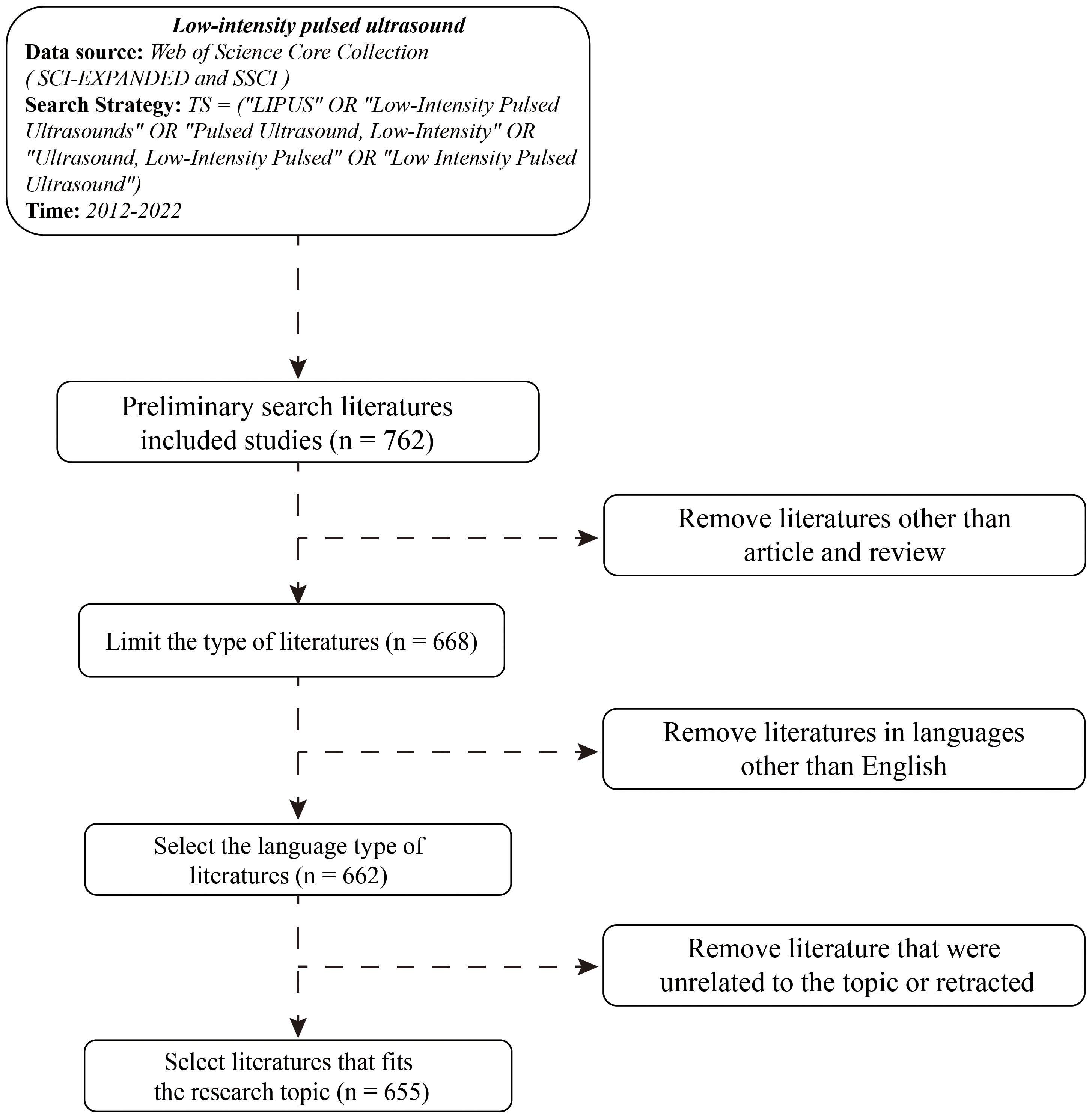
Figure 1 Literature search process and its inclusion and exclusion criteria.
3.2 Analysis of global trends
Through statistics of the annual publication volume (Figure 2), we found that the number of publications related to LIPUS research worldwide has shown a fluctuating upward trend. 2014 was a short-term peak period, and since 2015, the number of publications has been increasing year by year, especially in two periods from 2013-2014 and 2018-2019, where the publication speed increased rapidly, proving its rapid rise in global research popularity. In 2022, the publication volume showed a short-term decline, but the annual publication volume still exceeded 60 articles, which is at a relatively high level and consistent with the overall trend of fluctuating upward. In the past five years, the number of publications related to LIPUS research has remained at a high level, indicating that LIPUS is still a hot topic in current international research and continues to receive much attention.
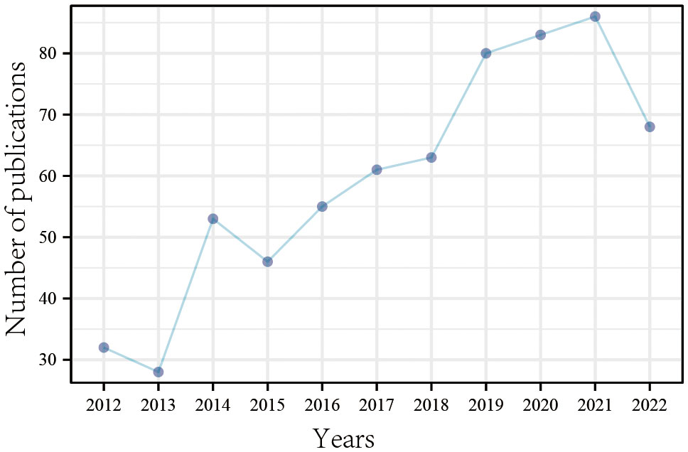
Figure 2 The number of publications per year from 2012 to 2022.
3.3 Analysis of the contributions of countries and regions
A total of 46 countries have contributed to international LIPUS research, among which China, the United States, and Japan are the top three countries with the highest number of related article publications, accounting for 81.2% (532/655) (Table 1). The top three countries with the highest average citation frequency of published articles are the Netherlands, France, and Italy. Stable cooperation and exchanges have been formed among the 46 countries, especially between China and the United States (Figure 3A). By analyzing the changes in time distribution, we found that early LIPUS research was mainly concentrated in countries such as Japan and Canada. However, it gradually gained attention and developed rapidly in other countries around 2018 (Figure 3B).
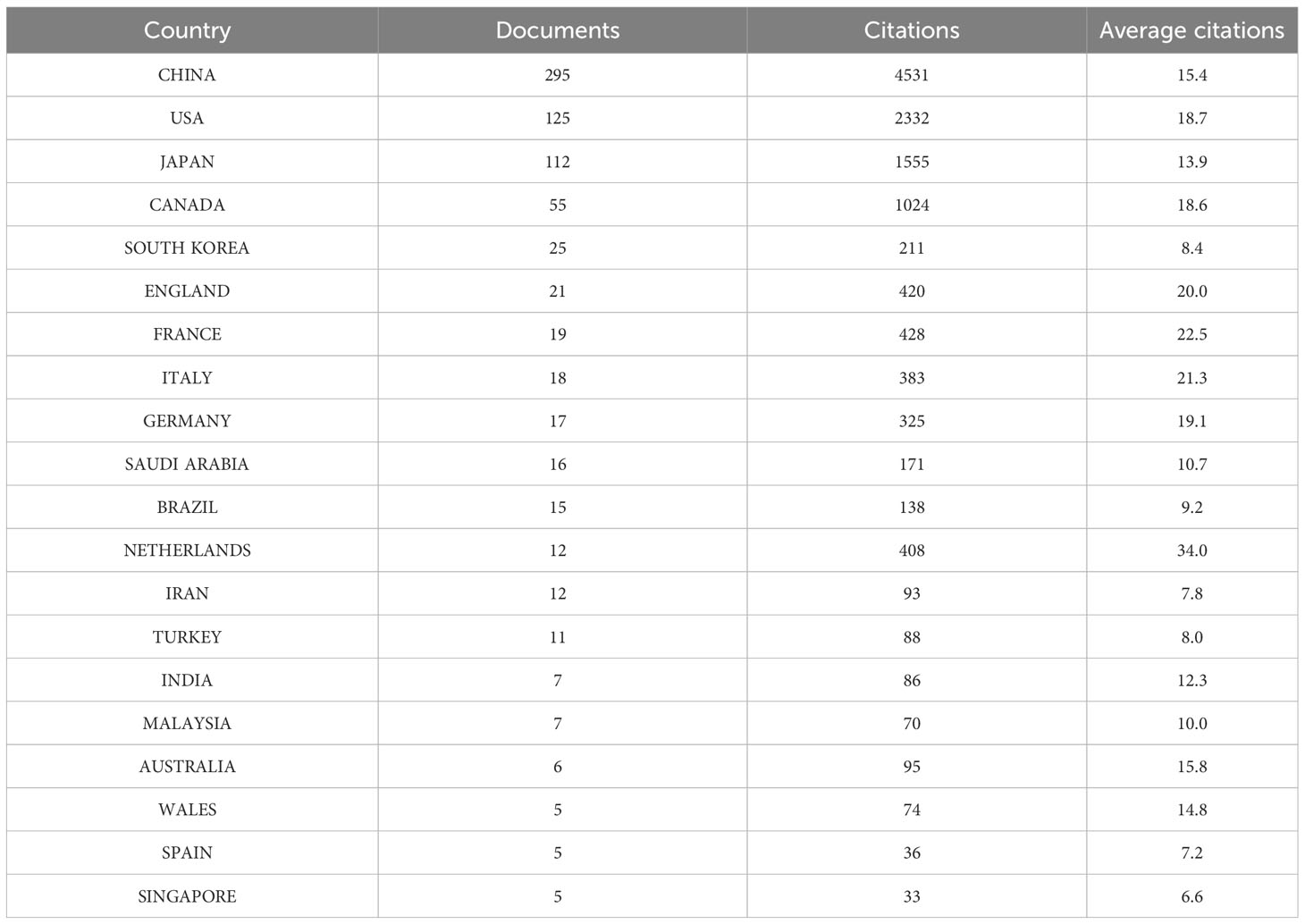
Table 1 The top 20 countries with the most LIPUS publications from 2012 to 2022.
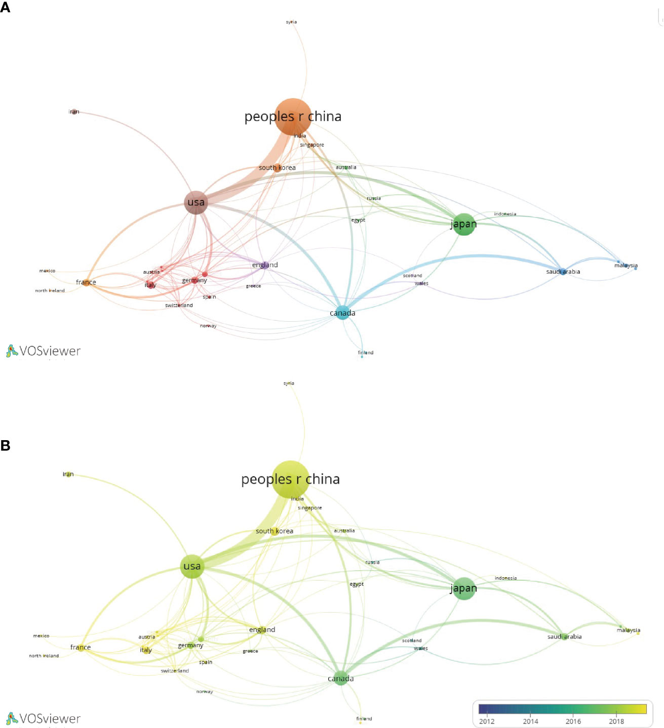
Figure 3 Collaboration network diagram of LIPUS literature publication countries. (A) Collaborative relationships in research in this field between different countries. (B) According to the time trend analysis of the relationship between cooperation among countries and years, the lighter the color represents the closer the year in which their cooperative relationship was developed.
3.4 Analysis of the contributions of universities and institutions
A total of 794 organizations have contributed to LIPUS research, among which Chongqing Medical University, University of Alberta, and Chinese Academy of Sciences are the top three organizations with the highest number of publications (Table 2). We selected the top 50 organizations in terms of publication volume for inter-organizational collaboration analysis and found that domestic Chinese organizations occupy the majority within the top 50 organizations, forming a relatively stable cooperative relationship (Figure 4A). Looking at the time trend, early LIPUS research was mainly carried out by related institutions in Japan and other countries. However, around 2018, research on LIPUS in China rose in popularity and produced many academic achievements (Figure 4B).
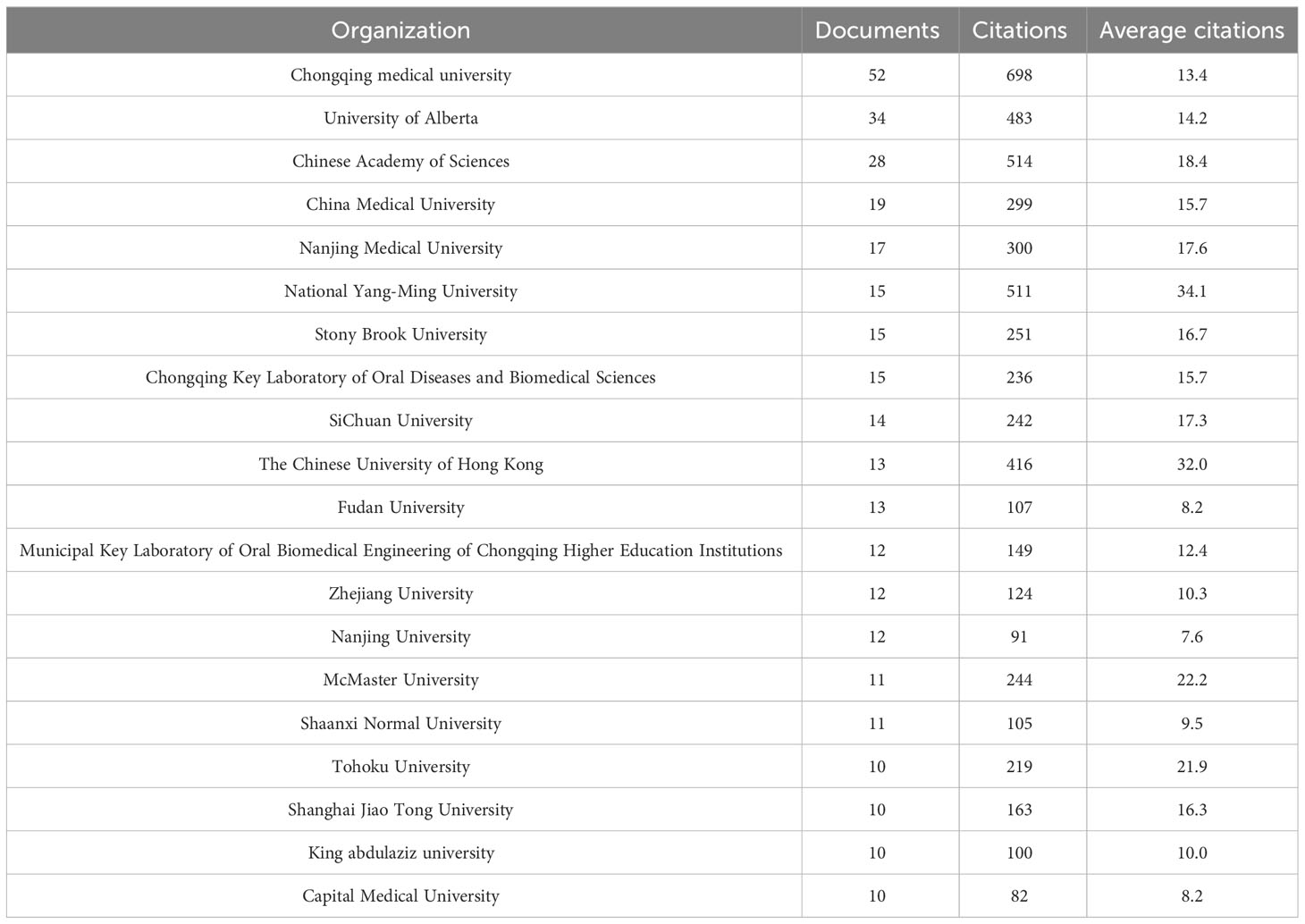
Table 2 The top 20 universities or institutions with the most LIPUS publications from 2012 to 2022.
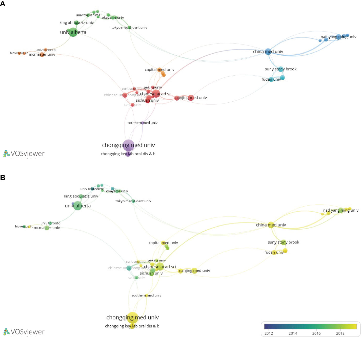
Figure 4 Collaboration network diagram of universities or institutions in LIPUS literature publication. (A) Collaborative relationships in research in this field among different universities or institutions. (B) According to the time trend analysis of the relationship between cooperation among universities or institutions and years, the lighter the color represents the closer the year in which their cooperative relationship was developed.
3.5 Analysis of the contributions of authors
A total of 2,973 authors have contributed to publications in this field, among which Tarek El-Bialy, Yan Wang, and Feng-Yi Yang are the top three authors with the highest number of publications (Table 3). Feng-Yi Yang is also the author with the highest number of citations. In terms of average citation frequency, Andrew Harrison, Kwok-Sui Leung, and Feng-Yi Yang are the top three scholars. Analyzing the collaborative relationship between authors, it can be found that international LIPUS research has formed a relatively stable collaborative group (Figure 5).
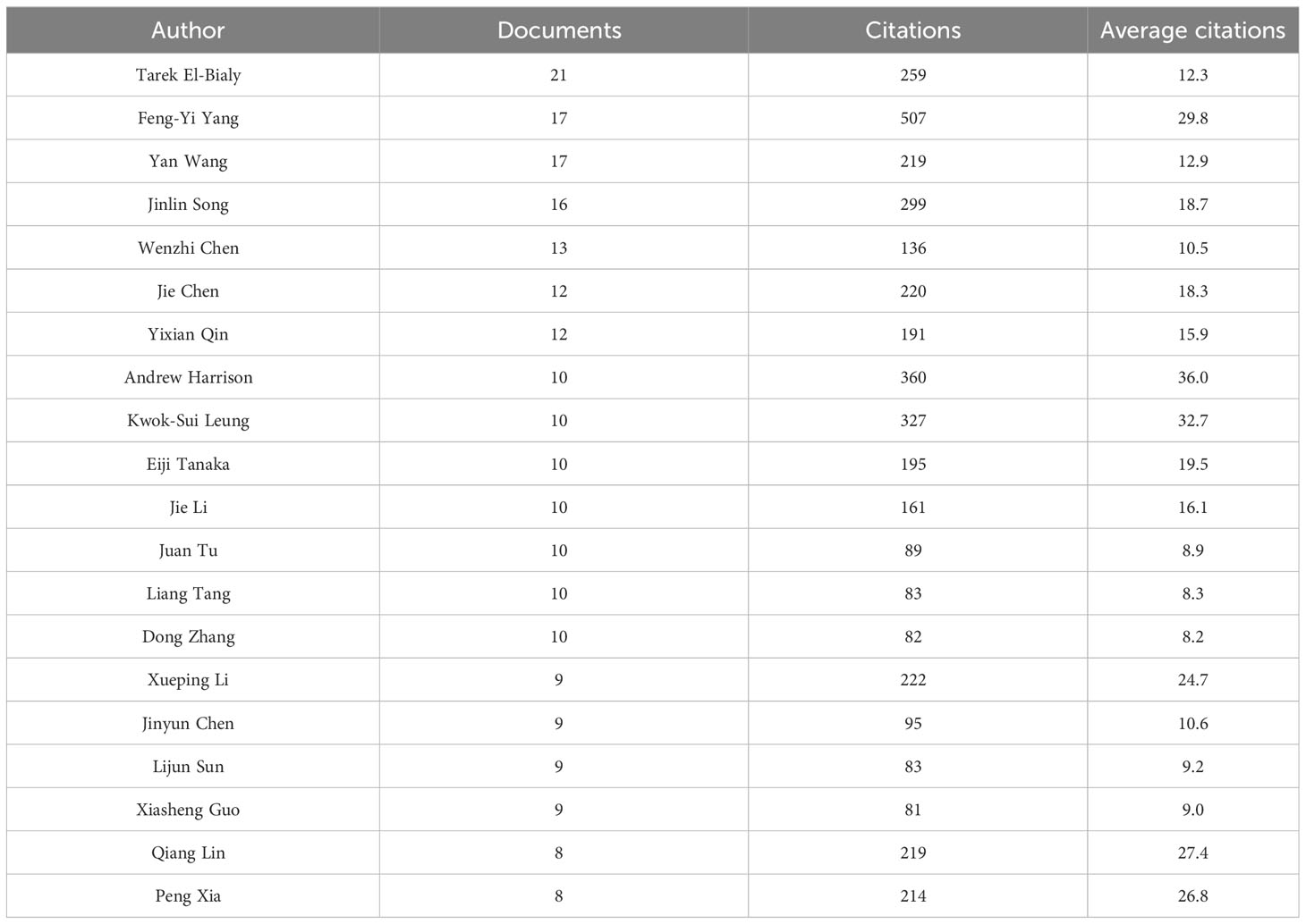
Table 3 The top 20 scholars with the most LIPUS publications from 2012 to 2022.
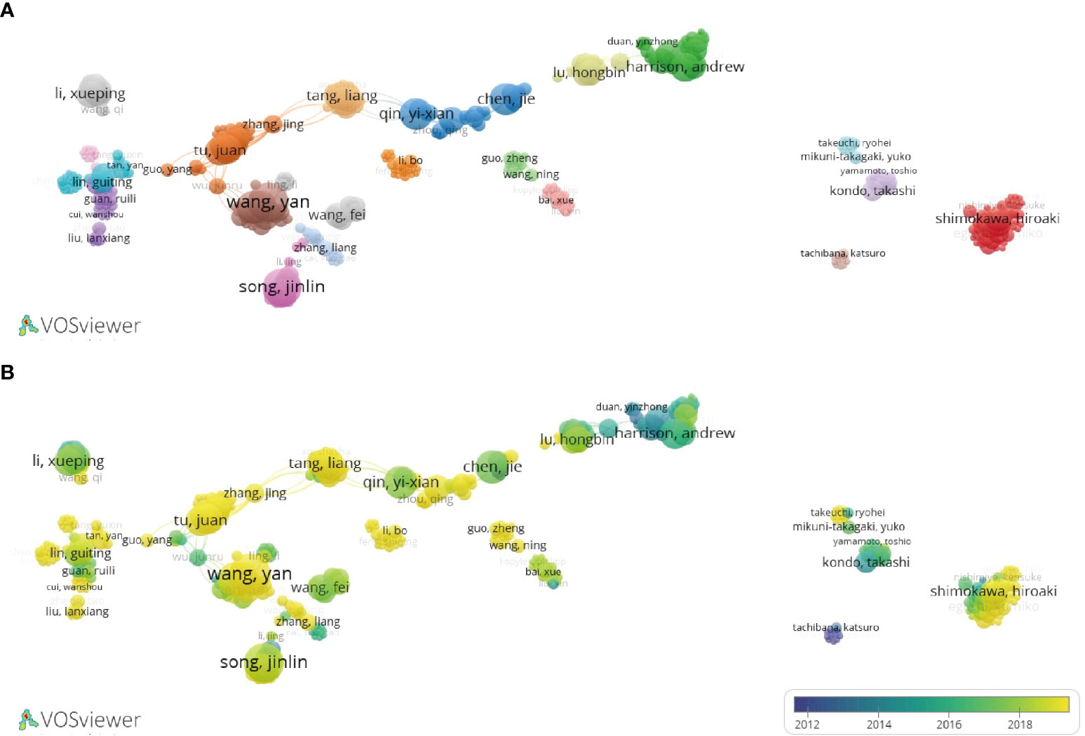
Figure 5 Collaboration network diagram of scholars in LIPUS literature publication. (A) Collaborative relationships in research in this field among different scholars. (B) According to the time trend analysis of the relationship between cooperation among scholars and years, the lighter the color represents the closer the year in which their cooperative relationship was developed.
3.6 Analysis of highly influential journals
This study indicates that a total of 292 journals have published research on LIPUS. The results show that the journal with the highest output is ULTRASOUND IN MEDICINE AND BIOLOGY (n=60). It is followed by ULTRASONICS (n=21), SCIENTIFIC REPORTS (n=18) and JOURNAL OF ULTRASOUND IN MEDICINE (n=18) (Table 4). ULTRASOUND IN MEDICINE AND BIOLOGY is also the most cited journal, with a total of 744 citations. This indicates that these journals have strong academic influence in the field of LIPUS.
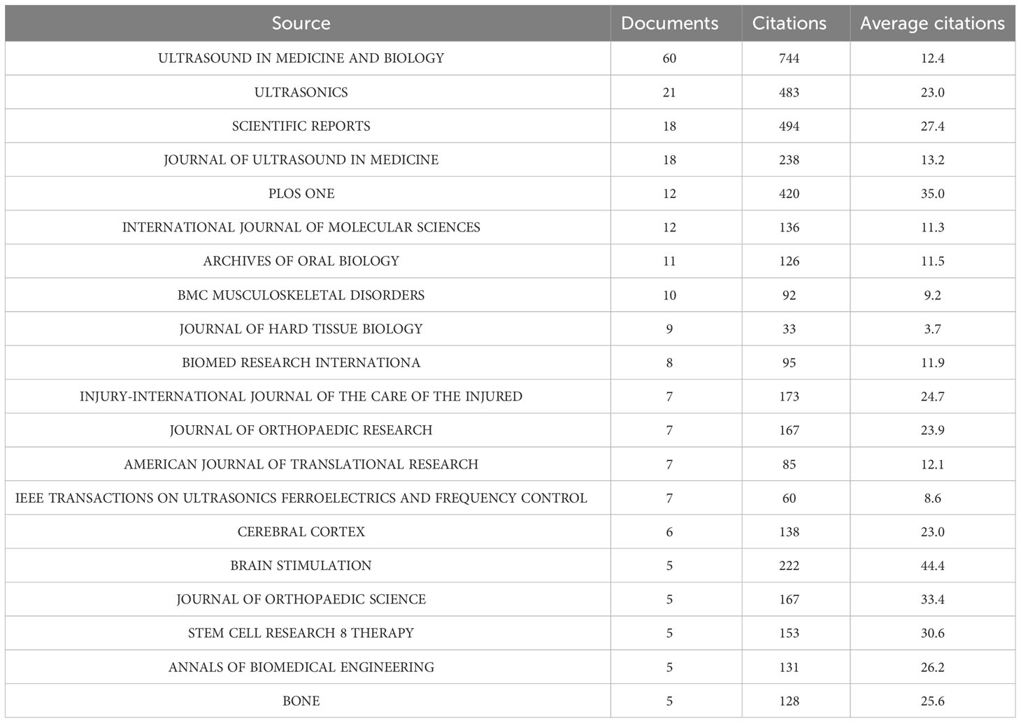
Table 4 The top 20 journals with the most LIPUS publications from 2012 to 2022.
3.7 Analysis of co-citation networks
We employed VOSviewer, a visualization software tool, to construct a co-citation network of references that encompasses 19,325 individual references across 3,832 distinct academic journals. Larger nodes represent references with higher citation frequencies, and thicker lines represent higher frequencies of co-citation. These co-cited articles can be divided into three main clusters (Figure 6A) and are mainly published in journals such as ULTRASOUND IN MEDICINE AND BIOLOGY, BONE, and JOURNAL OF ORTHOPAEDIC RESEARCH (Figure 6B).
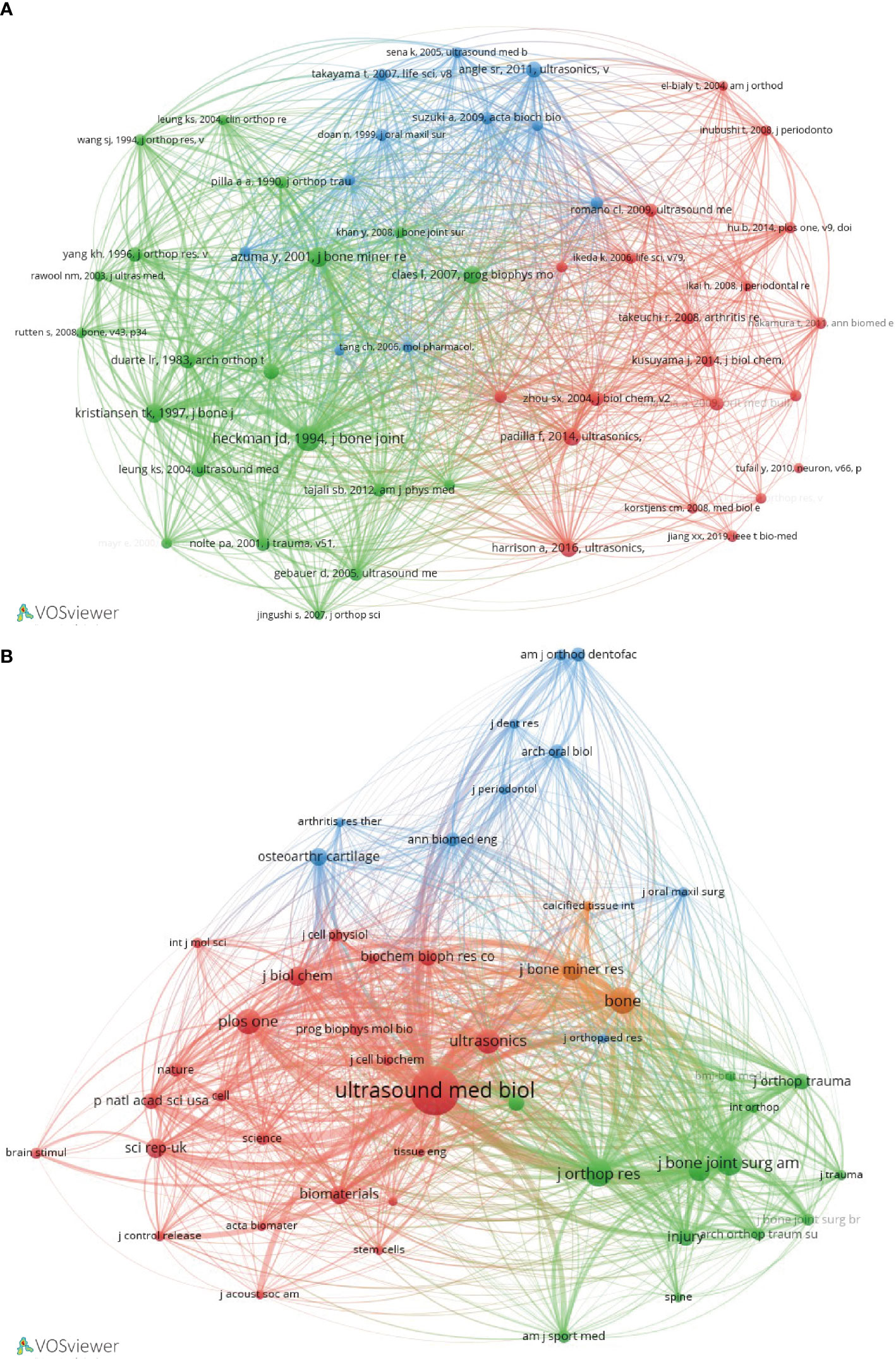
Figure 6 Co-citation network analysis of LIPUS literature. (A) Network cluster diagram of co-cited literature. (B) Cluster diagram of the main journals in which co-cited literatures were published.
3.8 Analysis of keywords
From the retrieved records, a total of 3,004 keywords were extracted. We selected 67 keywords with a frequency of more than 15 occurrences, which can be divided into four clusters (Figure 7A). The top five keywords are low-intensity pulsed ultrasound (195 times), expression (141 times), LIPUS (125 times), stimulation (117 times), and differentiation (103 times), which encompass the most core keywords in each cluster (Figure 7B).
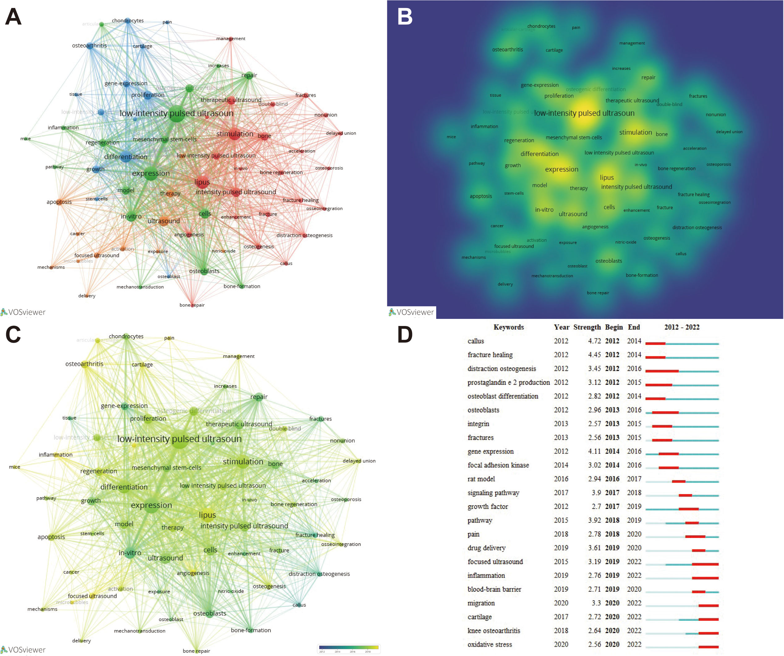
Figure 7 Analysis of keywords and burst word. (A) Cluster analysis of keywords and results. (B) Presentation of keyword occurrence frequency in the form of density plot. (C) According to the time trend analysis of the relationship between keywords and years, the lighter the color represents the closer the year in which the keyword appeared. (D) Identification of the most relevant keywords to LIPUS through burst words analysis.
The identification of burst words, defined as keywords that exhibit a significant rise in frequency during a condensed time frame, is indicative of research hotspots within the respective field. Through an examination of time trends and burst words, one can gain insight into overarching research trends and developmental directions. Through temporal trend analysis and burst word analysis, we found that migration, cartilage, knee osteoarthritis, and oxidative stress related to LIPUS have become research hotspots since 2020 and have a continuous trend, and may continue to lead the direction of LIPUS research in the future (Figures 7C, D).
3.9 Analysis of the latest research hotspots on LIPUS in endocrine and metabolic diseases
Currently, research on the application of LIPUS in endocrine and metabolic diseases is steadily increasing, but there is still a lack of systematic summarization. Therefore, we conducted further analysis on hypopituitarism, thyroid diseases, adrenal cortical diseases, pheochromocytoma, gout, osteoporosis, and other typical endocrine and metabolic diseases based on preliminary search results. Through further screening, we obtained a total of 326 relevant keywords, with 31 keywords appearing three or more times (Figure 8A). Among these keywords, “osteoporosis” had the highest frequency of occurrence. Time trend analysis reveals that osteoporosis serves as an early and mature example of LIPUS application, from basic research to clinical studies. And, in recent years, emerging areas such as stem cells and proliferation have become new research directions for LIPUS (Figure 8B).
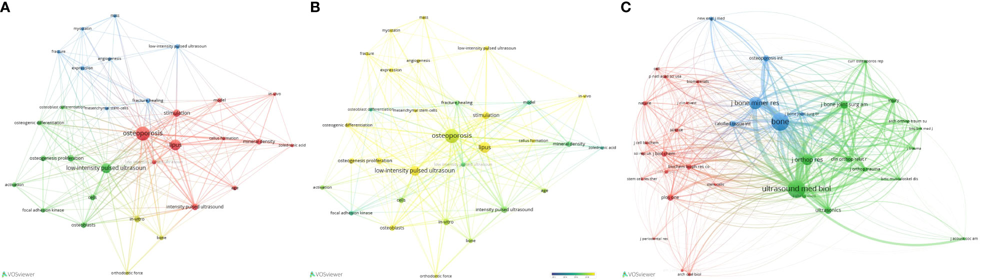
Figure 8 Analysis of keywords and co-citation network analysis of LIPUS in Endocrine and Metabolic Diseases. (A) Cluster analysis of keywords and results. (B) According to the time trend analysis of the relationship between keywords and years, the lighter the color represents the closer the year in which the keyword appeared. (C) Cluster diagram of the main journals in which co-cited literatures were published.
The co-citation cluster analysis revealed that the selected articles cited a total of 1697 references, which were derived from 658 journals. Among these journals, 37 had been cited more than 10 times. ULTRASOUND IN MEDICINE AND BIOLOGY (n=105), BONE (n=105), JOURNAL OF BONE AND MINERAL RESEARCH (n=69), JOURNAL OF ORTHOPAEDIC RESEARCH (n=59), and JOURNAL OF BONE and JOINT SURGERY-AMERICAN VOLUME (n=40) were the top five most frequently cited journals, primarily focused on ultrasound and orthopedic fields (Figure 8C).
4 Discussion
In this global study, we conducted bibliometric analysis to summarize the collaboration network, research trends and hotspots, as well as future directions of LIPUS research. A total of 655 records were retrieved and published in 292 journals by 2973 authors from 794 institutions in 46 countries/regions. This is the first systematic study using bibliometric methods to analyze the development patterns of LIPUS over the past decade and analyze its research in the field of endocrine and metabolic diseases, which will guide its future development direction.
The results of this study indicate that China has the highest number of publications (n=295), followed by the United States (n=125), Japan (n=112), Canada (n=55), and South Korea (n=25), suggesting that LIPUS-related research is still relatively dominated by developed countries, which also reflects the correlation between scientific research, economic investment, and national social economy. The citation frequency of published articles can represent the academic influence of the results to a certain extent. Currently, China (n=4531), the US (n=2332), Japan (n=1555), and Canada (n=1024) have the highest total citation frequencies, consistent with the publication ranking. However, in terms of the average citation frequency of published articles, the top five countries are the Netherlands, France, Italy, the UK, and Germany, indicating that these countries may have higher academic influence in this field.
Regarding the distribution of patterns of institutions publishing relevant research and their national origins, the overall trend is consistent. In terms of the number of publications, four of the top five institutions are from China, namely Chongqing Medical University (n=52), Chinese Academy of Sciences (n=28), China Medical University (n=19), and Nanjing Medical University (n=17), while the other is the University of Alberta in Canada (n=34). Moreover, Chongqing Medical University has the highest total citation frequency of published articles (n=698) worldwide. Scholars planning to conduct LIPUS-related research should pay close attention to the academic achievements of this institution in the future.
In terms of author distribution, Tarek El-Bialy ranks first with 21 publications, focusing on the application of LIPUS in oral medicine-related areas such as the maxilla, mandible, alveolar bone, and orthodontics (19–24). Feng-Yi Yang and Yan Wang follow closely with 17 publications each. Feng-Yi Yang has the highest total citation frequency and primarily focuses on combining LIPUS to treat neurological disorders. He proved that LIPUS stimulation can be a potential therapeutic method by accelerating myelin regeneration through weakening glial cell activation, enhancing mature oligodendrocyte density, and increasing the production of BDNF (25). Abdominal LIPUS stimulation can combat neuroinflammation by enhancing tight junction protein levels and inhibiting colonic inflammation (26). LIPUS stimulation can serve as a potential therapeutic tool against Parkinson’s disease, possibly combined with its neural regenerative and protective effects (27, 28), and has therapeutic effects on erectile dysfunction caused by cavernous nerve injury (29). The scholar with the highest average citation frequency is Professor Andrew Harrison from The Chinese University of Hong Kong. His main academic contributions include exploring the effects of different therapeutic ultrasound intensities on rat fracture healing (30), demonstrating the positive effect of far-field LIPUS on stimulating bone cells and promoting mechanical transmission between bone and osteoblasts (31), summarizing the patterns and mechanisms of LIPUS in repairing fractures and conducting clinical trials of LIPUS in patients with non-healing chronic fractures (32–35). Scholars planning to study relevant research on LIPUS in orthopedics should pay attention to the research progress of Professor Andrew Harrison in the future.
LIPUS has significant clinical value in the treatment of various diseases and has been recognized by numerous high-level journals and editors. Nearly 300 journals have published reports related to LIPUS, with the top three journals in terms of number of published studies being ULTRASOUND IN MEDICINE AND BIOLOGY, ULTRASONICS, and SCIENTIFIC REPORTS, which also have the highest total citation frequencies. The top three journals in terms of average citation frequency are BRAIN STIMULATION, PLOS ONE, and JOURNAL OF ORTHOPAEDIC SCIENCE.
High-frequency keywords and burst words help scholars clarify future research directions in related fields. After keyword network and burst word analysis, we found that LIPUS-related research has gradually progressed from previous growth factors (36, 37), fracture healing (38–41), and integrins (42–44) to fields such as oxidative stress (45), regeneration (46, 47), and tumors (48–50). This suggests that the exploration of molecular biological mechanisms for LIPUS is gradually deepening, and its application areas are gradually expanding to major diseases such as cancer, proving once again the broad application prospects and clinical value of LIPUS.
The value of LIPUS in endocrine and metabolic diseases has attracted increasing attention. Han et al. found that LIPUS could increase testosterone levels in animal models and alleviate Leydig cell aging in vitro (51). Furthermore, combining LIPUS with tadalafil effectively treats erectile dysfunction in patients with type 2 diabetes (52), and promotes nerve regeneration and improves erectile function by enhancing Schwann cell proliferation, migration, and expression of neurotrophic factors such as nerve growth factor (53). In terms of bone metabolism, Wu et al. have demonstrated that LIPUS can accelerate osteoblast differentiation and promote bone formation in a rat model of osteoporosis (54), and its ultrasound intensity is closely related to its therapeutic effect on osteoporosis (55, 56). Diabetes is a typical disease caused by glucose metabolism disorder, with a long course and many complications. LIPUS has unique effects in regulating glucose metabolism, and has been extensively studied. It has been shown to prevent muscle atrophy in rats with type 1 diabetes (57), and lower blood pressure in hypertensive patients with type 2 diabetes (58). LIPUS shows potential as a new therapy for type 2 diabetes (59). Our study stands at the forefront of summarizing the role and mechanisms of LIPUS from the perspective of endocrine and metabolic diseases, which will facilitate further research in this field by scholars in the future. However, our search has also revealed that, although research on LIPUS in this field is gradually gaining momentum, there is still significant room for further exploration, particularly in relation to specific diseases. The search results indicate that the most well-established application of LIPUS is in the study of osteoporosis. It has progressed from preliminary research on cellular and molecular mechanisms to practical clinical applications, and has shown promising therapeutic effects. There are also relevant studies on diabetes-related osteoporosis, muscle atrophy, erectile dysfunction, among others. However, research on specific diseases such as thyroid disorders and gout remains limited, with most studies focusing on the cellular and molecular levels, still some distance away from clinical application. However, it is well known that many endocrine and metabolic diseases are influenced by hormonal actions or metabolic disruptions. The specific regulatory mechanisms of LIPUS on substances such as glucose, lipids, hormones, and bone have been extensively documented, providing a foundation for its future clinical applications and driving advancements in the field.
Undoubtedly, this study, like other bibliometric studies, has certain limitations. Firstly, the data source mainly comes from the core dataset of WoSCC and lacks other databases, which may result in some omissions in the data we included. Secondly, the language type set for this study is only limited to English, which may cause relevant research in other languages to be missed. In addition, some newly published research results may be missed due to lack of inclusion in the database. Therefore, in the future, we will continue to update the data, pay attention to other non-English literature reports, and the latest published research.
5 Conclusion
Through comprehensive bibliometric analysis, we systematically analyzed global research on LIPUS over the past decade. Overall, the number of publications showed a fluctuating upward trend and the annual publication volume has now stabilized at a high level. In recent years, China and the United States have made the greatest contributions to LIPUS research, and their cooperation and exchanges have been the closest. The research interest in LIPUS for endocrine and metabolic diseases is steadily increasing, with a particular focus on areas such as regulation of glucose metabolism, lipid metabolism, and bone metabolism. Furthermore, LIPUS has shown promising applications in various diseases, with osteoporosis serving as a notable example. Based on the current development trend, it is expected that research on LIPUS will continue to increase globally, and exploring the related molecular mechanisms of LIPUS and expanding its clinical applications will be the future direction of research, especially in the field of endocrine and metabolic diseases.
Data availability statement
The original contributions presented in the study are included in the article/supplementary material. Further inquiries can be directed to the corresponding author.
Author contributions
GC designed the whole study, analyzed the data and drafted the manuscript. HN revised the manuscript. All authors read and approved the final manuscript. All authors contributed to the article and approved the submitted version.
Funding
This work was supported by the National Natural Science Foundation of China under grant numbers 82071750; Taishan Scholar Program of Shandong Province under grant number tstp20221165.
Acknowledgments
We are grateful to The Affiliated Hospital of Qingdao University for encouraging and supporting this work.
Conflict of interest
The authors declare that the research was conducted in the absence of any commercial or financial relationships that could be construed as a potential conflict of interest.
Publisher's note
All claims expressed in this article are solely those of the authors and do not necessarily represent those of their affiliated organizations, or those of the publisher, the editors and the reviewers. Any product that may be evaluated in this article, or claim that may be made by its manufacturer, is not guaranteed or endorsed by the publisher.
References
1. Xin Z, Lin G, Lei H, Lue TF, Guo Y. Clinical applications of low-intensity pulsed ultrasound and its potential role in urology. Transl Androl Urol (2016) 5(2):255–66. doi: 10.21037/tau.2016.02.04
2. Tan Y, Guo Y, Reed-Maldonado AB, Li Z, Lin G, Xia S-J, et al. Low-intensity pulsed ultrasound stimulates proliferation of stem/progenitor cells: what we need to know to translate basic science research into clinical applications. Asian J Androl (2021) 23(6):602–10. doi: 10.4103/aja.aja_25_21
3. Chen Y, Yang H, Wang Z, Zhu R, Cheng L, Cheng Q. Low-intensity pulsed ultrasound promotes mesenchymal stem cell transplantation-based articular cartilage regeneration via inhibiting the TNF signaling pathway. Stem Cell Res Ther (2023) 14(1):93. doi: 10.1186/s13287-023-03296-6
4. Claes L, Willie B. The enhancement of bone regeneration by ultrasound. Prog Biophys Mol Biol (2007) 93(1-3):384–98. doi: 10.1016/j.pbiomolbio.2006.07.021
5. Qin H, Luo Z, Sun Y, He Z, Qi B, Chen Y, et al. Low-intensity pulsed ultrasound promotes skeletal muscle regeneration via modulating the inflammatory immune microenvironment. Int J Biol Sci (2023) 19(4):1123–45. doi: 10.7150/ijbs.79685
6. Chan Y-S, Hsu K-Y, Kuo C-H, Lee S-D, Chen S-C, Chen W-J, et al. Using low-intensity pulsed ultrasound to improve muscle healing after laceration injury: an in vitro and in vivo study. Ultrasound Med Biol (2010) 36(5):743–51. doi: 10.1016/j.ultrasmedbio.2010.02.010
7. Hu J, Qu J, Xu D, Zhang T, Qin L, Lu H. Combined application of low-intensity pulsed ultrasound and functional electrical stimulation accelerates bone-tendon junction healing in a rabbit model. J Orthop Res (2014) 32(2):204–9. doi: 10.1002/jor.22505
8. Montalti CS, Souza NVCKL, Rodrigues NC, Fernandes KR, Toma RL, Renno ACM. Effects of low-intensity pulsed ultrasound on injured skeletal muscle. Braz J Phys Ther (2013) 17(4):343–50. doi: 10.1590/S1413-35552012005000101
9. Mykoniatis I, Kalyvianakis D, Zilotis F, Kapoteli P, Fournaraki A, Poulios E, et al. Evaluation of a low-intensity shockwave therapy for chronic prostatitis type IIIb/chronic pelvic pain syndrome: a double-blind randomized sham-controlled clinical trial. Prostate Cancer Prostatic Dis (2021) 24(2):370–9. doi: 10.1038/s41391-020-00284-2
10. Chen H, Li Z, Li X, Yang Y, Dai Y, Xie Z, et al. The Efficacy and Safety of Thrice vs Twice per Week Low-Intensity Pulsed Ultrasound Therapy for Erectile Dysfunction: A Randomized Clinical Trial. J Sex Med (2022) 19(10):1536–45. doi: 10.1016/j.jsxm.2022.06.009
11. Müller A, Akin-Olugbade Y, Deveci S, Donohue JF, Tal R, Kobylarz KA, et al. The impact of shock wave therapy at varied energy and dose levels on functional and structural changes in erectile tissue. Eur Urol (2008) 53(3):635–42. doi: 10.1016/j.eururo.2007.06.032
12. Yang B, Li M, Lei H, Xu Y, Li H, Gao Z, et al. Low intensity pulsed ultrasound influences the myogenic differentiation of muscle satellite cells in a stress urinary incontinence rat model. Urology (2019) 123:297.e1–.e8. doi: 10.1016/j.urology.2018.09.020
13. Tang H, Liu Y, Fan Y, Li C. Therapeutic effects of low-intensity pulsed ultrasound on premature ovarian insufficiency. Ultrasound Med Biol (2021) 47(8):2377–87. doi: 10.1016/j.ultrasmedbio.2021.04.022
14. Qin Y, Zhao X, Dong X, Liu J, Wang L, Wu X, et al. Low-intensity ultrasound promotes uterine involution after cesarean section: the first multicenter, randomized, controlled clinical trial. Int J Hyperthermia (2022) 39(1):181–9. doi: 10.1080/02656736.2022.2025924
15. Agarwal A, Durairajanayagam D, Tatagari S, Esteves SC, Harlev A, Henkel R, et al. Bibliometrics: tracking research impact by selecting the appropriate metrics. Asian J Androl (2016) 18(2):296–309. doi: 10.4103/1008-682X.171582
16. Wu F, Gao J, Kang J, Wang X, Niu Q, Liu J, et al. Knowledge mapping of exosomes in autoimmune diseases: A bibliometric analysis (2002-2021). Front Immunol (2022) 13:939433. doi: 10.3389/fimmu.2022.939433
17. Zhang L, Bao B, Guo J, Qin Z, Huang H, Chen L, et al. Current status and prospects of diabetes mellitus induced erectile dysfunction: A bibliometric and visualization study. Front Endocrinol (Lausanne) (2023) 14:1168744. doi: 10.3389/fendo.2023.1168744
18. Liang X, He H, Zeng H, Wei L, Yang J, Wen Y, et al. The relationship between polycystic ovary syndrome and coronary heart disease: a bibliometric analysis. Front Endocrinol (Lausanne) (2023) 14:1172750. doi: 10.3389/fendo.2023.1172750
19. Al-Daghreer S, Doschak M, Sloan AJ, Major PW, Heo G, Scurtescu C, et al. Effect of low-intensity pulsed ultrasound on orthodontically induced root resorption in beagle dogs. Ultrasound Med Biol (2014) 40(6):1187–96. doi: 10.1016/j.ultrasmedbio.2013.12.016
20. Tanaka E, Kuroda S, Horiuchi S, Tabata A, El-Bialy T. Low-intensity pulsed ultrasound in dentofacial tissue engineering. Ann BioMed Eng (2015) 43(4):871–86. doi: 10.1007/s10439-015-1274-y
21. Kaur H, El-Bialy T. Shortening of overall orthodontic treatment duration with low-intensity pulsed ultrasound (LIPUS). J Clin Med (2020) 9(5):1303. doi: 10.3390/jcm9051303
22. Bahammam M, El-Bialy T. Effect of low-intensity pulsed ultrasound (LIPUS) on alveolar bone during maxillary expansion using clear aligners. BioMed Res Int (2022) 2022:4505063. doi: 10.1155/2022/4505063
23. El-Bialy T, Farouk K, Carlyle TD, Wiltshire W, Drummond R, Dumore T, et al. Effect of low intensity pulsed ultrasound (LIPUS) on tooth movement and root resorption: A prospective multi-center randomized controlled trial. J Clin Med (2020) 9(3):804. doi: 10.3390/jcm9030804
24. Kaur H, Uludağ H, Dederich DN, El-Bialy T. Effect of increasing low-intensity pulsed ultrasound and a functional appliance on the mandibular condyle in growing rats. J Ultrasound Med (2017) 36(1):109–20. doi: 10.7863/ultra.15.06063
25. Yang F-Y, Huang L-H, Wu M-T, Pan Z-Y. Ultrasound neuromodulation reduces demyelination in a rat model of multiple sclerosis. Int J Mol Sci (2022) 23(17):10034. doi: 10.3390/ijms231710034
26. Yang F-Y, Chan W-H, Gao C-Y, Zheng Y-T, Ke C-H. Transabdominal ultrasound alleviates LPS-induced neuroinflammation by modulation of TLR4/NF-κB signaling and tight junction protein expression. Life Sci (2023) 325:121769. doi: 10.1016/j.lfs.2023.121769
27. Song W-S, Sung C-Y, Ke C-H, Yang F-Y. Anti-inflammatory and neuroprotective effects of transcranial ultrasound stimulation on Parkinson's disease. Ultrasound Med Biol (2022) 48(2):265–74. doi: 10.1016/j.ultrasmedbio.2021.10.001
28. Sung C-Y, Chiang P-K, Tsai C-W, Yang F-Y. Low-intensity pulsed ultrasound enhances neurotrophic factors and alleviates neuroinflammation in a rat model of Parkinson's disease. Cereb Cortex (2021) 32(1):176–85. doi: 10.1093/cercor/bhab201
29. Chiang P-K, Yang F-Y. A potential treatment of low intensity pulsed ultrasound on cavernous nerve injury for erectile dysfunction. Med Hypotheses (2019) 122:19–21. doi: 10.1016/j.mehy.2018.10.014
30. Fung C-H, Cheung W-H, Pounder NM, de Ana FJ, Harrison A, Leung K-S. Effects of different therapeutic ultrasound intensities on fracture healing in rats. Ultrasound Med Biol (2012) 38(5):745–52. doi: 10.1016/j.ultrasmedbio.2012.01.022
31. Fung C-H, Cheung W-H, Pounder NM, Harrison A, Leung K-S. Osteocytes exposed to far field of therapeutic ultrasound promotes osteogenic cellular activities in pre-osteoblasts through soluble factors. Ultrasonics (2014) 54(5):1358–65. doi: 10.1016/j.ultras.2014.02.003
32. Harrison A, Lin S, Pounder N, Mikuni-Takagaki Y. Mode & mechanism of low intensity pulsed ultrasound (LIPUS) in fracture repair. Ultrasonics (2016) 70:45–52. doi: 10.1016/j.ultras.2016.03.016
33. Zura R, Della Rocca GJ, Mehta S, Harrison A, Brodie C, Jones J, et al. Treatment of chronic (>1 year) fracture nonunion: heal rate in a cohort of 767 patients treated with low-intensity pulsed ultrasound (LIPUS). Injury (2015) 46(10):2036–41. doi: 10.1016/j.injury.2015.05.042
34. Harrison A, Alt V. Low-intensity pulsed ultrasound (LIPUS) for stimulation of bone healing - A narrative review. Injury (2021) 52(Suppl 2):S91–S6. doi: 10.1016/j.injury.2021.05.002
35. Leighton R, Watson JT, Giannoudis P, Papakostidis C, Harrison A, Steen RG. Healing of fracture nonunions treated with low-intensity pulsed ultrasound (LIPUS): A systematic review and meta-analysis. Injury (2017) 48(7):1339–47. doi: 10.1016/j.injury.2017.05.016
36. Toyama Y, K-i S, Tachibana K, Ueno T, Kajimoto H, Yokoyama S, et al. Ultrasound stimulation restores impaired neovascularization-related capacities of human circulating angiogenic cells. Cardiovasc Res (2012) 95(4):448–59. doi: 10.1093/cvr/cvs173
37. Moncion A, Lin M, O'Neill EG, Franceschi RT, Kripfgans OD, Putnam AJ, et al. Controlled release of basic fibroblast growth factor for angiogenesis using acoustically-responsive scaffolds. Biomaterials (2017) 140:26–36. doi: 10.1016/j.biomaterials.2017.06.012
38. Griffin XL, Parsons N, Costa ML, Metcalfe D. Ultrasound and shockwave therapy for acute fractures in adults. Cochrane Database Syst Rev (2014) 2014(6):CD008579. doi: 10.1002/14651858.CD008579.pub3
39. Busse JW, Bhandari M, Einhorn TA, Schemitsch E, Heckman JD, Tornetta P, et al. Re-evaluation of low intensity pulsed ultrasound in treatment of tibial fractures (TRUST): randomized clinical trial. BMJ (2016) 355:i5351. doi: 10.1136/bmj.i5351
40. Poolman RW, Agoritsas T, Siemieniuk RAC, Harris IA, Schipper IB, Mollon B, et al. Low intensity pulsed ultrasound (LIPUS) for bone healing: a clinical practice guideline. BMJ (2017) 356:j576. doi: 10.1136/bmj.j576
41. Schandelmaier S, Kaushal A, Lytvyn L, Heels-Ansdell D, Siemieniuk RAC, Agoritsas T, et al. Low intensity pulsed ultrasound for bone healing: systematic review of randomized controlled trials. BMJ (2017) 356:j656. doi: 10.1136/bmj.j656
42. Xia P, Wang X, Qu Y, Lin Q, Cheng K, Gao M, et al. TGF-β1-induced chondrogenesis of bone marrow mesenchymal stem cells is promoted by low-intensity pulsed ultrasound through the integrin-mTOR signaling pathway. Stem Cell Res Ther (2017) 8(1):281. doi: 10.1186/s13287-017-0733-9
43. Cheng K, Xia P, Lin Q, Shen S, Gao M, Ren S, et al. Effects of low-intensity pulsed ultrasound on integrin-FAK-PI3K/Akt mechanochemical transduction in rabbit osteoarthritis chondrocytes. Ultrasound Med Biol (2014) 40(7):1609–18. doi: 10.1016/j.ultrasmedbio.2014.03.002
44. Xu J-k, Chen H-j, Li X-d, Huang Z-l, Xu H, Yang H-l, et al. Optimal intensity shock wave promotes the adhesion and migration of rat osteoblasts via integrin β1-mediated expression of phosphorylated focal adhesion kinase. J Biol Chem (2012) 287(31):26200–12. doi: 10.1074/jbc.M112.349811
45. Ying S, Tan M, Feng G, Kuang Y, Chen D, Li J, et al. Low-intensity Pulsed Ultrasound regulates alveolar bone homeostasis in experimental Periodontitis by diminishing Oxidative Stress. Theranostics (2020) 10(21):9789–807. doi: 10.7150/thno.42508
46. Ye K, Li Z, Yin Y, Zhou J, Li D, Gan Y, et al. LIPUS-SCs-Exo promotes peripheral nerve regeneration in cavernous nerve crush injury-induced ED rats via PI3K/Akt/FoxO signaling pathway. CNS Neurosci Ther (2023) 00:1–20. doi: 10.1111/cns.14256
47. Acheta J, Stephens SBZ, Belin S, Poitelon Y. Therapeutic low-intensity ultrasound for peripheral nerve regeneration - A schwann cell perspective. Front Cell Neurosci (2021) 15:812588. doi: 10.3389/fncel.2021.812588
48. Carina V, Costa V, Pagani S, De Luca A, Raimondi L, Bellavia D, et al. Inhibitory effects of low intensity pulsed ultrasound on osteoclastogenesis induced in vitro by breast cancer cells. J Exp Clin Cancer Res (2018) 37(1):197. doi: 10.1186/s13046-018-0868-2
49. Kip B, Aydin O. Overcome of cisplatin resistance in ovarian cancer by combination of low-intensity ultrasound and Cisplatin. Curr Drug Delivery (2023) 20(9):1380–90. doi: 10.2174/1567201819666220627100857
50. Song S, Ma D, Xu L, Wang Q, Liu L, Tong X, et al. Low-intensity pulsed ultrasound-generated singlet oxygen induces telomere damage leading to glioma stem cell awakening from quiescence. iScience (2022) 25(1):103558. doi: 10.1016/j.isci.2021.103558
51. Han S, Luo J, Xu S, Zhao L, Yao C, Xu J, et al. Low-intensity pulsed ultrasound alleviates human testicular leydig cell senescence. In Vitro Int J Mol Sci (2022) 24(1):418. doi: 10.3390/ijms24010418
52. Verze P, Capece M, Creta M, La Rocca R, Persico F, Spirito L, et al. Efficacy and safety of low-intensity shockwave therapy plus tadalafil 5 mg once daily in men with type 2 diabetes mellitus and erectile dysfunction: a matched-pair comparison study. Asian J Androl (2020) 22(4):379–82. doi: 10.4103/aja.aja_121_19
53. Li Z, Ye K, Yin Y, Zhou J, Li D, Gan Y, et al. Low-intensity pulsed ultrasound ameliorates erectile dysfunction induced by bilateral cavernous nerve injury through enhancing Schwann cell-mediated cavernous nerve regeneration. Andrology (2023) 11(6):1188–202. doi: 10.1111/andr.13406
54. Wu S, Kawahara Y, Manabe T, Ogawa K, Matsumoto M, Sasaki A, et al. Low-intensity pulsed ultrasound accelerates osteoblast differentiation and promotes bone formation in an osteoporosis rat model. Pathobiology (2009) 76(3):99–107. doi: 10.1159/000209387
55. Sun S, Sun L, Kang Y, Tang L, Qin Y-X, Ta D. Therapeutic effects of low-intensity pulsed ultrasound on osteoporosis in ovariectomized rats: intensity-dependent study. Ultrasound Med Biol (2020) 46(1):108–21. doi: 10.1016/j.ultrasmedbio.2019.08.025
56. Sun S, Tang L, Zhao T, Kang Y, Sun L, Liu C, et al. Longitudinal effects of low-intensity pulsed ultrasound on osteoporosis and osteoporotic bone defect in ovariectomized rats. Ultrasonics (2021) 113:106360. doi: 10.1016/j.ultras.2021.106360
57. Tang L, Li N, Jian W, Kang Y, Yin B, Sun S, et al. Low-intensity pulsed ultrasound prevents muscle atrophy induced by type 1 diabetes in rats. Skelet Muscle (2017) 7(1):29. doi: 10.1186/s13395-017-0145-7
58. Nonogaki K, Yamazaki T, Murakami M, Satoh N, Hazama M, Takeda K, et al. Low-frequency and very low-intensity ultrasound decreases blood pressure in hypertensive subjects with type 2 diabetes. Int J Cardiol (2016) 215:147–9. doi: 10.1016/j.ijcard.2016.04.062
Keywords: low-intensity pulsed ultrasound, bibliometrics, visualization, endocrine and metabolic diseases, global trend
Citation: Chu G and Niu H (2023) Knowledge mapping and global trends in the field of low-intensity pulsed ultrasound and endocrine and metabolic diseases: a bibliometric and visual analysis from 2012 to 2022. Front. Endocrinol. 14:1237864. doi: 10.3389/fendo.2023.1237864
Received: 10 June 2023; Accepted: 21 August 2023;
Published: 05 September 2023.
Edited by:
Yan Wang, Chongqing Medical University, ChinaReviewed by:
Wei Deng, Chongqing Medical University, ChinaQian Ge, The First Affiliated Hospital of Chongqing Medical University, China
Copyright © 2023 Chu and Niu. This is an open-access article distributed under the terms of the Creative Commons Attribution License (CC BY). The use, distribution or reproduction in other forums is permitted, provided the original author(s) and the copyright owner(s) are credited and that the original publication in this journal is cited, in accordance with accepted academic practice. No use, distribution or reproduction is permitted which does not comply with these terms.
*Correspondence: Haitao Niu, bml1aHQwNTMyQDEyNi5jb20=