 Huan Feng
 Wei Peng
 Zhiyao Deng
 Jihong Liu
 Tao Wang
94% of researchers rate our articles as excellent or good
Learn more about the work of our research integrity team to safeguard the quality of each article we publish.
Find out more
REVIEW article
Front. Endocrinol., 09 March 2023
Sec. Reproduction
Volume 14 - 2023 | https://doi.org/10.3389/fendo.2023.1123383
This article is part of the Research TopicMetabolic Factors in Erectile DysfunctionView all 14 articles
Erectile dysfunction (ED), as a common male disease, can seriously reduce the life quality of men and their partners. With the improvement of human living standards, ED is considered to be an important health issue that plagues men. However, it is difficult for existing therapeutic approaches to meet the needs of all patients, so it is necessary to develop novel treatment strategies. Exosomes, as a class of vesicles secreted by cells with bilayer membrane structure, are involved in various physiological and pathological processes in human body and considered to have great therapeutic potentials. This review summarizes the recent advances on exosome therapy with animal models of ED, and proposes the prospect of future research in order to provide a basis for clinical trials and clinical translation.
More than 5,000 years ago, erectile dysfunction (ED) was mentioned in ancient Egyptian scriptures (1). It was defined until 1993 by the National Institutes of Health (NIH) as the persistent or recurrent failure to attain or maintain a sufficient penile erection for successful penetration (2, 3). ED is a widespread medical issue that seriously affects male health, with 150 million men suffering from it in varying degrees worldwide (4, 5). With males over the age of 40 having a higher prevalence of this issue, it is anticipated that there will be more than 322 million cases of ED worldwide by 2025 (1). In the past, it was frequently believed that psychiatric disorders contribute significantly to the development of ED, but many researches have revealed that organic etiology accounts for more than 80% of ED cases. Cardiovascular diseases, diabetes, dyslipidemia, hypogonadism and nerve damage are independent risk factors for ED (6). More importantly, ED is no longer just related to sexual dysfunction, it may also signal potential vascular endothelial dysfunction, acting as an early indicator of cardiovascular disease (7). Selective phosphodiesterase type 5 inhibitors (PDE5i), like sildenafil citrate, have been widely utilized as the first-line treatment for ED since they enhanced erectile function in 63% of patients and were exploited based on the role of nitric oxide (NO) in cavernous smooth muscle relaxation (8). However, because of the complexity of the pathway that regulates penile erection, up to 35% of patients do not respond to the pharmacological therapy (1). As current treatments do not provide maximum benefits to patients, it is crucial to investigate novel strategies for ED treatments (9).
Extracellular vesicles (EVs) are cell-derived membrane structure with a diameter of 40 nm to 1000 nm. Exosomes and microvesicles are two types of EVs that are released from the endosomal system or shed from plasma membrane, respectively (10). Exosomes are nanosized particles with a diameter of roughly 40-160 nm secreted by various cells under physiological or pathological conditions (11). Exosomes have a bilayer structure that is made by the plasma membrane through encapsulating extracellular components and membrane proteins as well as intersecting with other vesicles and organelles (10, 12). Therefore, exosomes contain many constituents such as metabolites, proteins, lipids and nucleic acids (13, 14). Exosomes are particularly heterogeneous population due to their origin from different cells, as well as the notable differences in their size, composition and effect on the function of recipient cells (15, 16). Exosomes are involved in a variety of physiological and pathological processes in the human body (17), and can be exploited as molecular and signal carriers in intercellular communication (18). Exosome biogenesis enables cells to rapidly and selectively remove proteins from the plasma membrane, facilitating procedures like sperm-egg binding (19). Exosomes are linked to the process of viral infection and are involved in the induction of innate and adaptive immune responses (20). Studies on exosomes and diseases have revealed that exosomes may be involved in cardiovascular and metabolic disorders, take a role in the pathogenesis of neurological disorders, and dynamically affect the growth of cancers (11, 21).
Exosomes are frequently investigated in clinical studies. Exosomal non-coding RNAs (ncRNAs) have been revealed to be expressed differently in most human disorders, which paves the way for their potential application as biomarkers in early stage of diseases (22). Exosomes also serve as drug delivery systems (DDS) for the treatment of cancer (23). For instance, exosomes containing cisplatin inhibited the progression of hepatocellular carcinoma (24). In animal experiments, transplanted bone marrow promoted the regeneration of injured β cell by releasing exosomes enriched with miR‐106b‐5p and miR‐222‐3p (25). Exosomal miRNAs produced by mesenchymal stem cells have been found to exhibit anti-atherosclerotic properties (26). Therefore, it is valuable to further explore the application of exosomes in ED patients, which might be caused by diabetes or cardiovascular disease.
In this review, we introduced the biosynthesis and contents of exosomes, and concentrated on the application of exosomes as well as other types of EVs in the treatment of ED.
The exact mechanisms of exosome formation are still unclear, but existing studies suggest that the process of exosome biogenesis may be similar in different cell types. Generally, the biogenesis of exosomes is a continuous dynamic process and consists mainly of two invaginations of plasma membrane and the formation of multivesicular bodies (MVBs) containing intraluminal vesicles (ILVs) (11).
First of all, exosomes originate from the inward budding of plasma membrane (that is the first invagination of plasma membrane) to form early sorting endosomes (ESEs) with cell surface proteins and extracellular constituents (27, 28). In some cases, the generated ESEs may be directly merged with the preformed ESEs (11). Afterwards, ESEs either fuse with the plasma membrane for the recycling of sequestered cargoes or convert into late sorting endosomes (LSEs), which can give rise to MVBs with the involvement of endoplasmic reticulum and Golgi complex (29, 30). During the maturation of MVBs, cytoplasmic cargoes including nucleic acids, proteins, lipids, amino acids and metabolites can enter into LSEs by inward budding of endosomal membrane (that is the second invagination of plasma membrane), which leads to the generation of ILVs (27, 29–31). Finally, some MVBs are degraded by the autophagosome or lysosome to maintain cellular homeostasis, while others fuse with the plasma membrane via the exocytotic pattern to secrete ILVs, which are also considered as exosomes (32, 33). Notably, exosomes derived from other cells can be taken up by the cells and further fused with ESEs (11).
However, in addition to this canonical model, several studies also revealed the other mechanisms involving in the biogenesis of exosomes. Exosomes can be formed immediately by outward budding through the plasma membrane or delayed released by budding through the deep invagination of plasma membrane (17). Electron microscopy experiments in mesenchymal stem cells and human immune cells have confirmed the existence of the above phenomenon (17, 34–36).
Many factors are associated with the process of exosome biogenesis. Endosomal-sorting complex required for transport (ESCRT) machinery is a key regulator for the conversion of ESEs to LSEs/MVBs, while sometimes this process is independent of ESCRT complex (10, 37). ESCRT machinery is composed of four complexes (ESCRT-0, ESCRT-I, ESCRT-II and ESCRT-III) and the associated proteins (VPS4, VTA1, ALIX and TSG101) (29, 31, 38). ESCRT-0 binds to the ubiquitinated cargoes on the membrane of MVBs followed by the recruitment of ESCRT-I and ESCRT-II thus initiating the invagination of endosomal membrane (10, 39, 40). ESCRT-III is responsible for the scission of ILVs into MVBs (10, 39, 40). Moreover, it is reported that Munc13-4, NEH6, heat shock protein αB-crystallin (HSPB5) and Rab GTPases are required for the endosome maturation and exosome release (41–43).
After exosomes are released by donor cells, they either fuse directly with the plasma membrane of recipient cells to transfer cargoes, or interact with the receptors of recipient cells to activate the corresponding signaling pathways, or undergo endocytosis by recipient cells to release cargoes or fuse with the endosomes to take part in the biogenesis of exosomes in recipient cells (11, 27, 31).
Exosomes contain not only extracellular substances and plasma membrane proteins, but also a variety of cytoplasmic substances such as DNAs, RNAs, proteins, lipids and metabolites, some of which can be used as biomarkers of exosome (27, 30, 31). The content of exosomes is not constant and varies widely in different cell microenvironments.
Exosomal proteins include: (i) proteins located on the membrane of exosome such as tetraspanins (CD9, CD63, CD81), integrins, MHC class I, II, glycoproteins and other signaling receptors (tumor necrosis factor (TNF) receptor, transferrin receptor), as well as (ii) proteins located in the lumen of exosome such as HSPs (HSP60, HSP70, HSP90), ESCRT machinery (ALIX, TSG101), cytoskeletal proteins (actin, tubulin), enzymes, growth factors and cytokines (27, 44, 45). For instance, transforming growth factor-beta 1 (TGF-β1) and bone morphogenetic protein 2 (BMP2) were detected in plasma exosomes from gastric cancer patients and gastric cancer cell-derived exosomes, respectively (45). These exosomal cytokines were demonstrated to activate the SMAD or PI3K/AKT signaling pathway and give rise to the differentiation of other cells to fibroblasts in gastric cancer (45). Prostate specific membrane antigens (PSMA; PSMA6 and PSMA7) were also identified in blood exosomes from atherosclerosis patients (46). Under the stimulation of lipopolysaccharide macrophage-derived exosomes contain TNF-α and interleukin-1β (IL-1β), which both regulate the inflammatory responses (47).
Exosomal nucleic acids include DNA, mRNA and non-coding RNA (miRNA, lncRNA and circRNA), among which miRNA is one of the most abundant nucleic acids in exosomes. More and more miRNAs contained in exosomes are identified in scientific researches, and they regulate the function of recipient cells or the expression of target mRNA in recipient cells (22, 32). The overexpression of miR-934 in colorectal cancer cell-derived exosomes induced M2 macrophage polarization, activated PI3K/AKT signaling pathway and decreased the PTEN expression (48). High levels of miR-205 were found in circulating exosomes from ovarian cancer patients promoting the angiogenesis and tumor growth (49). Moreover, it was reported that exosomal miRNA from M2 macrophages such as miR-328 promoted the proliferation of pulmonary interstitial fibroblasts and induced the development of lung fibrosis (50). In addition, lipids including cholesterol, ceramides, sphingomyelin and phosphatidylinositol are also contained in exosomes (27).
Most studies on exosomes have focused on the field of oncology, while the correlation between exosomes and ED is less studied. The investigation of effects of exosome on the amelioration of ED was analyzed for the first time in 2017 (51). Details of exosome therapy studies in ED models are displayed in Figure 1 and Table 1.

Figure 1 Schematic diagram of the molecular mechanism of exosome therapy for ED. CCSMC, corpora cavernosum smooth muscle cell; eNOS: endothelial nitric oxide synthase; nNOS: neuronal nitric oxide synthase; iNOS: inducible nitric oxide synthase; Ang 1, angiotensin 1; Ang 2, angiotensin 2; ANP: atrial natriuretic peptide; BNP: brain natriuretic peptide; TGF-β1, transforming growth factor-β 1; NT3: neurotrophin 3; NGF, nerve growth factor.
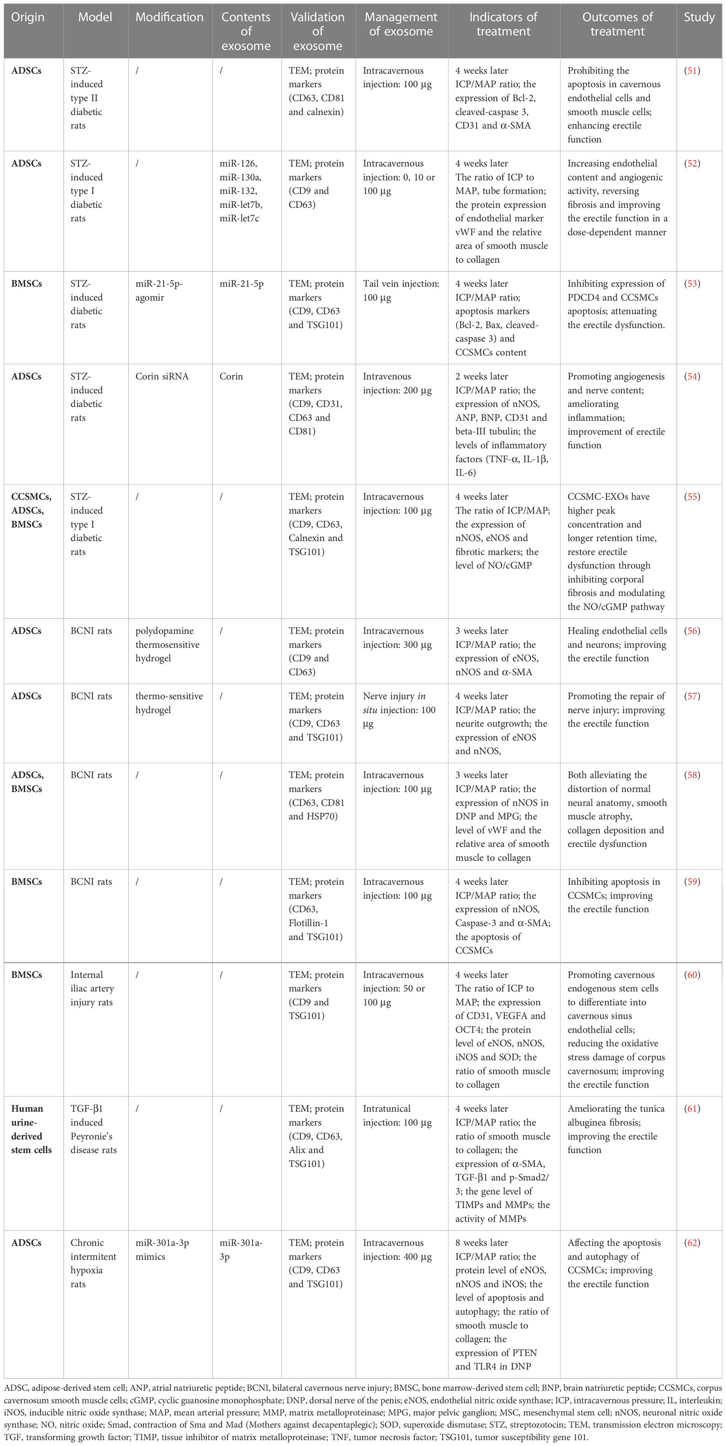
Table 1 Therapeutic strategies of exosomes in rat models of ED.
ED is considered as one of the long-term complications of diabetes, which increases the risk of developing ED by 2.5-fold, and more than 50% of people with diabetes are affected by ED (68). Diabetic patients are mainly manifested with hyperglycemia and insulin resistance, usually accompanied by the metabolic syndrome (obesity, hypertension, dyslipidemia), hypogonadism, cardiovascular diseases and neuropathy (69). These manifestations and comorbidities affect the levels of androgen, inflow of arteries, outflow of veins as well as nerve signaling, and ultimately influence the production of reactive oxygen species, NO, cyclic guanosine monophosphate (cGMP) and nitric oxide synthase (NOS), and the function of endothelial cells and corpus cavernosum smooth muscle cells (CCSMCs) during the erection of penis (69, 70).
Chen et al. established a rat model of type II diabetes and isolated exosomes from adipose-derived stem cells (ADSCs) by ultracentrifugation (51). They found that ADSC-derived exosomes promoted the recovery of erectile function by increasing the ratio of maximal intracavernous pressure (ICP) to mean arterial pressure (MAP), elevating the endothelium and smooth muscle contents and decreasing the apoptosis in cavernous endothelial cells and smooth muscle cells (51). In addition to type 2 diabetes, ED was also induced following type 1 diabetes. Zhu et al. injected type 1 diabetic ED rats with three different doses of ADSC-derived exosomes (0, 10 or 100 μg, respectively) (52). It was demonstrated that exosomes improved the erectile function by reversing fibrosis, increasing endothelial content and angiogenic activity in a dose-dependent manner. Interestingly, they also performed miRNA sequencing on the extracted exosomes and found that some functional miRNAs were contained in these exosomes, including proangiogenic miRNAs (miR-126, miR-130a, miRNA-132) and antifibrotic miRNAs (miR-let7b and miR-let7c). Although the role of these miRNAs in exosomes in the treatment of ED has not been validated in vitro or vivo, these findings revealed the potential mechanisms of exosome therapy in ED, which opened up a new perspective for the future investigation of exosomes in the treatment of ED.
Notably, donor cells can be modified by transgenic methods so that exosomes released by these cells can contain a large number of specific cargoes. For example, bone marrow-derived stem cells (BMSCs) treated with miR-21-5p-agomir resulted in the increase of miR-21-5p in BMSC-derived exosomes, which decreased the expression of target gene programmed cell death 4 (PDCD4) in CCSMCs, attenuating the erectile dysfunction through leading to the proliferation and apoptosis inhibition of these cells (53). Meanwhile, exosomes derived from ADSCs transfected with corin siRNA promoted the neurovascular function and suppressed the levels of inflammatory factors including TNF-α, IL-1β and IL-6 (54). Our group also investigated the therapeutic effects of exosomes in ED. Distinct from the above study, we isolated exosomes from CCSMCs and found that this type of exosomes was more easily retained in the corpus cavernosum and better ameliorated the diabetes-induced erectile dysfunction compared with exosomes from ADSCs and BMSCs (55).
Prostate cancer surgery and other pelvic surgeries often result in damages to the cavernous nerve, which originates from the major pelvic ganglion (MPG) and controls the relaxation and contraction of CCSMCs, thus regulating the erection of the penis (71). It was reported that up to 80% of prostate cancer patients suffer from ED after radical prostatectomy (72). Many molecules and signaling pathways contribute to the development of ED during the injury of cavernous nerve, for instance TGF-β, hydrogen sulfide, NO pathway, RhoA/ROCK pathway or oxidative stress-related pathway (73, 74). The dysregulation of these factors after BCNI leads to the tissue fibrosis, as well as phenotypic transformation and apoptosis of CCSMCs (73).
Exosomes isolated from ADSCs and BMSCs have been applied not only to diabetic ED but also to BCNI-induced ED. Ouyang et al. found that four weeks after injection with exosomes into the corpus cavernosum of BCNI-induced ED rats, the erectile function was obviously improved via inhibiting apoptosis in CCSMCs (59). Similarly, ADSC-derived and BMSC-derived exosomes have been shown to be effective in recovering erectile dysfunction in BCNI rat model (58). Both of them can alleviate the distortion of normal neural anatomy, smooth muscle atrophy and collagen deposition, which impaired the erection of penis.
In order to enhance the uptake of exosomes and the efficacy of treatment, some research teams have developed specific hydrogels and mixed them with exosomes for the treatment of ED. Liang et al. fabricated polydopamine thermosensitive hydrogels, which exhibited sol-gel transition at body temperature and allowed exosomes to be released slowly within two weeks (56). In vivo experiments confirmed that ADSC-derived exosomes loaded within polydopamine thermosensitive hydrogel improved the erectile function by healing the endothelial cells and neurons in penis. On the other side, ADSC-derived exosomes encapsulated into the thermosensitive hydrogel significantly repaired the cavernous nerves injury in rats, thus restoring erectile function (57).
In addition, a study showed that Schwann cell-derived exosomes promoted nerve regeneration of MPG and cavernous nerve with MPG, suggesting that they may provide potential therapeutic options for ED treatment (75). Since this study only involved ex vivo experiments, appropriate in vivo experiments are needed to further confirm the findings.
So far, exosomes have been introduced to treat some other types of ED in addition to diabetic ED and BCNI-induced ED. Vascular ED accounts for a large proportion in elderly patients with ED, which can be caused by atherosclerosis, trauma and surgery. It has been shown that in internal iliac artery injury-induced ED rats, BMSC-derived exosomes promoted cavernous endogenous stem cells to differentiate into cavernous sinus endothelial cells while effectively reduced the oxidative stress damage of corpus cavernosum (60). These findings provided a novel insight and strategy for the clinical treatment of severe arterial injury ED.
Since erectile dysfunction is often observed in patients with obstructive sleep apnea, the researchers developed a rat model of chronic obstructive hypoxia-induced ED. The miR-301a-3p-enriched ADSC-derived exosomes affected the apoptosis and autophagy of CCSMCs by targeting PTEN and TLR4, and ultimately improved erectile function (62). Furthermore, Yang et al. demonstrated that human urine-derived stem cell (HUSC)-derived exosomes ameliorated the fibrosis in tunica albuginea and restored erectile function in Peyronie’s disease rats (61).
Due to the low production of EVs, Kwon et al. developed embryonic stem cell (ESC)-derived EV-mimetic nanovesicles (ESC-NVs), and diabetic mice were received intravenous injection of 0.1, 0.5, 1, 2 or 5 μg ESC-NVs for 2 times, respectively. The results revealed that those ESC-NVs enhanced penile neurovascular regeneration by boosting the expression of angiogenic and neurotrophic factors (63). A team of researchers from South Korea extracted EV-NVs from cavernous pericytes, and they found that these NVs can significantly promote neurovascular regeneration and ameliorate erectile dysfunction in both diabetic and BCNI-induced ED rats (64, 65). The studies also compared the efficacy of different doses of NVs and found that the higher the dose, the better the recovery of pathological changes and erectile dysfunction. Moreover, EVs derived from HUSC (HUSC-EVs) have been proven to ameliorate erectile dysfunction effectively (66, 67). In contrast to the conventional intracavernous or intravenous injection, Zhuang et al. mixed HUSC-EVs with hyaluronic acid and smeared them on the glans of the rats several times (67). The results showed that this administration can also improve apoptosis, angiogenesis, and smooth muscle regeneration as the conventional injection Table 2.
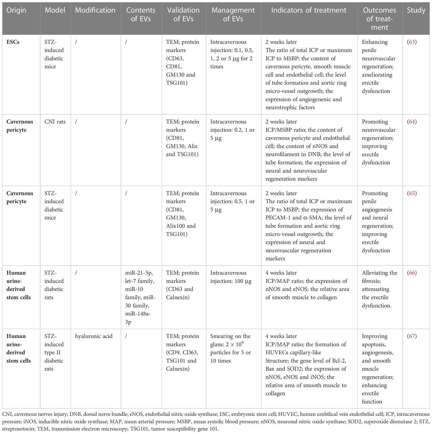
Table 2 Therapeutic strategies of other subsets of EVs in rat models of ED.
Basic and preclinical researches have shown that exosomes have the potential to treat ED. Since the research in this field is still at a preliminary stage, there are no ongoing or completed clinical trials. Although exosomes exhibit good therapeutic effects in rat and mouse, rodent models are insufficient to predict human clinical outcomes. Exosome therapy in ED needs to be validated in higher-order animals that more closely mimic the physiological and clinical characteristics of the human body. In fact, a large number of clinical trials based on exosomes have been completed in many other diseases (76–83), so we need to speed up the pace to more comprehensively confirm the therapeutic effect of exosomes on ED and promote the translation from animal studies to clinical trials. In addition, it is worth noting that most of the existing studies only focus on the therapeutic effects of exosomes on ED and related mechanisms, but there are relatively few explorations of exosome contents. In the future, we should take actions to further understand the role of key molecules identified in exosomes in the treatments of ED.
There are still several key issues to be resolved, including long-term safety, optimal source of exosomes, optimal therapeutic method, dose and course, appropriate delivery system and elucidation of the specific mechanism. Most of the studies used exosomes derived from allogeneic cells to treat ED through intracavernous injection. Tail vein injection, intratunical injection and nerve injury in situ injection are also working with exosomes during the treatment of ED, although there are only a few reports in the literature. Considering the low yield of human exosomes, how to obtain sufficient exosomes for future clinical use is worth thinking. The sources of exosomes and therapeutic doses need recognized standards, and the comparison of the effects of exosomes from different sources requires more rigorous evidence to support it. Meantime, the doses of exosomes in previous studies vary greatly, which makes the appropriate dose necessary. Most researchers have injected exosomes at a dose of 100 μg and achieved good results, but this only applies to rodent models. Despite the fact that exosomes do not express MHC proteins and will not cause immune tolerance and cell malignancies (84), it is still unknown whether there will be other adverse advents in the further research owing to the fact that the contents of exosomes have not been fully clarified. While exosomes may not be able to completely reverse the pathological changes in the corpus cavernosum, they can delay the progression of the disease, which is enough to improve sexual function and improve the quality of life. Due to the complexity of ED pathophysiology, combination therapy may be more effective, such as oral medications and physical therapy.
Moreover, exosomes have some applicative advantages. First, exosomes can be stored for a long time at low temperature, which is convenient for storage and transportation (84). Secondly, the protein, nucleic acid and other contents in exosomes are encapsulated by lipids, which is the structural basis for good stability (85). It is important to note that exosomes may circumvent many issues related to ethics. The exosome may be the future of ED treatment, nevertheless research is still at the preliminary stage. Basic researches and clinical trials of exosomes in the treatment of ED lay a solid foundation for the clinical translation of exosome therapy.
HF: Conception, methodology, data investigation & manuscript draft. WP: Conception, data investigation & manuscript draft. ZD: Conception, data investigation & manuscript draft. JL: Manuscript reviewing & project supervision. TW: Manuscript reviewing & project supervision. All authors contributed to the article and approved the submitted version.
This work was supported by the Natural Science Foundation of Shenzhen (JCYJ20210324141404010 & JCYJ20210324134400002) and Hubei Provincial Medical Youth Top Talent Project (2020LJRC009).
The authors declare that the research was conducted in the absence of any commercial or financial relationships that could be construed as a potential conflict of interest.
All claims expressed in this article are solely those of the authors and do not necessarily represent those of their affiliated organizations, or those of the publisher, the editors and the reviewers. Any product that may be evaluated in this article, or claim that may be made by its manufacturer, is not guaranteed or endorsed by the publisher.
1. Shamloul R, Ghanem H. Erectile dysfunction. Lancet (2013) 381(9861):153–65. doi: 10.1016/S0140-6736(12)60520-0
2. NIH Consensus Conference. Impotence. NIH consensus development panel on impotence. JAMA (1993) 270(1):83–90. doi: 10.1001/jama.1993.03510010089036
3. Matz EL, Terlecki R, Zhang Y, Jackson J, Atala A. Stem cell therapy for erectile dysfunction. Sex Med Rev (2019) 7(2):321–8. doi: 10.1016/j.sxmr.2017.12.008
4. Lewis RW, Fugl-Meyer KS, Corona G, Hayes RD, Laumann EO, Moreira ED Jr., et al. Definitions/epidemiology/risk factors for sexual dysfunction. J Sex Med (2010) 7(4 Pt 2):1598–607. doi: 10.1111/j.1743-6109.2010.01778.x
5. Burnett AL, Nehra A, Breau RH, Culkin DJ, Faraday MM, Hakim LS, et al. Erectile dysfunction: AUA guideline. J Urol. (2018) 200(3):633–41. doi: 10.1016/j.juro.2018.05.004
6. Yafi FA, Jenkins L, Albersen M, Corona G, Isidori AM, Goldfarb S, et al. Erectile dysfunction. Nat Rev Dis Primers. (2016) 2:16003. doi: 10.1038/nrdp.2016.3
7. Gandaglia G, Briganti A, Jackson G, Kloner RA, Montorsi F, Montorsi P, et al. A systematic review of the association between erectile dysfunction and cardiovascular disease. Eur Urol. (2014) 65(5):968–78. doi: 10.1016/j.eururo.2013.08.023
8. Xu W, Jiang H, Liu J, Li H. Non-coding RNAs: New dawn for diabetes mellitus induced erectile dysfunction. Front Mol Biosci (2022) 9:888624. doi: 10.3389/fmolb.2022.888624
9. Kim S, Cho MC, Cho SY, Chung H, Rajasekaran MR. Novel emerging therapies for erectile dysfunction. World J Mens Health (2021) 39(1):48–64. doi: 10.5534/wjmh.200007
10. van Niel G, D'Angelo G, Raposo G. Shedding light on the cell biology of extracellular vesicles. Nat Rev Mol Cell Biol (2018) 19(4):213–28. doi: 10.1038/nrm.2017.125
11. Kalluri R, LeBleu VS. The biology, function, and biomedical applications of exosomes. Science (2020) 367(6478):eaau6977. doi: 10.1126/science.aau6977
12. McAndrews KM, Kalluri R. Mechanisms associated with biogenesis of exosomes in cancer. Mol Cancer. (2019) 18(1):52. doi: 10.1186/s12943-019-0963-9
13. Bai R, Li Y, Jian L, Yang Y, Zhao L, Wei M. The hypoxia-driven crosstalk between tumor and tumor-associated macrophages: mechanisms and clinical treatment strategies. Mol Cancer. (2022) 21(1):177. doi: 10.1186/s12943-022-01645-2
14. Zhao K, Li X, Shi Y, Lu Y, Qiu P, Deng Z, et al. Exosomes in the tumor microenvironment of cholangiocarcinoma: current status and future perspectives. J Transl Med (2022) 20(1):117. doi: 10.1186/s12967-022-03294-x
15. Araldi RP, D'Amelio F, Vigerelli H, de Melo TC, Kerkis I. Stem cell-derived exosomes as therapeutic approach for neurodegenerative disorders: From biology to biotechnology. Cells (2020) 9(12). doi: 10.3390/cells9122663
16. Eldaly AS, Mashaly SM, Fouda E, Emam OS, Aglan A, Abuasbeh J, et al. Systemic anti-inflammatory effects of mesenchymal stem cells in burn: A systematic review of animal studies. J Clin Transl Res (2022) 8(4):276–91. doi: 10.18053/jctres.08.202204.003
17. Pegtel DM, Gould SJ. Exosomes. Annu Rev Biochem (2019) 88:487–514. doi: 10.1146/annurev-biochem-013118-111902
18. Li D, Zhang J, Liu Z, Gong Y, Zheng Z. Human umbilical cord mesenchymal stem cell-derived exosomal miR-27b attenuates subretinal fibrosis via suppressing epithelial-mesenchymal transition by targeting HOXC6. Stem Cell Res Ther (2021) 12(1):24. doi: 10.1186/s13287-020-02064-0
19. Bianchi E, Doe B, Goulding D, Wright GJ. Juno Is the egg izumo receptor and is essential for mammalian fertilization. Nature (2014) 508(7497):483–7. doi: 10.1038/nature13203
20. Robbins PD, Morelli AE. Regulation of immune responses by extracellular vesicles. Nat Rev Immunol (2014) 14(3):195–208. doi: 10.1038/nri3622
21. Rajagopal C, Harikumar KB. The origin and functions of exosomes in cancer. Front Oncol (2018) 8:66. doi: 10.3389/fonc.2018.00066
22. Li C, Ni YQ, Xu H, Xiang QY, Zhao Y, Zhan JK, et al. Roles and mechanisms of exosomal non-coding RNAs in human health and diseases. Signal Transduct Target Ther (2021) 6(1):383. doi: 10.1038/s41392-021-00779-x
23. Zhou Y, Zhang Y, Gong H, Luo S, Cui Y. The role of exosomes and their applications in cancer. Int J Mol Sci (2021) 22(22). doi: 10.3390/ijms222212204
24. Maji S, Matsuda A, Yan IK, Parasramka M, Patel T. Extracellular vesicles in liver diseases. Am J Physiol Gastrointest Liver Physiol (2017) 312(3):G194–200. doi: 10.1152/ajpgi.00216.2016
25. Castano C, Novials A, Parrizas M. Exosomes and diabetes. Diabetes Metab Res Rev (2019) 35(3):e3107. doi: 10.1002/dmrr.3107
26. Zheng D, Huo M, Li B, Wang W, Piao H, Wang Y, et al. The role of exosomes and exosomal MicroRNA in cardiovascular disease. Front Cell Dev Biol (2020) 8:616161. doi: 10.3389/fcell.2020.616161
27. Gurung S, Perocheau D, Touramanidou L, Baruteau J. The exosome journey: from biogenesis to uptake and intracellular signalling. Cell Commun Signal (2021) 19(1):47. doi: 10.1186/s12964-021-00730-1
28. Hessvik NP, Llorente A. Current knowledge on exosome biogenesis and release. Cell Mol Life Sci (2018) 75(2):193–208. doi: 10.1007/s00018-017-2595-9
29. Mashouri L, Yousefi H, Aref AR, Ahadi AM, Molaei F, Alahari SK. Exosomes: composition, biogenesis, and mechanisms in cancer metastasis and drug resistance. Mol Cancer. (2019) 18(1):75. doi: 10.1186/s12943-019-0991-5
30. He C, Zheng S, Luo Y, Wang B. Exosome theranostics: Biology and translational medicine. Theranostics (2018) 8(1):237–55. doi: 10.7150/thno.21945
31. Dai J, Su Y, Zhong S, Cong L, Liu B, Yang J, et al. Exosomes: key players in cancer and potential therapeutic strategy. Signal Transduct Target Ther (2020) 5(1):145. doi: 10.1038/s41392-020-00261-0
32. Yue B, Yang H, Wang J, Ru W, Wu J, Huang Y, et al. Exosome biogenesis, secretion and function of exosomal miRNAs in skeletal muscle myogenesis. Cell Prolif. (2020) 53(7):e12857. doi: 10.1111/cpr.12857
33. Jadli AS, Ballasy N, Edalat P, Patel VB. Inside(sight) of tiny communicator: exosome biogenesis, secretion, and uptake. Mol Cell Biochem (2020) 467(1-2):77–94. doi: 10.1007/s11010-020-03703-z
34. Casado S, Lobo M, Paino CL. Dynamics of plasma membrane surface related to the release of extracellular vesicles by mesenchymal stem cells in culture. Sci Rep (2017) 7(1):6767. doi: 10.1038/s41598-017-07265-x
35. Booth AM, Fang Y, Fallon JK, Yang JM, Hildreth JE, Gould SJ. Exosomes and HIV gag bud from endosome-like domains of the T cell plasma membrane. J Cell Biol (2006) 172(6):923–35. doi: 10.1083/jcb.200508014
36. Fang Y, Wu N, Gan X, Yan W, Morrell JC, Gould SJ. Higher-order oligomerization targets plasma membrane proteins and HIV gag to exosomes. PloS Biol (2007) 5(6):e158. doi: 10.1371/journal.pbio.0050158
37. Vietri M, Radulovic M, Stenmark H. The many functions of ESCRTs. Nat Rev Mol Cell Biol (2020) 21(1):25–42. doi: 10.1038/s41580-019-0177-4
38. Subra C, Grand D, Laulagnier K, Stella A, Lambeau G, Paillasse M, et al. Exosomes account for vesicle-mediated transcellular transport of activatable phospholipases and prostaglandins. J Lipid Res (2010) 51(8):2105–20. doi: 10.1194/jlr.M003657
39. Ju Y, Bai H, Ren L, Zhang L. The role of exosome and the ESCRT pathway on enveloped virus infection. Int J Mol Sci (2021) 22(16):9060. doi: 10.3390/ijms22169060
40. Liu J, Ren L, Li S, Li W, Zheng X, Yang Y, et al. The biology, function, and applications of exosomes in cancer. Acta Pharm Sin B (2021) 11(9):2783–97. doi: 10.1016/j.apsb.2021.01.001
41. Messenger SW, Woo SS, Sun Z, Martin TFJ. A Ca(2+)-stimulated exosome release pathway in cancer cells is regulated by Munc13-4. J Cell Biol (2018) 217(8):2877–90. doi: 10.1083/jcb.201710132
42. Pescosolido MF, Ouyang Q, Liu JS, Morrow EM. Loss of christianson syndrome Na(+)/H(+) exchanger 6 (NHE6) causes abnormal endosome maturation and trafficking underlying lysosome dysfunction in neurons. J Neurosci (2021) 41(44):9235–56. doi: 10.1523/JNEUROSCI.1244-20.2021
43. Gangalum RK, Bhat AM, Kohan SA, Bhat SP. Inhibition of the expression of the small heat shock protein alphaB-crystallin inhibits exosome secretion in human retinal pigment epithelial cells in culture. J Biol Chem (2016) 291(25):12930–42. doi: 10.1074/jbc.M115.698530
44. Murao A, Brenner M, Aziz M, Wang P. Exosomes in sepsis. Front Immunol (2020) 11:2140. doi: 10.3389/fimmu.2020.02140
45. Fu M, Gu J, Jiang P, Qian H, Xu W, Zhang X. Exosomes in gastric cancer: roles, mechanisms, and applications. Mol Cancer. (2019) 18(1):41. doi: 10.1186/s12943-019-1001-7
46. Lu M, Yuan S, Li S, Li L, Liu M, Wan S. The exosome-derived biomarker in atherosclerosis and its clinical application. J Cardiovasc Transl Res (2019) 12(1):68–74. doi: 10.1007/s12265-018-9796-y
47. Hu Y, Wang Y, Chen T, Hao Z, Cai L, Li J. Exosome: Function and application in inflammatory bone diseases. Oxid Med Cell Longev (2021) 2021:6324912. doi: 10.1155/2021/6324912
48. Zhao S, Mi Y, Guan B, Zheng B, Wei P, Gu Y, et al. Tumor-derived exosomal miR-934 induces macrophage M2 polarization to promote liver metastasis of colorectal cancer. J Hematol Oncol (2020) 13(1):156. doi: 10.1186/s13045-020-00991-2
49. He L, Zhu W, Chen Q, Yuan Y, Wang Y, Wang J, et al. Ovarian cancer cell-secreted exosomal miR-205 promotes metastasis by inducing angiogenesis. Theranostics (2019) 9(26):8206–20. doi: 10.7150/thno.37455
50. Kishore A, Petrek M. Roles of macrophage polarization and macrophage-derived miRNAs in pulmonary fibrosis. Front Immunol (2021) 12:678457. doi: 10.3389/fimmu.2021.678457
51. Chen F, Zhang H, Wang Z, Ding W, Zeng Q, Liu W, et al. Adipose-derived stem cell-derived exosomes ameliorate erectile dysfunction in a rat model of type 2 diabetes. J Sex Med (2017) 14(9):1084–94. doi: 10.1016/j.jsxm.2017.07.005
52. Zhu LL, Huang X, Yu W, Chen H, Chen Y, Dai YT. Transplantation of adipose tissue-derived stem cell-derived exosomes ameliorates erectile function in diabetic rats. Andrologia (2018) 50(2). doi: 10.1111/and.12871
53. Huo W, Li Y, Zhang Y, Li H. Mesenchymal stem cells-derived exosomal microRNA-21-5p downregulates PDCD4 and ameliorates erectile dysfunction in a rat model of diabetes mellitus. FASEB J (2020) 34(10):13345–60. doi: 10.1096/fj.202000102RR
54. Wang J, Mi Y, Wu S, You X, Huang Y, Zhu J, et al. Exosomes from adipose-derived stem cells protect against high glucose-induced erectile dysfunction by delivery of corin in a streptozotocin-induced diabetic rat model. Regener Ther (2020) 14:227–33. doi: 10.1016/j.reth.2020.03.002
55. Song J, Sun T, Tang Z, Ruan Y, Liu K, Rao K, et al. Exosomes derived from smooth muscle cells ameliorate diabetes-induced erectile dysfunction by inhibiting fibrosis and modulating the NO/cGMP pathway. J Cell Mol Med (2020) 24(22):13289–302. doi: 10.1111/jcmm.15946
56. Liang L, Shen Y, Dong Z, Gu X. Photoacoustic image-guided corpus cavernosum intratunical injection of adipose stem cell-derived exosomes loaded polydopamine thermosensitive hydrogel for erectile dysfunction treatment. Bioact Mater (2022) 9:147–56. doi: 10.1016/j.bioactmat.2021.07.024
57. Liu S, Li R, Dou K, Li K, Zhou Q, Fu Q. Injectable thermo-sensitive hydrogel containing ADSC-derived exosomes for the treatment of cavernous nerve injury. Carbohydr Polym. (2023) 300:120226. doi: 10.1016/j.carbpol.2022.120226
58. Li M, Lei H, Xu Y, Li H, Yang B, Yu C, et al. Exosomes derived from mesenchymal stem cells exert therapeutic effect in a rat model of cavernous nerves injury. Andrology (2018) 6(6):927–35. doi: 10.1111/andr.12519
59. Ouyang X, Han X, Chen Z, Fang J, Huang X, Wei H. MSC-derived exosomes ameliorate erectile dysfunction by alleviation of corpus cavernosum smooth muscle apoptosis in a rat model of cavernous nerve injury. Stem Cell Res Ther (2018) 9(1):246. doi: 10.1186/s13287-018-1003-1
60. Liu Y, Zhao S, Luo L, Wang J, Zhu Z, Xiang Q, et al. Mesenchymal stem cell-derived exosomes ameliorate erection by reducing oxidative stress damage of corpus cavernosum in a rat model of artery injury. J Cell Mol Med (2019) 23(11):7462–73. doi: 10.1111/jcmm.14615
61. Yang Q, Chen W, Han D, Zhang C, Xie Y, Sun X, et al. Intratunical injection of human urine-derived stem cells derived exosomes prevents fibrosis and improves erectile function in a rat model of peyronie's disease. Andrologia (2020) 52(11):e13831. doi: 10.1111/and.13831
62. Liang L, Zheng D, Lu C, Xi Q, Bao H, Li W, et al. Exosomes derived from miR-301a-3p-overexpressing adipose-derived mesenchymal stem cells reverse hypoxia-induced erectile dysfunction in rat models. Stem Cell Res Ther (2021) 12(1):87. doi: 10.1186/s13287-021-02161-8
63. Kwon MH, Song KM, Limanjaya A, Choi MJ, Ghatak K, Nguyen NM, et al. Embryonic stem cell-derived extracellular vesicle-mimetic nanovesicles rescue erectile function by enhancing penile neurovascular regeneration in the streptozotocin-induced diabetic mouse. Sci Rep (2019) 9(1):20072. doi: 10.1038/s41598-019-54431-4
64. Yin GN, Park SH, Ock J, Choi MJ, Limanjaya A, Ghatak K, et al. Pericyte-derived extracellular vesicle-mimetic nanovesicles restore erectile function by enhancing neurovascular regeneration in a mouse model of cavernous nerve injury. J Sex Med (2020) 17(11):2118–28. doi: 10.1016/j.jsxm.2020.07.083
65. Anita L, Yin GN, Hong SS, Kang JH, Gho YS, Suh JK, et al. Pericyte-derived extracellular vesicle-mimetic nanovesicles ameliorate erectile dysfunction via lipocalin 2 in diabetic mice. Int J Biol Sci (2022) 18(9):3653–67. doi: 10.7150/ijbs.72243
66. Ouyang B, Xie Y, Zhang C, Deng C, Lv L, Yao J, et al. Extracellular vesicles from human urine-derived stem cells ameliorate erectile dysfunction in a diabetic rat model by delivering proangiogenic MicroRNA. Sex Med (2019) 7(2):241–50. doi: 10.1016/j.esxm.2019.02.001
67. Zhuang J, Gao P, Chen H, Fang Z, Zheng J, Zhu D, et al. Extracellular vesicles from human urine-derived stem cells merged in hyaluronic acid ameliorate erectile dysfunction in type 2 diabetic rats by glans administration. Andrology (2022) 10(8):1673–86. doi: 10.1111/andr.13293
68. Kouidrat Y, Pizzol D, Cosco T, Thompson T, Carnaghi M, Bertoldo A, et al. High prevalence of erectile dysfunction in diabetes: a systematic review and meta-analysis of 145 studies. Diabetes Med (2017) 34(9):1185–92. doi: 10.1111/dme.13403
69. Defeudis G, Mazzilli R, Tenuta M, Rossini G, Zamponi V, Olana S, et al. Erectile dysfunction and diabetes: A melting pot of circumstances and treatments. Diabetes Metab Res Rev (2022) 38(2):e3494. doi: 10.1002/dmrr.3494
70. Cignarelli A, Genchi VA, D'Oria R, Giordano F, Caruso I, Perrini S, et al. Role of glucose-lowering medications in erectile dysfunction. J Clin Med (2021) 10(11). doi: 10.3390/jcm10112501
71. Jiang N, Wu C, Zhou X, Zhai G, Wu J. Cavernous nerve injury resulted erectile dysfunction and regeneration. J Immunol Res (2021) 2021:5353785. doi: 10.1155/2021/5353785
72. Emanu JC, Avildsen IK, Nelson CJ. Erectile dysfunction after radical prostatectomy: prevalence, medical treatments, and psychosocial interventions. Curr Opin Support Palliat Care (2016) 10(1):102–7. doi: 10.1097/SPC.0000000000000195
73. Song G, Hu P, Song J, Liu J, Ruan Y. Molecular pathogenesis and treatment of cavernous nerve injury-induced erectile dysfunction: A narrative review. Front Physiol (2022) 13:1029650. doi: 10.3389/fphys.2022.1029650
74. Campbell JD, Burnett AL. Neuroprotective and nerve regenerative approaches for treatment of erectile dysfunction after cavernous nerve injury. Int J Mol Sci (2017) 18(8). doi: 10.3390/ijms18081794
75. Peng D, Reed-Maldonado AB, Zhou F, Tan Y, Yuan H, Banie L, et al. Exosome released from schwann cells may be involved in microenergy acoustic pulse-associated cavernous nerve regeneration. J Sex Med (2020) 17(9):1618–28. doi: 10.1016/j.jsxm.2020.05.018
76. Escudier B, Dorval T, Chaput N, Andre F, Caby MP, Novault S, et al. Vaccination of metastatic melanoma patients with autologous dendritic cell (DC) derived-exosomes: results of thefirst phase I clinical trial. J Transl Med (2005) 3(1):10. doi: 10.1186/1479-5876-3-10
77. Morse MA, Garst J, Osada T, Khan S, Hobeika A, Clay TM, et al. A phase I study of dexosome immunotherapy in patients with advanced non-small cell lung cancer. J Transl Med (2005) 3(1):9. doi: 10.1186/1479-5876-3-9
78. Dai S, Wei D, Wu Z, Zhou X, Wei X, Huang H, et al. Phase I clinical trial of autologous ascites-derived exosomes combined with GM-CSF for colorectal cancer. Mol Ther (2008) 16(4):782–90. doi: 10.1038/mt.2008.1
79. Besse B, Charrier M, Lapierre V, Dansin E, Lantz O, Planchard D, et al. Dendritic cell-derived exosomes as maintenance immunotherapy after first line chemotherapy in NSCLC. Oncoimmunology (2016) 5(4):e1071008. doi: 10.1080/2162402X.2015.1071008
80. Squillaro T, Peluso G, Galderisi U. Clinical trials with mesenchymal stem cells: An update. Cell Transplant. (2016) 25(5):829–48. doi: 10.3727/096368915X689622
81. Bagno L, Hatzistergos KE, Balkan W, Hare JM. Mesenchymal stem cell-based therapy for cardiovascular disease: Progress and challenges. Mol Ther (2018) 26(7):1610–23. doi: 10.1016/j.ymthe.2018.05.009
82. Kwon HH, Yang SH, Lee J, Park BC, Park KY, Jung JY, et al. Combination treatment with human adipose tissue stem cell-derived exosomes and fractional CO2 laser for acne scars: A 12-week prospective, double-blind, randomized, split-face study. Acta Derm Venereol (2020) 100(18):adv00310. doi: 10.2340/00015555-3666
83. Chu M, Wang H, Bian L, Huang J, Wu D, Zhang R, et al. Nebulization therapy with umbilical cord mesenchymal stem cell-derived exosomes for COVID-19 pneumonia. Stem Cell Rev Rep (2022) 18(6):2152–63. doi: 10.1007/s12015-022-10398-w
84. Tkach M, Thery C. Communication by extracellular vesicles: Where we are and where we need to go. Cell (2016) 164(6):1226–32. doi: 10.1016/j.cell.2016.01.043
Keywords: erectile dysfunction, exosome, extracellular vesicle, diabetes, cavernous nerve injury
Citation: Feng H, Peng W, Deng Z, Liu J and Wang T (2023) Erectile dysfunction and exosome therapy. Front. Endocrinol. 14:1123383. doi: 10.3389/fendo.2023.1123383
Received: 14 December 2022; Accepted: 27 February 2023;
Published: 09 March 2023.
Edited by:
Xianghu Meng, First Affiliated Hospital, Nanjing Medical University, ChinaReviewed by:
Muhammad Nawaz, University of Gothenburg, SwedenCopyright © 2023 Feng, Peng, Deng, Liu and Wang. This is an open-access article distributed under the terms of the Creative Commons Attribution License (CC BY). The use, distribution or reproduction in other forums is permitted, provided the original author(s) and the copyright owner(s) are credited and that the original publication in this journal is cited, in accordance with accepted academic practice. No use, distribution or reproduction is permitted which does not comply with these terms.
*Correspondence: Jihong Liu, amhsaXVAdGpoLnRqbXUuZWR1LmNu; Tao Wang, dHdhbmdAdGpoLnRqbXUuZWR1LmNu
†These authors have contributed equally to this work
Disclaimer: All claims expressed in this article are solely those of the authors and do not necessarily represent those of their affiliated organizations, or those of the publisher, the editors and the reviewers. Any product that may be evaluated in this article or claim that may be made by its manufacturer is not guaranteed or endorsed by the publisher.
Research integrity at Frontiers
Learn more about the work of our research integrity team to safeguard the quality of each article we publish.