94% of researchers rate our articles as excellent or good
Learn more about the work of our research integrity team to safeguard the quality of each article we publish.
Find out more
ORIGINAL RESEARCH article
Front. Endocrinol., 25 January 2023
Sec. Endocrinology of Aging
Volume 14 - 2023 | https://doi.org/10.3389/fendo.2023.1096723
This article is part of the Research TopicMetabolic Factors in Erectile DysfunctionView all 14 articles
 Yinghao Yin1,2†
 Yihong Zhou1,2†
 Jun Zhou1,2
 Liangyu Zhao1,2
 Hongji Hu1,2
 Ming Xiao1,2
 Bin Niu2
 Jingxuan Peng3
 Yingbo Dai1,2*
 Yuxin Tang1,2*
Introduction: Cisplatin (cis‐diamminedichloroplatinum II, CDDP), a drug widely used for cancer worldwide, may affect erectile function, but its side effects have not received enough attention. To investigate the effect of CDDP on erectile function and its possible mechanism.
Methods: Sprague−Dawley rats were intraperitoneally administered CDDP (CDDP group) or the same volume of normal saline (control group). Erectile function was evaluated after a one-week washout. Then, histologic changes in the corpus cavernosum and cavernous nerve (CN) were measured. Other Sprague-Dawley rats were used to isolate the major pelvic ganglion and cavernous nerve (MPG/CN). RSC96 cells were then treated with CDDP. SA-β-gal staining was used to identify senescent cells, and qPCR was used to detect the senescence-associated secretory phenotype (SASP). Finally, the supernatant of RSC96 cells was used to culture MPG/CN. Erectile function was measured after administration of CDDP. The cavernosum levels of α-SMA, CD31, eNOS, and γ-H2AX, the apoptosis rate and the expression of p16, p21 and p53 in CN were also assayed. The senescent phenotype of RSC96 cells treated with CDDP was identified, and neurite growth from the MPG/CN was photographed and measured.
Results: The CDDP group had a significantly lower ICP/MAP ratio than the control group. Compared to the control group, the CDDP group exhibited significantly lower α-SMA, CD31 and eNOS levels and significantly higher γ-H2AX and apoptosis rates in corpus cavernosum. In addition, CDDP increased some senescence markers p16, p21 and p53 in CN. In vitro, CDDP induced RSC96 senescence and SASP, and the supernatant of senescent cells slowed neurite outgrowth of MPG/CN.
Discussions: CDDP treatment could induce erectile dysfunction, by affecting the content of endothelial and smooth muscle and causing SASP in CN. The results indicate that CDDP treatment should be considered as a risk factor for ED. Clinicians should pay more attention to the erectile function of cancer patients who receive CDDP treatment.
In 2020, there were 19.3 million new cases of cancer and 10.0 million cancer deaths worldwide. The global cancer burden is expected to rise 47% from 2020, with 28.4 million cases in 2040 (1). Given the increasing incidence and prevalence of cancer, chemotherapy is a well-established treatment strategy. The role of chemotherapy will continue to expand and play a more important role in improving both the survival and quality of life of patients. CDDP is a widely used drug to treat various solid cancers such as testicular (2), ovarian (3), head and neck (4), bladder (5), lung (6), cervical cancer (7), lymphomas (8) and several others (9). However, the adverse effects of CDDP limit its effectiveness and become an inherent challenge for its application.
Erectile dysfunction is not usually mentioned as a side effect. However, 42% of colorectal cancer patients who were treated with oxaliplatin had erectile problem (10). Tomoya Kataoka et al. reported that oxaliplatin causes erectile dysfunction in rats due to endothelial dysfunction (11), providing further confirmation that platinum-based drugs can indeed induce ED and that this problem is often overlooked by clinicians. CDDP, a main chemotherapy agent for testicular tumors, has been reported to be associated with erectile dysfunction (12, 13). Moreover, testicular tumors are the most common tumors in young men. Therefore, it is necessary to clarify the relationship between CDDP and erectile function.
In this study, we studied erectile function in rats after administration of CDDP. Illustrating the mechanism, underlying the effect of CDDP on erectile function, will help doctors take measures to protect erectile function in cancer survivors.
RSC96, the Schwann cell line of the rat sciatic nerve, was purchased from Procell (Wuhan, China). Briefly, cells were maintained in DMEM (Gibco Life Technologies, CA, USA) supplemented with 10% fetal bovine serum (Gibco Life Technologies, CA, USA) and 1% penicillin-streptomycin (Gibco Life Technologies, CA, USA) at 37 ˚C in a humidified atmosphere of 5% CO2. CDDP was acquired from APExBIO (Houston, USA) and dissolved in normal saline.
Cell viability was evaluated at different time points (48 hours, 72 hours) using the 3-(4,5-dimethylthiazol-2-yl)-2,5-dip-henyltetrazolium bromide (MTT) assay. RSC96 cells were seeded in 96-well plates at a density of 3×10³ cells/well and cultured for 24 hours. Then, the cells were cotreated with different concentrations of CDDP in the medium. At different time points, 10 μL of MTT was added to each well and incubated at 37°C for an additional 4 hours. Then, the supernatant was carefully removed, and dimethyl sulfoxide was added to each well to dissolve the crystals by gentle agitation for 10 minutes. For each well, the absorbance at 490 nm was estimated on a microplate reader (Bio-Tek, VT, USA).
Cells were seeded in 6-well plates at a density of approximately 20 000 cells/well for 24 hours, and then treated with different concentrations of CDDP for 2 days. Thereafter, the medium was removed and replaced with complete medium (without CDDP) for another 2 days. Then, the cells were fixed and stained using a Senescence β-Galactosidase Staining Kit (Solarbio Life Science, Beijing, China).
For qPCR, total RNA was extracted using the RNA Kit (Omega Bio-Tek, USA) according to the manufacturer’s instructions. Reverse transcription was performed using a HiScript II 1st strand cDNA synthesis kit (Vazyme, China). qPCR was performed on a step-one plus real-time PCR system (Bio-Rad, CA, USA) using ChamQ universal SYBR qPCR master mix (Vazyme, China). All primers were synthesized by Ribobio Co., Ltd. (Guang Zhou, China). Primer sequences are listed in Table 1. β-actin was used as an internal control. The relative levels of RNAs were calculated using the 2-ΔΔCt method.
Bilateral MPG/CN (entire MPG with a 2-mm length CN attached) complexes from each rat were isolated and excised intact. Each MPG/CN complex was placed in a 6-well plate and then covered with 50 μL of Matrigel (Corning, NY, USA) as previously described (14). After incubation at 37 °C for 5 minutes, 2.0 mL of complete culture medium and 2.0 ml supernatant from RSC96 treated with or without CDDP were added according to the grouping. RSC96 cells were treated with CDDP for 2 days, and the medium was removed and replaced with complete culture medium (without CDDP) for another 2 days. The supernatant was then used to culture the MPG/CN complex.
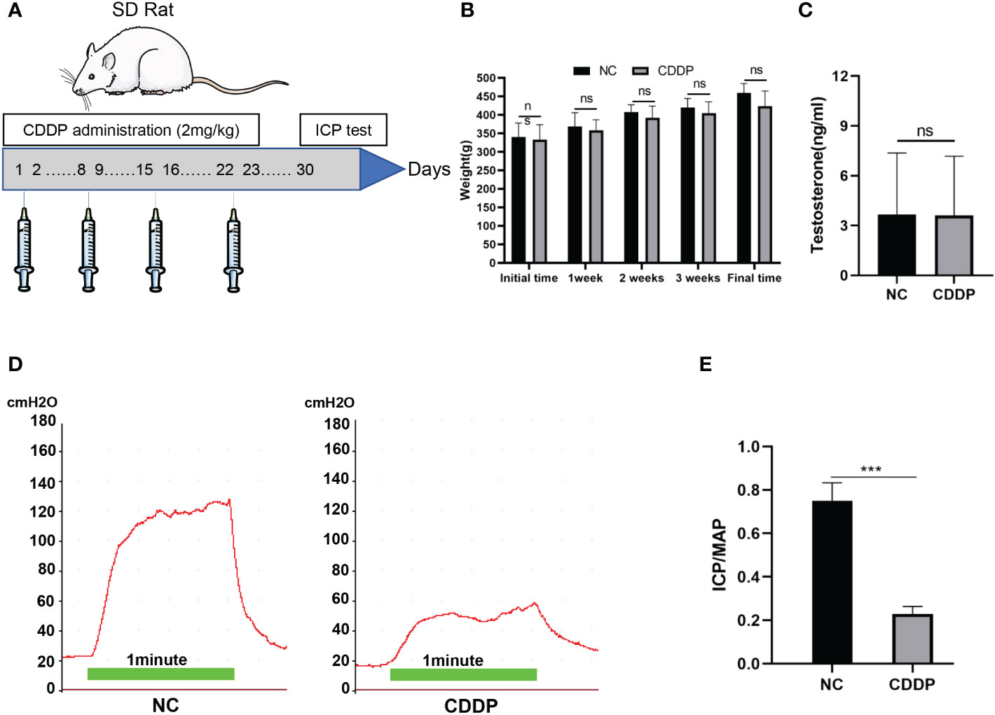
All animal experiments in this study were approved by the Institutional Animal Care and Use Committee of the Fifth Affiliated Hospital of Sun Yat-sen University.Twenty 12-week-old male Sprague−Dawley rats were used for this study. The rats were divided into 2 groups: The CDDP group rats (n = 10) were intraperitoneally administered 2 mg/kg body weight of CDDP on Days 1, 2, 8, 9, 15, 16, 22, and 23 (Figure 1A). And the others (n=10) injected with normal saline as the control group. The dose was based on some scholars’ recommendation about the dose translation from animals to humans (15, 16). The erectile function was evaluated after a one-week washout.

Figure 1 Characterization of the CIED rat model. (A) Experimental scheme for the development of the CIED rat model. CDDP was administered i.p. twice a week for 4 weeks. After a week washout, animals were evaluated to assess erectile function. (B) Changes in rat body weight over time. Initial weight levels in the 2 groups of rats after 7 days of adaptive feeding; final weight after 7 days of washout at 4 weeks. (C) Testosterone levels of each group were shown. (D) Representative images of ICP in response to electrical stimulation of the cavernosal nerve; (E) Results of the ratio of MaxICP to MAP in each group. ns: not significant, ***P < 0.001.
Intracavernous pressure (ICP) was measured by electrical stimulation, as previously reported (17, 18). All rats were anesthetized by injection with pentobarbital (40 mg/kg). Then, the carotid artery was exposed and cannulated with a PE-50 tube which was connected to a pressure transducer to continuously monitor arterial pressure. A 25-gauge needle containing 100 IU/mL heparin solution was inserted into the right penile crura and the other end of the tube was also connected to a pressure transducer to monitor intracavernous pressure (ICP). The cavernous nerve was electrically stimulated at 1 V, 20 Hz, with a pulse width of 5 ms for 1 minute and a 5-minute interval before subsequent stimulation. The ratios of maximal ICP (MaxICP) to mean arterial pressure (MAP) were calculated to evaluate erectile function in vivo.
After the ICP test was completed, clinical needles and vacuum tubes were used to collect blood through the inferior vena cava.The serum was separated immediately and stored at 80°C. The levels of testosterone were assessed using ELISA kits from Solarbio (Beijing, China).
The staining procedures were reported in our previous study (18, 19). Slides containing tissue sections were deparaffinized using a dry oven at 60°C for 30 minutes and in 2 changes of xylene for 10 minutes, and then rehydrated in a series of decreasing concentrations of alcohol. Finally, the slides were washed in tap water. After that, Harris’s hematoxylin reagent was used to stain for 8 minutes. The slides were then destained in 0.5% acid-alcohol for 3 seconds and washed with running water. The slides were counterstained with 0.5% eosin for another 1 minute. After dehydration with different concentrations of alcohol and xylene, the slides were observed with a microscope for histopathologic examination.
The staining procedures were reported in our previous study (18). Tissue sections of the middle part of the penis were stained according to Masson kit instructions (Solarbio, Beijing, China). The collagen tissues (stained blue) of the penile sponge and smooth muscle tissues (stained red) were observed under a light microscope. The cavernous smooth muscle/collagen ratio was analyzed using ImageJ software.
Slides containing tissue sections were deparaffinized with xylene and rehydrated with ethanol. Then, antigen retrieval was performed by placing the slides in Tris/EDTA buffer (Solarbio Life Science, Beijing, China) before heating for 10 minutes. The slides were then treated with endogenous peroxidase blocker for 10 minutes and normal 10% goat serum (Solarbio Life Science, Beijing, China) was utilized for 30 minutes to block nonspecific binding sites. Different antibodies were applied to the slides and incubated overnight at 4 °C. After the hybridization of secondary antibodies, DAPI was used to stain the cell nucleus. The slides were observed using a fluorescence microscope (OLYMPUS, Tokyo, Japan). Antibodies against γ-H2AX, p16, p21 and p53 were obtained from Abclonal Biotechnology (Wuhan, China); CD31 antibody was purchased from Bioworld Technology (Nanjing, China); eNOS antibody was obtained from Abcam Biotechnology (Cambridge, UK). α-SMA antibody was purchased from Affinity (OH, USA).
TUNEL staining was carried out according to previous studies (18). Apoptosis of the corpus cavernosum of each group was detected using a TUNEL apoptosis detection kit (KeyGEN BioTECH, Nanjing, China) following the manufacturer’s instructions. Each sample was randomly selected from 4 fields of view. Cells with green staining were counted as apoptotic, and the ratio of apoptotic cells to the total number of cells in the field of view yielded the rate of apoptosis.
Results were analyzed using GraphPad Prism 8.0 (GraphPad Software, San Diego, CA, USA) and expressed as the mean ± standard deviation (SD). Statistical analyses were performed using the two-tailed Student’s t test. Differences among groups were considered significant at a P value less than 0.05.
As shown in Figures 1B, C, the mean body weight and testosterone levels did not differ significantly between the two groups, but erectile function had obvious difference (Figures 1D, E). Erectile function was assessed by MaxICP and MaxICP/MAP. The results showed that both MaxICP and MaxICP/MAP revealed a significant decrease in the CDDP group compared to control rats.
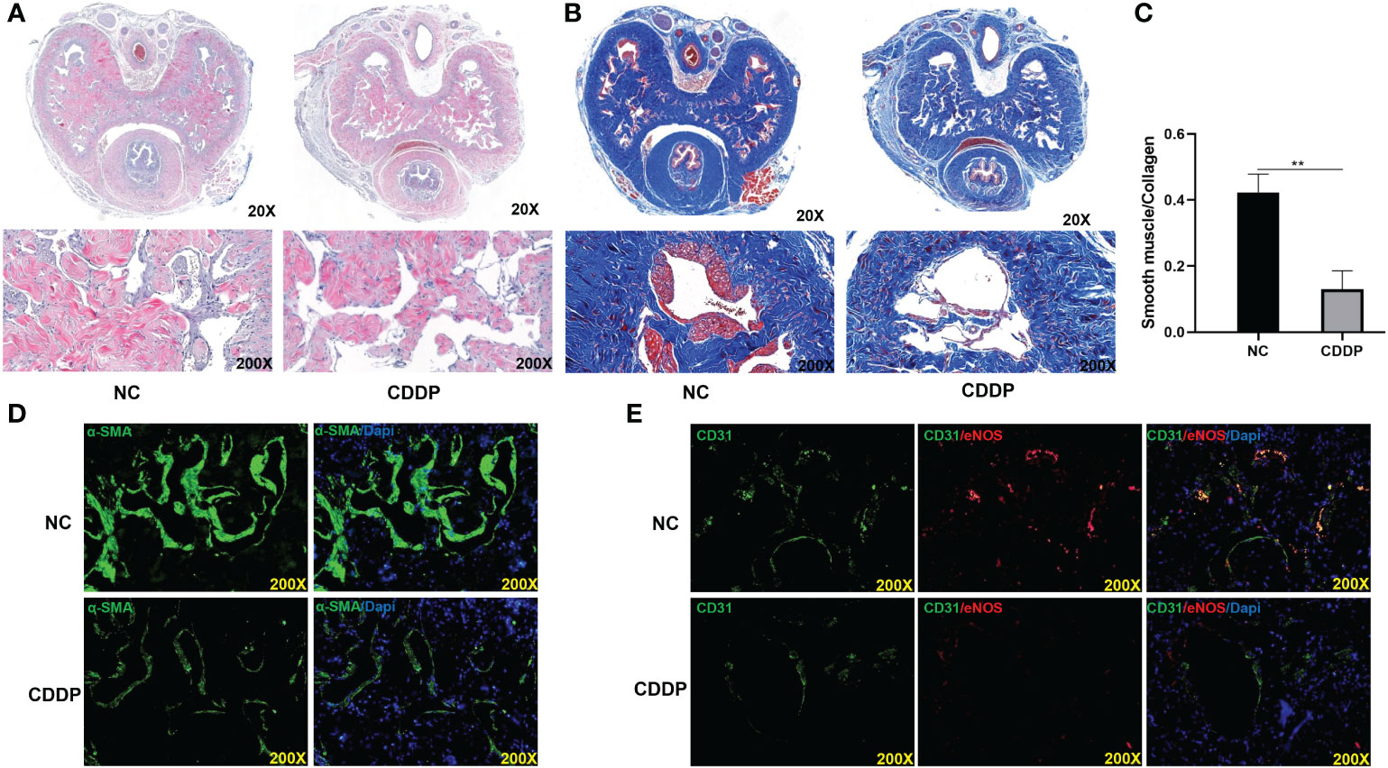
The morphological changes and smooth muscle (SM)-to-collagen ratios of the different groups were detected with H&E and Masson’s trichrome staining, as shown in Figures 2A–C. CDDP significantly caused structural disorders and decreased smooth muscle mass in the corpus cavernosum. Moreover, Immunofluorescence staining with an anti-α-SMA antibody showed a significant reduction in smooth muscle content in CDDP-treated rats compared with normal control rats (Figure 2D). Cavernosal endothelial dysfunction is recognized as a hallmark of the disease pathology. Tomoya Kataoka found that oxaliplatin causes erectile dysfunction in rats due to endothelial dysfunction (11). Consistent with the findings, we found the endothelial cell content and eNOS was severely decreased in the CDDP group (Figure 2E).

Figure 2 Effect of CDDP on smooth muscle and endothelium contents in the corpus cavernosum (A) H&E staining. A thinner smooth muscle layer was found in all CDDP-treated rats; (B) Masson trichrome staining. Smooth muscle manifested as red, and connective tissue was blue. (C) The ratio of smooth muscle to collagen. (D) Representative images of immunofluorescence staining of α-SMA in each group. (E) Representative images of immunofluorescence staining of CD31(green) and eNOS (red) in the corpus cavernosum from age-matched control and CDDP-treated rats. **P < 0.01.
CDDP binds to DNA to cause a biological effect by forming CDDP-DNA adducts and induceing DNA damage response. Phosphorylated H2AX (γ-H2AX) is known to be a marker to investigate the effects on DNA damage (20). As shown in Figure 3A, CDDP treatment significantly increased γ-H2AX levels compared with the control group in the corpus cavernosum. The apoptotic pathway may be triggered in cells after CDDP treatment. We measured the apoptosis level in corpus cavernosum by TUNEL (Figures 3B, C). In the CDDP group, there were a dramatically larger percentage of apoptosis cells than the control group. The aforementioned results indicates that CDDP can induces DNA damage and apoptosis in corpus cavernosum.

Figure 3 CDDP increased DNA damage and apoptosis in the corpus cavernosum. (A) Representative images of immunofluorescence staining of γ-H2AX, a DNA damage marker, in the corpus cavernosum from age-matched control and CDDP-treated rats. (B) Representative images of TUNEL staining in the corpus cavernosum from age matched control and CDDP-treated rats; (C) The apoptotic index, the percentage of apoptotic cells (stained green) of all cells, to quantify the cavernous apoptosis level. Red arrows showed the positive signals, ***P < 0.001.
ED is considered a complication of CDDP-induced peripheral neuropathy (12). Aina Calls found that CDDP-induced peripheral neuropathy was associated with neuronal senescence-like response (21). In senescent cells, the levels of cell cycle inhibitors, including p16, p21, and p53 were augmented. Following CDDP treatment, the expression levels of the p16, p21, and p53 were increased in CN (Figure 4).

Figure 4 CDDP induced senescence in CN. Representative images of immunofluorescence staining of p16 (A), p21 (B) and p53 (C) in each group.
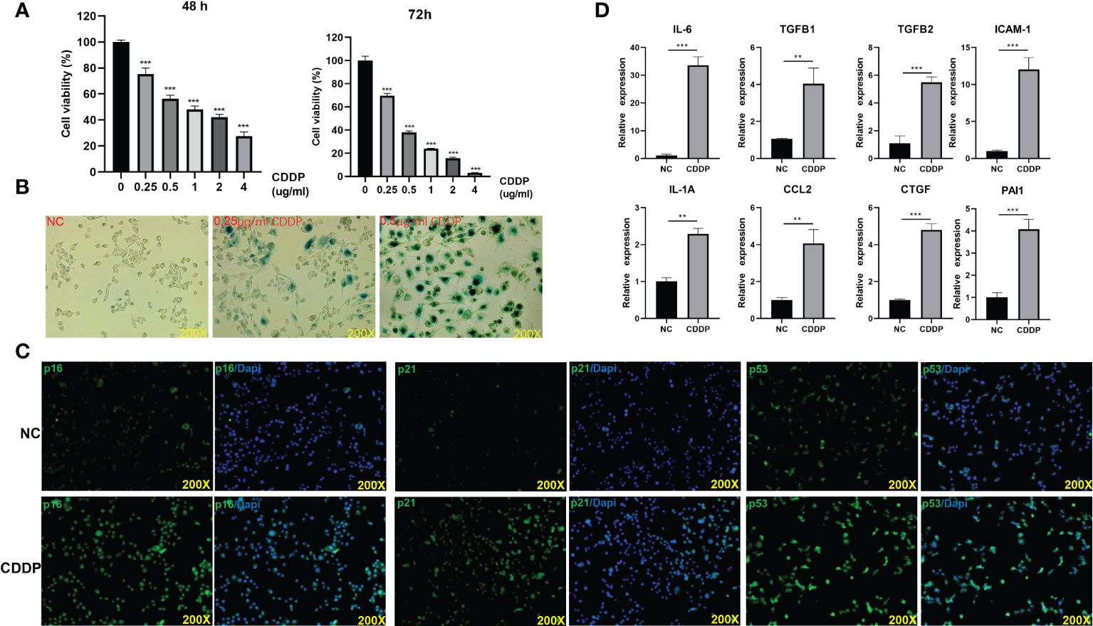
To determine RSC96 cells response to CDDP, an MTT assay was performed. Notably, the proliferation viability of cells decreased with increasing concentration (Figure 5A). The SA-β-gal staining assay confirmed our hypothesis that senescence occurred in RSC96 cells treated with CDDP (Figure 5B). Immunofluorescence illustrated that CDDP upregulated the expression of senescence-related genes: p16, p21 and p53 (Figure 5C). SASP is another typical feature of senescent cells. CDDP promoted the expression of SASP-related genes, IL-6, TGFβ1, ICAM1, TGFβ2, CCL2, IL-1α and CXCL1 (Figure 5D).

Figure 5 CDDP could induce senescence in RSC96 cells. (A) The effect of different concentrations of CDDP on the viability of RSC96 cells was detected by MTT at 48 and 72 hours. (B) RSC96 cells were treated with the indicated concentrations and then stained for SA-β-gal activity at 96 hours. (C) After RSC96 cells were treated with CDDP (0.5 μg/ml), immunofluorescence staining was performed to detect the expression of p53, p21 and p16 in RSC96 cells. (D) qPCR was used to detect the SASP-related genes after RSC96 cells were treated with CDDP (0.5 μg/ml). **P < 0.01, ***P < 0.001.
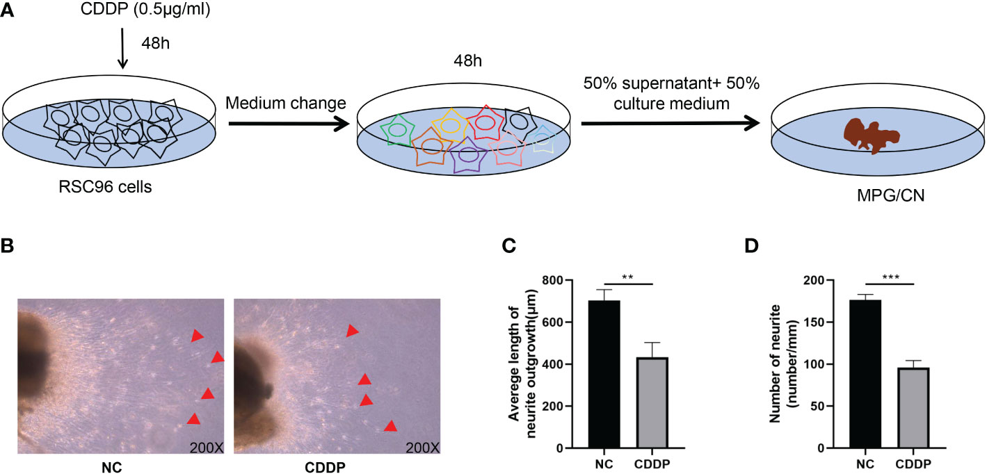
To investigate the effect of senescent Schwann cells on axonal growth of the CN, we used an in vitro model of MPG/CN culture (14). As shown in Figure 6A, the supernatant of RSC9 cells was used to treat the MPG/CN system. We dissected the MPG with 2 mm of CN attached and cultured the tissue in vitro. After 120 hours, new neurite outgrowths from the end of the CN were measured. CDDP significantly restrained the neurite outgrowth of CN (Figures 6B–D).

Figure 6 Senescent Schwann cells reduced neurite outgrowth from CN in vitro. (A) A schematic diagram of supernatant isolation and MPG treated with supernatant. (B) Neurite outgrowth from CN 120 hours after seeding. Original magnification x200. The red arrow indicates outgrowing fibers, (C). The supernatant of senescent cells inhibited fiber outgrowth from CN. (D). The supernatant of senescent cells decreased the number of fibers from CN. **P < 0.01, ***P < 0.001.
Many anticancer agents used in cancer chemotherapy possess either cytotoxic or cytocidal activity. However, they can injure both the cancer cells and the normal tissue and cells of patients because of their nonselectivity. The dysfunctions of normal cells and organs caused by these agents are called side effects. A variety of complications that cause suffering and lower quality of life make it harder for doctors. Many side effects have been reported and could be overcome. However, there are still problems to be resolved (22). Several studies have demonstrated that platinum-based chemotherapy can induce ED (10, 12, 13, 23, 24), but ED has not gained the attention of clinicians as a complication. Some scholars have found that oxaliplatin can induce ED in rats by inducing damage to the endothelium of the corpus cavernosum (11). CDDP is a conventional chemotherapeutic agent for numerous tumors. In this study, administration of CDDP to rats resulted in a decrease in the MaxICP/MAP ratio, endothelium and smooth muscle content and eNOS level. In addition, CDDP induces senescence in Schwann cells, which triggers the SASP phenotype, thereby affecting CN function. Therefore, CDDP is confirmed to be able to induce ED by affecting the corpus cavernosum and CN. CDDP exposure can cause testicular damage and a significant reduction in testosterone level (25, 26). In our study, the CDDP-treated rats showed no different serum testosterone levels than the control group, which might be caused by the use of a lower concentration of CDDP. eNOS has an indispensable role in the erectile response (27) and endothelial dysfunction is common in ED induced by diabetes mellitus (28), bilateral cavernous nerve injury (29) and oxaliplatin (11). Long-term cardiovascular events have often been reported to increase in patients with platinum-based chemotherapy due to vascular toxicity and endothelial dysfunction. In our results, CD31 staining and eNOS levels in the corpus cavernosum were decreased after CDDP treatment. CDDP can induce apoptosis in HUVECs (30). Furthermore, CDDP also upregulates NF-κB/ICAM-1 to affect the production of nitric oxide (NO) and cGMP, which leads to endothelial dysfunction (31). Moreover, CDDP also caused loss of smooth muscle. CDDP exerts anticancer activity via multiple mechanisms but the generation of DNA lesions is the most acceptable mechanism, followed by activation of the apoptosis pathway (9, 32). Correspondingly, our results showed that γ-H2AX, a DNA damage marker, and TUNEL staining were augmented in corpus cavernosum of CDDP group. Hence, we hypothesized that CDDP might damage DNA and cause endothelial and smooth muscle cell apoptosis, which impairs the erectile function of rats. Of course, the specific mechanism by which CDDP affects the endothelial and smooth muscle cells of the corpus cavernosum deserves further study.
Peripheral neurotoxicity is a common side effect of platinum-based chemotherapy (33). Some researchers have even considered ED as a neuropathic subscale-related symptom (10). A recent study found that CDDP-induced peripheral neuropathy is associated with neuronal senescence-like response (21). In our results, the expression of senescence-related markers was up-regulated in CN after CDDP treatment. Schwann cells help nerve cells to transmit information faster by wrapping their long extensions in myelin. Abnormal production of myelin can perturb signal transmission between nerve cells, which leads to neurological defects (34). Schwann cells in the peripheral nervous system play a pivotal role in nerve repair (35). In addition, dysfunction of Schwann cells plays an important role in the pathogenesis of diabetic peripheral neuropathy (36, 37). In vitro, we found that CDDP was indeed able to induce senescence in RSC96 cells and generate SASP. Inflammation is a key driver of pathological changes in many peripheral neuropathy (38). We used the supernatant of RSC96 cells to culture MPG/CN and indeed found that the growth of CN decelerated in the CDDP group, which indirectly indicated that the substances secreted by senescent Schwann cells could affect the function of CN.
In summary, our findings indicate that the CDDP-induced reduction in endothelial and smooth muscle content and SASP, which leads to CN dysfunction, are responsible for ED in rats.
In this study, we used normal rats administered CDDP, and the absence of cancer model experiments was one limitation of the present study. In addition, the effects of different dose and frequency of administration of CDDP on erectile function should be evaluated in the future. Another limitation of this study is that the direct effects of CDDP on nerves were not investigated. The underlying mechanism of effects of CDDP on CN needs to be further explored.
Our study revealed that CDDP treatment could induce erectile dysfunction by affecting the content of endothelial and smooth muscle and causing SASP in CN. Thus, erectile function in cancer survivors receiving CDDP chemotherapy should receive the attention of clinicians.
The original contributions presented in the study are included in the article/supplementary material. Further inquiries can be directed to the corresponding authors.
The animal study was reviewed and approved by the Ethics Committee of the Fifth Affiliated Hospital, Sun Yat-sen University.
Conceptualization, YY, YD and YT; Data curation, YY, JZ, LZ and YD, Formal analysis, YY, JZ, LZ, BN and JP, Funding acquisition, LZ, YZ and YT, Methodology, YY, HH, MX, and JP, Project administration, YD and YT, Supervision, YD and YT, Validation, YY, BN and YZ; Writing – Original Draft, YY, YD and YT, Writing – Review & Editing, YY, YD and YT. All authors contributed to the article and approved the submitted version.
This work was supported by National Natural Science Foundations of China (#82071636, #81571432),GuangDong Basic and Applied Basic Research Foundation (#2021A1515010436), China Postdoctoral Science Foundation (2021M703747), GuangDong Basic and Applied Basic Research Foundation (2021A1515111109) and Science and Technology Program of Zhuhai City (No. 2220004000276).
The authors declare that the research was conducted in the absence of any commercial or financial relationships that could be construed as a potential conflict of interest.
All claims expressed in this article are solely those of the authors and do not necessarily represent those of their affiliated organizations, or those of the publisher, the editors and the reviewers. Any product that may be evaluated in this article, or claim that may be made by its manufacturer, is not guaranteed or endorsed by the publisher.
1. Sung H, Ferlay J, Siegel RL, Laversanne M, Soerjomataram I, Jemal A, et al. Global cancer statistics 2020: GLOBOCAN estimates of incidence and mortality worldwide for 36 cancers in 185 countries. CA Cancer J Clin (2021) 71(3):209–49. doi: 10.3322/caac.21660
2. Hellesnes R, Myklebust TA, Fossa SD, Bremnes RM, Karlsdottir A, Kvammen O, et al. Testicular cancer in the cisplatin era: Causes of death and mortality rates in a population-based cohort. J Clin Oncol (2021) 39(32):3561–73. doi: 10.1200/JCO.21.00637
3. Parmar MK, Ledermann JA, Colombo N, du Bois A, Delaloye JF, Kristensen GB, et al. Paclitaxel plus platinum-based chemotherapy versus conventional platinum-based chemotherapy in women with relapsed ovarian cancer: the ICON4/AGO-OVAR-2. 2 trial Lancet (2003) 361(9375):2099–106. doi: 10.1016/s0140-6736(03)13718-x
4. Freyer DR, Brock PR, Chang KW, Dupuis LL, Epelman S, Knight K, et al. Prevention of cisplatin-induced ototoxicity in children and adolescents with cancer: a clinical practice guideline. Lancet Child Adolesc Health (2020) 4(2):141–50. doi: 10.1016/S2352-4642(19)30336-0
5. Mitin T, Hunt D, Shipley WU, Kaufman DS, Uzzo R, Wu CL, et al. Transurethral surgery and twice-daily radiation plus paclitaxel-cisplatin or fluorouracil-cisplatin with selective bladder preservation and adjuvant chemotherapy for patients with muscle invasive bladder cancer (RTOG 0233): a randomised multicentre phase 2 trial. Lancet Oncol (2013) 14(9):863–72. doi: 10.1016/S1470-2045(13)70255-9
6. Yang JC, Wu YL, Schuler M, Sebastian M, Popat S, Yamamoto N, et al. Afatinib versus cisplatin-based chemotherapy for EGFR mutation-positive lung adenocarcinoma (LUX-lung 3 and LUX-lung 6): analysis of overall survival data from two randomised, phase 3 trials. Lancet Oncol (2015) 16(2):141–51. doi: 10.1016/S1470-2045(14)71173-8
7. Hougardy BM, Maduro JH, van der Zee AG, Willemse PH, de Jong S, de Vries EG. Clinical potential of inhibitors of survival pathways and activators of apoptotic pathways in treatment of cervical cancer: changing the apoptotic balance. Lancet Oncol (2005) 6(8):589–98. doi: 10.1016/S1470-2045(05)70281-3
8. Batgi H, Basci S, Dal MS, Kizil Cakar M, Uncu Ulu B, Yigenoglu TN, et al. Gemcitabine, dexamethasone and cisplatin (GDP) is an effective and well-tolerated mobilization regimen for relapsed and refractory lymphoma: a single center experience. Turk J Med Sci (2021) 51(2):685–92. doi: 10.3906/sag-2008-114
9. Ghosh S. Cisplatin: The first metal based anticancer drug. Bioorg Chem (2019) 88:102925. doi: 10.1016/j.bioorg.2019.102925
10. Mols F, Beijers T, Lemmens V, van den Hurk CJ, Vreugdenhil G, van de Poll-Franse LV. Chemotherapy-induced neuropathy and its association with quality of life among 2- to 11-year colorectal cancer survivors: results from the population-based PROFILES registry. J Clin Oncol (2013) 31(21):2699–707. doi: 10.1200/JCO.2013.49.1514
11. Kataoka T, Mori T, Suzuki J, Kawaki Y, Kito Y, Hotta Y, et al. Oxaliplatin, an anticancer agent, causes erectile dysfunction in rats due to endothelial dysfunction. J Sex Med (2021) 18(8):1337–45. doi: 10.1016/j.jsxm.2021.06.004
12. Kerns SL, Fung C, Monahan PO, Ardeshir-Rouhani-Fard S, Abu Zaid MI, Williams AM, et al. Cumulative burden of morbidity among testicular cancer survivors after standard cisplatin-based chemotherapy: A multi-institutional study. J Clin Oncol (2018) 36(15):1505–12. doi: 10.1200/JCO.2017.77.0735
13. Bandak M, Lauritsen J, Johansen C, Kreiberg M, Skott JW, Agerbaek M, et al. Sexual function in a nationwide cohort of 2,260 survivors of testicular cancer after 17 years of followup. J Urol (2018) 200(4):794–800. doi: 10.1016/j.juro.2018.04.077
14. Peng D, Reed-Maldonado AB, Zhou F, Tan Y, Yuan H, Banie L, et al. Exosome released from schwann cells may be involved in microenergy acoustic pulse-associated cavernous nerve regeneration. J Sex Med (2020) 17(9):1618–28. doi: 10.1016/j.jsxm.2020.05.018
15. Staff NP, Cavaletti G, Islam B, Lustberg M, Psimaras D, Tamburin S. Platinum-induced peripheral neurotoxicity: From pathogenesis to treatment. J Peripher Nerv Syst (2019) 24 Suppl 2(Suppl 2):S26–39. doi: 10.1111/jns.12335
16. Reagan-Shaw S, Nihal M, Ahmad N. Dose translation from animal to human studies revisited. FASEB J (2008) 22(3):659–61. doi: 10.1096/fj.07-9574LSF
17. Li Z, Yin Y, He K, Ye K, Zhou J, Qi H, et al. Intracavernous pressure recording in a cavernous nerve injury rat model. J Vis Exp (2021) 175:e63024. doi: 10.3791/63024
18. Yin Y, Peng J, Zhou J, Chen H, Peng D, Li D, et al. Tetrathiomolybdate partially alleviates erectile dysfunction of type 1 diabetic rats through affecting Ceruloplasmin/eNOS and inhibiting corporal fibrosis and systemic inflammation. Sex Med (2022) 10(1):100455. doi: 10.1016/j.esxm.2021.100455
19. Girma S, Avanzi C, Bobosha K, Desta K, Idriss MH, Busso P, et al. Evaluation of auramine O staining and conventional PCR for leprosy diagnosis: A comparative cross-sectional study from Ethiopia. PLoS Negl Trop Dis (2018) 12(9):e0006706. doi: 10.1371/journal.pntd.0006706
20. Kinner A, Wu W, Staudt C, Iliakis G. Gamma-H2AX in recognition and signaling of DNA double-strand breaks in the context of chromatin. Nucleic Acids Res (2008) 36(17):5678–94. doi: 10.1093/nar/gkn550
21. Calls A, Torres-Espin A, Navarro X, Yuste VJ, Udina E, Bruna J. Cisplatin-induced peripheral neuropathy is associated with neuronal senescence-like response. Neuro Oncol (2021) 23(1):88–99. doi: 10.1093/neuonc/noaa151
22. Taguchi T. [Side effects of cancer chemotherapy and steps to deal with them]. Gan To Kagaku Ryoho (1995) 22(14):2017–28.
23. van Basten JP, Hoekstra HJ, van Driel MF, Koops HS, Droste JH, Jonker-Pool G, et al. Sexual dysfunction in nonseminoma testicular cancer patients is related to chemotherapy-induced angiopathy. J Clin Oncol (1997) 15(6):2442–8. doi: 10.1200/JCO.1997.15.6.2442
24. Abu Zaid M, Dinh PC, Monahan PO, Fung C, El-Charif O, Feldman DR, et al. Adverse health outcomes in relationship to hypogonadism after chemotherapy: A multicenter study of testicular cancer survivors. J Natl Compr Canc Netw (2019) 17(5):459–68. doi: 10.6004/jnccn.2018.7109
25. Rauf N, Nawaz A, Ullah H, Ullah R, Nabi G, Ullah A, et al. Therapeutic effects of chitosan-embedded vitamin c, e nanoparticles against cisplatin-induced gametogenic and androgenic toxicity in adult male rats. Environ Sci Pollut Res Int (2021) 28(40):56319–32. doi: 10.1007/s11356-021-14516-y
26. Abdel-Wahab BA, Walbi IA, Albarqi HA, Ali FEM, Hassanein EHM. Roflumilast protects from cisplatin-induced testicular toxicity in male rats and enhances its cytotoxicity in prostate cancer cell line. role of NF-kappaB-p65, cAMP/PKA and Nrf2/HO-1, NQO1 signaling. Food Chem Toxicol (2021) 151:112133. doi: 10.1016/j.fct.2021.112133
27. Musicki B, Burnett AL. eNOS function and dysfunction in the penis. Exp Biol Med (Maywood) (2006) 231(2):154–65. doi: 10.1177/153537020623100205
28. Castela A, Costa C. Molecular mechanisms associated with diabetic endothelial-erectile dysfunction. Nat Rev Urol (2016) 13(5):266–74. doi: 10.1038/nrurol.2016.23
29. Li M, Yuan YM, Yang BC, Gu SJ, Li HX, Xin ZC, et al. Comparative study of intracavernous pressure and cavernous pathology after bilateral cavernous nerve crushing and resection in rats. Asian J Androl (2020) 22(6):629–35. doi: 10.4103/aja.aja_10_20
30. Chen CH, Chen MC, Hsu YH, Chou TC. Far-infrared radiation alleviates cisplatin-induced vascular damage and impaired circulation via activation of HIF-1alpha. Cancer Sci (2022) 113(6):2194–206. doi: 10.1111/cas.15371
31. Bodiga VL, Bathula J, Kudle MR, Vemuri PK, Bodiga S. Andrographolide suppresses cisplatin-induced endothelial hyperpermeability through activation of PI3K/Akt and eNOS -derived nitric oxide. Bioorg Med Chem (2020) 28(23):115809. doi: 10.1016/j.bmc.2020.115809
32. Dasari S, Tchounwou PB. Cisplatin in cancer therapy: molecular mechanisms of action. Eur J Pharmacol (2014) 740:364–78. doi: 10.1016/j.ejphar.2014.07.025
33. Santos N, Ferreira RS, Santos ACD. Overview of cisplatin-induced neurotoxicity and ototoxicity, and the protective agents. Food Chem Toxicol (2020) 136:111079. doi: 10.1016/j.fct.2019.111079
34. Wiltbank AT, Steinson ER, Criswell SJ, Piller M, Kucenas S. Cd59 and inflammation regulate schwann cell development. Elife (2022) 11:e76640. doi: 10.7554/eLife.76640
35. Modrak M, Talukder MAH, Gurgenashvili K, Noble M, Elfar JC. Peripheral nerve injury and myelination: Potential therapeutic strategies. J Neurosci Res (2020) 98(5):780–95. doi: 10.1002/jnr.24538
36. Zhang X, Zhao S, Yuan Q, Zhu L, Li F, Wang H, et al. TXNIP, a novel key factor to cause schwann cell dysfunction in diabetic peripheral neuropathy, under the regulation of PI3K/Akt pathway inhibition-induced DNMT1 and DNMT3a overexpression. Cell Death Dis (2021) 12(7):642. doi: 10.1038/s41419-021-03930-2
37. Cheng YC, Chu LW, Chen JY, Hsieh SL, Chang YC, Dai ZK, et al. Loganin attenuates high glucose-induced schwann cells pyroptosis by inhibiting ROS generation and NLRP3 inflammasome activation. Cells (2020) 9(9):1948. doi: 10.3390/cells9091948
Keywords: cisplatin, erectile dysfunction, apoptosis, senescence, cancer survivor
Citation: Yin Y, Zhou Y, Zhou J, Zhao L, Hu H, Xiao M, Niu B, Peng J, Dai Y and Tang Y (2023) Cisplatin causes erectile dysfunction by decreasing endothelial and smooth muscle content and inducing cavernosal nerve senescence in rats. Front. Endocrinol. 14:1096723. doi: 10.3389/fendo.2023.1096723
Received: 12 November 2022; Accepted: 10 January 2023;
Published: 25 January 2023.
Edited by:
Ke Rao, Huazhong University of Science and Technology, ChinaReviewed by:
Ming Wang, Zhengzhou University, ChinaCopyright © 2023 Yin, Zhou, Zhou, Zhao, Hu, Xiao, Niu, Peng, Dai and Tang. This is an open-access article distributed under the terms of the Creative Commons Attribution License (CC BY). The use, distribution or reproduction in other forums is permitted, provided the original author(s) and the copyright owner(s) are credited and that the original publication in this journal is cited, in accordance with accepted academic practice. No use, distribution or reproduction is permitted which does not comply with these terms.
*Correspondence: Yingbo Dai, ZGFpeWluZ2JvMDYyMkAxMjYuY29t; Yuxin Tang, dGFuZ3l4MzZAbWFpbC5zeXN1LmVkdS5jbg==
†These authors have contributed equally to this work and share first authorship
Disclaimer: All claims expressed in this article are solely those of the authors and do not necessarily represent those of their affiliated organizations, or those of the publisher, the editors and the reviewers. Any product that may be evaluated in this article or claim that may be made by its manufacturer is not guaranteed or endorsed by the publisher.
Research integrity at Frontiers

Learn more about the work of our research integrity team to safeguard the quality of each article we publish.