
95% of researchers rate our articles as excellent or good
Learn more about the work of our research integrity team to safeguard the quality of each article we publish.
Find out more
ORIGINAL RESEARCH article
Front. Ecol. Evol. , 08 March 2021
Sec. Evolutionary Developmental Biology
Volume 9 - 2021 | https://doi.org/10.3389/fevo.2021.624306
This article is part of the Research Topic Evolution of Postembryonic Development View all 12 articles
Developmental diet is known to exert long-term effects on adult phenotypes in many animal species as well as disease risk in humans, purportedly mediated through long-term changes in gene expression. However, there are few studies linking developmental diet to adult gene expression. Here, we use a full-factorial design to address how three different larval and adult diets interact to affect gene expression in 1-day-old adult fruit flies (Drosophila melanogaster) of both sexes. We found that the largest contributor to transcriptional variation in young adult flies is larval, and not adult diet, particularly in females. We further characterized gene expression variation by applying weighted gene correlation network analysis (WGCNA) to identify modules of co-expressed genes. In adult female flies, the caloric content of the larval diet associated with two strongly negatively correlated modules, one of which was highly enriched for reproduction-related processes. This suggests that gene expression in young adult female flies is in large part related to investment into reproduction-related processes, and that the level of expression is affected by dietary conditions during development. In males, most modules had expression patterns independent of developmental or adult diet. However, the modules that did correlate with larval and/or adult dietary regimes related primarily to nutrient sensing and metabolic functions, and contained genes highly expressed in the gut and fat body. The gut and fat body are among the most important nutrient sensing tissues, and are also the only tissues known to avoid histolysis during pupation. This suggests that correlations between larval diet and gene expression in male flies may be mediated by the carry-over of these tissues into young adulthood. Our results show that developmental diet can have profound effects on gene expression in early life and warrant future research into how they correlate with actual fitness related traits in early adulthood.
Developmental diet can exert long-term effects on adult phenotypes and disease risk (Schlichting and Pigliucci, 1998). For example, human and rodent studies have demonstrated that inadequate developmental diet can increase the risk of heart disease, obesity, stroke, and hypertension in adulthood (reviewed in Bruce and Hanson, 2010; Burdge and Lillycrop, 2010; Langley-Evans, 2015). Several evolutionary theories have been proposed to explain the mechanisms underlying this link including the silver spoon hypothesis, the developmental programming hypothesis, and the predictive adaptive response (PAR) hypothesis (reviewed in Lindström, 1999; Monaghan, 2008). The silver spoon hypothesis proposes that sub-optimal developmental conditions directly constrain overall adult size or quality, resulting in decreased fitness across adult environments (e.g., Emlen, 1997; Lanet and Maurange, 2014), while the developmental programming hypothesis extends this idea and suggests that the negative effects of sub-optimal development conditions will be more pronounced the more the developmental and adult environment differ (Fernandez-Twinn and Ozanne, 2006). Finally, the PAR hypothesis goes one step further and proposes that the developmental environment can serve as a cue anticipating the adult environment, allowing individuals to adjust their phenotypes during development to “match” their predicted adult conditions. However, when these predictions are incorrect a mismatch occurs, resulting in an increased risk of metabolic disease (Ravelli et al., 1998; Gluckman and Hanson, 2004; Gluckman et al., 2007; Bateson et al., 2014).
Regardless of the mechanism, developmental influences on adult phenotypes are expected to result in gene-expression changes at the level of the whole organism (Burdge et al., 2007; Burdge and Lillycrop, 2010). Yet, despite the widespread incidence of variation in the quality of the developmental environment, relatively few studies have addressed the extent to which developmental diet influences gene expression in adulthood. In rats, the adult offspring of protein-restricted mothers have altered expression of metabolic genes (Bertram et al., 2001; Maloney et al., 2003; Lillycrop et al., 2005), while high protein larval diets in fruit flies affect immune gene expression in young adults (Fellous and Lazzaro, 2010). We have previously shown that subtle effects of developmental diet on the transcriptome persist into middle- and old-age in fruit flies (May and Zwaan, 2017), and that the expression of particular ribosome, transcription, and translation-related genes correlate with observed patterns of lifespan and fecundity across diets (May and Zwaan, 2017). However, to our knowledge, no study has addressed the global effect of developmental diet on the transcriptome in early life, nor if and how it depends on the adult environment.
Here, we use the fruit fly, Drosophila melanogaster, to determine to what extent developmental diet affects whole-body, whole-genome, gene expression in 1-day old adult male and female flies, and how this interacts with adult dietary conditions. We chose 1-day old flies in order to establish a baseline effect of developmental diet on adult gene-expression. We first assess broad-scale patterns of expression variation as obtained from principle component analysis (PCA). Subsequently, we use weighted gene correlation network analysis (WGCNA v1.69. Langfelder and Horvath, 2008) to identify modules of genes whose expression is affected by larval and/or adult dietary conditions. We chose to focus on gene co-expression modules as these more accurately reflect the nature of genes as components of hierarchically structured regulatory networks (Zhang and Horvath, 2005). As such, co-expression often reflects co-regulation (Allocco et al., 2004), or tissue-specific function (Boutanaev et al., 2002) and can provide more biologically meaningful insights as compared to single-gene analysis (e.g., Oldham et al., 2006; Hilliard et al., 2012; Xue et al., 2013). After identifying modules of co-expressed genes affected by dietary conditions, we annotate the modules using several external databases and address whether there is evidence for specific functions [gene ontology (GO)], tissue-specific roles (FlyAtlas and FlyGut databases), or co-regulation of the modules by specific transcription factors (TFs) (DroID Database).
We find that both larval and adult diet affect the whole-body transcriptome of young adults, however, the effect of larval diet is considerably larger than that of adult diet, especially in females. In females, larval diet primarily affects relative expression of reproduction vs non-reproduction related processes, while in males, larval diet modulates the expression of modules associated with nutrient sensing and metabolic functions, containing genes highly expressed in the gut and fat body. This suggests that there is scope for long-term effects of developmental conditions on adult gene expression. We discuss the results in the context of theories and hypotheses linking early life conditions to late life health as they relate to epigenetic mechanisms.
We used the laboratory stock population described in May et al. (2015). This population was derived from six wild populations collected across Europe in 2006. To ensure equal representation of existing genetic variation, the populations were subjected to four rounds of crossing to generate the final population, This population has been maintained in the laboratory for more than 60 generations under standard laboratory conditions: 25°C, 65% humidity, 12:12 h light:dark cycle, 14 day generation time, and a standard diet (1SY) of 100 grams sugar, 70 grams yeast, 20 grams agar, 15 mL nipagin, and 3 mL propionic acid per liter of water. We modified the sugar (S) and yeast (Y) content of our standard laboratory diet (1SY) by 0.25 times and 2.5 times, respectively to obtain three diets (0.25SY, 1SY, and 2.5SY) spanning a 10-fold range of sugar and yeast concentrations. For the experiment, we raised larvae on these three diets at a density of 100 eggs per vial (6 mL medium) and immediately after eclosion randomly distributed the resulting adults across these three diets again in a full factorial design (Supplementary Figure 1). To obtain virgin flies we sexed the adults immediately after the cuticle had hardened (∼3 h post eclosion) and maintained them at a density of 10 adults per vial. Twenty-four hours post-eclosion, we flash froze the flies in groups of five using liquid nitrogen.
We extracted RNA from four replicates per combination of sex, larval diet, and adult diet (4 replicates × 2 sexes × 3 larval diets × 3 adult diets = 72 arrays), using the Machery Nagel Nucleospin II kit (Machery and Nagel). For each sample we homogenized five whole flies together in order to minimize the effect of random variation between individuals. Biotin labeling, cRNA synthesis, hybridization to Affymetrix Drosophila 2.0 GeneChips and array readouts were performed by ServiceXS1.
Prior to analysis we excluded ten female and four male arrays from analysis due to either evidence of mating (5 female samples) or outlier status as defined by WGCNA (z.k. score > 2.5; Horvath, 2011). Excluded arrays were randomly distributed across the nine treatment groups. Subsequently we performed background adjustment, quantile normalization and summarization using the robust multi-array average (RMA) algorithm (Irizarry et al., 2003). When normalizing males and females together, 96% of the variation in expression was due to sex, so we normalized male and female samples separately to emphasize the effects of larval and adult diet rather than ubiquitous and well-documented sex-specific differences (e.g., Ayroles et al., 2009). We performed all subsequent analysis steps separately for each sex. To gain an understanding of the broad patterns of variation in the data we applied principal components analysis to the normalized expression data (PCA; Pearson 1901). The data quality check and all other analyses were performed using R (version 4.0.3) and Bioconductor (v1.30.10; Gentleman et al. 2004).
We applied WGCNA to the normalized expression data for each sex. We used signed modules and applied the recommended default settings for detection of signed modules (Zhang and Horvath, 2005). WGCNA first determines expression correlations for all pairs of genes and weights them by the connection strength. These correlations are then clustered using hierarchical clustering, and modules are defined as branches of the clustering tree. The expression profile of each module can be summarized by its eigengene (E), which is defined as the first principal component of the expression matrix. The eigengene can be thought of as a weighted average expression profile and thus for a particular sample, the value of its eigengene is representative of the overall expression of the module (Langfelder and Horvath, 2008). For each module, eigengenes can be compared across samples to detect significant correlations between treatments and module eigengenes (i.e., expression). By convention, modules are named by colors, with unassigned genes grouped together in the “gray” module. Implementation of WGCNA is freely available in the WGCNA R package (v1.69., Langfelder and Horvath, 2008).
We applied several additional analysis steps to biologically characterize the modules. First, for each module we fitted an analysis of variance (ANOVA) model that partitions the variance in module eigengene expression explained by larval diet (L), adult diet (A), and their interaction (L by A). We set a false discovery rate (FDR) threshold of 0.001 (Benjamini and Hochberg, 1995) and focused the remaining analysis on the modules that showed a significant effect of dietary treatment on their eigengene value.
We next submitted the probe list associated with each module to DAVID (Database for Annotation, Visualization and Integrated Discovery; Huang et al., 2008) for GO enrichment analysis focusing on GO FAT terms. GO FAT terms exclude broader GO terms and eliminate term redundancy. We adjusted p-values using the linear step-up method of Benjamini and Hochberg (1995) to correct for multiple comparisons. To facilitate interpretation, we submitted lists of more than 20 terms to the REVIGO online tool which uses semantic similarity to reduce large lists of terms to a representative subset of terms (REduce and VIsualize Gene Ontology: Supek et al., 2011).
We used two external databases: FlyAtlas (Chintapalli et al., 2007) and FlyGut (Buchon et al., 2013) to address whether modules showed evidence of tissue-specific expression profiles. FlyAtlas contains tissue-specific gene expression data for both larval and adult tissues of the fruit fly, while FlyGut contains region-specific expression patterns in the gut. For each of our modules, we calculated the median expression across all of its probes in each tissue available in the FlyAtlas database and in each gut region available in the FlyGut database.
We used the Drosophila Interactions Database (DroID; Murali et al., 2010) to address whether there was evidence that modules were co-regulated by certain TFs. DroID is a merged dataset of empirically validated interactions between TFs and genes from the “Regulatory Element Database for Drosophila and other insects” (REDfly; Halfon et al., 2008) and “model organism ENCyclopedia Of DNA Elements” databases (modENCODE; Roy et al., 2010). We first filtered the data set to exclude TFs that are known to interact with less than five genes. We then performed a Fisher’s exact test to identify modules enriched for genes regulated by the same TFs. We considered a module enriched for probes binding a certain TF at an odds-ratio of greater than 1.5-fold and p-value of <0.05, although at this threshold, not all interactions remained significant after applying the Bonferroni correction for multiple testing (Neyman and Pearson, 1928). Source code and links to data for all analyses are available at https://github.com/transcriptome-in-transition/Transcriptome-in-transition.
The first principal component in both sexes (PC1) is driven by larval diet (females: Figure 1A; males: Figure 1B). The proportion of variation explained is much higher in females than in males (females: 43%, males: 13%), suggesting female gene expression is more strongly dependent on larval conditions. In both sexes, PC1 separates flies raised on the 0.25SY and 1SY diets as larvae, while those raised on 2.5SY fall in between (females: Figure 1C, males: Figure 1D). PC2, by contrast, appears to reflect more subtle effects of adult diet in both sexes. In females, PC2 separates flies transferred to the 0.25SY diet as adults from the rest, suggesting a rapid transcriptional response to this dietary condition (Figure 1E). In males, PC2 appears to separate flies transferred to the 2.5SY diet as adults from the rest (Figure 1F). Additional PC’s did not explain more than 5% of variation individually and did not show clear effects of diet, suggesting that any additional effects are too subtle to be detected at the global level.
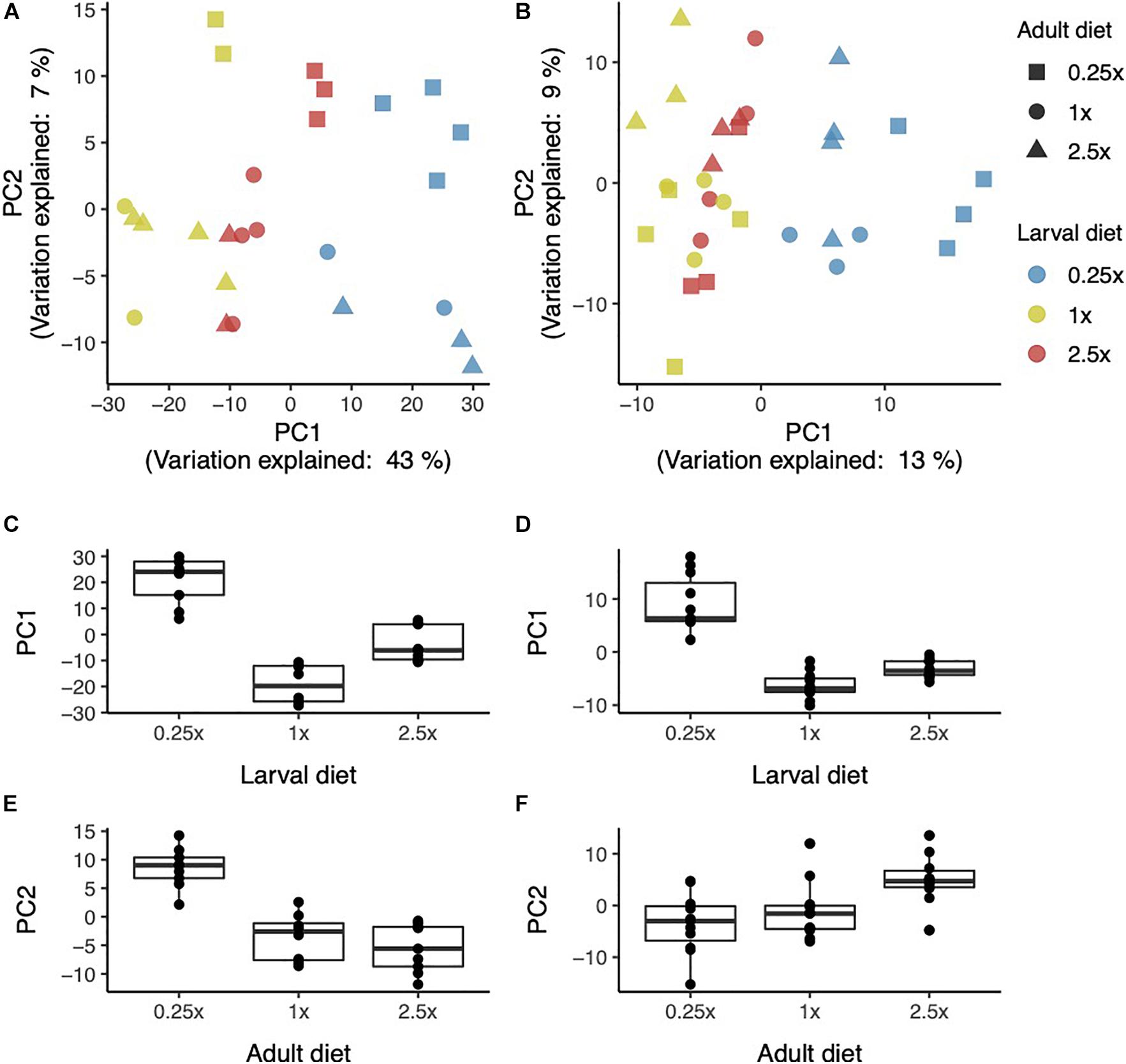
Figure 1. Principle component analysis of global gene expression. PC1 vs. PC2 in females (A) and males (B). Larval diet vs. PC1 in females (C) and males (D) and adult diet vs. PC2 in females (E) and males (F).
As an alternative to PCA, we also identified modules of co-expressed genes using WGCNA. In females, WGCNA identified two very large co-expression modules: blue (5,509 probes) and turquoise (5,695 probes) while the gray module (7,565) contained genes not assigned to a module. The eigengenes of the blue and turquoise modules were nearly perfectly anti-correlated (r2 = −0.96, n = 26, p < 0.0001; Figure 2A), thus increased expression of one module implies decreased expression of the other. We could identify only subtle tissue specific effects for these two modules (Figures 3A–C): a relatively high expression in the larval central nervous system (CNS) for the turquoise module (and low for blue; Figure 3A), and higher expression in the adult ovary for the turquoise module (and thus low for blue; Figure 3B).
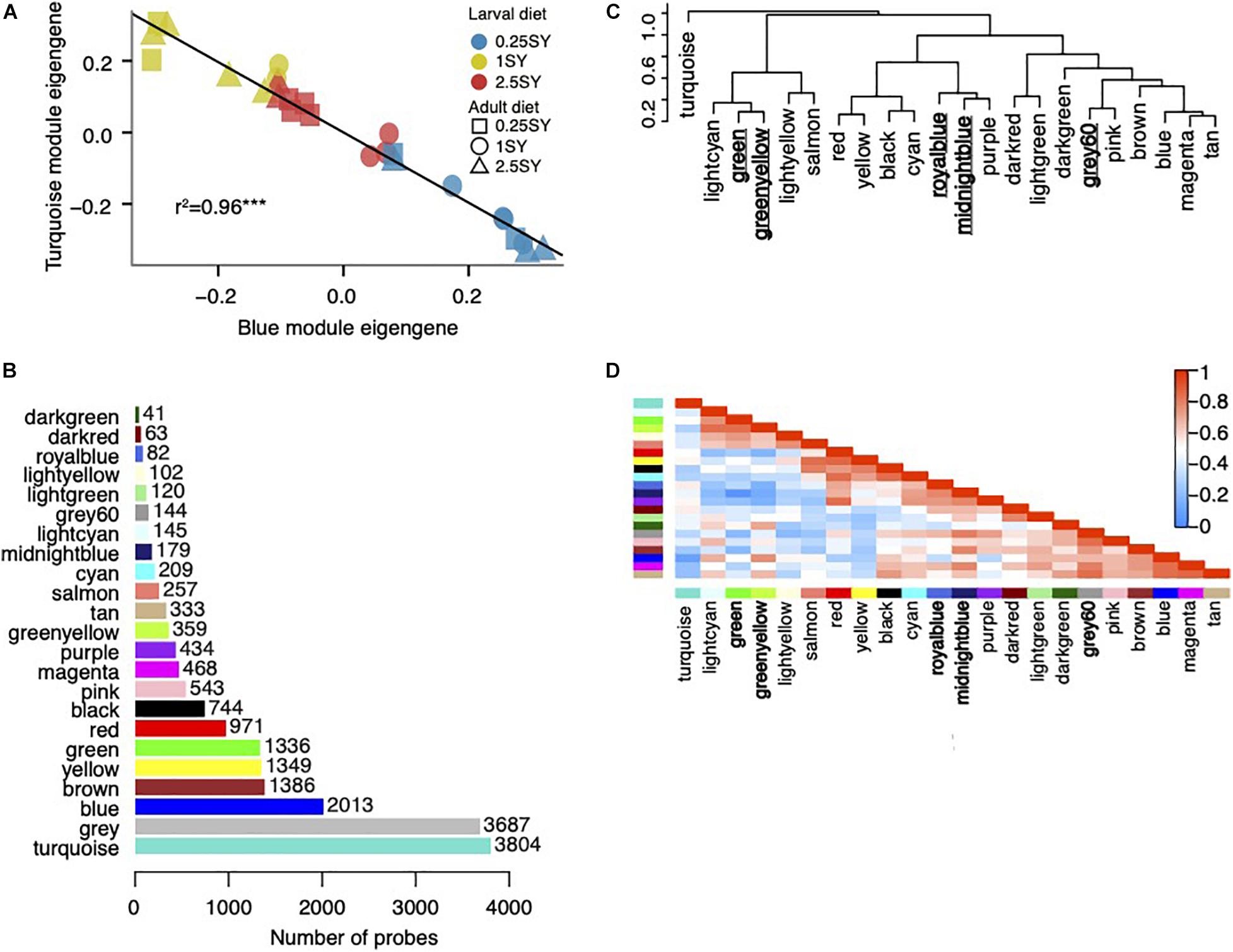
Figure 2. Modules detected by WGCNA. (A) Module eigengenes of the female blue module plotted against eigengenes of the female turquoise module. Eigengenes of the two modules are nearly perfectly negatively correlated (r2 = 0.96). (B) Module sizes in males. (C) Hierarchical clustering dendrogram of male module eigengenes (labeled by their colors). (D) Heatmap plot of the adjacencies in the eigengene network. Each row and column in the heatmap corresponds to one module eigengene (labeled by color). A value of 1 (red) represents high adjacency (perfect positive correlation), while 0 (blue) represents low adjacency (perfect negative correlation). Modules with high adjacency tend to have similar tissue-specific expression profiles in the FlyAtlas and FlyGut datasets. For both sexes, the “gray” module contains probes that were unassigned to a module.
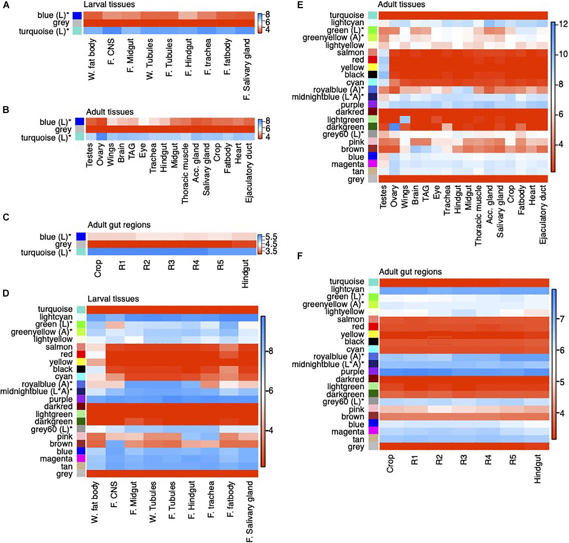
Figure 3. Heatmaps of module median expression in the FlyAtlas and FlyGut tissue-specific gene expression databases. Median expression of female modules per tissue in (A) larval tissues in FlyAtlas, (B) adult tissues in FlyAtlas and (C) across gut regions in FlyGut. Median expression of male modules per tissue in (D) larval tissues in FlyAtlas, (E) adult tissues in FlyAtlas, and (F) across gut regions in FlyGut. W: wandering, F: feeding, CNS: central nervous system, Acc. gland: accessory gland. For larval tissues “W.” refers to tissues collected during the wandering larval stage (late third instar) and “F.” refers to tissues collected while third instar larvae were still feeding. For FlyGut data (C,F) R1 through R5 refer to five sequential regions of the gut which are delineated by the combination of an anatomical constriction between one region and the next, changes in histology and changes in gene expression (Buchon et al., 2013). Modules are shown in the same order as they appear in the hierarchical clustering dendrogram. Modules whose expression is affected by larval or adult diet are indicated by a “*” and annotated with whether they are affected by larval diet (L), adult diet (A), or their interaction (L*A).
In males, we identified 22 co-expression modules ranging in size from 41 to 3,805 probes (Figure 2B). A total of 15,082 probes (80% of total) were assigned to a module while 3,687 probes were unassigned (gray module). Nearly all modules possessed significant GO annotation and enrichment for binding of specific TFs (Supplementary File 1), as well as distinct tissue-specific expression profiles in the FlyAtlas and FlyGut databases (Figures 3D–F) suggesting that WGCNA was able to identify biologically relevant groups of co-expressed genes. For example, the salmon, red, yellow, black, and cyan modules contain genes whose expression is restricted to the adult testes and wandering larval fat body in external databases (Figures 3D,E). These two pieces of external information are consistent with each other, as the testes are embedded within the wandering fat body during larval development (Gärtner et al., 2014).
We next assessed which modules were affected by larval or adult diet. ANOVA analysis of module eigengenes showed that the expression of both female modules and five out of 22 male modules were affected by dietary conditions (Table 1), thus we focus our remaining analysis on these modules.
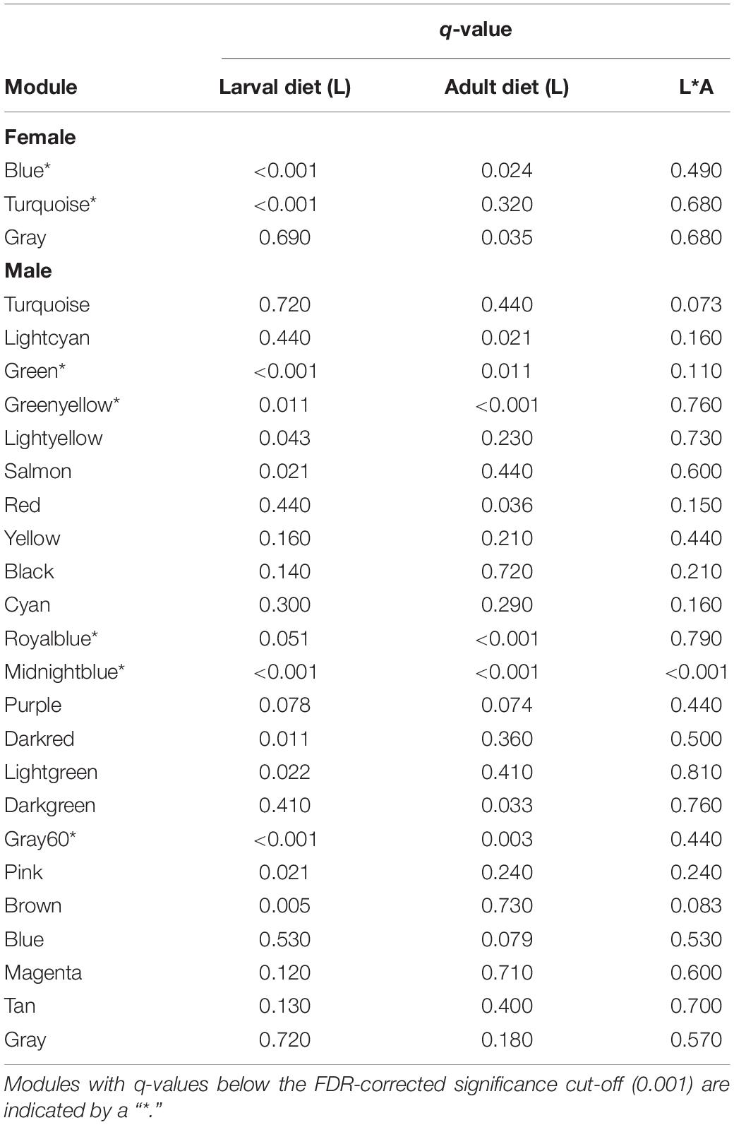
Table 1. q-values for per-module ANOVAs assessing the effect of larval diet (L), adult diet (A), and their interaction (L*A) on module eigengene values.
In females, the eigengene value of both modules (i.e., expression) was strongly dependent on larval diet (Table 1). In both modules, the 0.25SY and 1SY-raised flies form the two extremes of expression, while the 2.5SY-raised flies fall in between (Figures 4A,B), similar to the differences observed for the PCA analysis (Figure 1). The blue module is up-regulated in 0.25SY raised flies (Figure 4A), and the turquoise module is down-regulated (Figure 4B), consistent with the tight negative correlation of the module eigengene values (Figure 2A). Furthermore, the blue module also shows increased expression in flies transferred to the 0.25SY diet as adults, suggesting a rapid transcriptional response in the same direction as the effect of larval diet (Figure 4A).
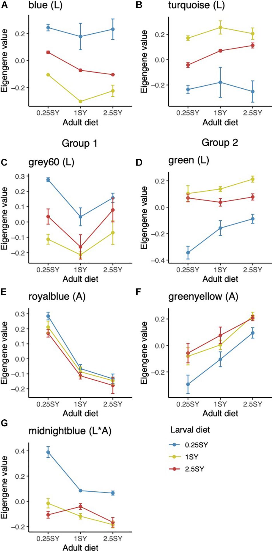
Figure 4. Module eigengene (expression) values across larval (colors) and adult (x-axis) diets. Module eigengenes of blue (A) and turquoise (B) female modules. Both modules are significantly affected by larval diet. Module eigengenes of gray60 (C) and green (D) male modules. The eigengene values of both modules are significantly affected by larval diet. Module eigengenes of royal blue (E) and green yellow (F) male modules. The eigengene values of both modules are significantly affected by adult diet. (G) Module eigengenes of the midnight blue module. Expression of the midnight blue module is significantly affected by the interaction between larval and adult diet. Male modules can be divided into two groups based on their expression correlation: Group 1 (C,E,G) and Group 2 (D,F). Within a group eigengene expression is highly correlated, but across groups, eigengene expressions are negatively correlated. All error bars are 95% C.I’s.
Both modules have extensive GO enrichment (Blue module: 373 terms; Turquoise module: 564 terms; Supplementary File 1). Summarization with REVIGO (Supek et al., 2011) shows the blue module (up-regulated in 0.25SY) is enriched for a broad spectrum of terms including mesoderm development, detection of external stimuli, leg and limb morphogenesis, energy derivation by oxidation of organic compounds, and generation of precursor metabolites and energy (Figure 5A), while the turquoise module (down-regulated in 0.25SY) is annotated with terms related to cell division, cell cycle, oogenesis and reproduction (Figure 5B).
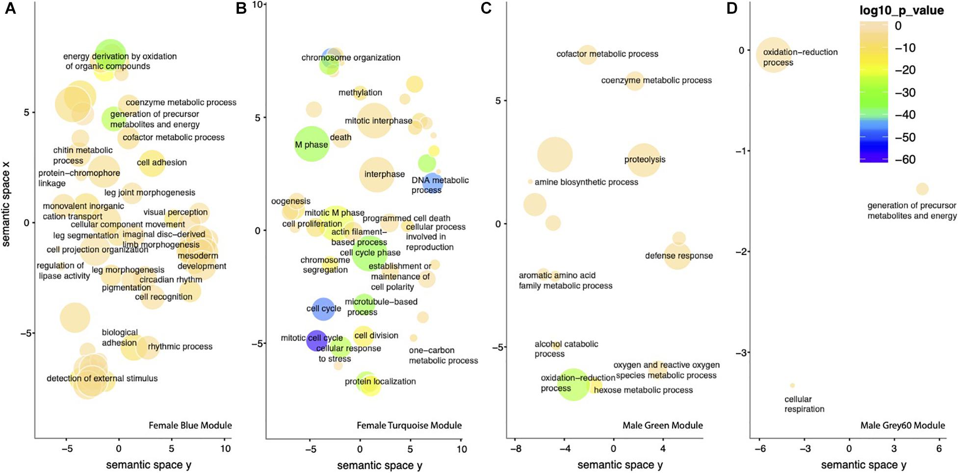
Figure 5. Summarization of module GO annotation using REVIGO. Representative GO terms summarizing (A) the 373 GO terms significantly associated with the blue female module, (B) the 564 GO terms associated with the turquoise female module, (C) the 75 GO terms associated with the green male module, and (D) the 21 terms associated with the gray60 male module. Each scatterplot shows the cluster representatives (i.e., terms remaining after the redundancy reduction) in a two dimensional space derived by applying multidimensional scaling to a matrix of the GO terms’ semantic similarities. Bubble size indicates the frequency of the GO term in the underlying GO database; more general terms have larger bubbles. Bubble color indicates the significance of the enrichment in the DAVID analysis (log10 p-value).
The turquoise module has relatively high overall expression across tissues (Figures 3A,C), and is particularly highly expressed in the adult ovary (Figure 3B), consistent with its enrichment for cell cycle and reproduction related GO terms, while the blue module has relatively low median expression overall (Figures 3A,C) and in the ovary in particular (Figure 3B), consistent with its lack of reproduction and cell-cycle related GO terms. Assessment of enrichment of TF binding probes found enrichment for 19 TFs in the turquoise module, but none in the blue module (Table 2). These 19 TFs show similar tissue-specific transcription patterns to the turquoise module overall (i.e., they are highly expressed in general, and especially in the ovary; Supplementary File 2a,c) and also share very similar GO annotation with the turquoise module, with 89 of the 92 terms enriched for the TF list also enriched in the turquoise module. Furthermore, REVIGO summary of these 92 terms shows a similar emphasis on reproduction, oogenesis, and early embryonic development (Supplementary File 2d).
Overall, these findings suggest that larval diet drives relative investment into reproduction and cell-cycle related processes (turquoise) versus non-reproduction related processes (blue) in young adult females. The low calorie 0.25SY larval (and to some extent adult) diet shifts the balance toward higher expression of the blue, non-reproduction related module, while the 1SY larval diet does the opposite.
In males, we found a significant effect of diet in five out of 22 modules (Table 1). Thus, in contrast to females, a significant proportion of gene expression in young males is independent of dietary conditions, as was suggested by the principal component analysis (Figure 1B). Of the five modules, two were affected by larval diet (gray60, green), two by adult diet (royal blue, green yellow), and one by their interaction (midnight blue). The modules could be broadly divided into two groups whose eigengenes were highly positively correlated within groups, but negatively correlated across groups (Group 1: gray60, royalblue and midnight blue; Group 2: green and greenyellow; Figures 2C,D). Modules within a group tend to have similar expression profiles (Figures 4C,G) and tissue specific expression levels (Figures 3D,F) while across groups, these two metrics are roughly inverse.
The modules in Group 1 are most highly expressed in flies either raised on (gray60, midnight blue) or transferred to (royal blue, midnight blue) the 0.25SY diet (Figures 4C,E,G). The midnight blue module shows an interaction between these two effects as it is up-regulated in flies raised on the 0.25SY larval diet, but this effect is especially pronounced when combined with transfer to the 0.25SY adult diet (Figure 4G). The modules in Group 2 are down-regulated in flies raised on (green) or transferred to (green–yellow) the 0.25SY diet (Figures 4D,F). For the two modules affected by larval diet (gray60, green), flies raised on the 2.5SY diet have intermediate expression between 0.25SY and 1SY raised flies, similar to the pattern observed for the modules affected by larval diet in female flies (Figures 4C,D). For the two modules affected by adult diet (royal blue, green yellow) the effect of adult diet tends to be linear, thus expression either decreases (green yellow) or increases (royal blue) with increasing adult diet (Figures 4E,F). For the interacting module, midnight blue, there is no consistent difference between the flies raised on or transferred to the 1SY and 2.5SY diet, suggesting that this module is driven by changes in expression particular to experiencing the 0.25SY diet, especially when experienced during both development and early adulthood (Figure 4G).
Each of the five modules are enriched for GO terms (Supplementary File 1). The two modules affected by larval diet, gray60, and green, possess the most extensive annotation. The gray60 module, up-regulated in flies raised on the 0.25SY larval diet (Figure 4C), is enriched for 21 terms, and REVIGO summary of these identifies three main functional categories: oxidation-reduction process, generation of precursor metabolites and energy, and cellular respiration (Figure 5D). The green module, which is down-regulated in 0.25SY raised flies (Figure 4D), is significantly enriched for 75 terms related to metabolism (hexose metabolic process, amine biosynthetic process, cofactor metabolic process et cetera) and defense response (Figure 5C). By contrast, the two modules affected by adult diet possess relatively little annotation: the royal blue module, which is strongly up-regulated in flies transferred to the 0.25SY adult diet (Figure 4E), is solely enriched for the term alkaline phosphatase activity and contains five out of the 13 putative Drosophila alkaline phosphatases. The green–yellow module, whose expression increases linearly with increasing adult diet (Figure 4F), is annotated with the two general terms proteolysis and peptidase activity. These terms are also enriched in the green module, which falls in the same group. Finally, the midnight blue module is annotated with the general terms “response to nutrients” and “response to extracellular stimulus.”
Of the 101 GO terms enriched in all five male modules, none overlapped with the terms enriched for the female turquoise reproduction and cell-cycle related module, but nearly 60% (58/101) overlapped with the female blue module. This suggests that the response to diet in males is not directly related to (female) reproduction and cell-cycle related processes, and furthermore, processes that show more subtle regulation in males are subsumed in females into the blue “non-reproduction” module.
In addition to having correlated eigengenes (Figure 2D), tissue specific expression profiles are also correlated within groups (Figures 3D,F). In larval tissues in FlyAtlas, the Group 2 modules (green, green yellow) which are down-regulated in response to 0.25SY larval or adult diet are highly expressed overall, and they are most highly expressed in the feeding and wandering fat body (Figure 3D). By contrast, the Group 1 modules, which are up-regulated in response to 0.25SY larval or adult diet show generally high expression across tissues, particularly in the midgut, hindgut and malpighian tubules, but have their lowest expression in the feeding and wandering fat body (Figure 3D). Expression of the royal blue module in particular is restricted to the midgut, hindgut and malpighian tubules in FlyAtlas. In adult tissues of FlyAtlas, the Group 2 modules (green and green yellow) show generally lower expression than in larval tissues, but are most highly expressed in the fat body, heart and trachea (Figure 3E). The Group 1 modules are also generally less highly expressed in adult tissues than in larval tissues. Expression of the royal blue module in adult tissues is also constrained to the hindgut and midgut similar to its expression in larval tissues (Figure 3E). The gray60 module shows a similar pattern, while the midnight blue module shows expression across all tissues, but a slight tendency for higher expression in the crop (Figure 3E). Furthermore, two of the three modules (midnight blue, gray60) have their lowest expression in the adult fat body (Figure 3E). Finally, we looked at expression in the FlyGut database (Figure 3F). Consistent with their relatively high expression in gut tissues, the Group 1 modules are relatively highly expressed across gut regions, while the Group 2 modules show intermediate expression. It is noteworthy that the royal blue module, whose expression is most strongly restricted to the gut and malpighian tubules, contains five of 13 known alkaline phosphatases (APH; Pletcher et al., 2005), which are known to be expressed almost exclusively in the midgut and malpighian tubules (Yang et al., 2000). ALPs respond strongly to changes in available nutrition (Pletcher et al., 2002, 2005; Landis et al., 2004) and play roles in phosphate uptake and in secretory processes in epithelia (Cabrero et al., 2004). By contrast, the green module (Group 2) is annotated with terms related to metabolism (hexose metabolic process, amine biosynthetic process, cofactor metabolic process) and defense response, canonical functions of the insect fat body (Arrese and Soulages, 2010; Zhang and Xi, 2015).
The Group 1 modules which were up-regulated in response to 0.25SY larval or adult diet were each enriched for probes binding TFs known to be involved in gut development (Table 2), which is consistent with their tissue-specific expression profiles. Both the royal blue and midnight blue modules were enriched for probes binding two related TFs (Table 2): bagpipe (bap) and biniou (bin). Both of these TFs are important regulators of the development of the visceral musculature of the midgut (Azpiazu and Frasch, 1993; Zaffran et al., 2001). The midnight blue module was also enriched for nine additional TFs (Table 2) which are enriched for GO terms related to mesoderm development/morphogenesis, heart development, formation of the primary germ layer, and regulation of transcription from RNA polymerase II (Supplementary File 2e). Finally, the gray60 module was enriched for the TFs runt and invected (Table 2), both of which are important in the embryonic development of the hindgut (Iwaki and Lengyel, 2002; Nam et al., 2002).
Of the Group 2 modules, which were down-regulated in response to 0.25SY larval or adult diet, only the green yellow module was enriched for TF binding (Table 2). It was enriched for three seemingly unrelated TFs (Table 2): gooseberry-neuro (gsb-n), Negative Elongation Factor Complex Member B (NELF-B), and tinman (tin). gsb-n is known to play an important role in the patterning of the CNS (He and Noll, 2013) while NELF-B is a component of the NELF complex which negatively regulates the elongation of transcription by RNA II polymerase (Wu et al., 2005). tinman is essential to the development of visceral and cardiac mesoderm during development (Azpiazu and Frasch, 1993).
Here, we show that both larval and adult diet affect the whole-body transcriptome of young adults, however, the effect of larval diet is considerably larger than that of adult diet, especially in females. Thus flies do not begin life with a clean slate, but rather, retain a considerable legacy of their developmental conditions in their whole-body transcriptome, setting the stage not only for early life (life history) differences in phenotypic trait values, but also for potential long-term effects of developmental diet on adult gene expression and late-life phenotypes. Indeed, we previously analyzed data from flies raised in the same experiment that were allowed to survive to middle- and old-age (May and Zwaan, 2017). At these time points, larval diet explained less than 5% of gene expression variation. Furthermore, there is little overlap between the tissues and processes affected here and those affected later in life. This suggests that while the effects observed here are substantial, they are largely lost and may be more strongly related to ensuring fitness in early life. Such early-life effects of “post-embryonic” development may be unique to species with completely distinct developmental stages, as in the fruit fly. In that respect, it is notable that that the genes affected by larval diet in males are highly expressed in the larval gut, fat body and malpighian tubules, tissues that are known to avoid complete histolysis during pupation and development into adulthood (Riddiford, 1993; Klapper, 2000; Nelliot et al., 2006; Aguila et al., 2007, 2013), suggesting a true physiological link between “post-embryonic” development and early adulthood.
We found that development on the 2.5SY larval diet, rather than leading to the most extreme expression, actually lead to intermediate expression in both sexes (Figures 1A,B). Therefore, there is no linear correlation between available calories during development and global gene expression profiles in young adults. We have previously shown that the effects of these diets on larval phenotypes follows a similar pattern: flies raised on the 0.25SY larval diet develop slower and are smaller as adults, while flies raised on the 2.5SY larval diet are intermediate between 0.25SY and 1SY (May et al., 2015). This suggests that both the 0.25SY and 2.5SY larval diet may reflect sub-optimal developmental conditions, and that this is reflected in the relative similarity of their gene expression profiles. Interestingly, the long-term phenotypic effects of the larval diets follow a different pattern: as caloric content of the larval diet increases, adult lifespan and fecundity decreases (May et al., 2015). Thus, the global patterns of gene expression patterns at the beginning of adult life do not appear to translate to patterns of variation in lifespan or fecundity. So, while it is clear that the developmental nutritional environment is reflected to a large extent in whole-body gene expression early in life, this effect is much smaller late in life and this variation is likely causally related to adult life history and fitness-related traits for only a small number of genes (May and Zwaan, 2017). Indeed, we have argued before (May and Zwaan, 2017), that “developmental imprints” on gene expression (or other epigenetic marks) are more likely to be “ghosts of development past” than the result of evolved mechanisms as suggested by hypotheses such as the predictive-adaptive-responses (c.f., Van Den Heuvel et al., 2016).
Female fruit flies begin life primed for egg-laying and reproduction, being both most receptive to mating and most fecund within the first week of life (Sgrò and Partridge, 2000; Fricke et al., 2013). We show here that this immediate drive for reproduction is clearly visible in the transcriptome; while in males we could identify many different co-expression modules, the female transcriptome could only be broken down into two highly negatively correlated modules, one highly related to reproduction related processes (turquoise module) and the other not (blue module). Furthermore, the relative expression of the two modules was strongly dependent on larval diet. This suggests that, (i) at the beginning of life, there is scope for a large effect of developmental diet on reproductive output, and (ii) that developmental diet can also have long-term effects on the transcriptome, a prerequisite for each of the hypotheses that link development to adult late-life phenotypes (e.g., silver spoon, developmental programming, and PAR).
There are several plausible mechanisms by which larval diet may affect the extent of reproduction related gene expression. First, larval diet may determine the overall energy available to reproduction. In fruit flies, larval-derived carbon is known to contribute to egg production in the first week of adult life (Min et al., 2006), and furthermore, considerable energy for early reproduction comes from the larval fat body (Aguila et al., 2013) which dissociates during pupation and persists into early adulthood as free-floating fat cells (Nelliot et al., 2006; Aguila et al., 2007, 2013). Thus, the 0.25SY larval diet may simply decrease the relative size of energy stores, and in so doing decrease overall reproduction related expression. However, such an effect is not likely to persist, as the larval fat body dissociates and after 1 week larval-derived carbon is no longer detectable in eggs (Min et al., 2006). Alternatively, larval diet may affect the relative size of reproductive tissues. Ovariole number is set during development, and females raised on nutritionally poor larval diets often have fewer ovarioles as adults, putting an upper limit on their potential fecundity (Hodin and Riddiford, 2000; Tu and Tatar, 2003). Thus, it is also plausible that the 0.25SY larval diet leads to fewer ovarioles, and a concomitant decrease in overall fecundity and reproduction related gene expression. However, given the (non-significant) trend toward decreased expression of the turquoise module observed in flies transferred to the poor larval diet as adults (whose ovariole number is set), ovariole number alone is unlikely to account for the entire response. This assessment is borne out by the observation that females resulting from larvae raised on poor-calorie food actually have increased early and total life fecundity (May et al., 2015; May and Zwaan, 2017). Another possibility is that ovary size has been affected by larval diet. In contrast to ovariole number, ovary size retains plasticity into adulthood and is not permanently set during development (Poças et al., 2020), however, larval diet does affect the overall size of the ovaries at the beginning of adulthood (Hodin and Riddiford, 2000; Tu and Tatar, 2003). This may also result in the observed differences in reproduction related gene expression. Further work looking at the expression of the individual tissues may further help to elucidate whether allometric changes, or changes in gene expression regulation per se underlie the observed differences.
In contrast to females, male gene expression could be decomposed into a substantially larger number of co-expression modules, most of which were unaffected by dietary conditions. For the modules that were affected by diet, GO annotation showed that they were primarily annotated with terms related to metabolism and nutrient sensing, and furthermore, while they overlapped considerably with the female blue “non-reproduction” module, they did not overlap at all with the female turquoise “reproduction-related” module. This suggests a much smaller role for diet in reproduction related processes in males and indicates that processes that show more subtle regulation in males are subsumed into the dichotomy of “reproduction” versus “non-reproduction” related gene expression in females. In general, these patterns and our interpretation aligns well with the much lower direct costs to reproduction in males than in females (i.e., sperm is much less “costly” than eggs).
It is becoming increasingly clear that some tissues are not histolysed during metamorphosis in D. melanogaster, but rather persist into adulthood. These tissues include the visceral musculature of the gut (Klapper, 2000), the larval fat body (Nelliot et al., 2006; Aguila et al., 2007, 2013), and the malpighian tubules (Riddiford, 1993). Thus, one intriguing hypothesis for the relatively high expression of the male modules affected by larval diet in these tissues is that they simply represent the carry-over of these tissues from development into adulthood, and that the size and/or the regulation of the tissue has been affected by larval diet. For example, the green module, which is highly expressed in the larval fat body, is strongly down-regulated in flies raised on the 0.25SY larval diet, suggesting that they may have accumulated less fat during development. The modules affected by adult diet then could reflect modifications of the expression of these “carried over” tissues to adapt to the current adult conditions. For example, the green yellow module, which is also highly expressed in the larval fat body (but with less specificity than the green module), is strongly down-regulated in flies transferred to the 0.25SY adult diet. Because the adult fat body only forms several days after eclosion (Hoshizaki et al., 1995; Aguila et al., 2007), one hypothesis suggested by the annotation of proteolysis and peptidase activity of this module is that the green yellow module may relate to the breakdown of the larval fat body, and that this process depends on the adult dietary conditions. It has been shown that inhibition of programmed cell death (PCD) of the larval fat body in adults increases starvation resistance (Aguila et al., 2007), thus it seems plausible that PCD of the larval fat body may also be inhibited in response to poor adult dietary conditions, and up-regulated in plentiful adult conditions where stores can easily be replenished. The two modules affected by adult diet and highly expressed in the gut (royal blue, midnight blue) may reflect the adaptation of the visceral musculature to adult dietary conditions, or adaptation of other regions of the gut. Regional patterning of the Drosophila gut is not complete until approximately 3 days after eclosion (Buchon et al., 2013), thus early adulthood may provide an optimal time to adjust the gut to prevailing dietary conditions. This opens up the possibility that long-term phenotypic effects would not be the direct result of variation in gene expression (or other epigenetic marks) at the time these phenotypes are assessed, but rather the result of structural tissue remodeling as part of post-embryonic development driven by early-life gene expression variation. This warrants a closer look at allometric variation in explaining the link between developmental perturbation and adult phenotypes, in addition to measuring molecular marks of epigenetic effects such as gene expression, DNA methylation, or histone modifications (c.f., May and Zwaan, 2017).
The question then remains: why do we not observe any evidence for a role of the gut or fat body in females? This can either be because there is no such effect in females, or because we are unable to detect it. The latter explanation is simpler: because of the effect of reproduction on the female transcriptome is so large, more subtle changes may be obscured, especially at the level of the whole-body transcriptome. However, the former explanation is also plausible. Regan et al. (2016) showed that the male and female gut of Drosophila are fundamentally different, and that these differences may underlie observed differences between the sexes in the lifespan response to adult diet. Thus, the male and female gut may also respond differently to developmental and early adult diet. Furthermore, the female gut retains extensive plasticity in adult life, showing extensive remodeling in response to mating for instance (Reiff et al., 2015). Thus, there may be no incentive for females to “remodel” their gut early in life, as we hypothesize may be the case in males.
Here, we show that developmental diet continues to affect the whole-body transcriptome of young adult flies, especially females. This finding is relevant for the potential of the flies to respond to early life environments and their capacity for reproduction. Moreover, there is scope for long-term effects of developmental diet on gene expression, which is necessary for all hypothesized mechanisms that link developmental conditions to late-life health and disease. In females, larval diet modulates the relative expression levels of reproduction versus non-reproduction related genes, while in males a large portion of the transcriptome is unaffected by dietary conditions, suggesting a lesser role for both larval and adult diet in affecting gene expression. The modules affected by diet in males relate primarily to nutrient sensing and metabolism and show no evidence of the reproduction and cell-cycle related processes identified in females, however, their expression in external tissue specific data sets suggests a role for the gut and fat body in mediating the effects of diet in males. Given the oft-cited hypothesis that long-term phenotypic effects of developmental diet may be mediated by changes in gene expression, these findings suggest that such effects are possible, and furthermore, at least in Drosophila, are likely to be largely sex specific.
The original contributions presented in the study are publicly available. This data can be found here: GEO repository, accession number: GSE101882 (https://www.ncbi.nlm.nih.gov/geo/query/acc.cgi?acc=GSE101882), and all code in the github repository (https://github.com/transcriptome-in-transition/Transcriptome-in-transition).
CM and BZ designed the experiment. CM performed the experiments. EV and CM analyzed the data. CM, EV, and BZ wrote, edited, and approved the manuscript. All authors contributed to the article and approved the submitted version.
This work was supported by the European Union’s FP6 Program (Network of Excellence LifeSpan FP6/036894) and the EU’s FP7 Program (IDEAL FP7/2007-2011/259679).
The authors declare that the research was conducted in the absence of any commercial or financial relationships that could be construed as a potential conflict of interest.
We would like to thank Dennie Helmink, Marijke Slakhorst, and Bertha Koopmanschap (†) for help with fly rearing and the set-up of the experiment. We would also like to thank Fons Debets and Jelle Zandveld for helpful discussions on the experimental design and interpretation of the results.
The Supplementary Material for this article can be found online at: https://www.frontiersin.org/articles/10.3389/fevo.2021.624306/full#supplementary-material
Supplementary Figure 1 | Experimental design. Eggs developed from larvae to adults under three diets, 0.25SY, 1SY, and 2.5SY, that differed only in their concentrations of sugar and yeast. Emerging adults were immediately divided across these same three diets resulting in a total of nine different treatment groups. Flies were collected for gene expression analysis 24 h after eclosion as adults. Adapted with permission from May and Zwaan (2017).
Supplementary File 1 | Module membership, GO enrichment and TF enrichment. Excel File containing module membership of all probes for both sexes (Tab 1), significant GO terms for each module (Tab 2) and results of the hypergeometric test for transcription factor enrichment for each TF in each module (Tab 3).
Supplementary File 2 | Expression and annotation of enriched transcription factors. Expression profiles of TFs enriched per module in (a) adult FlyAtlas data, (b) FlyGut regions, and (c) larval FlyAtlas data. REVIGO analysis of GO terms associated with TFs enriched in (e) the female turquoise module and (f) the male midnight blue module. For panels (e) and (f), each scatterplot shows the cluster representatives (i.e., terms remaining after the redundancy reduction) in a two dimensional space derived by applying multidimensional scaling to a matrix of the GO terms’ semantic similarities. Bubble size indicates the frequency of the GO term in the underlying GO database; more general terms have larger bubbles. Bubble color indicates the significance of the enrichment in the DAVID analysis (log10 p-value).
Aguila, J. R., Hoshizaki, D. K., and Gibbs, A. G. (2013). Contribution of larval nutrition to adult reproduction in Drosophila melanogaster. J. Exp. Biol. 216, 399–406. doi: 10.1242/jeb.078311
Aguila, J. R., Suszko, J., Gibbs, A. G., and Hoshizaki, D. K. (2007). The role of larval fat cells in adult Drosophila melanogaster. J. Exp. Biol. 210, 956–963. doi: 10.1242/jeb.001586
Allocco, D. J., Kohane, I. S., and Butte, A. J. (2004). Quantifying the relationship between co-expression, co-regulation and gene function. BMC Bioinformatics 5:18. doi: 10.1186/1471-2105-5-18
Arrese, E. L., and Soulages, J. L. (2010). Insect fat body: energy, metabolism, and regulation. Ann. Rev. Entomol. 55, 207–225. doi: 10.1146/annurev-ento-112408-085356
Ayroles, J. F., Carbone, M. A., Stone, E. A., Jordan, K. W., Lyman, R. F., Magwire, M. M., et al. (2009). Systems genetics of complex traits in Drosophila melanogaster. Nat. Genet. 41, 299–307. doi: 10.1038/ng.332
Azpiazu, N., and Frasch, M. (1993). Tinman and bagpipe: two homeo box genes that determine cell fates in the dorsal mesoderm of drosophila. Genes Dev. 7, 1325–1340. doi: 10.1101/gad.7.7b.1325
Bateson, P., Gluckman, P., and Hanson, M. (2014). The biology of developmental plasticity and the predictive adaptive response hypothesis. J. Physiol. 592, 2357–2368. doi: 10.1113/jphysiol.2014.271460
Benjamini, Y., and Hochberg, Y. (1995). Controlling the false discovery rate: a practical and powerful approach to multiple testing. J. Royal Stat. Soc. Series B 57, 289–300. doi: 10.1111/j.2517-6161.1995.tb02031.x
Bertram, C., Trowern, A., Copin, N., Jackson, A., and Whorwood, C. (2001). The maternal diet during pregnancy programs altered expression of the glucocorticoid receptor and type 2 11β-hydroxysteroid dehydrogenase: potential molecular mechanisms underlying the programming of hypertension in utero 1. Endocrinology 142, 2841–2853. doi: 10.1210/endo.142.7.8238
Boutanaev, A. M., Kalmykova, A. I., Shevelyov, Y. Y., and Nurminsky, D. I. (2002). Large clusters of co-expressed genes in the drosophila genome. Nature 420, 666–669. doi: 10.1038/nature01216
Bruce, K. D., and Hanson, M. A. (2010). The developmental origins, mechanisms, and implications of metabolic syndrome. J. Nutr. 140, 648–652. doi: 10.3945/jn.109.111179
Buchon, N., Osman, D., David, F. P., Fang, H. Y., Boquete, J. P., Deplancke, B., et al. (2013). Morphological and molecular characterization of adult midgut compartmentalization in drosophila. Cell. Rep. 3, 1725–1738. doi: 10.1016/j.celrep.2013.04.001
Burdge, G. C., Hanson, M. A., Slater-Jefferies, J. L., and Lillycrop, K. A. (2007). Epigenetic regulation of transcription: a mechanism for inducing variations in phenotype (fetal programming) by differences in nutrition during early life? Br. J. Nutr. 97, 1036–1046. doi: 10.1017/S0007114507682920
Burdge, G. C., and Lillycrop, K. A. (2010). Nutrition, epigenetics, and developmental plasticity: implications for understanding human disease. Ann. Rev. Nutr. 30, 315–339. doi: 10.1146/annurev.nutr.012809.104751
Cabrero, P., Pollock, V. P., Davies, S. A., and Dow, J. A. T. (2004). A conserved domain of alkaline phosphatase expression in the malpighian tubules of dipteran insects. J. Exp. Biol. 207, 3299–3305. doi: 10.1242/jeb.01156
Chintapalli, R. V., Wang, J., and Dow, J. A. T. (2007). Using FlyAtlas to identify better Drosophila melanogaster models of human disease. Nat. Genet. 39, 715–720. doi: 10.1038/ng2049
Emlen, D. J. (1997). Diet alters male horn allometry in the beetle Onthophagus acuminatus (Coleoptera: Scarabaeidae). Proc. Biol. Sci. 264, 567–574. doi: 10.1098/rspb.1997.0081
Fellous, S., and Lazzaro, B. P. (2010). Larval food quality affects adult (but not larval) immune gene expression independent of effects on general condition. Mol. Ecol. 19, 1462–1468. doi: 10.1111/j.1365-294X.2010.04567.x
Fernandez-Twinn, D. S., and Ozanne, S. E. (2006). Mechanisms by which poor early growth programs type-2 diabetes, obesity and the metabolic syndrome. Physiol. Behav. 88, 234–243. doi: 10.1016/j.physbeh.2006.05.039
Fricke, C., Green, D., Mills, W. E., and Chapman, T. (2013). Age-dependent female responses to a male ejaculate signal alter demographic opportunities for selection. Proc. Biol. Sci. 280:20130428. doi: 10.1098/rspb.2013.0428
Gärtner, S. M. K., Rathke, C., Renkawitz-Pohl, R., and Awe, S. (2014). Ex vivo culture of Drosophila pupal testis and single male germ-line cysts: dissection, imaging, and pharmacological treatment. J. Vis. Exp. 91:51868. doi: 10.3791/51868
Gluckman, P. D., and Hanson, M. A. (2004). The developmental origins of the metabolic syndrome. Trends Endocrinol. Metab. 15, 183–187. doi: 10.1016/j.tem.2004.03.002
Gluckman, P. D., Hanson, M. A., and Beedle, A. S. (2007). Early life events and their consequences for later disease: a life history and evolutionary perspective. Am. J. Hum. Biol. 19, 1–19. doi: 10.1002/ajhb.20590
Halfon, M. S., Gallo, S. M., and Bergman, C. M. (2008). REDfly 2.0: an integrated database of cis-regulatory modules and transcription factor binding sites in drosophila. Nucleic Acids Res. 36, D594–D598. doi: 10.1093/nar/gkm876
He, H., and Noll, M. (2013). Differential and redundant functions of gooseberry and gooseberry neuro in the central nervous system and segmentation of the drosophila embryo. Dev. Biol. 382, 209–223. doi: 10.1016/j.ydbio.2013.05.017
Hilliard, A. T., Miller, J. E., Fraley, E. R., Horvath, S., and White, S. A. (2012). Molecular microcircuitry underlies functional specification in a basal ganglia circuit dedicated to vocal learning. Neuron 73, 537–552. doi: 10.1016/j.neuron.2012.01.005
Hodin, J., and Riddiford, L. M. (2000). Different mechanisms underlie phenotypic plasticity and interspecifc variation for a reproductive character in drosophilids (insecta: diptera). Evolution 54, 1638–1653. doi: 10.1111/j.0014-3820.2000.tb00708.x
Horvath, S. (2011). Weighted network analysis: applications in genomics and systems biology. Berlin, Germany: Springer Science & Business Media. doi: 10.1007/978-1-4419-8819-5
Hoshizaki, D. K., Lunz, R., Johnson, W., and Ghosh, M. (1995). Identification of fat-cell enhancer activity in drosophila melanogaster using P-element enhancer traps. Genome 38, 497–506. doi: 10.1139/g95-065
Huang, D. W., Sherman, B. T., and Lempicki, R. A. (2008). Systematic and integrative analysis of large gene lists using DAVID bioinformatics resources. Nat. Protocols 4, 44–57. doi: 10.1038/nprot.2008.211
Irizarry, R. A., Hobbs, B., Collin, F., Beazer-Barclay, Y. D., Antonellis, K. J., Scherf, U., et al. (2003). Exploration, normalization, and summaries of high density oligonucleotide array probe level data. Biostatistics 4, 249–264. doi: 10.1093/biostatistics/4.2.249
Iwaki, D. D., and Lengyel, J. A. (2002). A Delta–Notch signaling border regulated by engrailed/invected repression specifies boundary cells in the drosophila hindgut. Mech. Dev. 114, 71–84. doi: 10.1016/S0925-4773(02)00061-8
Klapper, R. (2000). The longitudinal visceral musculature of drosophila melanogaster persists through metamorphosis. Mech. Dev. 95, 47–54. doi: 10.1016/S0925-4773(00)00328-2
Landis, G., Abdueva, D., Skvortsov, D., Yang, J., Rabin, B., Carrick, J., et al. (2004). Similar gene expression patterns characterize aging and oxidative stress in Drosophila melanogaster. Proc. Natl. Acad. Sci. U S A. 101, 7663–7668. doi: 10.1073/pnas.0307605101
Lanet, E., and Maurange, C. (2014). Building a brain under nutritional restriction: insights on sparing and plasticity from drosophila studies. Front. Physiol. 5:117. doi: 10.3389/fphys.2014.00117
Langfelder, P., and Horvath, S. (2008). WGCNA: an R package for weighted correlation network analysis. BMC Bioinformatics 9:559. doi: 10.1186/1471-2105-9-559
Langley-Evans, S. C. (2015). Nutrition in early life and the programming of adult disease: a review. J. Hum. Nutr. Diet 28, 1–14. doi: 10.1111/jhn.12212
Lillycrop, K. A., Phillips, E. S., Jackson, A. A., Hanson, M. A., and Burdge, G. C. (2005). Dietary protein restriction of pregnant rats induces and folic acid supplementation prevents epigenetic modification of hepatic gene expression in the offspring. J. Nutr. 135, 1382–1386. doi: 10.1093/jn/135.6.1382
Lindström, J. (1999). Early development and fitness in birds and mammals. Trends Ecol. Evol. 14, 343–348. doi: 10.1016/S0169-5347(99)01639-0
Maloney, C. A., Gosby, A. K., Phuyal, J. L., Denyer, G. S., Bryson, J. M., and Caterson, I. D. (2003). Site-specific changes in the expression of fat-partitioning genes in weanling rats exposed to a low-protein diet in utero. Obesity Res. 11, 461–468. doi: 10.1038/oby.2003.63
May, C. M., Doroszuk, A., and Zwaan, B. J. (2015). The effect of developmental nutrition on life span and fecundity depends on the adult reproductive environment in drosophila melanogaster. Ecol. Evol. 5, 1156–1168. doi: 10.1002/ece3.1389
May, C. M., and Zwaan, B. J. (2017). Relating past and present diet to phenotypic and transcriptomic variation in the fruit fly. BMC Genom. 18:640. doi: 10.1186/s12864-017-3968-z
Min, K.-J., Hogan, M. F., Tatar, M., and O’brien, D. M. (2006). Resource allocation to reproduction and soma in drosophila: a stable isotope analysis of carbon from dietary sugar. J. Insect Physiol. 52, 763–770. doi: 10.1016/j.jinsphys.2006.04.004
Monaghan, P. (2008). Early growth conditions, phenotypic development and environmental change. Philos. Trans. R Soc. Lond B Biol. Sci. 363, 1635–1645. doi: 10.1098/rstb.2007.0011
Murali, T., Pacifico, S., Yu, J., Guest, S., Roberts, G. G., and Finley, R. L. (2010). DroID 2011: a comprehensive, integrated resource for protein, transcription factor, RNA and gene interactions for drosophila. Nucleic Acids Res. 39, 736–743. doi: 10.1093/nar/gkq1092
Nam, S., Jin, Y.-H., Li, Q.-L., Lee, K.-Y., Jeong, G.-B., Ito, Y., et al. (2002). Expression pattern, regulation, and biological role of runt domain transcription factor, run, in caenorhabditis elegans. Mol. Cell. Biol. 22, 547–554. doi: 10.1128/MCB.22.2.547-554.2002
Nelliot, A., Bond, N., and Hoshizaki, D. K. (2006). Fat-body remodeling in drosophila melanogaster. Genesis 44, 396–400. doi: 10.1002/dvg.20229
Neyman, J., and Pearson, E. S. (1928). On the use and interpretation of certain test criteria for purposes of statistical inference: part I. Biometrika 20, 175–240. doi: 10.1093/biomet/20A.1-2.175
Oldham, M. C., Horvath, S., and Geschwind, D. H. (2006). Conservation and evolution of gene coexpression networks in human and chimpanzee brains. Proc. Natl. Acad. Sci. 103, 17973–17978. doi: 10.1073/pnas.0605938103
Pletcher, S., Macdonald, S., Marguerie, R., Certa, U., Stearns, S., Goldstein, D., et al. (2002). Genome-wide transcript profiles in aging and calorically restricted drosophila melanogaster. Curr. Biol. 12, 712–723. doi: 10.1016/S0960-9822(02)00808-4
Pletcher, S. D., Libert, S., and Skorupa, D. (2005). Flies and their golden apples: the effect of dietary restriction on drosophila aging and age-dependent gene expression. Ageing Res. Rev. 4, 451–480. doi: 10.1016/j.arr.2005.06.007
Poças, G. M., Crosbie, A. E., and Mirth, C. K. (2020). When does diet matter? the roles of larval and adult nutrition in regulating adult size traits in drosophila melanogaster. J. Insect Physiol. 2020:104051. doi: 10.1016/j.jinsphys.2020.104051
Ravelli, A. C. J., Van Der Meulen, J. H. P., Michels, R. P. J., Osmond, C., Barker, D. J. P., Hales, C. N., et al. (1998). Glucose tolerance in adults after prenatal exposure to famine. Lancet 351, 173–177. doi: 10.1016/S0140-6736(97)07244-9
Regan, J. C., Khericha, M., Dobson, A. J., Bolukbasi, E., Rattanavirotkul, N., and Partridge, L. (2016). Sex difference in pathology of the ageing gut mediates the greater response of female lifespan to dietary restriction. eLife 5:e10956. doi: 10.7554/eLife.10956
Reiff, T., Jacobson, J., Cognigni, P., Antonello, Z., Ballesta, E., Tan, K. J., et al. (2015). Endocrine remodelling of the adult intestine sustains reproduction in drosophila. Elife 4:e06930. doi: 10.7554/eLife.06930.012
Riddiford, L. M. (1993). Hormones and drosophila development. Dev. Drosophila Melanogaster 2, 899–939.
Roy, S., Ernst, J., Kharchenko, P. V., Kheradpour, P., Negre, N., Eaton, M. L., et al. (2010). Identification of functional elements and regulatory circuits by drosophila modencode. Science 330, 1787–1797. doi: 10.1126/science.1198374
Schlichting, C., and Pigliucci, M. (1998). Phenotypic Evolution: A Reaction Norm Perspective. Sunderland, MA: Sinauer.
Sgrò, C. M., and Partridge, L. (2000). Evolutionary responses of the life history of wild-caught drosophila melanogaster to two standard methods of laboratory culture. Am. Nat. 156, 341–353. doi: 10.1086/303394
Supek, F., Bošnjak, M., Škunca, N., and Šmuc, T. (2011). REVIGO summarizes and visualizes long lists of gene ontology terms. PloS One 6:e21800. doi: 10.1371/journal.pone.0021800
Tu, M. P., and Tatar, M. (2003). Juvenile diet restriction and the aging and reproduction of adult drosophila melanogaster. Aging Cell. 2, 327–333. doi: 10.1046/j.1474-9728.2003.00064.x
Van Den Heuvel, J., English, S., and Uller, T. (2016). Disposable soma theory and the evolution of maternal effects on ageing. PLoS One 11:e0145544. doi: 10.1371/journal.pone.0145544
Wu, C.-H., Lee, C., Fan, R., Smith, M. J., Yamaguchi, Y., Handa, H., et al. (2005). Molecular characterization of drosophila NELF. Nucleic Acids Res. 33, 1269–1279. doi: 10.1093/nar/gki274
Xue, Z., Huang, K., Cai, C., Cai, L., Jiang, C.-Y., Feng, Y., et al. (2013). Genetic programs in human and mouse early embryos revealed by single-cell RNA[thinsp]sequencing. Nature 500, 593–597. doi: 10.1038/nature12364
Yang, M. Y., Wang, Z., Macpherson, M., Dow, J. A., and Kaiser, K. (2000). A novel Drosophila alkaline phosphatase specific to the ellipsoid body of the adult brain and the lower malpighian (renal) tubule. Genetics 154, 285–297.
Zaffran, S., Küchler, A., Lee, H.-H., and Frasch, M. (2001). biniou (FoxF), a central component in a regulatory network controlling visceral mesoderm development and midgut morphogenesis in drosophila. Genes Dev. 15, 2900–2915.
Zhang, B., and Horvath, S. (2005). A general framework for weighted gene co-expression network analysis. Stat. Appl. Genet. Mol. Biol. 4:7. doi: 10.2202/1544-6115.1128
Keywords: phenotypic plasticity, transcriptomics, diet, development, nutrition, Drosophila melanogaster
Citation: May CM, Van den Akker EB and Zwaan BJ (2021) The Transcriptome in Transition: Global Gene Expression Profiles of Young Adult Fruit Flies Depend More Strongly on Developmental Than Adult Diet. Front. Ecol. Evol. 9:624306. doi: 10.3389/fevo.2021.624306
Received: 31 October 2020; Accepted: 12 February 2021;
Published: 08 March 2021.
Edited by:
Nico Posnien, University of Göttingen, GermanyReviewed by:
Sonja Grath, Ludwig Maximilian University of Munich, GermanyCopyright © 2021 May, Van den Akker and Zwaan. This is an open-access article distributed under the terms of the Creative Commons Attribution License (CC BY). The use, distribution or reproduction in other forums is permitted, provided the original author(s) and the copyright owner(s) are credited and that the original publication in this journal is cited, in accordance with accepted academic practice. No use, distribution or reproduction is permitted which does not comply with these terms.
*Correspondence: Bas J. Zwaan, YmFzLnp3YWFuQHd1ci5ubA==; orcid.org/0000-0002-8221-4998
†These authors share first authorship
‡ORCID: Christina M. May, orcid.org/0000-0003-2691-505X; Erik B. Van den Akker, orcid.org/0000-0002-7693-0728
Disclaimer: All claims expressed in this article are solely those of the authors and do not necessarily represent those of their affiliated organizations, or those of the publisher, the editors and the reviewers. Any product that may be evaluated in this article or claim that may be made by its manufacturer is not guaranteed or endorsed by the publisher.
Research integrity at Frontiers

Learn more about the work of our research integrity team to safeguard the quality of each article we publish.