
94% of researchers rate our articles as excellent or good
Learn more about the work of our research integrity team to safeguard the quality of each article we publish.
Find out more
ORIGINAL RESEARCH article
Front. Earth Sci., 26 April 2022
Sec. Paleontology
Volume 10 - 2022 | https://doi.org/10.3389/feart.2022.862679
This article is part of the Research TopicCo-Evolution of Life and Environment During the Ediacaran-Cambrian TransitionView all 6 articles
 Yichen Wu1,2,3
 Jianni Liu1,2,3*
The Guanshan Biota (Cambrian Series 2, Stage 4) is a typical Burgess Shale-type biota and is one of the most significant Cambrian Konservat-Lagerstätten from China. Tuzoia is a relatively common non-biomineralized bivalved arthropod from the Guanshan Biota and, stratigraphically, ranges from Cambrian Series 2 through the Miaolingian Series. Based on new specimens from the Longbaoshan Section of the Wulongqing Formation, this study distinguished and described in detail the Tuzoia in the Guanshan Biota. Supplemental details about the larval stage of Tuzoia tylodesa were obtained, and the ontogenetic pattern of T. tylodesa was revised. The confirmation of the presence of Tuzoia retifera and the first report of Tuzoia cf. canadensis in the Guanshan biota, as well as the confirmation of the presence of T. retifera and the first report of T. cf. canadensis out of Laurentia (in Gondwana), indicated that species communication between paleogeographic plates is possible.
Tuzoia (Walcott, 1912) was one of the largest known Cambrian bivalved arthropods, occurring widely in the Burgess Shale-type Lagerstätten, ranging from the Cambrian Series 2 to the Miaolingian Series (Vannier et al., 2007; Robison et al., 2015; Ma et al., 2021), with remains in many parts of North America, Europe, Asia, and Australia (Figures 1, 2). According to Yuan and Zhao (1999), Vannier et al. (2007), and Wen et al. (2015), Tuzoia was a swimming animal as it occurred in continental shelf environments and deeper slope environments; it is possible that Tuzoia was benthic or nektobenthic. The stratigraphic range of Tuzoia was rather limited (Cambrian Stage 3 to Droudman Stage), but it was geographically widespread and played a minor role in the correlation of intercontinental biostratigraphy. The known biogeographic distribution of Tuzoia is shown in Figure 1.
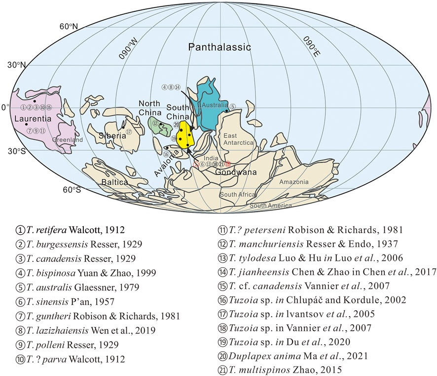
FIGURE 1. Paleogeographical distribution of Tuzoia. Paleogeographical reconstruction in earlier and middle Cambrian modified from Ma et al. (2021). Numbers 1 and 15 are reported for the first time in Gondwana.
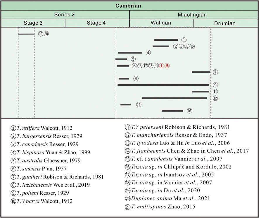
FIGURE 2. Stratigraphical distribution of Tuzoia; numbers match those in Figure 1. Numbers 1 and 15 are reported for the first time in the Guanshan Biota.
Since Walcott first described Tuzoia in 1912, more than 20 species of Tuzoia have been recorded prior to this study (Vannier et al., 2007; Wen et al., 2019; Ma et al., 2021) (Figure 2). The first record of Tuzoia was a single specimen collected from the Burgess Shale (Miaolingian Series, Wuliuan Stage) in British Columbia, Canada, named T. retifera by Walcott in 1912. The stratigraphy of this specimen was likely equivalent to that of the Raymond Quarry Shale Member (Fletcher and Collins, 1998). Resser (1929) redescribed T. retifera using additional material and established three new species from the Burgess Shale: Tuzoia canadensis, Tuzoia burgessensis, and Tuzoia polleni. These and other species were later studied by Vannier et al. (2007). Following Walcott (1912) and Resser (1929), Tuzoia was discovered in many localities in North America (Resser and Howell, 1938; Briggs, 1977; Robison and Richards, 1981; Lieberman, 2003; Vannier et al., 2007; Moore and Lieberman, 2009; Robison et al., 2015), Europe (Czech Republic) (Chlupac and Kordule, 2002; Fatka and Herynk, 2016), Siberia (Ivantsov et al., 2005), China (Endo and Resser, 1937; P’an, 1957; Shu, 1990; Luo et al., 1999; Yuan and Zhao, 1999; Chen et al., 2005; Peng et al., 2007; Yao et al., 2009; He et al., 2012; Wen et al., 2015, 2019; Chen et al., 2017; Du et al., 2020), and Australia (Glaessner, 1979; Nedin, 1995; Paterson et al., 2008; Garcıa-Bellido et al., 2009).
This study reported on Tuzoia from the Guanshan Biota (Wulongqing Formation, Cambrian Series 2, Stage 4), a representative Burgess Shale-type biota from southwestern China that included several categories of soft-bodied metazoans (Luo et al., 2005; Li et al., 2006; Luo et al., 2006; Hu et al., 2007; Luo et al., 2007; Yang et al., 2008.; Hu et al., 2010a; Hu et al., 2010b; Liu et al., 2012; Hu et al., 2013; Liu et al., 2015; Liu et al., 2016; Li et al., 2017; Yang et al., 2018; Wu and Liu. 2019; Chen et al., 2020; Zhao et al., 2020). More than 60 taxa have been described from this biota, belonging to more than ten animal groups and algae, including arthropods, brachiopods, vetulicolians, lobopodians, sponges, eocrinoid echinoderms, hyolithids, polychaetes, priapulids, chancelloriids, eldonoids, and problematic fossils (Luo et al., 2005; Li et al., 2006; Luo et al., 2006; Hu et al., 2007; Luo et al., 2007; Yang et al., 2008.; Hu et al., 2010a; Hu et al., 2010b; Liu et al., 2012; Hu et al., 2013; Liu et al., 2015; Liu et al., 2016; Li et al., 2017; Yang et al., 2018; Zhao et al., 2018; Wu and Liu. 2019; Chen et al., 2020; Zhao et al., 2020). Among the large bivalved arthropods from the Wulongqing Formation, Tuzoia ranked second, after Isoxys.
The materials were collected from the Longbaoshan and Xinglongcun sections. Five species were identified, two of which were reported for the first time from South China. In addition, two comparative species of this genus were illustrated from the Burgess Shale Formation of British Columbia, Canada (Cambrian Wuliuan Stage) (Vannier et al., 2007).
All new specimens discussed here were excavated from the Longbaoshan Section (Luo et al., 2008) between 2015 and 2021. Specimens were observed and prepared with fine needles under a microscope’s high magnification. Fossils were photographed with a Canon EOS 5D Mark II digital camera. Figures were prepared using Adobe Photoshop CS6 and CorelDraw X4.
All specimens were deposited in the Shaanxi Key Laboratory of Early Life and Environments, Northwest University (NWU), Xi’an, China.
The morphological terminology used in this study follows that of Scott (1961), Briggs (1978), Robison and Richards (1981), Lieberman (2003), Vannier et al. (2007), and Wen et al. (2015). A list of relevant abbreviations is presented in Table 1.
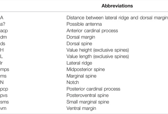
TABLE 1. List of abbreviations.
Phylum Arthropoda Siebold and Stannius, 1845 (total group).
Class Uncertain
Order Tuzoiida Simonetta and Delle, 1975
Family Tuzoiidae Raymond, 1935
Genus Tuzoia Walcott, 1912
Tuzoia retifera Walcott, 1912, Middle Cambrian Burgess Shale (Middle Cambrian: Miaolingian Series, Wuliuan Stage), British Columbia, Canada.
Tuzoia australis Glaessner, 1979; T. bispinosa Yuan and Zhao, 1999; T. burgessensis Resser, 1929; T. canadensis Resser, 1929; T. cf. canadensis Vannier et al., 2007; T. guntheri Robison and Richards, 1981; T. jianheensis Chen and Zhao in Chen et al., 2017; T. lazizhaiensis Wen et al., 2019. T. manchuriensis Endo and Resser, 1937; T. multispinos Zhao, 2015; T. polleni Resser, 1929; T. sinensis P’an, 1957; T. tylodesa Luo and Hu in Luo et al., 2006; T. praemorsa Resser, 1929; Tuzoia sp. Chlupáč and Kordule, 2002; Tuzoia sp. Ivantsov et al., 2005; Tuzoia sp. Vannier et al., 2007; Tuzoia sp. Du et al., 2020; Duplapex anima Ma et al., 2021; and questionably T.? parva Walcott 1912; T.? peterseni Robison and Richards, 1981.
Large carapace bivalved arthropod, non-biomineralized, or weakly biomineralized. Valve hemispherical, folded dorsally (no true articulated hinge), surface showing reticulate ornament. Dorsal margin straight or slightly convex, with or without spines. Anterior and posterior cardinal processes (ACP and PCP, respectively) along the dorsal margin. The ventral margin spinose typically had a mid-posterior spine (MPS) and posteroventral spine (PVS). The lateral ridge spinose extended medially along the entire length of the valves. Paired stalked eyes protruded anteriorly through the notch; eye stalks annulated. Gut with possible digestive glands.
Cambrian Stage 3 to the Drumian Stage of North America (Canada, United States), North and South China, Australia, Russia, and the Czech Republic.
Tuzoia was a diverse genus widely distributed across Laurentia, Bohemia, Siberia, and Gondwana. With at least 14 valid species, it ranks among the most diverse non-biomineralized or lightly biomineralized genera of arthropods known from the Cambrian. Tuzoia was first described in Laurentia and, subsequently, elsewhere. Among the described species, six distinctive forms (T. bispinosa Yuan and Zhao, 1999, T. Sinensis P’an, 1957, T. tylodesa Luo and Hu in Luo et al., 2006, T. multispinos Zhao, 2015, T. lazizhaiensis Wen et al., 2019, and Duplapex anima Ma et al., 2021), were all reported from North and South China. In this study, two additional distinctive species from South China were added to the list.
Tuzoia sinensis P’an, 1957 Figure 3, 7C; 1957 Tuzoia sinensis P’an, pp. 523–526, pl. 1 figs 1–2, pl. 2 figs 1–2; 1999 Tuzoia sinensis P’an; Luo et al., pp. 67–68, pl. 32 figs 4–5; 2005 Tuzoia kunmingensis Chen et al., pp. 152–154, fig. 1, pl. 2 figs 1–4; 2006 Tuzoia sinensis P’an; Luo et al., pp. 460–472, pl. 2 figs 1–3. 2007 Tuzoia sinensis P’an; Vannier et al., pp. 445–471, fig. 26; 2008 Tuzoia sinensis P’an; Luo et al., pp. 86, pl. 25 figs 1–6. 2012 Tuzoia sinensis P’an; He et al., pp. 10–13, pl. 1 figs 1–5; 2013 Tuzoia sinensis P’an; Hu et al., pp. 122–123, figs 158–161; 2019 Tuzoia sinensis P’an; Wen et al., pp. 719–742, pl. 4–5, pl. 12 figs E–F.
Forty-nine specimens were collected from the Wulongqing Formation (Cambrian Stage 4) in the Longbaoshan and Xinglongcun sections of Yunnan, China.
The original diagnoses (Luo et al., 2006; Hu et al., 2013) are followed here. A carapace with an ovoid, slightly postplete outline was observed. T. sinensis had a relatively short and straight dorsal margin bearing numerous spines, which occurred along the ventral and posterior margins. The ACP was small, pointing slightly upward. The PCP was broad-based and subtriangular. The lateral ridge was oblique to the dorsal margin. A reticulate ornament was present over the entire lateral surface (Figure 3A–D, Figure 7).
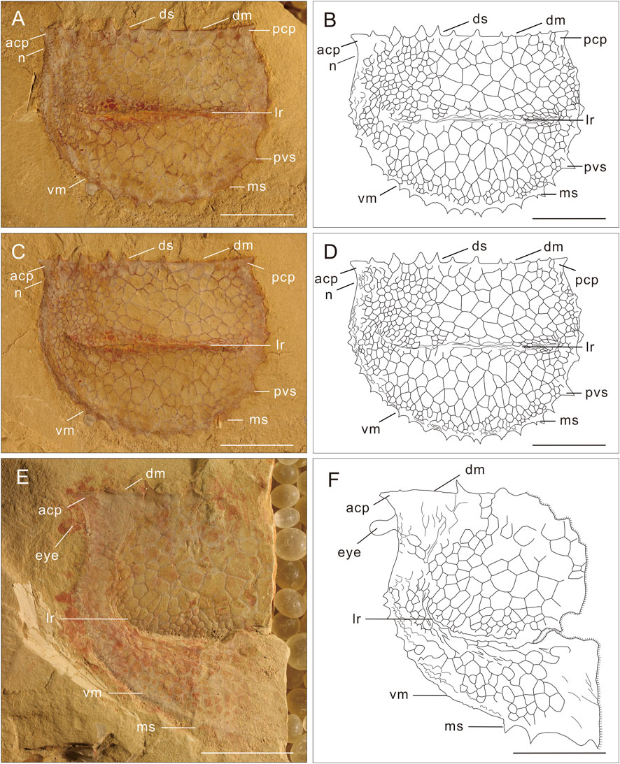
FIGURE 3. Tuzoia sinensis P’an, 1957 from the Wulongqing Formation in the Longbaoshan Section, Yunnan, China. (A,B) Lateral aspect views of specimen CG0901A. (C,D) Lateral aspect views of specimen CG0901B, showing the T. sinensis from Longbaoshan Section. ACP is small, PCP is broad-based, and sub-triangular, numerous spines occur along the ventral and the dorsal margins. (E,F) Lateral aspect views of specimen ELI-LBST-0024, showing the eye of T. sinensis, with the dorsal spines and ventral spines, with few tumor-like protrusions on the surface. The scale bars are 10 mm. For abbreviations, see Table 1.
The specimens from the Wulongqing Formation of Kunming described herein showed morphology similar to the original material of T. sinensis from the Balang Formation of Guizhou (Wen et al., 2019). The specimens described in this study range in length from 35 to 61 mm and showed the lateral ridge sub-parallel to the dorsal margin, similar to specimens from the Balang Biota.
One specimen of T. sinensis bore a stalked eye protruding from the notch below the anterior cardinal process. The eye was large, oval-shaped, and approximately 2 mm in diameter, extending from a stalk (Figures 3E,F, 7).
Wulongqing Formation (Cambrian Stage 4) of Yunnan Province, South China, and Balang Formation (Cambrian Stage 4) of Guizhou Province, South China.
Tuzoia tylodesa Luo et Hu et al., 2006 Figure 4, 7D; 2006 Tuzoia tylodesa Luo et Hu, pp. 460–472, pl. 1 figs 3–5, pl. 2 figs 4–6; 2015 Tuzoia tylodesa Luo et Hu, Zhao et al., pp. 1–62, pl. 1 figs. 3–7; 2019 Tuzoia tylodesa Luo et Hu, Wen et al., pp. 719–742, pl. 12 figs A–D.
Twenty-seven specimens, most of which were preserved in the lateral aspect on the surface of the bedding planes, were all from the Wulongqing Formation (Cambrian Stage 4) in the Longbaoshan Section, Yunnan, China.
Carapace bivalved arthropod, thin and non-biomineralized, with an ovoid outline, truncated dorsally by a straight convex hinge, apparently without accurate articulation. The valve length ranged from 8 to 60 mm; the valve length was two times the height. The dorsal margin was straight, with 6–10 small subtriangular marginal spines extending along the dorsal margin length. ACP and PCP were along the dorsal margin, straight or slightly upward; ACP was usually more prominent than PCP and with an underlying notch.
The anterior margin was inflated more prominently than the ACP; the PCP was more prominent than the posterior margin. The posterior margin was relatively straight and extended beyond the anterior margin. There were at least 20 subtriangular marginal spines, more than three of which were located at the anterior margin. Dorsal margin spines were the strongest, while anterior margin spines were the weakest. The surface was covered with a tumor-like protrusion, 0.4–1.2 mm in diameter and numbering 43–100. The lateral ridge was relatively distinct and protruded, extending downward and backward. The lateral ridge was decorated with tiny protrusions, similar to the margin spines.
The present study argues that the morphological stage of Tuzoia was distinguished primarily based on its size and characteristics. The larval stage of T. tylodesa, which was less than 2 cm in length, had no spines on the dorsal and ventral margins. New larvae specimens can be classified into two categories: early larval stage and late larval stage. The early larval stage specimens (Figures 4A,B) had a tiny ovoid outline, with a length less than 1 cm, ACP and PCP along the straight dorsal margin, and three spines between the ACP and PCP, closer to the PCP. The ventral margin had no spine. The lateral ridge was slightly (Figures 4C,D) oblique to the dorsal margin. A few tumor-like protrusions were present over the entire lateral surface. The late larval stage was bigger than the early stage; the length was 1–2 cm, ACP and PCP were along the straight dorsal margin, and three spines occurred between the ACP and PCP, closer to the PCP. The ventral margin had a few tiny spines. The lateral ridge was slightly oblique to the dorsal margin. A few tumor-like protrusions were present over the entire lateral surface. Based on these details, the ontogenetic patterns of T. tylodesa were supplemented and revised (Figure 4E).
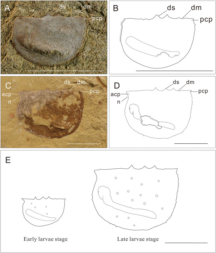
FIGURE 4. Tuzoia tylodesa Luo et Hu et al., 2007 from the Wulongqing Formation in the Longbaoshan Section, Yunnan, China. (A,B) Lateral aspect views of specimen ELI-LBST-0079A, showing the early larval stage of T. tylodesa, with three dorsal spines, no ventral spines, with few tumor-like protrusions on the surface; (C,D) Lateral aspect views of specimen ELI-LBST-0012A, showing the later larval stage of T. tylodesa, with three dorsal spines, some tiny spines among the ventral margin, with more tumor-like protrusions in the surface; (E) Two categories of T. tylodesa: Early larval stage and late larval stage. The scale bar is 10 mm. For abbreviations, see Table 1.
Longbaoshan Section, Wulongqing Formation (Cambrian Stage 4) of Yunnan Province, South China.
Tuzoia multispinosa Zhao, MS. 2015 Figure 7B; 2015 Tuzoia multispinosa Zhao, pp. 1–62, pl. 1 figs. 1–7, pl. 2 figs 1–6.
Large bivalved arthropod, valve outline, carapace thin, non-mineralized, folded dorsally (no true articulated hinge) into two hemispherical valves (L: H approximately 1.68). ACP and PCP along the straight dorsal margin. The ACP was small, pointing straight forward. The PCP was also small, pointing slightly upward. The angle formed between the dorsal margin and the posterior ventral margin was 23.6°. The posterior margin was inflated and more prominent than the PCP; the lateral ridge was at mid-distance between the dorsal and ventral margins and slightly oblique to the dorsal margin. The lateral ridge was armed with at least 31 small spines, all of which were aligned compactness and indented; the dorsal margin had ten spines equally distributed along the length of the dorsal margin. The ventral margin bore, at most, 25 minuscule subtriangular marginal spines, two of which were located at the anterior margin. The ventral marginal spines showed the form of many small spines between the two large spines primarily. There were fewer spines along the posterior margins, typically with an MPS and PVS, and the PVS was slightly longer than the MPS (Figure 7B).
T. multispinosa was first described by Zhao in 2015, based on only three specimens. This study obtained no additional material. More specimens should be collected for further studies.
Longbaoshan Section, Wulongqing Formation (Cambrian Stage 4) of Yunnan Province, South China.
Tuzoia retifera Walcott, 1912 Figure 5, 7A; 1912 Tuzoia retifera Walcott, pp. 187, pl. 33 fig. 2; 1929 Tuzoia retifera Walcott; Resser, pp. 7–8, pl. 1 fig. 2; 1975 Tuzoia retifera Walcott; Simonetta and Delle Cave, p. 8, pl. 47 fig. 2; 2003 Tuzoia retifera Walcott; Lieberman, pp. 679; 2007 Tuzoia retifera Walcott; Vannier et al., pp. 445–471, fig. 26.
Thirteen specimens were preserved in lateral aspect on the surface of the bedding planes, all from the Wulongqing Formation (Cambrian Stage 4) in the Longbaoshan Section, Yunnan, China.
This specimen had an ovoid, slightly postplete/replete outline (L = 95 mm, H = 65 mm, L:H approximately 1.46). The dorsal margin was slightly convex. The ACP was broad-based, very short, and directed slightly downward, overhanging a well-developed anterior notch. The PCP was blunt and very small in size. No dorsal spines. MPS and PVS were present but were very short. There were additional marginal spines between the MPS and the PVS. Low-relief lateral ridge ran almost parallel to the dorsal margin (H:A approximately 2.0), underlined by a narrow frill with very small crenulation. The reticulate pattern was dense, uniform, and small along the anterior, posterior, and dorsal margins (Figures 5A,D, 7A).
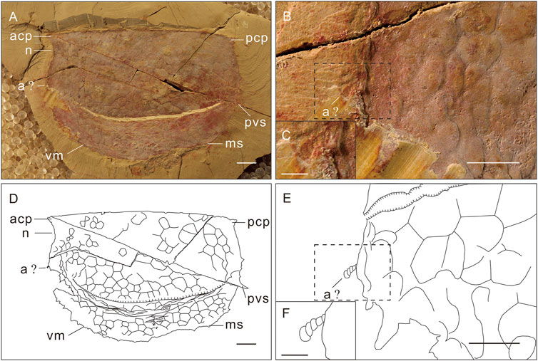
FIGURE 5. Tuzoia retifera Walcott, 1912 from the Wulongqing Formation in the Longbaoshan Section, Yunnan, China. (A,D) Lateral aspect views of specimen ELI-LBST-0045A, showing a mature stage, no dorsal spines. MPS and PVS are present but very short, with additional marginal spines between MPS and PVS. (B,E) Enlarged plot of (A,C,D,F) Enlarged plot of (B,E) showing the antenna of the T. sinensis. The scale bars of (A,B,D,E) are 10 mm, and the scale bars of (C,F) are 1 mm. For abbreviations, see Table 1.
T. cf. retifera was described by Hu et al. in 2013. Because the size of their specimens was too small and there was only one spine at the ventral margin, the specimen could not be considered as T. retifera. The details of the new specimen described herein (Figures 5A,D) were identical to those of T. retifera. The study inferred that T. cf. retifera (Hu et al., 2013) might be the juvenile stage of T. retifera. This is not only the first time that T. retifera has been confirmed in Guanshan Biota (Series 2, Stage 4) but also the first time that T. retifera was reported out of Laurentia (in Gondwana). This report indicated that the communication of species between paleogeographic plates was possible, providing new evidence for the study of the communication of species between paleogeographic plates.
A small, segmented structure under the notch was interpreted as a fragment of the antenna (Figures 5B,C,E,F, 7A). The fragment was extremely short, multi-segmented, and composed of several articles. As this fragment was too small to be a specimen, confirmation from more specimens with well-preserved antennae remains a goal.
Burgess Shale Formation in British Columbia, Canada; Wulongqing Formation (Cambrian Stage 4) in Yunnan Province, South China.
Tuzoia cf. canadensis Resser, 1929 Figure 6, 7E.
Three specimens were preserved in lateral aspect in shale, one of which was well-preserved. The other two specimens were preserved as fragments from the Wulongqing Formation (Cambrian Stage 4) Longbaoshan Section, Yunnan, China.
This form was represented by a single specimen with both valves preserved in a lateral position (NWU-LBS-0137). It had an ovoid outline (L = 45 mm, H=28 mm, L:H approximately 1.61) and well-developed cardinal processes, SMS, MPS, and PVS. The ACP pointed straight forward and overhung the notch. The PCP was longer than the ACP and was directed upward and backward. The ventral spines (at least eight) were slender, and their length increased toward the front. The dorsal margin had at least six long spines. The lateral ridge was at mid-distance between the dorsal and ventral margins and bore spines. The surface showed a reticulate pattern formed by polygons; most polygons had 4–5 sides and were 2–4 mm in diameter (Figures 6A–F, 7E).
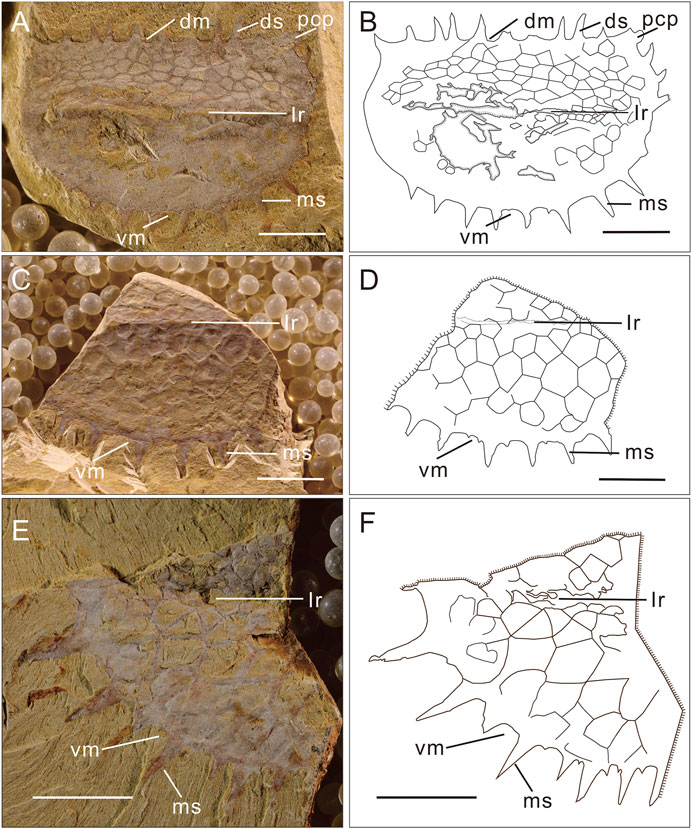
FIGURE 6. Tuzoia cf. Canadensis Resser, 1929 from the Wulongqing Formation in the Longbaoshan Section, Yunnan, China. (A,B) Lateral aspect views of specimen ELI-LBST-0156A, a new species in the Guanshan biota, the T. cf. canadensis. The ventral spines (at least eight) are slender, and the dorsal margin has at least six long spines. The typical character of this species is the relatively longer and more prominent spines. (C,D) Lateral aspect views of specimen ELI-LBST-0032, also with longer and prominent spines. (E,F) Lateral aspect views of specimen ELI-LBST-0197A, also with longer and prominent spines. The scale bar is 10 mm. For abbreviations, see Table 1.
These specimens were similar to T. canadensis and T. cf. canadensis (Vannier et al., 2007), both of which were from the Cambrian Wuliuan Stage. These specimens differ from T. canadensis in having more spines along the dorsal margin, in missing the secondary spines along their dorsal spines margins, in having a shorter ACP and PCP along the dorsal margin, and in having relatively longer and more prominent spines, especially in the ventral margin. Compared with T. cf. canadensis, except for the lack of awareness caused by the incomplete specimen, the two of them almost touched the same. These specimens differ from T. sinensis in having longer and more prominent spines, especially in the ventral margin. These specimens differ from T. tylodesa in having relatively longer and more prominent spines, especially in the dorsal-ventral margin, in the position of the lateral ridge, in the decoration on the surface, in the outline, and the outline angle formed between the dorsal margin and the posterior ventral margin. These specimens differ from T. multisinosa in having relatively longer and thinner spines, in the missing of the secondary spines between the dorsal/ventral spines and in the angle formed between the dorsal margin and the posterior ventral margin. These specimens differ from T. retifera in having relatively longer and more prominent spines in the dorsal and ventral margin (Figure 7).
The species broadly resembled T. canadensis in outline and was an additional material of T. cf. canadensis. It is possible that these specimens represented a new species. However, full confirmation requires the collection of complete material.
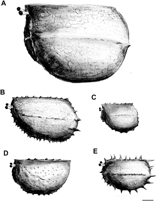
FIGURE 7. Reconstructions of the Tuzoia from Guanshan Biota, Yunnan, China. (A) Reconstruction of the Tuzoia retifera Walcott, 1912; (B) Reconstruction of the T. multispinosa Zhao, 2015; (C) Reconstruction of the T. sinensis P’an; (D) Reconstruction of the T. tylodesa Luo et Hu et al., 2007; (E) Reconstruction of the T. cf. canadensis Resser, 1929. The scale bar is 10 mm.
Longbaoshan Section, Wulongqing Formation, Kunming, Yunnan, China.Geographic and stratigraphic distribution. Longbaoshan Section in Kunming, Southwest China (Cambrian Series 2, Stage 3; local Wulongqing Formation).
1. This study distinguished and described the following species of Tuzoia from the Guanshan Biota in detail: 1. T. sinensis P’an, 1957; 2. T. tylodesa Luo et Hu et al., 2007; 3. T. multispinosa Zhao, 2015; 4. T. retifera Walcott, 1912; and 5. T. cf. canadensis Resser, 1929.
2. The specimens we described herein definitely represent that it is T. retifera. We inferred that T. cf. retifera is the juvenile stage of T. retifera, rather than the conformis species. It is the first time that T. retifera reported in the Guanshan Biota.
3. This study also described new material about T. cf. canadensis and provided supplementary details about this species. This was the first report of T. cf. canadensis in the Guanshan Biota.
4. The presence of T. retifera and T. cf. canadensis in the Guanshan Biota demonstrated communication of the species between paleogeographic plates, providing new evidence for the study of “species communication between paleogeographic plates.”
5. Finally, the study provided additional material related to T. tylodesa and supplemented and revised the ontogenetic pattern of T. tylodesa, shedding new light on the ontogenetic pattern of Tuzoia.
The raw data supporting the conclusion of this article will be made available by the authors, without undue reservation.
JL conceived the project. YW interpreted the fossil material and wrote the draft manuscript. Both the authors were involved in developing observations and discussed the results.
This research was supported by the Key Scientific and Technological Innovation Team Project in Shaanxi Province, the National Natural Science Foundation of China (42130206, 41890845, 41222014, 41172023, 41621003, and 41102012), the 111 Project, and the Ministry of Education of China for Changjiang Scholars to JL.
The authors declare that the research was conducted in the absence of any commercial or financial relationships that could be construed as a potential conflict of interest.
All claims expressed in this article are solely those of the authors and do not necessarily represent those of their affiliated organizations, or those of the publisher, the editors, and the reviewers. Any product that may be evaluated in this article, or claim that may be made by its manufacturer, is not guaranteed or endorsed by the publisher.
We are grateful to Feifei Chen, Yi Ding, Jinshu Li, Shengxiang Luo, Yingyue Ma, Li Yan, and Wenyu Zhao, at Northwest University (Xi’an, China), for their assistance in both field and laboratory work.
Briggs, D. E. G. (1977). Bivalved Arthropods from the Cambrian Burgess Shale of British Columbia. Palaeontology 22, 631–664. (North America).
Briggs, D. E. G. (1978). The Morphology, Mode of Life, and Affinities of Canadaspis Perfecta (Crustacea: Phyllocarida), Middle Cambrian, Burgess Shale, British Columbia. Philosophical Trans. R. Soc. Lond. B 281, 439–487.
Chen, G. Y., Han, N. Y., and Zhang, J. Z. (2005). New Materials of Tuzoia from the Lower Cambrian, Yunnan Province. J. Guilin Univ. Technology 2, 152–154. doi:10.1360/gs050302
Chen, H., Legg, D., Liu, Y., Hou, X.-g., and Hou, X. G. (2020). New Data on the Anatomy of Fuxianhuiid Arthropod Guangweicaris Spinatus from the Lower Cambrian Guanshan Biota, Yunnan, China. App 65 (1), 139–148. doi:10.4202/app.00508.2018
Chen, W. Y., Zhao, Y. L., Yang, X. L., and Wen, R. Q. (2017). Tuzoia Walcott, 1912 from the Cambrian ‘Tsinghsutung Formation’ of Guzihou, China. Acta Palaeontologica Sinica 56, 301–311. [in Chinese with English abstract].
Chlupáč, I., and Kordule, V. (2002). Arthropods of Burgess Shale Type from the Middle Cambrian of Bohemia (Czech Republic). Bull. Czech Geol. Surv. 77, 167–182. doi:10.3140/bull.geosci.2002.03.167
Du, K.-s., Ortega-Hernández, J., Yang, J., Yang, X.-y., Guo, Q.-h., Li, W., et al. (2020). A New Early Cambrian Konservat-Lagerstätte Expands the Occurrence of Burgess Shale-type Deposits on the Yangtze Platform. Earth-Science Rev. 211, 103409. doi:10.1016/j.earscirev.2020.103409
Endo, R., and Resser, C. E. (1937). The Sinian and Cambrian Formations and Fossils of Southern Manchoukuo. Bull. Manchurian Sci. Mus. 406.
Fletcher, T. P., and Collins, D. H. (1998). The Middle Cambrian Burgess Shale and its Relationship to the Stephen Formation in the Southern Canadian Rocky Mountains. Can. J. Earth Sci. 35, 413–436. doi:10.1139/e97-120
Glaessner, M. F. (1979). Lower Cambrian Crustacea and Annelid Worms from Kangaroo Island, South Australia. Alcheringa: Australas. J. Palaeontology 3, 21–31. doi:10.1080/03115517908565437
He, T. J., Liu, J. N., and Han, J. (2012). Tuzoia Sinensis from the Lower Cambrian Gaunshan Fauna of Eastern Yunnan Province. Geology. Gansu Province 21, 10–13. [in Chinese with English abstract].
Hu, S., Luo, H., Hou, S., and Erdtmann, B.-D. (2007). Eocrinoid Echinoderms from the Lower Cambrian Guanshan Fauna in Wuding, Yunnan, China. Chin. Sci Bull 52 (5), 717–719. doi:10.1007/s11434-007-0083-6
Hu, S. X., Li, X. K., Tan, X. H., Zhan, D. Q., and Luo, H. L. (2010b). Phlogites from the Early Cambrian Guanshan Fauna. Acta Palaeontologica Sinica 49 (3), 360–364. doi:10.19800/j.cnki.aps.2010.03.008
Hu, S. X., Zhu, M., Steiner, M., Luo, H., Zhao, F., and Liu, Q. (2010a). Biodiversity and Taphonomy of the Early Cambrian Guanshan Biota, Eastern Yunnan. Sci. China Earth Sci. 53 (12), 1765–1773. doi:10.1007/s11430-010-4086-9
Hu, S. X., Zhu, M. Y., Luo, H. L., Steiner, M., Zhao, F. C., Li, G. X., et al. (2013). The Guanshan Biota. Kunming, Yunnan Science and Technology Press, 106–109.
Huilin, L., Xiaoping, F., Shixue, H., Yong, L., Liangzhong, C., Ting, Y., et al. (2005). New Vetulicoliids from the Lower Cambrian Guanshan Fauna, Kunming. Acta Geologica Sinica 79, 1–6. doi:10.1111/j.1755-6724.2005.tb00860.x
Ivantsov, A. I., Zhuravlev, A. I., Krasilov, V. A., Leguta, A. V., Melnikova, L. M., Urbanek, A., et al. (2005). Unique Sinsk Localities of Early Cambrian Organisms (Siberia Platform). Moscow: Nauka. Rossiyskaya Akademia Nauk, 143. [in Russian].
Li, J., Liu, J., and Ou, Q. (2017). New Observations on Vetulicola Longbaoshanensis from the Lower Cambrian Guanshan Biota (Series 2, Stage 4), South China. Sci. China Earth Sci. 60, 1795–1804. doi:10.1007/s11430-017-9088-y
Li, Y., Chen, L. Z., Luo, H. L., Fu, X. P., Hu, S. X., You, T., et al. (2006). New Advances in the Study of the Early Cambrian Guanshan Fauna in the Kunming Area, Yunnan, China. Geol. Bull. China 25 (3), 415–418. doi:10.3969/j.issn.1671-2552.2006.03.012
Lieberman, B. S. (2003). A New Soft-Bodied Fauna: the Pioche Formation of Nevada. J. Paleontol. 77, 674–690. doi:10.1666/0022-3360(2003)077<0674:ansftp>2.0.co;2
Liu, J., Han, J., Li, J., Wu, Y., Peng, J., Qi, N., et al. (2016). New Localities and Palaeoscolecid Worms from the Cambrian (Stage 4, Series 2) Guanshan Biota in Kunming, Yunnan, South China. Acta Geologica Sinica - English Edition 90 (6), 1939–1945. doi:10.1111/1755-6724.13013
Liu, J. N., Ou, Q., Han, J., Zhang, Z. F., He, T. J., Yao, X. Y., et al. (2012). New Occurence of the Cambrian (Stage 4, Series 2) Guanshan Biota in Huize, Yunnan, South China. Bull. Geosci. 87 (1), 125–132. doi:10.3140/bull.geosci.1229
Liu, J., Ou, Q., Han, J., Li, J., Wu, Y., Jiao, G., et al. (2015). Lower Cambrian Polychaete from China Sheds Light on Early Annelid Evolution. Sci. Nat. 102, 34. doi:10.1007/s00114-015-1285-4
Luo, H. L., Fu, X. P., Hu, S. X., Li, Y., Chen, L. Z., You, T., et al. (2006). New Bivalved Arthropods from the Early Cambrian Guanshan Fauna in the Kunming and Wuding Area. Acta Palaeontologica Sinica 45 (4), 460–472. doi:10.1016/S1004-4132(06)60002-9
Luo, H. L., Fu, X. P., Hu, S. X., Li, Y., Hou, S. G., You, T., et al. (2007). A new Arthropod, Guangweicaris Luo, Fu et Hu gen. nov. from the Early Cambrian Guanshan Fauna, Kunming, China. Acta Geologica Sinica (English Edition) 8l, 1–7. doi:10.3321/j.issn:1000-9515.2007.01.001
Luo, H. L., Hu, S. X., Chen, L. Z., Zhang, S. S., and Tao, Y. H. (1999). Early Cambrian Chengjiang Fauna from Kunming Region. ChinaKunming: Yunnan Science & Technology Press, 162. [in Chinese with English summary].
Luo, H. L., Li, Y., Hu, S. X., Fu, X. P., Hou, S. G., Liu, X. R., et al. (2008). The Malong Fauna and the Guanshan Fuana from the Lower Cambrian Eastern Yunan. Yunnan Science & Technology Press, 134. [in Chinese with English summary].
Ma, J., Lin, W., Liu, C., Sun, A., Wu, Y., Wu, Y., et al. (2021). A New Bivalved Arthropod from the Cambrian (Stage 3) Qingjiang Biota Expands the Palaeogeographical Distribution and Increases the Diversity of Tuzoiidae. J. Geol. Soc. 179, jgs2020–229. doi:10.1144/jgs2020-229
Moore, R. A., and Lieberman, B. S. (2009). Preservation of Early and Middle Cambrian Soft-Bodied Arthropods from the Pioche Shale, Nevada, USA. Palaeogeogr. Palaeoclimatol. Palaeoecol. 277, 57–62. doi:10.1016/j.palaeo.2009.02.014
Nedin, C. (1995). The Emu Bay Shale, a Lower Cambrian Fossil Lagerstätten, Kangaroo Island, South Australia. Mem. Assoc. Australas. Palaeontologists 18, 31–40.
P’an, J. (1957). On the Discovery of Homopoda from South China. Acta Palaeontologica Sinica 5, 523–526. [in Chinese with English summary].
Peng, J., Fen, H. Z., Zhao, Y. L., Fu, X. P., and Wang, Y. X. (2007). The Discovery of Tuzoia from Balang Formation Lower Cambrian Eastern Guizhou. Geology. Rev. 53, 397–402. [in Chinese with English summary]. doi:10.1016/S1872-5791(07)60044-X
Raymond, P. E. (1935). Leanchoilia and Other Mid-cambrian Arthropoda. Bull. Mus. Comp. Zoolog. 76 (6), 205–230. Harvard University.
Resser, C. E., and Howell, B. F. (1938). Lower Cambrian Olenellus Zone of the Appalachians. Geol. Soc. America Bull. 49, 195–248. doi:10.1130/gsab-49-195
Resser, C. E. (1929). New Lower and Middle Cambrian Crustacea. Proc. U.S. Natl. Mus. 76, 1–18. doi:10.5479/si.00963801.76-2806.1
Robison, R. A., Babcock, L. E., and Gunther, V. G. (2015). Exceptional Cambrian Fossils from Utah: A Window into the Age of Trilobites, 15-1. Utah Geological Survey, Miscellaneous Publication, 97.
Robison, R. A., and Richards, B. C. (1981). Larger Bivalve Arthropods from the Middle Cambrian of Utah, 106. Paleontological Contributions of the University of Kansas, 1–19.
Scott, H. V. (1961). “Shell Morphology of Ostracoda. Q21– Q43,” in Treatise on Invertebrate Paleontology. Part Q. Arthropoda 3. Crustacea, Ostracoda. Editors R. C. MOORE, and C. TEICHERT (Geological Society of America & University of Kansas Press).
Shu, D. G. (1990). Cambrian and Lower Ordovician Bradoriida from Zhejiang. Xian, Hunan and Shaanxi Provinces: Northwest University Press, 95.
Siebold, K. T. E., and Stannius, H. F. (1845). Lehrbuch der vergleichenden Anatomieder wirbellosen Tiere. Berlin: Von Veit.
Simonetta, A., and Delle, C. L. (1975). The Cambrian Non-trilobite Arthropods from the Burgess Shale of British Columbia. A Study of Their Comparative Morphology, Taxonomy and Evolutionary Significance. Palaeontographica Italica 69, 1–37.
Vannier, J., Caron, J.-B., Yuan, J.-L., Briggs, D. E. G., Collins, D., Zhao, Y.-L., et al. (2007). Tuzoia: Morphology and Lifestyle of a Large Bivalved Arthropod of the Cambrian Seas. J. Paleontol. 81, 445–471. doi:10.1666/pleo05070.1
Walcott, C. D. (1912). Cambrian Geology and Paleontology II: Middle Cambrian Branchiopoda, Malacostraca, Trilobita and Merostomata. Smithsonian Miscellaneous Collections 57, 145–228.
Wen, R.-Q., Zhao, Y.-L., and Peng, J. (2015). Morphology and Ontogeny of Tuzoia Bispinosa from the Kaili Biota (Cambrian Stage 5) of Eastern Guizhou, China. Palaeoworld 24, 61–70. doi:10.1016/j.palwor.2014.12.005
Wen, R., Babcock, L. E., Peng, J., Liu, S., and Liang, B. (2019). The Bivalved Arthropod Tuzoia from the Balang Formation (Cambrian Stage 4) of Guizhou, China, and New Observations on Comparative Species. Pap. Palaeontol. 5 (4), 719–742. doi:10.1002/spp2.1262
Wu, Y. C., and Liu, J. N. (2019). Anatomy and Relationships of the Fuxianhuiid Euarthropod Guangweicaris from the Early Cambrian Guanshan Biota in Kunming, Yunnan, Southwest China. Acta Palaeontologica Pol. 64 (3), 543–548. doi:10.4202/app.00542.2018
Yang, J., Hou, X. G., and Dong, W. (2008). Restudy of Guangweicaris Luo, Fu et Hu, 2007 from the lower Cambrian Canglangpu Formation in Kunming area. Acta Palaeontogica Sinica 47, 115–122. [in Chinese with English abstract]. doi:10.3969/j.issn.0001-6616.2008.01.008
Yang, J., Ortega-Hernández, J., Legg, D. A., Lan, T., Hou, J.-b., and Zhang, X.-g. (2018). Early Cambrian Fuxianhuiids from China Reveal Origin of the Gnathobasic Protopodite in Euarthropods. Nat. Commun. 9, 470. doi:10.1038/s41467-017-02754-z
Yao, L., Peng, J., Fu, X. P., and Zhao, Y. L. (2009). Ontogeny of Tuzoia Bispinosa from the Middle Cambrian Kaili (Guizhou China) Fauna. Acta Palaeontologica Sinica 48, 56–64. [in Chinese with English summary]. doi:10.3969/j.issn.0001-6616.2009.01.007
Yuan, J. L., and Zhao, Y. L. (1999). Tuzoia (Bivalved Arthropods) from the Lower-Middle Cambrian Kaili Formation of Taijiang, Guizhou. Acta Palaeontologica Sinica 38, 88–93. [In Chinese with English abstract].
Zhao, J., Li, G. B., and Selden, P. A. (2018). New Well-Preserved Scleritomes of Chancelloriida from Early Cambrian Guanshan Biota, Eastern Yunnan, China. J. Paleontol. 92 (6), 955–971. doi:10.1017/jpa.2018.43
Zhao, J. (2015).The Revision of Tuzoia and Longquania from Early Cambrian Guanshan Biota of Eastern Yunnan, China. Published Master Degree thesis. China: Northwest University, 56. [in Chinese with English abstract].
Keywords: Tuzoia, bivalved arthropod, Wulongqing Formation, Guanshan Biota, Yunnan, South China
Citation: Wu Y and Liu J (2022) New Data on the Bivalved Arthropod Tuzoia From the Cambrian (Series 2, Stage 4) Guanshan Biota in Kunming, Yunnan, Southwest China. Front. Earth Sci. 10:862679. doi: 10.3389/feart.2022.862679
Received: 26 January 2022; Accepted: 21 March 2022;
Published: 26 April 2022.
Edited by:
Haijun Song, China University of Geosciences, Wuhan, ChinaReviewed by:
Shixue Hu, Chengdu Geological Survey Center, ChinaCopyright © 2022 Wu and Liu. This is an open-access article distributed under the terms of the Creative Commons Attribution License (CC BY). The use, distribution or reproduction in other forums is permitted, provided the original author(s) and the copyright owner(s) are credited and that the original publication in this journal is cited, in accordance with accepted academic practice. No use, distribution or reproduction is permitted which does not comply with these terms.
*Correspondence: Jianni Liu, ZWxpbGpuQG53dS5lZHUuY24=
Disclaimer: All claims expressed in this article are solely those of the authors and do not necessarily represent those of their affiliated organizations, or those of the publisher, the editors and the reviewers. Any product that may be evaluated in this article or claim that may be made by its manufacturer is not guaranteed or endorsed by the publisher.
Research integrity at Frontiers
Learn more about the work of our research integrity team to safeguard the quality of each article we publish.