
95% of researchers rate our articles as excellent or good
Learn more about the work of our research integrity team to safeguard the quality of each article we publish.
Find out more
METHODS article
Front. Earth Sci. , 30 March 2022
Sec. Paleontology
Volume 10 - 2022 | https://doi.org/10.3389/feart.2022.810406
This article is part of the Research Topic Precambrian Paleontology View all 13 articles
 Matheus Denezine1*
 Rodrigo Rodrigues Adôrno1,2Dermeval Aparecido Do Carmo1Edi Mendes Guimarães1Detlef Hans Gert Walde1Carlos José Souza De Alvarenga1Gerard Germs3Lucas Silveira Antonietto1
 Christian Gianfranco Valdivia Rodríguez4Osvaldo De Oliveira Nunes Junior1
The recovery of microfossils from Proterozoic rocks is commonly challenging because of metamorphism. In this study, an application of different methods usually applied on Phanerozoic rocks to test efficiency on recovering microfossil from Proterozoic units is presented. Chemical, physical, and biological factors can influence the recovery of microfossils, thereby becoming a barrier for biostratigraphic and paleoecological studies. Furthermore, low-cost projects with a reduced amount of sample collected, such as drill core sampling, need to optimize the preparation time and sample needed for different analyses. To overcome this challenge, the classical procedure of mineralized microfossil preparation, the palynological technique, and the study of clay mineralogy with the analyses of diagenetic alteration and the search for possible microfossils in thin sections were combined. Three Proterozoic lithostratigraphic units were selected to develop an integrated procedure for preparing samples for micropaleontologic and sedimentologic studies: the Paranoá Group, Mesoproterozoic, and the Bambuí Group, Ediacarian-Cambrian, Brazil, and Nama Group, Ediacaran-Cambrian, Namibia. Recovering individual microfossils from the Paranoá and Bambuí groups has been a challenge for paleontologists. Therefore, most micropaleontological studies have been done as a part of microbiofacies analyses in thin sections. All sediment fractions were studied in trial for the examination (and picking) of mineralized microfossils, even the finest ones. The microfossil picking was conducted using a stereomicroscope. Three species were recovered following this procedure: Vetronostocale aff V. amoenum Schopf and Blacic, 1971, Myxococcoides sp., and Melanocyrillium sp. Analyses in whole rock samples of residues from water (H2O) and hydrogen peroxide 30% (H2O2) procedures showed similar results when the clay fraction studied was obtained as part of micropaleontological preparation compared with the results from the standard clay mineral preparation method. The clay fraction diffractograms showed that the micropaleontological preparation with H2O and H2O2 caused an increase in the intensity of the quartz reflections compared with untreated samples. Moreover, detailed protocols for organic-walled microfossil preparation and low concentrated acetic and formic acids attacks for mineralized microfossil extraction were presented.
The diversity and preservation of fossil specimens from the Precambrian have been considered rare compared with those recovered from the Phanerozoic (Knoll, 1985; Schopf, 1995). Among other causes, such as taphonomic alterations, which greatly influence the fossil record, the preparation methodology also plays an essential role in recovering. Therefore, this barrier in the study of the Precambrian strata requires methodological considerations because, depending on the method applied, the fossil record may be lost. The present study proposes a protocol to increase microfossil recovery based on a combined methodology focused on micropaleontological and sedimentological analysis integration (Alves, 1987; Campos, 2012; Horne and Siveter, 2016; Leite et al., 2018). Samples from Paranoá and Bambuí groups, Brazil, and Nama Group, Namibia, were analyzed to assess all methods presented in this study.
Because of distinctive micropaleontological recoveries procedures on samples from Phanerozoic to other strata, it is necessary to formalize preparation methodologies for recovery of organic-walled and mineralized microfossils from Precambrian lithostratigraphic units. With mineralized micropaleontological analyses, the residues from the same preparation can be used for clay mineral analyses. This combination accelerates the whole research besides reducing the costs of preparation procedures. The application of this protocol could improve the recovery of microfossils from Precambrian units and, consequently, improve biostratigraphic studies besides combining analyses for micropaleontology and sedimentology for integration and reduction of costs. In the present case, at least three laboratories are working together, Laboratory of Mineralized Microfossils, Laboratory of Organic-walled Microfossils, and Laboratory of X-ray Diffraction, so curatorial procedures must be shared and followed to promote efficiency on data acquisition and analysis integration.
Moreover, it also detailed the curatorship procedures, identification, allocation by collection category, packaging, and housing samples under the policy of the Museum of Geosciences, University of Brasilia. In addition to the management methodology, rules for the transit of samples between laboratories are also described.
Two localities in Minas Gerais State, Brazil, were studied: the Buritis Municipality, which is part of the Brasília belt within the Tocantins province, and the Januária Municipality, which is located in a nondeformed domain of the São Francisco craton (Figure 1). A thick interval of Meso-Neoproterozoic sedimentary rocks was deposited along the west portion of the San Francisco craton. These rocks were separated into three stratigraphic units, from bottom to top: Paranoá Group, Jequitaí Formation, and Bambuí Group.
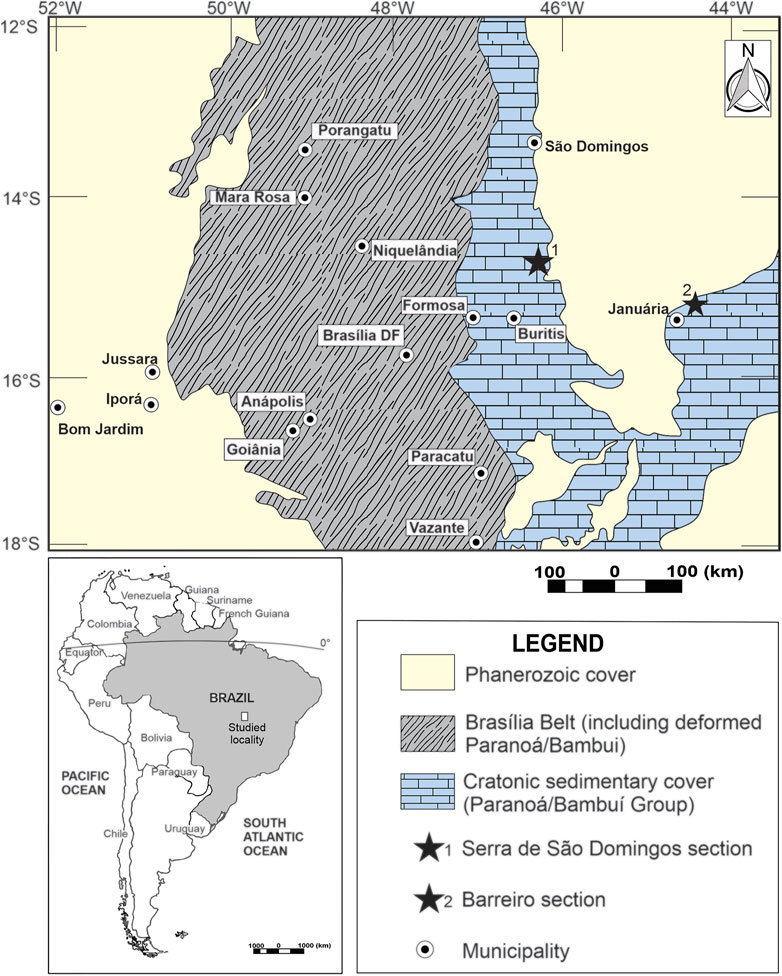
FIGURE 1. Geological map of studied areas in Brazil. (1) Serra de São Domingos section, Buritis Municipality, Minas Gerais State; (2) Barreiro section, Januária Municipality, Minas Gerais State.
The deposition of terrigenous and chemical sedimentary rocks belonging to the Paranoá Group dates from the Mesoproterozoic when the separation of the Rodinia supercontinent generated a passive rift-margin basin, West of the São Francisco craton (Alvarenga et al., 2014). Faria (1995) studied the stratigraphy of the Paranoá Group in the type locality of Alto Paraíso de Goiás and São João D'Aliança municipalities, Goiás State, Brazil; however, the study did not formalize the units according to any stratigraphic code. Thereafter, Campos et al. (2013) formalized 11 stratigraphic units within the Paranoá Group according to the Brazilian Code of Stratigraphic Nomenclature in order to adjust the informal units proposed by Faria (1995). The Paranoá Group consists of, in ascending stratigraphic order, the Ribeirão São Miguel, Córrego Cordovil, Serra da Boa Vista, Serra Almécegas, Serra do Paranã, Ribeirão Piçarrão, Ribeirão do Torto, Serra da Meia Noite, Ribeirão Contagem, Córrego do Sansão, and Córrego do Barreiro formations (Campos et al., 2013) (Figure 3).
After the deposition of the Paranoá Group, because of climatic changes, the Jequitaí Formation was deposited under glacial conditions, and their records remain in erosional contact with the Paranoá Group (unconformity) (Uhlein et al., 1995; Caxito et al., 2012). Right above in conformable contact, the carbonated-terrigenous Bambuí Group was deposited in a foreland-type basin generated from the flexure caused by tectonics in the Brasília belt. The Bambuí Group consists of five lithostratigraphic units, from base to top, the Sete Lagoas, Serra de Santa Helena, Lagoa do Jacaré, Serra da Saudade, and Três Marias formations (Dardenne, 1978) (Figure 3). Lately, the Bambuí Group has been attributed to the Ediacaran/Cambrian interval (Pimentel et al., 2011; Warren et al., 2014; Paula-Santos et al., 2015; Moreira et al., 2020; Sanchez et al., 2021).
The Nama Group, Namibia (Figure 2), represents the deposition in a shallow water foreland system; the deposition of the basal portion started around 550 Ma, followed by the deposition of siliciclastic Molasse sediments from the upper portion deposited in 540 Ma (Germs, 1983; Germs and Gresse, 1991). In the central and southern part of Namibia, the Nama Group rests discordantly on the crystalline basement. Its basal portion is represented by a succession of siliciclastic and carbonate rocks with occurrences of skeletal fossils of Cloudina lucianoi and other fossils with carbonate skeletons, as well as ichnofossils and palynomorphs in the Kuibis Formation (Germs, 1995; Gaucher et al., 2005). The upper portion of the Nama Group is represented by the Schwarzrand subgroup, which contains the ichnofossil Phycodes pedum, Cloudina, and palynomorphs (Figure 3) (Germs, 1983; Germs and Gresse, 1991; Gaucher et al., 2005).
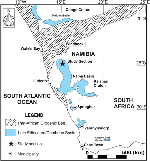
FIGURE 2. Map of the Precambrian expositions in Namibia and the studied area positioning (after Gaucher et al., 2005).
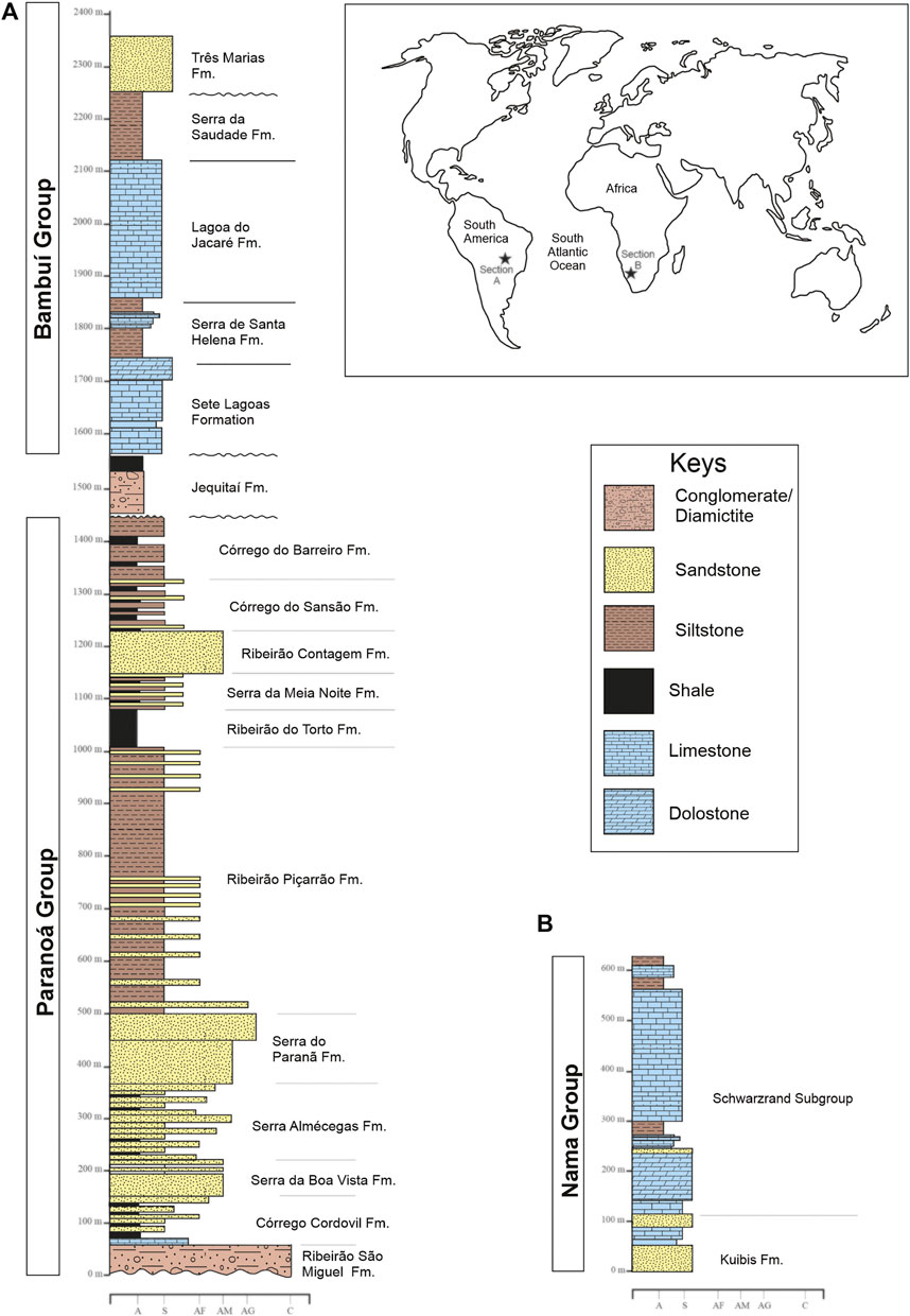
FIGURE 3. (A) Regional stratigraphy of the Paranoá and Bambui groups, adapted from Campos et al. (2013); (B) Nama Group (after Gaucher et al., 2005).
The studied material is from three Precambrian units: Paranoá (Mesoproterozoic) and Bambuí (Ediacaran-Cambrian) groups, São Francisco craton, Brazil, and Nama Group (Ediacaran-Cambrian), Namibia. The samples from Brazil were collected in outcrops from Buritis and Januária municipalities, Minas Gerais State (Table 1). Detailed methodological processes for microfossiliferous recovery are discussed in Preparation Methodologies.
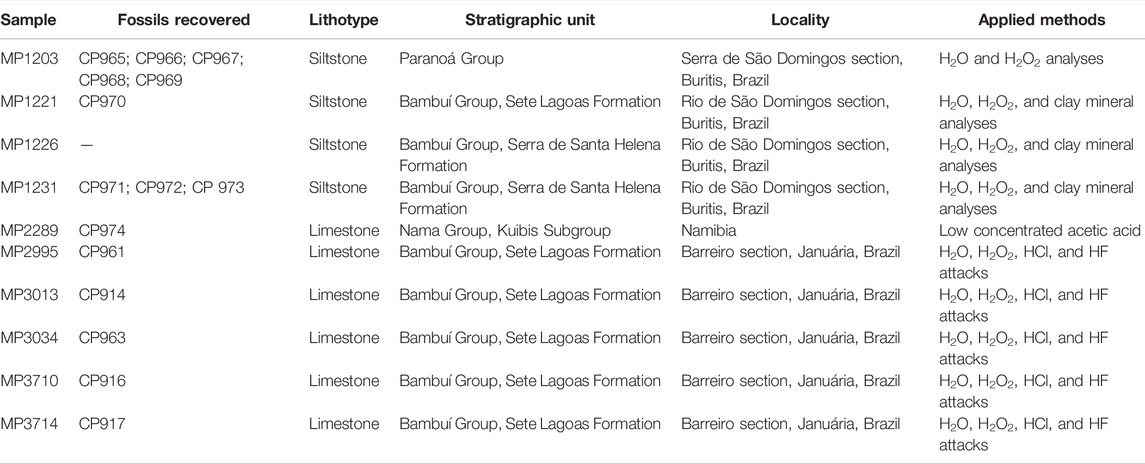
TABLE 1. Samples from Ediacaran units analyzed for specific preparations.
The same sample was analyzed throw different ways to obtain clay minerals information: (1) using the residues from water (H2O) and hydrogen peroxide 30% (H2O2) micropaleontological preparations; (2) using the standard clay preparation, which initially included material disaggregation with a hammer and powdering in the Planetary Mill pulverisette by Fritsch for 5 min with 400 revolutions/min. X-ray powder diffraction was carried out on clay fractions. Clay fractions (<2 µm) were separated by centrifugation routine at LARIX described by Campos (2012) and modified from Alves (1987). The measurements were undertaken in oriented clay fractions in air-dried conditions. Analyses were performed in a RIGAKU Ultima IV diffractometer equipped with CuKα radiation, Ni filter, under 35 kV and 15 mA. The samples were scanned at 5°/min velocity, 0.05 stepping ranging from 2 to 40°2Ɵ for clay fraction. Mineral phases were identified using Jade XRD 9.0 (Materials Data) with PC-PDF (Powder Diffraction File—PDF for PC—ICDD). Major (M), minor (m), and trace (tr) components were established by comparing the reflection intensities in d: 4.26 Å for quartz, 10 Å for illite, and 7 Å for chlorite.
Curatorship procedures must rule the studied material (rocks and fossils samples) management when multiple analysis is performed in different laboratories. This procedure aims to share information about samples, data acquisition, and analysis integration. In this study, the protocol used at the Micropaleontology Laboratory of the University of Brasilia (LabMicro), on curatorship of geological and paleontological samples that become housed at the Museum of Geosciences, was presented. The LabMicro is currently responsible for the Paleontological Collection of the Museum of Geosciences of the University of Brasilia (MGeo), which is subdivided into seven collections: (1) field collection, 2) residual samples, (3) recovered collection, (4) research collection, (5) special collection, (6) didactical collection, and (7) macrofossil collection (Table 2).
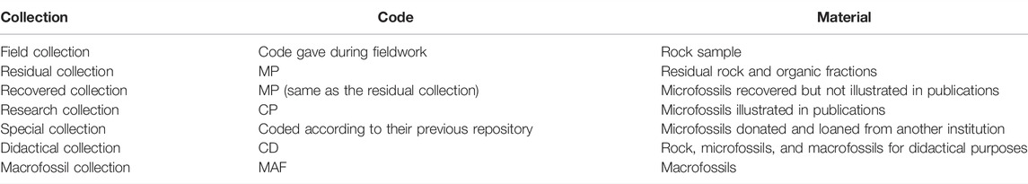
TABLE 2. Collections into the paleontological collection of the Museum of Geosciences, University of Brasilia.
Sample curatorship starts during fieldwork. Field sampling is always accompanied by labeling to identify collected samples once they arrive at the laboratory. This is guaranteed by the mandatory completion of an individual sample tag containing data about their recollection site (Figure 4). The field collection comprises materials that have recently arrived at the LabMicro through fieldwork, independent of its immediate use (or not) as research, teaching, and/or training material. If they generate such interest, samples are due to be processed through laboratory work, which will result in both a residual sample and possible recovered fossil assemblage. The residual sample left from preparation is stored in the residual collection in field bags inside storage cabinets, whereas the recovered fossil assemblage is encased in micropaleontological slides to be held in specific fossil cabinets, consisting of the recovered collection. Research macrofossil and microfossil specimens, used to illustrate taxa in publications such as articles, theses, and reports, are isolated from others either in macrofossil cabinets or microslides that will be deposited at their specific fossil cabinets. In this case, the specimen is relocated into the research collection and recoded with a CP prefix.
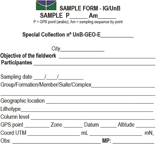
FIGURE 4. Sample datasheet used to identify samples during the Laboratory of Micropaleontology fieldwork, University of Brasília, Brazil. The datasheet contains all information needed for further curatorship.
Special collection covers fossil material of scientific interest donated or temporarily transferred to the MGeo by partner institutions such as universities, private companies, and other museums. The didactical collection is used in undergraduate and graduate courses given by the Institute of Geosciences, University of Brasília (IG); it comprises fossil material from other collections at the LabMicro and those collected by professors and students at the IG, as well as third-party direct donations. Finally, the macrofossil collection comprises macrofossil samples that require special conditions for safekeeping because of their size; therefore, they are stored in a cabinet of their own.
Samples arriving at the LabMicro initially get separated into three collections: field, macrofossils, or residual collections (the latter to be prepared for possible microfossil recovery). Once the fossil content is recovered from analyzed samples by picking, it is deposited either on multicelled micropaleontological slides (carbonate/siliceous fossils separated from rock through sieving) or glass microscope slides (organic-walled microfossils concentrated through organic preparation). The possible use of any microfossils on publications requires their relocation into single-celled micropaleontological slides to be stored in the research collection cabinet or the relocation of the entire glass microscope slide (with microfossils of scientific relevance properly marked) into the same space.
Once the initial steps of the curatorial procedure are completed, thin-section slides of the samples are produced for sedimentological/paleontological studies. Subsequently, a mechanical fragmentation of samples can be performed by using several possible methodologies, including soaking them in H2O and/or chemical attack with H2O2, acetic acid 4%–10%, formic acid 10%, hydrochloric acid 36% (HCl), and hydrofluoric acid 40% (HF).
In the present work, both water and oxygen peroxide preparations were performed for mineralized microfossiliferous recovery. After washing both preparations on a sieve set (composed of 630-, 250-, 120-, 80-, and 50-µm mesh sieves plus a collecting bucket underneath), each fraction was analyzed on a stereoscopic microscope to pick for mineralized fossil remains.
The preparation presented herein aims to recover mineralized fossiliferous remains from disaggregating 30 g of sedimentary rock samples. This method is commonly used with Quaternary and Cretaceous units (Horne and Siveter, 2016; Leite et al., 2018; Machado et al., 2020). Two distinct sieving procedures were conducted on the same sample for mineralized microfossil recovery and clay mineral analyses: (1) treatment with water before sieving and (2) attack with hydrogen peroxide before sieving. Both methods aim to disaggregate the rock sample. After sieving both products from the water treatment and hydrogen peroxide attack, the sedimentary fractions were dried in a laboratory drying oven, and then analyses were performed under a stereoscope microscope to pick microfossils.
After mechanical disaggregation, a single sample followed two preparation routes: (1) left in beaker for 48 h with H2O and (2) left in beaker for 48 h with H2O2 30% (PV). After these procedures, the samples were washed in a battery of sieves (630, 250, 160, 80, and 50 µm) (Figure 5). The fraction smaller than 50 µm were kept in an appropriate container. All fractions were dried in a laboratory drying oven at 60°C and then examined under a stereoscope microscope to pick microfossils. This drying temperature prevents unwanted fragmentation of microfossils. The finest fraction (>50 µm) from both preparations was also analyzed through X-ray diffraction for clay minerals studies.
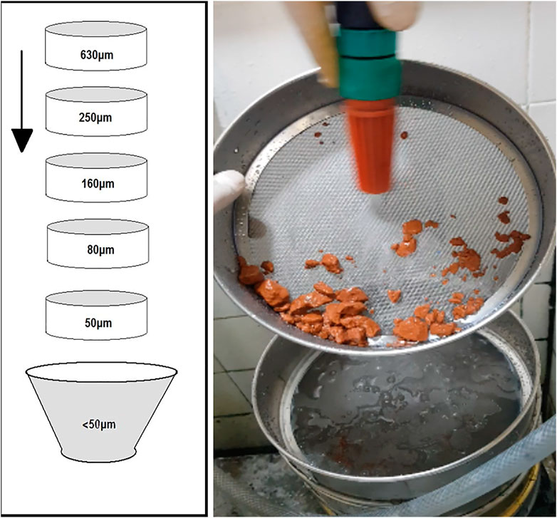
FIGURE 5. Battery of sieves (630, 250, 160, 80, and 50 µm) and final recipient to store sediments smaller than 50 µm.
The traditional study of Cloudina species and other tubular carbonate fossils hosted in limestone is performed preferably in two-dimensional (2D) views. This analysis uses polish or thin sections due to the ease of this methodology and quick preparation, although studying fossils in 2D views make the 3D morphology reconstruction more complex and less accurate. In some cases, phosphatization processes offer an opportunity to know more about its morphology. The fossil can be easily isolated from the rock matrix by acid attack without destroying the specimen (Hua et al., 2003). In contrast, when the composition of the fossil and that of the matrix are both carbonates, it becomes a challenge to separate the specimen from the rock. This work shows a new methodology of fossil extraction using a low concentrated acetic acid such as vinegar (∼4% acetic acid).
The preparation returned a positive result because acetic acid (4%) attacks the carbonate matrix preferentially, to the detriment of the carapace. Its slightly larger magnesium content is dissolved more slowly than the carbonate matrix. The dissolved fraction of the sample can be separated to analyze the palynological content (Figure 6).
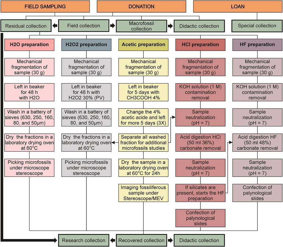
FIGURE 6. Flowchart of curatorship management and sample preparations of the Laboratory of Micropaleontology, University of Brasília. The great black arrows indicate the possibilities of relocation of samples.
The methodology consists of selecting a fossiliferous sample and introducing acetic acid solution 4% concentration. As the shell composition varies slightly from the matrix’s, it allows the acid to act differently, releasing the specimens following the reaction: CaCO3(s) + 2CH3COOH(aq) → Ca(CH3-COO)2(aq) + H2O(l) + CO2(g). A similar, but slower, process occurs in the outcrops of these carbonate fossiliferous rocks, where the carbonic acid (H2CO3) of the rain erodes the matrix resulting in the eventual exposition of the skeleton. The reaction can be controlled daily by observing the acid’s reaction on the fossiliferous sample. The entire preparation cycle takes approximately 15–20 days. The acid must be replaced every 3 days. At the end of the preparation, the sample must be gently and thoroughly washed with running water for approximately 5 min.
After the preparation mentioned previously, the fragments retained in sieves with mesh equal to or greater than 160 µm undergo a new preparation, this time using weak acids, such as acetic acid, to attack limestone, and formic acid, to attack dolomites, both at 10% concentration, with the aim of disaggregating the sample. For each sample to be prepared, it is recommended to use 1 L of 10% diluted acid solution for 200 g of sample. The sample is then placed in a hood, where it will remain until the chemical reaction is complete.
Periodically, after every 24 h of acid attack, it is recommended to change this acidic solution, as it loses its reaction power as the limestone is attacked. The solution that would initially be discarded during the exchange process, as it is a methodological evaluation, is separated for testing in micropaleontology. These tests are carried out with an emphasis on permineralized palynomorphs and for those microfossils that may be sorted with the aid of a stereoscopic microscope (any particle in suspension).
When weak acids are used, the preparation can take up to 2 months to be completed, but instead, the risk of destruction of mineralized microfossils is reduced. After being disaggregated, the material is washed in a battery of sieves. The fraction retained in each sieve is dried in an oven at 60°C and then examined under a stereoscopic microscope.
Approximately 30 g of sample is used for preparation to recover organic microfossils. Here, the mineral components of the rock are dissolved using two acids: HCl and HF (Figure 6). First, fragmented samples are put in a 400-mL beaker, adding 50 mL of HCl at 36% concentration during 24 h to dissolve carbonates. The next step is to bring the sample solution to a neutral pH value, using distilled water in periodic washings. The neutralization procedure involves the addition of distilled water to beaker capacity, waiting for the decantation of the organic extract, carefully removing the acid solution; the process is repeated until neutral pH is reached. Then 50 mL of HF at 40% concentration is added to dissolve silicates for 48 h. Then, the washing procedure is repeated. All recovered organic residues are placed in polypropylene tubes and distilled water at pH 7 to further conserve these residues.
After the acid attack process, the final remains are named palynological extract. This material is kept in water solution and, sometimes, when following the classic procedure, needs to be sieved before preparing palynological slides. In synthesis, this traditional procedure uses aleatory organic remains distributed in this solution to prepare palynological slides. Nevertheless, an approach to this classic procedure on picking palynological remains under a stereoscope microscope is presented. Using a very liner brush (000), it is possible to select specimens to prepare palynological slides with this procedure. There are two ways of making palynological slides: (1) palynological slides created after picking microfossils under a stereoscope microscope; (2) palynological slides without preanalyses under stereoscope microscope, which involves placing a few drops of the recovered organic residue and distilled water on a glass cover. Both types of slides are prepared after putting on a heating plate at 30°C. After the water had evaporated, a few drops of Entelan® resin were added to the coverslip to be completely sealed after contact with the blade. The resin used has the function of drying together with the material mounted on the blade and preventing oxidation of the organic matter and its degradation.
The results presented comprehend micropaleontological, mineralized, organic-walled microfossils, and sedimentological data, specifically clay mineral analyses. The oxidation attack was conducted to promote the complete or partial disaggregation of sedimentary rock. When sedimentary rock is composed of organic matrix, the H2O2 reacts with it and may result in a full or partial disaggregation. In this case, it is possible to recover microfossils in the H2O2 preparation, even in samples with oxidation considered ineffective (MP1226) and low efficiency (MP1203, MP1221, and MP1231). After the chemical reaction, the sedimentary material was sieved and by picking finest fractions. The coccoidal structures, as well as tubular and vase-shaped structures, recovered (Figure 7) were recognized as fossil content due to their similar morphological and size features assigned to well-described species and genus commonly found in Precambrian units. Besides that, they are very distinct from other grain particles analyzed from the same sample. The species recovered from the Paranoá Group, Mesoproterozoic, from sample MP1203, comprehends Myxococcoides sp. (CP965, CP966, CP967, CP968) (Figures 7.1–4) and Vetronostocale aff. V. amoenum Schopf and Blacic, 1971 (CP969) (Figure 7.5). One species was recovered in the Sete Lagoas Formation, Bambuí Group, from sample MP1221: Melanocyrillium sp. (CP970) (Figure 7.6), and one species was recovered from the Serra de Santa Helena Formation, Bambuí Group, from sample MP1231: Myxococcoides sp. (CP971, CP972, CP973) (Figures 7.7–9).
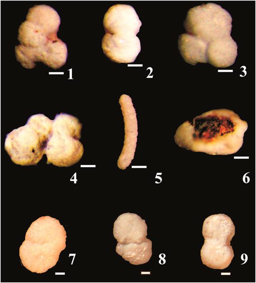
FIGURE 7. Recovered specimens from the Proterozoic units of the São Francisco craton, São Domingos River section (samples from the Sete Lagoas, MP1221, and Serra de Santa Helena, MP1231, formations) and from São Domingos Hill section (sample from the Paranoá Group, MP1203), Municipality of Buritis, Minas Gerais State, Brazil. (1–5) Specimens from the Paranoá Group; (6) Specimens from the Sete Lagoas Formation, Bambuí Group; (7–9) Specimens from the Serra de Santa Helena, Bambuí Group. (1–4,7–9) Myxococcoides sp., respectively, CP965, CP966, CP967, CP968, CP971, CP972, CP 973; (5) Vetronostocale aff. V. amoenum Schopf and Blacic, 1971, CP969; (6) Melanocyrillium sp., CP970. Scale bar: 10 µm.
The limestone samples of Sete Lagoas Formation, Januária Municipality, did not show a considerable disaggregation effectiveness. The H2O2 disaggregation method shows more effectiveness on siliciclastic rocks when compared with carbonate rocks. This could be due to the difference in permeability of those two lithotypes. The more permeable the rock, the easier the H2O2 reacts with the organic matter content. In this context, metamorphism can also affect the H2O2 disaggregation process as, depending on the metamorphic grade, it could change the rock permeability because of rock compaction.
The finest fraction (>50 µm) sieved from samples MP1226, MP1221, and MP1231 from three distinct micropaleontological preparations procedures were analyzed: (1) treatment with tap water before sieving, (2) treatment with deionized water before sieving, and (3) hydrogen peroxide attack before sieving. Analyses in whole rock from all three procedures showed similar results when the clay fraction studied was obtained as part of micropaleontological preparation compared with the results from the standard clay mineral preparation method. The total similarities between diffractograms could be verified when both oxidized (H2O2) and nonoxidized (tap water and deionized water) preparations of the same sample (Table 3) are compared. The mineral composition of the whole rock sample, determined by X-ray diffraction, shows that all samples have quartz as their major constituent, besides the sample MP1221, which also has calcite as the major component. Illite and albite are minor constituents of all samples.
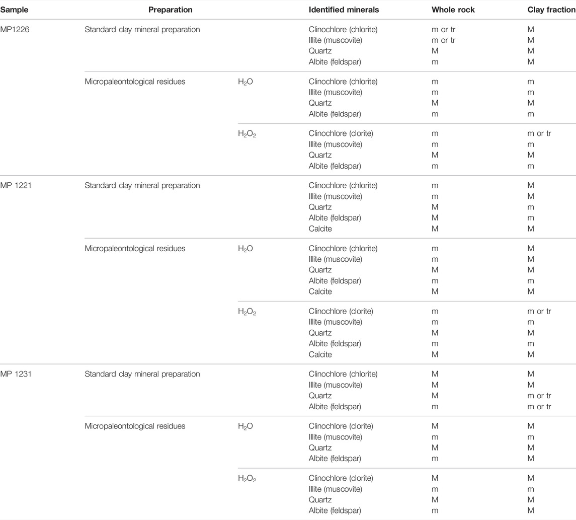
TABLE 3. Mineral composition of siltstones in whole rock and clay fraction, indicating the major constituents (M), minor (m), and trace (tr).
The standard clay mineral preparation results present changes in reflection intensities compared with the whole rock: the phyllosilicates have higher reflection intensity, which becomes major constituents, whereas the quartz reflection intensity decreases, which becomes a minor constituent. When the standard clay mineral preparation results are analyzed, the clay fraction shows the same composition as the whole rock, but (except for calcite in MP1221) the reflection intensities are opposite to those of the whole rock. Chlorite and illite are major constituents in the clay fraction, whereas quartz and albite are minor constituents (Figure 8). The diffractograms of samples MP1226 and MP1221 show a low and ill-defined band at the d∼28 position that expands slightly under treatment with glycerol, indicating the presence of interstratified clay mineral, possibly illite/vermiculite. Clay residues obtained from samples treated with H2O and H2O2 during the micropaleontological preparation do not maintain this trend. The clay fraction maintains the same intensities as the total sample: quartz remains a major constituent, whereas phyllosilicates are presented as minor or trace constituents (Figure 9). This effect can be explained as the effect of disaggregation, dispersion, or release of quartz particles during micropaleontological treatment.
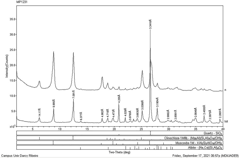
FIGURE 8. Diffractograms of sample MP1231 from Bambuí Group, Municipality of Buritis, Minas Gerais State, Brazil. Whole rock (tot) and clay fraction (n); note the variation in reflection intensities.
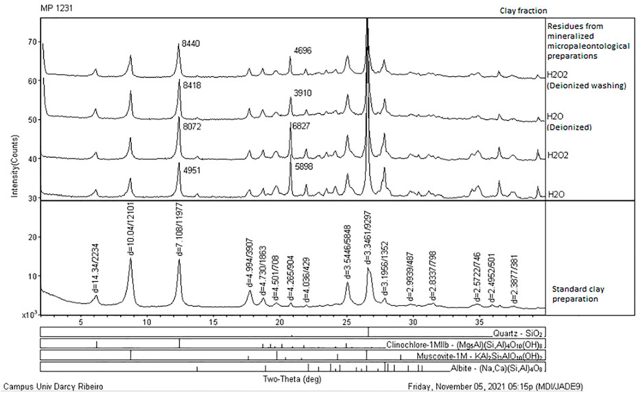
FIGURE 9. Diffractogram of the sample MP1231 from Bambuí Group, Municipality of Buritis, Minas Gerais State, Brazil. Clay fraction (n); note the variation in reflection intensities between the diffractogram of untreated sample (base) and treated samples; note that all treatments have the same effect on the clay fraction.
The procedure of analyzing the same sample residue for both micropaleontological and sedimentological approaches as a combined preparation reduces time of maceration and costs, besides being sure that both analyses comprehend the same depositional interval. This association leads to a more precise paleoenvironmental interpretation. In addition, it can save samples when a small amount is available for multiple analyses.
The acetic acid preparation was conducted on a sample from the Nama Group, Namibia; it showed efficient extraction of carbonate C. lucianoi skeleton within a carbonate matrix. This extraction technique allowed 3D imaging of the carbonate skeleton (Figure 10). This preparation shows similar results compared with phosphatized skeleton preparations from Dengying Formation in China (Hua et al., 2005). Researchers might use this easy, accessible, and environmentally friendly method to conduct 3D studies on carbonate skeleton fossils within limestone rocks. This extraction is possible when the skeleton composition is slightly or entirely different compared with the host carbonate matrix, such as the case of (1) the slightly richer magnesium C. lucianoi skeleton from Namibia and (2) the complete different composition of the phosphatized C. lucianoi skeleton from Dengying Formation, China (Hua et al., 2005).
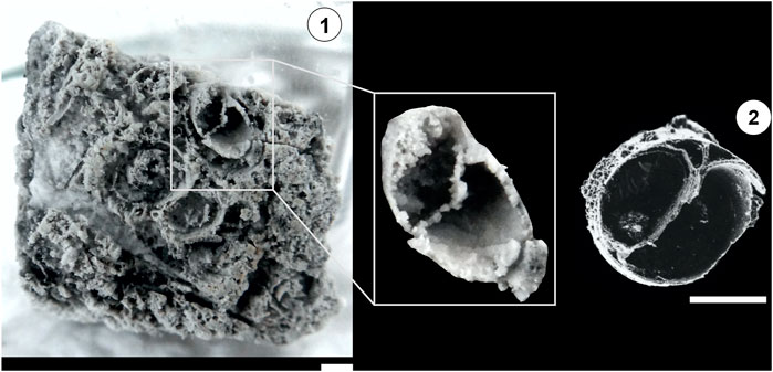
FIGURE 10. Comparison between results of different preparations. (1) Cloudina lucianoi (Beurlen and Sommer, 1957), CP974. Sample MP2289, carbonate from the type-section of Cloudina riemkeae Germs, 1972, Nomtsas Formation, Namibia, (UTM Coord. 33 k 0667883 7358829); (2) Phosphatized skeleton of Cloudina lucianoi from Dengying Formation, China (Hua et al., 2005). Scale bars: 400 µm.
The organic-walled microfossil preparation of limestones from Sete Lagoas Formation, Bambuí Group, Januária Municipality, Minas Gerais State, Brazil (Table 1), which followed the protocol presented in this work, led to the recovery of exquisitely specimens of organic-walled microfossils. The recovered assemblage comprises Leiosphaeridia minutissima (Naumova, 1949), CP963 (Figures 11.1, 2) and Leiosphaeridia tenuissima Eisenack, 1958, CP914 (Figure 11.3), one acritarch: Germinosphaera bispinosa Mikhailova, 1986, CP917 (Figure 11.4), and two cyanobacteria species: Ghoshia sp., CP916 (Figure 11.5), and Siphonophycus robustum (Schopf, 1968), CP961 (Figure 11.6).
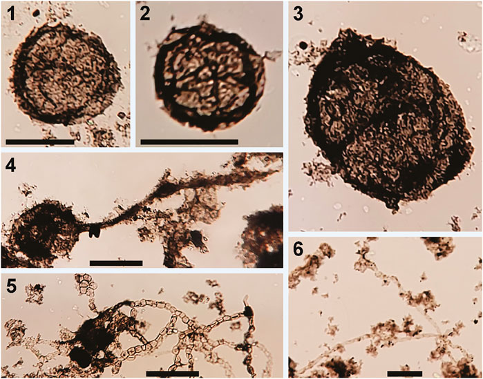
FIGURE 11. Organic-walled microfossils recovered from Sete Lagoas Formation, Bambuí Group, Januária Municipality. (1–2) Leiosphaeridia minutissima (Naumova, 1949)—CP963; (3) Leiosphaeridia tenuissima Eisenack, 1958—CP914; (4) Germinosphaera bispinosa Mikhailova, 1986 - CP917; (5) Ghoshia sp.—CP916; (6) Siphonophycus robustum (Schopf, 1968)—CP961. Scale bars: 25 µm.
(1) Efficiency of mineralized microfossiliferous disaggregation using H2O2: differences in disaggregation efficiency were observed, varying from ineffective (MP1226) to low efficiency (MP1203, MP1221, and MP1231). The lithotype, the amount of organic matter within the matrix, and the metamorphic grade can influence the disaggregation efficacy. The H2O2 disaggregation method shows more effectiveness on siliciclastic rocks when compared with carbonate rocks. This could be due to the difference in permeability of those two lithotypes. The more permeable the rock, the easier the H2O2 reacts with the organic matter content. In this context, metamorphism can also affect the H2O2 disaggregation process as, depending on the metamorphic grade, it could change the rock permeability due to rock compaction.
(2) Mineralized microfossils recovered using the H2O2 preparation: three permineralized species were recovered: Vetronostocale aff V. amoenum Schopf and Blacic, 1971 (from Paranoá Group), Myxococcoides sp. (from Paranoá Group and Lagoa do Jacaré Formation, Bambuí Group), and Melanocyrillium sp. (from Sete Lagoas Formation, Bambuí Group).
(3) Organic-walled microfossils recovered from Sete Lagoas Formation, Bambuí Group, using HCl and HF preparation: L. minutissima (Naumova, 1949), L. tenuissima Eisenack, 1958, G. bispinosa Mikhailova, 1986, Ghoshia sp., S. robustum (Schopf, 1968). The organic residue can integrate organic carbon isotopic studies.
(4) Mineralized microfossils recovered using acetic acid preparation: C. lucianoi (Beurlen and Sommer, 1957).
(5) Integration of clay mineral and micropaleontology preparations methods: the whole rock diffractograms of siltstones without treatments (standard preparation for clay mineral analyses) or treated in micropaleontological preparation with H2O (tap water or deionized water) and H2O2 did not show differences and allow the determination of mineral composition.
(6) Clay fraction diffractograms of residues from micropaleontological preparation: The clay fraction diffractograms showed that the micropaleontological preparation caused an increase in the intensity of the quartz reflections compared with untreated samples. Samples obtained after micropaleontological treatment may not be suitable for assessing the intensity of diagenesis using the Kübler Index, but they are useful for identifying the mineral assemblage.
(7) 3D extraction of a skeletal fossil can be possible even when the skeleton is carbonate in a carbonate matrix using weak acetic acid dissolution. This extraction is possible when the skeleton composition is slightly or entirely different from the host carbonate matrix. The organic material released by this preparation can be integrated into palynology studies.
The original contributions presented in the study are included in the article/Supplementary Material, further inquiries can be directed to the corresponding author.
MD—Fieldwork, data collection and interpretation, drawing of the figures and writing of the manuscript. RA—Fieldwork, data collection and interpretation, drawing of the figures and writing of the manuscript. DC—Ph.D. supervisor of MD, fieldwork, data interpretation and writing of the manuscript. EG—Data interpretation, drawing of the figures and writing of the manuscript. DW—Fieldwork and review of the paleontological and stratigraphic aspects of the manuscript. CA—Fieldwork and review of the stratigraphic aspects of the manuscript. GG—Fieldwork, data collection. LA—Curator of the Paleontology Collection of the Museum of Geosciences, University of Brasília, review the curatorship protocol and the mineralized microfossiliferous preparation protocol. CV—Data interpretation and review of the manuscript. OJ—Data collection and interpretation of the mineralized microfossils.
This study was financed in part by the Coordination for the Improvement of Higher Education Personnel—Brazil (CAPES)—Finance Code 001. Part of the studied samples was obtained from the Ediacariano Project, funded by the National Agency for Petroleum, Gas, and Biofuels (ANP) and the Brazilian Petroleum Corporation (PETROBRAS).
The authors declare that the research was conducted in the absence of any commercial or financial relationships that could be construed as a potential conflict of interest.
All claims expressed in this article are solely those of the authors and do not necessarily represent those of their affiliated organizations, or those of the publisher, the editors and the reviewers. Any product that may be evaluated in this article, or claim that may be made by its manufacturer, is not guaranteed or endorsed by the publisher.
We want to thank all institutions that participated in the development of this research: the National Council for Scientific and Technological Development (CNPq), the Coordination for the Improvement of Higher Education Personnel (CAPES), CAPES/IODP—Brazil Program via project 88887.091704/2014-01, the Brazilian Petroleum Corporation (PETROBRAS), the National Agency for Petroleum, Gas, and Biofuels (ANP) and the University of Brasília (UnB). We thank FINATEC for assistance in administrative affairs supporting scientific projects in Brasília. We would like to specially thank Dr. Norma Maria da Costa Cruz for approaching and sharing the palynological preparation method with students. We thank the two reviewers whose comments and suggestions helped improve and clarify this manuscript.
Alvarenga, C. J. S., Santos, R. V., Vieira, L. C., Lima, B. A. F., and Mancini, L. H. (2014). Meso-Neoproterozoic Isotope Stratigraphy on Carbonates Platforms in the Brasilia Belt of Brazil. Precambrian Res. 251, 164–180. doi:10.1016/j.precamres.2014.06.011
Alves, D. B. (1987). Desenvolvimento da metodologia de preparação de amostras para análise difratométrica de argilominerais no Centro de Pesquisas da Petrobrás. Bol. Geociências da Petrobras 1, 157–175.
Beurlen, K., and Sommer, F. W. (1957). Observações estratigráficas e paleontológicas sobre o calcário Corumbá. Rio de Janeiro: Departamento Nacional de Produção Mineral, Divisão de Geologia e Mineralogia, Boletim, Vol. 168, 1–35.
Campos, J. E. G., Dardenne, M. A., Freitas-Silva, F. H., and Martins-Ferreira, M. A. C. (2013). Geologia Do Grupo Paranoá na porção externa da Faixa Brasília. Braz. J. Geol. 43, 461–476. doi:10.5327/Z2317-48892013000300004
Campos, L. F. B. (2012). Diagênese das sequências Proterozóicas com base na caracterização de argilominerais – topo do Grupo Paranoá e base do Grupo Bambuí – Norte do Distrito Federal Masters Dissertation. Brasília: Institute of Geosciences, University of Brasília, 145.
Caxito, F. d. A., Halverson, G. P., Uhlein, A., Stevenson, R., Gonçalves Dias, T., and Uhlein, G. J. (2012). Marinoan Glaciation in East central Brazil. Precambrian Res. 200-203, 38–58. doi:10.1016/j.precamres.2012.01.005
Dardenne, M. A. (1978). “Síntese sobre a estratigrafia Do Grupo Bambuí no Brasil Central,” in XXX Congresso Brasileiro de Geologia (Recife: Trabalho completo), 597–610.
Eisenack, A. (1958). Microfossilien aus dem Ordovizium des Baltikums, 1, Markasitschicht, Dictyonema-Scheifer, Glaukonitsand, Glaukonitkalk. Senckenbergian Lethaea 39, 389–404.
Faria, A. (1995). Estratigrafia e sistemas deposicionais do Grupo Paranoá nas áreas de Cristalina. Distrito Federal e São João D’aliança-Alto Paraíso de Goiás. Doctoral Thesis. Brasília: Institute of Geosciences, University of Brasília, 199.
Gaucher, C., Frimmel, H. E., and Germs, G. J. B. (2005). Organic-walled Microfossils and Biostratigraphy of the Upper Port Nolloth Group (Namibia): Implications for Latest Neoproterozoic Glaciations. Geol. Mag. 142, 539–559. doi:10.1017/S0016756805001123
Germs, G. J. B., and Gresse, P. G. (1991). The Foreland basin of the Damara and Gariep Orogens in Namaqualand and Southern Namibia: Stratigraphic Correlations and basin Dynamics. South Afr. J. Geol. 94, 159–169.
Germs, G. J. B. (1983). “Implications of a Sedimentary Facies and Depositional Environmental Analysis of the Nama Group in South West Africa/Namibia,” in Evolution of the Damara Orogen of South Africa/Namibia. Editor R. M. Miller (South Africa: Geological Society of South Africa), 89–114.
Germs, G. J. B. (1972). New Shelly Fossils from Nama Group, South West Africa. Am. J. Sci. 272, 752–761. doi:10.2475/ajs.272.8.752
Germs, G. (1995). The Neoproterozoic of Southwestern Africa, with Emphasis on Platform Stratigraphy and Paleontology. Precambrian Res. 73, 137–151. doi:10.1016/0301-9268(94)00075-3
Horne, D. J., and Siveter, D. J. (2016). Collecting and Processing Fossil Ostracods. J. Crustac. Biol. 36, 841–848. doi:10.1163/1937240X-00002487
Hua, H., Chen, Z., Yuan, X., Zhang, L., and Xiao, S. (2005). Skeletogenesis and Asexual Reproduction in the Earliest Biomineralizing Animal Cloudina. Geol 33, 277–280. doi:10.1130/G21198.1
Hua, H., Pratt, B. R., and Zhang, L.-Y. (2003). Borings in Cloudina Shells: Complex Predator-Prey Dynamics in the Terminal Neoproterozoic. Palaios 18, 454–459. doi:10.1669/0883-1351(2003)018<0454:bicscp>2.0.co;2
Knoll, A. H. (1985). Patterns of Evolution in the Archean and Proterozoic Eons. Paleobiology 11, 53–64. doi:10.1017/s0094837300011398
Leite, A. M., Do Carmo, D. A., Ress, C. B., Pessoa, M., Caixeta, G. M., Denezine, M., et al. (2018). Taxonomy of Limnic Ostracoda (Crustacea) from the Quiricó Formation, Lower Cretaceous, São Francisco basin, Minas Gerais State, Southeast Brazil. J. Paleontol. 92, 661–680. doi:10.1017/jpa.2018.1
Machado, C. P., Coimbra, J. C., and Bergue, C. T. (2020). Provinciality of Ostracoda (Crustacea) in the Northeastern and Eastern Brazilian Shelves Based on Neontological and Paleontological Analyses. Rev. Bras. Paleontol. 23, 3–31. doi:10.4072/rbp.2020.1.01
Mikhailova, N. S. (1986). “Novye Nakhodki Mikrofitofossilij Iz Otlozhenij Verkhnego Rifeya Krasnoyarskogo Kraya,” in Aktual ’nye Voprosy Sovremennoj, Naukova Dumka. Editor B. S. Sokolov (Kiev: Nauka), 31–37.
Moreira, D. S., Uhlein, A., Dussin, I. A., Uhlein, G. J., and Pimentel Misuzaki, A. M. (2020). A Cambrian Age for the Upper Bambuí Group, Brazil, Supported by the First U-Pb Dating of Volcaniclastic Bed. J. South Am. Earth Sci. 99, 102503–102515. doi:10.1016/j.jsames.2020.102503
Paula-Santos, G. M., Babinski, M., Kuchenbecker, M., Caetano-Filho, S., Trindade, R. I., and Pedrosa-Soares, A. C. (2015). New Evidence of an Ediacaran Age for the Bambuí Group in Southern São Francisco Craton (Eastern Brazil) from Zircon U-Pb Data and Isotope Chemostratigraphy. Gondwana Res. 28, 702–720. doi:10.1016/j.gr.2014.07.012
Pimentel, M. M., Rodrigues, J. B., DellaGiustina, M. E. S., Junges, S., Matteini, M., and Armstrong, R. (2011). The Tectonic Evolution of the Neoproterozoic Brasília Belt, central Brazil, Based on SHRIMP and LA-ICPMS U-Pb Sedimentary Provenance Data: A Review. J. South Am. Earth Sci. 31, 345–357. doi:10.1016/j.jsames.2011.02.011
Sanchez, E. A. M., Uhlein, A., and Fairchild, T. R. (2021). Treptichnus Pedum in the Três Marias Formation, South-central Brazil, and its Implications for the Ediacaran-Cambrian Transition in South America. J. South Am. Earth Sci. 105, 102983–102989. doi:10.1016/j.jsames.2020.102983
Schopf, J. W., and Blacic, J. M. (1971). New Microorganisms from the Bitter Springs Formation (Late Precambrian) of the north-central Amadeus Basin, Australia. J. Paleontol. 45, 105–114.
Schopf, J. W. (1995). “Disparate Rates, Differing Fates: Tempo and Mode of Evolution Changed from the Precambrian to the Phanerozoic,” in Tempo and Mode in Evolution. Editors W. M. Fitch, and F. J. Ayala (Washington, D.C.: National Academy of Sciences), 41–62. Available at: https://www.ncbi.nlm.nih.gov/books/NBK232208/
Schopf, J. W. (1968). Microflora of the Bitter Springs Formation, Late Precambrian, Central Australia. J. Paleontol. 42, 651–688.
Uhlein, A., Trompette, R., and Egydio-Silva, M. (1995). Rifteamentos Superpostos E Tectônica De Inversão Na Borda Sudeste Do Cráton Do São Francisco. Revista Geonomos 3, 99–107. doi:10.18285/geonomos.v3i1.219
Keywords: micropaleontological preparation, sedimentalogical preparation, proterozoic microfossils, clay minerals, curatorship protocol
Citation: Denezine M, Adôrno RR, Do Carmo DA, Guimarães EM, Walde DHG, De Alvarenga CJS, Germs G, Antonietto LS, Valdivia Rodríguez CG and Nunes Junior ODO (2022) Methodological Development of a Combined Preparation for Micropaleontological and Sedimentological Studies of Samples From the Proterozoic Record. Front. Earth Sci. 10:810406. doi: 10.3389/feart.2022.810406
Received: 06 November 2021; Accepted: 18 January 2022;
Published: 30 March 2022.
Edited by:
Juliana Leme, University of São Paulo, BrazilReviewed by:
Luana Morais, University of São Paulo, BrazilCopyright © 2022 Denezine, Adôrno, Do Carmo, Guimarães, Walde, De Alvarenga, Germs, Antonietto, Valdivia Rodríguez and Nunes Junior. This is an open-access article distributed under the terms of the Creative Commons Attribution License (CC BY). The use, distribution or reproduction in other forums is permitted, provided the original author(s) and the copyright owner(s) are credited and that the original publication in this journal is cited, in accordance with accepted academic practice. No use, distribution or reproduction is permitted which does not comply with these terms.
*Correspondence: Matheus Denezine, bWF0aGV1c2RlbmV6aW5lQGdtYWlsLmNvbQ==
Disclaimer: All claims expressed in this article are solely those of the authors and do not necessarily represent those of their affiliated organizations, or those of the publisher, the editors and the reviewers. Any product that may be evaluated in this article or claim that may be made by its manufacturer is not guaranteed or endorsed by the publisher.
Research integrity at Frontiers
Learn more about the work of our research integrity team to safeguard the quality of each article we publish.