
95% of researchers rate our articles as excellent or good
Learn more about the work of our research integrity team to safeguard the quality of each article we publish.
Find out more
SYSTEMATIC REVIEW article
Front. Dent. Med. , 27 July 2021
Sec. Reconstructive Dentistry
Volume 2 - 2021 | https://doi.org/10.3389/fdmed.2021.689537
This article is part of the Research Topic Tooth Bleaching View all 6 articles
 Alexandre Henrique dos Reis-Prado1
 Isadora Rodrigues Grossi1
 Hebertt Gonzaga dos Santos Chaves1
 Carolina Bosso André1
 Luís Fernando dos Santos Alves Morgan1André Luiz Fraga Briso2Luciano Tavares Angelo Cintra2
 Francine Benetti1*
Background: Dental bleaching agents show the ability to permeate through dental hard tissues, which may lead to pulp tissue changes. This systematic review (PROSPERO register: CRD42020213767) is aimed at understanding the effects of bleaching agents on the process of mineralization of the pulp tissue.
Methods: Only in vitro studies evaluating the influence of hydrogen peroxide (HP) on mineralization in dental pulp cells were included. Studies without a non-bleached control group or cells after co-treatment with a bleaching agent other than HP and/or carbamide peroxide were excluded. The primary outcomes evaluated were alkaline phosphatase (ALP) activity and mineralized nodule deposition. The mineralization markers analysis in dental pulp cells and the cell viability were considered secondary outcomes. Two independent authors conducted a systematic search (PubMed/MEDLINE, Scopus, Embase, Cochrane Library, and OpenGrey until January 2021) with no language restrictions and performed data extraction. The quality assessment was appraised according to a modified Joanna Briggs Institute critical appraisal checklist.
Results: The search resulted in 473 studies, and 11 were considered eligible. Overall, a reduction in the process of mineralization was observed among pulp cells after bleaching. A reduction in the ALP activity was reported in the mostly bleached groups using different protocols and analysis periods of nine studies. Regarding mineralized nodule deposition, 6 studies reported a significant reduction from 7 to 21 days among bleached groups. Of those three studies that investigated other mineralization markers, two found a reduction in the expression of dentin matrix acidic phosphoprotein (DMP)-1, dentin sialophosphoprotein (DSPP), and matrix extracellular phosphoglycoprotein (MEPE) among some bleaching gel concentrations. In contrast, one study showed a greater expression of osteopontin (OPN) and osteocalcin (OCN) in 100 μmol/L HP after 5 or 10 min of exposure, and another study showed significant induction of DSPP in concentrations of up to 0.5 mmol/L HP.
Conclusion: Especially, high concentrations of bleaching gel reduce the potential of mineralization in pulp cells in in vitro studies; however, different HP concentrations, bleaching protocols, and analysis periods can influence this outcome.
Dental bleaching using hydrogen peroxide (HP), the main active agent of most bleaching products, is considered a popular treatment to achieve esthetical bleaching. This procedure is also considered safe and successful under the supervision of dentists, and it is credited to promote color change by the interaction of oxygen free radicals by HP with intrinsic and extrinsic pigments (1), and has one of the main benefits, fostering positive changes in patients, such as smiling, laughing, and showing teeth without embarrassment (2); however, this procedure is related to having effects on enamel and dentin mineral loss and can lead to pulp tissue changes (3), especially in human mandibular incisor teeth (4), that is still not fully understood and regarded as one of the main concerns about this treatment (3–6).
Bleaching agents with different HP concentrations can diffuse through interprismatic space and promote some enamel surface morphology alterations, for instance, reducing enamel microhardness and increasing the surface roughness (1, 7). Besides that, the reactive oxygen species (ROS) released by HP can permeate through enamel and dentin, reaching the pulp tissue and leading to inflammation, decreasing cellularity and cell metabolism (3, 8–12), protein denaturation (4, 13, 14), and areas of tissue necrosis (5, 6, 12, 15).
From preliminary studies simulating clinical conditions, severe inflammation and necrosis areas in the pulp tissue of rats were observed after one to five bleaching sessions with 3 applications of 15 min each of 35% HP gel, which was accentuated following the number of bleaching sessions (5). These histological observations can be related to the tooth sensitivity reported by the patients after the bleaching (16). After 30 days, the inflammation was ceased, and the pulp tissue was reorganized; however, another study showed an intense production of tertiary dentin directly proportional to the increase in HP concentration (20–35% HP gel) and time of application (5–45 min) of bleaching gel (10). It was also observed the existence of apoptotic cells, which are fundamental for the development of tissues and their recovery after internal and external stimuli (17, 18) and intense cell proliferation in regions below the areas of necrosis (18). Therefore, the pulp tissue can recover after HP cell damage but the long-term consequences of this damage are not fully understood.
A previous study evaluated the immunolabeling of mineralization proteins on bleached molar pulp tissue in rats using a single application of 30 min of the 35% HP (19). The authors observed that osteopontin (OPN) is present in the pulp tissue during the repair process after bleaching and is more immunolabeled after 7 and 15 days (19); however, osteocalcin (OCN) was significantly immunolabeled from 7 days on, but it was more significant after 30 days of the bleaching treatment (19). The Jun-D transcription factor of odontoblasts was significantly identified after 7 days of similar bleaching treatment with 35% HP, indicating differentiation of these cells (6). Another study also reported the expression of OCN and OPN in cell cultures after 10 min of contact with minimal concentrations of HP (100 μmol/L) (20).
Several laboratory studies have reported the effects of distinct concentrations (0.025–0.3 mM or 17.5–35%) of HP on the mineralization of different cell lines (21–23). A favorable outcome of HP on odontoblasts suggests an increase in dentin production capability (21). The studies conducted so far indicate a process involving dentinogenesis and mineralization of pulp tissue or pulp cells after contact with HP. Although several systematic reviews have evaluated the adverse clinical effects of bleaching (16, 24), only one recent systematic review assessed the impact of this procedure on the pulp tissue (12). Thus, the objective of this study is to carry out a systematic review to understand the immediate and long-term effects of bleaching agents on the process of mineralization of the pulp tissue.
The present study was reported according to the Preferred Reporting Items for Systematic Reviews and Meta-Analyses (PRISMA) statement (25, 26). This systematic review was registered in the International Prospective Register of Systematic Reviews (PROSPERO) under the registration number CRD42020213767.
The inclusion criteria were as follows: (1) studies that evaluated the effects of the bleaching gel on the process of mineralization of dental pulp cells and proteins involved in this process; (2) studies that present a non-bleached control group/without bleaching gel; and (3) in vitro studies. The exclusion criteria were studies that analyzed pulp cells after co-treatment with bleach or another agent rather than HP and/or carbamide peroxide.
The population-intervention-comparison-outcome (PICO) approach was used to address the following question: “Can the bleaching agent influence the mineralization process of dental pulp cells?” In this process, the population (P) was dental pulp cells. The intervention (I) was pulp cells after exposure to the bleaching agent. The comparison (C) was pulp cells being not exposed to the bleaching agent. The primary outcomes (O) evaluated were alkaline phosphatase (ALP) activity and mineralized nodule deposition. The mineralization markers analysis in dental pulp cells and the cell viability were considered secondary outcomes.
Electronic searches were conducted in PubMed/MEDLINE, Scopus, Embase, and Cochrane Library until January 2021. The gray literature was also consulted through OpenGrey. The search strategy was firstly defined for the MEDLINE database via Pubmed using a controlled vocabulary (MeSH terms) and free keywords. The MEDLINE search was also adapted to the other databases as shown in Supplementary Table 1. Manual searches were also performed in the reference lists of the included articles to find additional studies. No restrictions to publication date or language were considered.
The articles retrieved by the literature search were selected by two independent authors (IGR and AHRP) in a two-step procedure. In Step 1, the two authors appraised titles and abstracts of studies that met the eligibility criteria. The studies were arranged alphabetically by title, and duplicates were identified and removed manually. In Step 2, the two authors assessed the full texts of each study. Only studies in which the full text fulfilled the proposed eligibility criteria were included in this review. Any disagreements between the two authors were resolved through discussion, and when necessary, a third author (LFSAM) was consulted.
One author collected data (IGR) from the included studies and tabulated it to analyze the results. The following data were retrieved: author and year, experimental model, groups and bleaching protocol, period of analysis, and outcomes of evaluating the ALP activity, Alizarin red or von Kossa staining, mineralization markers, and cell viability. The second author (AHRP) revised the collected data.
Two investigators (AHRP and HGSC) independently assessed the methodological quality of the selected studies according to their levels of evidence as proposed by a modified version as described previously (26) in Joanna Briggs Institute critical appraisal checklist for experimental studies (27). To further characterize bleaching reporting in the selected studies, the authors also assessed if a clear bleaching protocol was present. The following were the other items in the checklist: clearly stated aim, justification of sample size, sample randomization, the possibility of comparison between controls and treatment groups, baseline equivalence of control and treatment groups, measurement method, measurement standardization, and adequate statistical analysis. Each item was scored using a 2-point scale: 0, not reported or reported inadequately; 1, reported and adequate. Doubts and discrepancies between both investigators were discussed, and if not solved, a third examiner (CBA) was consulted.
The selection process of the articles is presented in Figure 1. A total of 473 articles were found on databases search. After the first screening and removal of duplicates, 12 studies were selected. These studies were then subjected to full-text evaluation that resulted in the exclusion of one study that had no control group (28). Finally, 11 studies were included in this review (3, 20–23, 29–34).
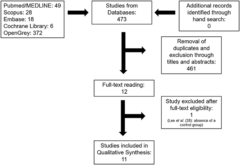
Figure 1. Flow diagram of the search strategy of the systematic review following the Preferred Reporting Items for Systematic Reviews and Meta-analyses (PRISMA) guidelines.
The assessed Cohen kappa coefficient value for the inter-investigator agreement was equal to 0.959 for PubMed, 0.837 for Scopus, 0.889 for Embase, 1.000 for the Cochrane Library, and 1.000 for OpenGrey. These values indicated an almost perfect agreement among reviewers according to the scale of Landis and Koch (35).
The characteristics of the studies are depicted in Table 1. Most studies (a total of seven) used tooth discs for the application of bleaching treatments with artificial pulp chambers and different cell types, such as dental pulp stem cells (DPSCs), human dental pulp cells (HDPCs), and odontoblast-like MDPC-23 (22, 23, 29–33). Of these, six studies used tooth discs from bovine incisors. Four studies performed the analyses exposing only some cell cultures to the different concentrations of extracts of bleaching gel (3, 20, 21, 23).
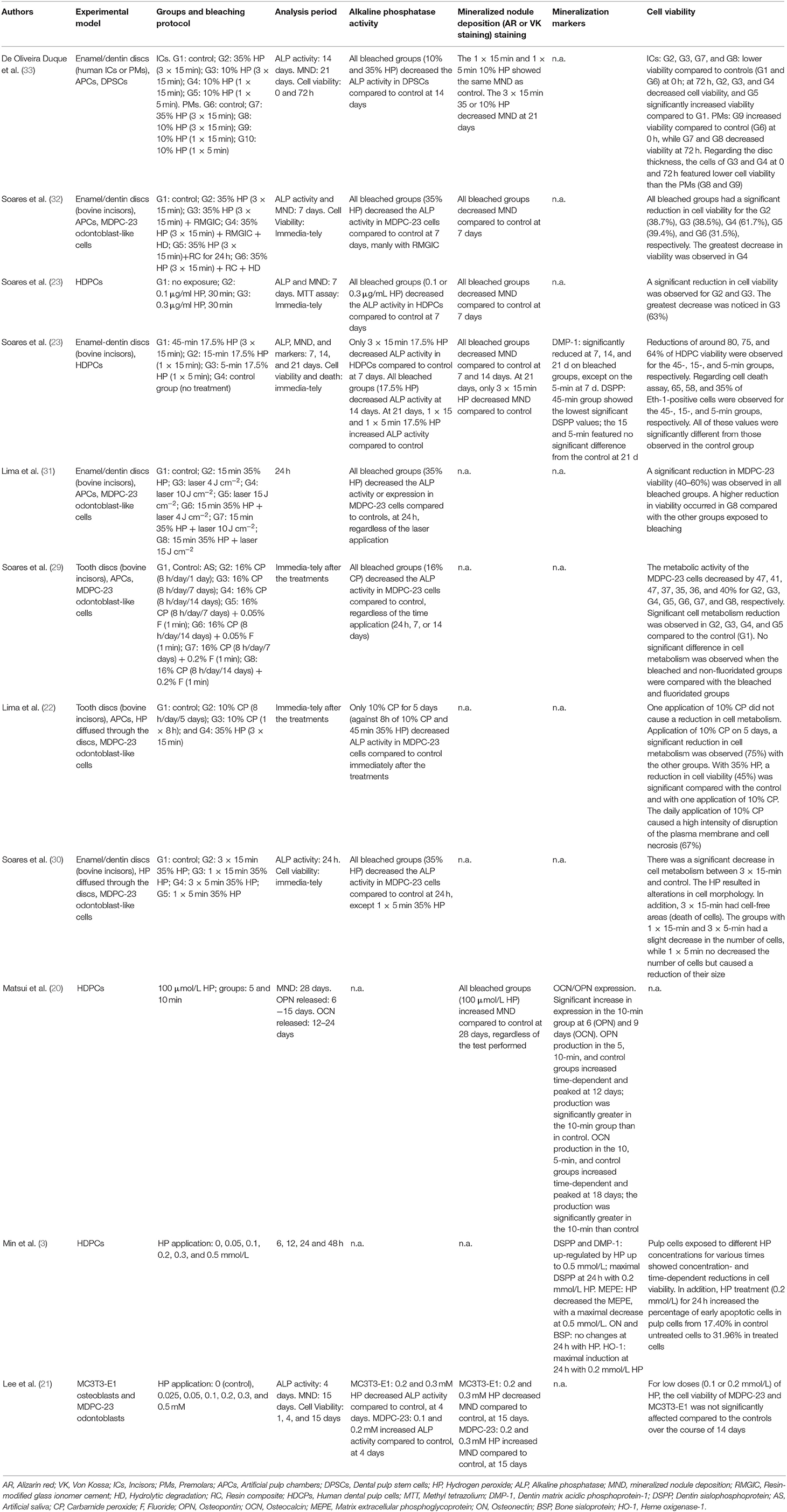
Table 1. Characteristics of the studies included in the review and synthesis of outcomes.
The bleaching agent with 35% HP concentration was used in five studies (22, 30–33), with different application times. The bleaching agent with 10% HP concentration was used in only one study (33), as well as bleaching agents with 17.5% HP concentration (23), 16% carbamide peroxide (CP) (29), and 10% CP (22). Other studies used extracts that were previously prepared with lower HP concentrations (3, 20, 21).
A total of nine studies evaluated ALP activity. The period of analysis was immediately after the treatments (22, 29), after 24 h (3, 30, 31), after 4 days (21), after 7 days (23, 32), after 14 days (23, 33), and after 21 days (23). Regarding mineralized nodule deposition, six studies performed this analysis after 7 (23, 32), 14 (23), 15 (21), 21 (23), and 28 days (20). All studies used Alizarin red staining for this analysis, but one study used von Kossa staining (20).
Regarding the mineralization markers, three studies evaluated the mRNA gene expression of these markers by using reverse transcriptase-PCR (3, 20, 23). The markers evaluated were dentin matrix acidic phosphoprotein-1 (DMP-1) and dentin sialophosphoprotein (DSPP) in two studies (3, 23), OCN and OPN in another study (20), and matrix extracellular phosphoglycoprotein (MEPE), osteonectin (ON), and bone sialoprotein (BSP) in one last study (3).
Each of the included studies was analyzed in terms of similarities to determine whether a meta-analysis could be applied for these records; however, despite a favorable number of studies in this review, considerable heterogeneity was found between the studied groups. There were several bleaching gel concentrations or protocols, types of analysis performed, experimental periods, besides different cell types; therefore, a meta-analysis was not performed. The data regarding studies outcomes are summarized in Table 1 and are described below.
Nine studies that evaluated ALP activity (21–23, 23, 29–33) found a decrease in this enzyme for approximately all bleached groups using different protocols and HP concentrations for the majority of the assessed period of analysis; however, two of these studies also reported an increased APL activity with 0.1 mM and 0.2 mM HP after 4 days (21) and after one session of 17.5% HP for 5 min or 15 min and after 21 days (23), using MDPC-23 cells and HDPCs, respectively.
From the selected studies, only six performed the mineralized nodule deposition analysis. These studies reported that bleached groups reduced the mineralized nodule deposition by using 35% HP in MDPC-23 cells (32), 0.1 and 0.3 μg/mL HP (23), or 17.5% HP (23) in HDPCs after 7 days. At 14 or 15 days, a reduction of mineralized nodule deposition with 17.5% HP in HPDCs (23) and with 0.2- and 0.3-mM HP in MC3T3-E1 (21) was observed. In addition to these data, a decrease in the mineralized nodule formation was also observed in 3 sessions of 10% HP (33) or 35 % HP (23, 33), after 21 days. On the contrary, two studies reported a significant induction of mineralized nodules in 0.2- or 0.3-mM (21) and 100 μmol/L HP (20) for MDPC-23 cells and HDPCs after 15 and 28 days, respectively.
From the three articles that evaluated the mineralization markers in cells after bleaching, DMP-1 and DSPP markers were investigated in two of these studies (3, 23). One study reported a reduction in the expression of DMP-1 after 7, 14, and 21 days on bleached groups using 17.5% HP (23), while the other study revealed that up to 0.5 mmol/L HP stimulates DMP-1 expression (3). Regarding the expression of DSPP, the application of 17.5% HP for 45 min was associated with a reduction in its expression in one study (23), while up to 0.5 mmol/L HP increased DSPP values in another study (3).
Only one study evaluated OPN and OCN expressions (20). The study showed that 100 μmol/L HP for 5 or 10 min promoted a greater expression of these mineralization markers in HDPCs after 6 (OPN), 9 (OCN), 12 (OPN), or 18 (OCN) days compared with control (20). One study (3) that investigated more mineralization markers demonstrated a reduction in MEPE at 0.5 mmol/L HP, a significant induction of HO-1 with 0.2 mmol/L after 24 h, and no significant differences in the production of ON and BSP among the groups.
Figure 2 and Table 2 summarize the results of the critical appraisal of the eligible studies. Only one study (31) reported the presence of sample randomization. All the included articles showed a possibility of comparison between control and treatment groups in the beginning and a reliable measurement method. Low risk of bias was also observed in the clearly stated aim, baseline equivalence of control and treatment groups, clear bleaching protocol, measurement standardization, and appropriate statistical approach. Conversely, a high risk of bias was noticed for some specific items, such as sample randomization and justification of sample size.
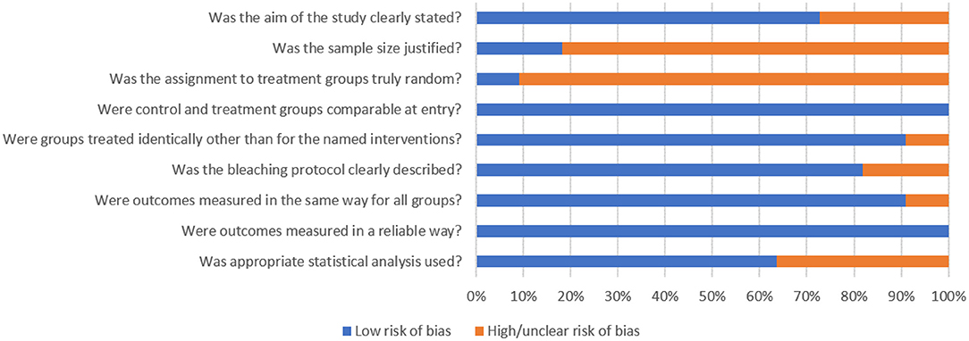
Figure 2. Assessment of the risk of bias in the included studies according to the percentage of the scores attributed to each evaluated item.
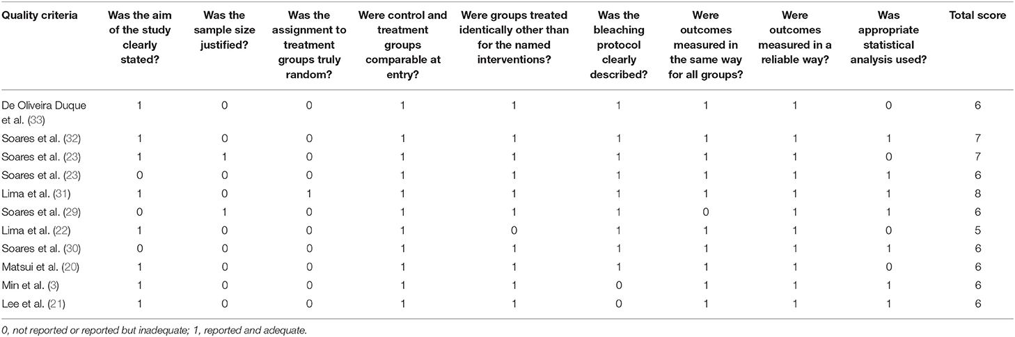
Table 2. Risk of bias of included studies.
This systematic review investigated if the bleaching gel can influence the mineralization process of dental pulp cells. A total of 11 in vitro studies were included in this review. Overall, the evaluated data showed that the bleaching gel decreases the ALP activity and mineralized nodules deposition in dental pulp cells. In addition, it was possible to understand that, particularly, the high concentrations of HP after different times of exposure are capable of reducing the potential of mineralization in pulp cells. Meta-analysis was not performed due to heterogeneity among the bleaching protocols or analysis periods of the included studies, besides different cell types.
It is known that in the presence of an aggressor, such as the HP of bleaching gel, the pulp tissue responds with the production of tertiary dentin (36, 37). When there is mild aggression, the odontoblastic cells themselves are responsible for producing the reactionary dentin. If intense damage occurs, the odontoblasts are lost, and the pulp tissue stem cells generate new odontoblastic-like cells, which will produce reparative dentin (19, 36, 37). In this way, both differentiated and non-differentiated cells are useful for the mechanisms of tertiary dentin production (38).
Thus, it was expected that HP could induce the mineralizing potential of pulp cells; however, this review shows that in most studies, HP reduced ALP activity and deposition of mineralized nodules, which could indicate a loss in the mineralization potential of pulp cells after dental bleaching. These data disagree with those observed in in vivo studies, where a large formation of tertiary dentin is observed in bleached teeth (11, 12, 17, 21). This may be due to the differences of the models in in vitro and in vivo evaluations. In in vitro studies, an absence of cytoplasm extensions of odontoblast and dentin fluid that act as a physical barrier to the penetration of HP is found (10), besides the lack of HP-degrading enzymes (39). Thus, when damage occurs to the cell monolayer, it is expected to increase cell death, which can reduce the potential for remaining cells to respond against the aggressor.
In addition, HP causes damage mainly to the occlusal third of the coronary pulp in in vivo studies, meaning that the underlying pulp tissue receives the lowest concentration of HP, and it is induced to proliferate and replenish the lost tissue, depositing tertiary dentin (18). Then, bleaching agents containing a low HP concentration and the underlying pulp tissue can be both associated with the expression of proteins related to the mineralization process, which occurs later in animal models (19). Thus, the three-dimensional (3D) system can be more appropriated than the traditional two-dimensional system for in vitro analysis because it can better represent in vivo cellular conditions using scaffold supports of cell growth and cell-to-cell interactions (40); however, the 3D model was not used by any of the selected studies in this review.
The production of tertiary dentin in in vivo studies after dental bleaching was observed from the 7th day (6, 19); however, most studies in this review found a reduction in ALP activity in this period (23, 23, 30, 32). ALP is an enzyme expressed by odontoblasts with an important role in the repair and regeneration of pulp tissue (29). Only one study in this review showed that despite the reduction of ALP activity at 7 and 14 days, induced by 17.5% HP, the ALP activity increased after 21 days when compared to the control group (23). In addition, this was the only study that assessed ALP activity over a prolonged period (21 days), in contrast with other studies that performed this analysis for up to 15 days. Thus, further analysis of ALP activity after a prolonged period is still necessary to fully understand this process.
Another study that evaluated a single application of 8 h of 10% CP or 45 min of 35% HP found no significant difference with the control regarding ALP activity immediately after bleaching (22). These results are in contrast with the study of Soares et al. (29), who observed a reduction in ALP activity after 8 h of 16% CP. These were the only studies that used CP and may indicate that the increase in concentration from 10 to 16% was enough to influence the results of ALP activity. In addition, regarding 45 min of 35% HP, the other three studies that evaluated this concentration performed ALP activity analysis after 24 h or more, and all showed a reduction in ALP activity (30, 32, 33).
The decrease in ALP activity can be related to the cytotoxicity observed in the studies since high concentrations of HP are used in most of them. It is observed, for example, in two studies (3, 20) of this review, that the low concentration of HP led to an increase in the expression of markers of mineralization. But these results were not correlated with the analysis of the ALP. Matsui et al. (20) explained that the oxidative stress induced by HP to the cells results in the formation of calcium phosphate in them, which induces the expression of mineralization markers.
On the contrary, Soares et al. (23) found a reduction in the expression of mineralization markers when a high concentration of HP was used. A previous study in molars of bleached rats showed that a high concentration of HP can cause severe damage to the dental pulp, while low concentrations of HP can induce proliferation in the pulp tissue, which could explain these results. In addition, two studies showed an increase in OCN in the pulp tissue after dental bleaching (19, 41), which demonstrates that despite the importance of in vitro studies, different results can be observed from those that occur in in vivo models. In addition, the ROS can accumulate during oxidative stress, but due to their reactivity, this accumulation occurs in a transitory way; however, it can damage essential biomolecules, which can be challenging to recover in cell culture, but reversible in living tissue (42).
Regarding mineralized nodules deposition, the results followed those of the ALP activity; however, one study showed that minimal time of the application of 10% HP did not change the mineralized nodules deposition in dental pulp cells compared with control (33), despite reducing ALP activity. Further, these results were obtained in different analysis periods, which may indicate that in more prolonged periods, the production of mineralization nodules may occur. In addition, only one study evaluated the presence of mineralized nodules at 28 days after bleaching and revealed an increased number of mineralized nodules compared with the control group (20). Thus, studies with long periods of analysis for ALP activity and deposition of mineralized nodules would be important to clarify the results.
It should be noted that most of the studies located in this systematic review were carried out by the same group of authors (23, 23, 29, 30, 32). This can be considered a limitation of the present study due to a strong influence of methodological characteristics of a single research group on the results found in this review. Still, only in vitro studies were selected, requiring further systematic analysis of in vivo studies; however, this systematic review presents studies with a low risk of bias (Figure 2), where most of them revealed that high concentrations of HP could impair the mineralization potential of dental pulp cells. Some methodological limitations, such as the absence of randomization and no justification of sample size, were noticed. Future research should address these issues, including well-described sample randomization and suitable calculation of the sample size. In addition, although few studies have evaluated low HP concentrations or longer periods of exposure, there seems to be a tendency for induction of mineralization to occur under these conditions, which needs to be further investigated.
It is also important to investigate adequate protocols and methods of evaluation that analyze the effect of this intervention in the mineralization of pulp tissue in clinical trials. Additional randomized and longitudinal investigations considering the different age ranges of participants, bleaching protocols, and anatomical variations should be considered to confirm these results; however, caution is highlighted when using high concentrations of HP, mainly used in in-office bleaching, since cytotoxic effects can be observed in pulp cells. This is in accordance with the European Union Council Directive 2011/84 /EU (amending EU Council Directive 76/768 /EEC) that stated that bleaching gel used for dental clinical practice might only contain up to 6% HP (43). Based on these results, we enhance the importance of dentist supervision under both home and in-office bleaching, mainly, when high concentrations of bleaching gel are used. Accelerated aging of the pulp tissue, in addition to inflammation, may be a consequence of the use of a high concentration of HP (15, 19).
Within the limitations of this review of in vitro studies, it can be concluded that mostly high concentrations of bleaching gels could reduce the potential of mineralization in pulp cells; however, different HP concentrations, bleaching protocols, and analysis periods can influence this outcome.
The original contributions presented in the study are included in the article/Supplementary Material, further inquiries can be directed to the corresponding author/s.
FB, CA, and LM: conceptualization. IG, AR-P, and LM: study selection. IG and AR-P: data collection. AR-P, HC, and CA: quality assessment. FB, AR-P, HC, and CA: methodology. FB, AB, and LC: project administration. LC and FB: supervision. AB, LM, and FB: validation. CA, AB, and LC: visualization. IG, AR-P, and HC: writing—original draft. LM, AB, LC, FB, and CA: writing—review and editing. All authors read and approved the final manuscript.
The authors declare that the research was conducted in the absence of any commercial or financial relationships that could be construed as a potential conflict of interest.
All claims expressed in this article are solely those of the authors and do not necessarily represent those of their affiliated organizations, or those of the publisher, the editors and the reviewers. Any product that may be evaluated in this article, or claim that may be made by its manufacturer, is not guaranteed or endorsed by the publisher.
The Supplementary Material for this article can be found online at: https://www.frontiersin.org/articles/10.3389/fdmed.2021.689537/full#supplementary-material
1. Kwon SR, Wertz PW. Review of the mechanism of tooth whitening. J Esthet Restor Dent. (2015) 27:240–57. doi: 10.1111/jerd.12152
2. Kothari S, Gray AR, Lyons K, Tan XW, Brunton PA. Vital bleaching and oral-health-related quality of life in adults: a systematic review and meta-analysis. J Dent. (2019) 84:22–9. doi: 10.1016/j.jdent.2019.03.007
3. Min KS, Lee HJ, Kim SH, Lee SK, Kim HR, Pae HO, et al. Hydrogen peroxide induces heme oxygenase-1 and dentin sialophosphoprotein mRNA in human pulp cells. J Endod. (2008) 34:983–9. doi: 10.1016/j.joen.2008.05.012
4. Costa CA, Riehl H, Kina JF, Sacono NT, Hebling J. Human pulp responses to in-office tooth bleaching. Oral Surg Oral Med Oral Pathol Oral Radiol Endod. (2010) 109:59–64. doi: 10.1016/j.tripleo.2009.12.002
5. Cintra LT, Benetti F, da Silva Facundo AC, Ferreira LL, Gomes-Filho JE, Ervolino E, et al. The number of bleaching sessions influences pulp tissue damage in rat teeth. J Endod. (2013) 39:1576–80. doi: 10.1016/j.joen.2013.08.007
6. Benetti F, Briso ALF, de Araújo Lopes JM, Carminatti M, Conti LC, Gallinari MO, et al. In vivo analysis of the presence of heme oxygenase-1, transcription factor Jun-D and CD90+/CD73+/CD105+/CD45- cells in the pulp of bleached teeth. Int Endod J. (2019) 52:1723–37. doi: 10.1111/iej.13190
7. Chen HP, Chang CH, Liu JK, Chuang SF, Yang JY. Effect of fluoride containing bleaching agents on enamel surface properties. J Dent. (2008) 36:718–25. doi: 10.1016/j.jdent.2008.05.003
8. Soares DG, Basso FG, Hebling J, Costa CAS. Concentrations of and application protocols for hydrogen peroxide bleaching gels: effects on pulp cell viability and whitening efficacy. J Dent. (2014) 42:185–98. doi: 10.1016/j.jdent.2013.10.021
9. Cintra LT, Benetti F, Ferreira LL, Gomes-Filho JE, Ervolino E, Gallinari MO, et al. Penetration capacity, color alteration and biological response of two in-office bleaching protocols. Braz Dent J. (2016) 27:169–75. doi: 10.1590/0103-6440201600329
10. Cintra LT, Benetti F, Ferreira LL, Rahal V, Ervolino E, Jacinto RC, et al. Evaluation of an experimental rat model for comparative studies of bleaching agents. J Appl Oral Sci. (2016) 24:171–80. doi: 10.1590/1678-775720150393
11. Benetti F, Gomes-Filho JE, Ferreira LL, Sivieri-Araújo G, Ervolino E, Briso ALF, et al. Concentration-dependent effect of bleaching agents on the immunolabeling of interleukin-6, interleukin-17, and CD5-positive cells in the dental pulp. Int Endod J. (2018) 51:789–99. doi: 10.1111/iej.12891
12. Benetti F, Lemos CAA, Gallinari MO, Terayama AM, Briso ALF, Jacinto RC, et al. Influence of different types of light on the response of the pulp tissue in dental bleaching: a systematic review. Clin Oral Investig. (2018) 22:1825–37. doi: 10.1007/s00784-017-2278-9
13. Caviedes-Bucheli J, Lombana N, Azuero-Holguín MM, Munoz HR. Quantification of neuropeptides (calcitonin gene-related peptide, substance P, neurokinin A, neuropeptide Y and vasoactive intestinal polypeptide) expressed in healthy and inflamed human dental pulp. Int Endod J. (2006) 39:394–400. doi: 10.1111/j.1365-2591.2006.01093.x
14. Camargo SEA, Valera MC, Camargo CHR, Mancini MNG, Menezes MM. Penetration of 38% hydrogen peroxide into the pulp chamber in bovine and human teeth submitted to office bleach technique. J Endod. (2007) 33:1074–7 doi: 10.1016/j.joen.2007.04.014
15. Cintra LTA, Ferreira LL, Benetti F, Gastélum AA, Gomes-Filho JE, Ervolino E, et al. The effect of dental bleaching on pulpal tissue response in a diabetic animal model. Int Endod J. (2017) 50:790–8. doi: 10.1111/iej.12692
16. Maran BM, Matos TP, de Castro ADS, Vochikovski L, Amadori AL, Loguercio AD, et al. In-office bleaching with low/medium vs. high concentrate hydrogen peroxide: A systematic review and meta-analysis. J Dent. (2020) 103:103–499. doi: 10.1016/j.jdent.2020.103499
17. Mitsiadis TA, De Bari C, About I. Apoptosis in developmental and repair-related human tooth remodeling: a view from the inside. Exp Cell Res. (2008) 314:869–77. doi: 10.1016/j.yexcr.2007.11.001
18. Benetti F, Gomes-Filho JE, Ferreira LL, Ervolino E, Briso ALF, Sivieri-Araújo G, et al. Hydrogen peroxide induces cell proliferation and apoptosis in the dental pulp after bleaching in vivo. Arch Oral Biol. (2017) 81:103–9. doi: 10.1016/j.archoralbio.2017.04.013
19. Benetti F, Briso ALF, Carminatti M, Lopes JMA, Barbosa JG, Ervolino E, et al. The presence of osteocalcin, osteopontin and reactive oxygen species-positive cells in pulp tissue after dental bleaching. Int Endod J. (2019) 52:665–75. doi: 10.1111/iej.13049
20. Matsui S, Takahashi C, Tsujimoto Y, Matsushima K. Stimulatory effects of low-concentration reactive oxygen species on calcification ability of human dental pulp cells. J Endod. (2009) 35:67–72. doi: 10.1016/j.joen.2008.08.034
21. Lee DH, Lim BS, Lee YK, Yang HC. Effects of hydrogen peroxide (H2O2) on alkaline phosphatase activity and matrix mineralization of odontoblast and osteoblast cell lines. Cell Biol Toxicol. (2006) 22:39–46. doi: 10.1007/s10565-006-0018-z
22. Lima AF, Ribeiro APD, Soares DGS, Sacono NT, Hebling J, Costa CAS. Toxic effects of daily applications of 10% carbamide peroxide on odontoblast-like MDPC-23 cells. Acta Odontol Scand. (2013) 71:1319–25. doi: 10.3109/00016357.2012.762992
23. Soares DG, Basso FG, Hebling J, Costa CAS. Effect of hydrogen-peroxide-mediated oxidative stress on human dental pulp cells. J Dent. (2015) 43:750–6. doi: 10.1016/j.jdent.2014.12.006
24. Pontes M, Gomes J, Lemos C, Leão RS, Moraes S, Vasconcelos B, et al. Effect of bleaching gel concetration on tooth color and sensitivity: a systematic review and meta-analysis. Oper Dent. (2020) 45:265–75. doi: 10.2341/17-376-L
25. Page MJ, McKenzie JE, Bossuyt PM, Boutron I, Hoffmann TC, Mulrow CD, et al. Updating guidance for reporting systematic reviews: development of the PRISMA 2020 statement. J Clin Epidemiol. (2021) 134:103–12. doi: 10.1016/j.jclinepi.2021.02.003
26. Dos Reis-Prado AH, Abreu LG, Tavares WLF, Peixoto IFDC, Viana ACD, de Oliveira EMC, et al. Comparison between immediate and delayed post space preparations: a systematic review and meta-analysis. Clin Oral Investig. (2021) 25:417–40. doi: 10.1007/s00784-020-03690-x
27. Aminoshariae A, Kulild J. Master apical file size—smaller or larger: a systematic review of microbial reduction. Int Endod J. (2015) 48:639–47. doi: 10.1111/iej.12370
28. Lee YH, Kang YM, Heo MJ, Kim GE, Bhattarai G, Lee NH, et al. The survival role of peroxisome proliferator-activated receptor gamma induces odontoblast differentiation against oxidative stress in human dental pulp cells. J Endod. (2013) 39:236–41. doi: 10.1016/j.joen.2012.11.006
29. Soares DG, Ribeiro APD, Lima AF, Sacono NT, Hebling J, Costa CAS. Effect of fluoride-treated enamel on indirect cytotoxicity of a 16% carbamide peroxide bleaching gel to pulp cells. Braz Dent J. (2013) 24:121–7. doi: 10.1590/0103-6440201302161
30. Soares DG, Ribeiro APD, Vargas FS, Hebling J, Costa CAS. Efficacy and cytotoxicity of a bleaching gel after short application times on dental enamel. Clin Oral Investig. (2013) 17:1901–19. doi: 10.1007/s00784-012-0883-1
31. Lima AF, Basso FG, Ribeiro AP, Bagnato VS, Hebling J, Marchi GM, et al. Effects of laser irradiation on pulp cells exposed to bleaching agents. Photochem Photobiol. (2014) 90:201–6. doi: 10.1111/php.12155
32. Soares DG, Marcomini N, Basso FG, Pansani TN, Hebling J, Costa CAS. Influence of restoration type on the cytotoxicity of a 35% hydrogen peroxide bleaching gel. Oper Dent. (2016) 41:293–304. doi: 10.2341/14-325-L
33. De Oliveira Duque CC, Soares DG, Basso FG, Hebling J, Costa CAS. Influence of enamel/dentin thickness on the toxic and esthetic effects of experimental in-office bleaching protocols. Clin Oral Investig. (2017) 21:2509–20. doi: 10.1007/s00784-017-2049-7
34. Soares DG, Basso FG, Scheffel DS, Hebling J, Costa CAS. Responses of human dental pulp cells after application of a low-concentration bleaching gel to enamel. Arch Oral Bio. (2015) 60:1428–36.
35. Landis JR, Kock GG. The measurement of observer agreement for categorical data. Biometrics. (1977) 33:159–74. doi: 10.2307/2529310
36. Lee YL, Liu J, Clarkson BH, Lin CP, Godovikova V, Ritchie HH. Dentin-pulp complex responses to carious lesions. Caries Res. (2006) 40:256–64. doi: 10.1159/000092235
37. Yuan G, Yang G, Song G, Chen Z, Chen S. Immunohistochemical localization of the NH2-terminal and COOH-terminal fragments of dentin sialoprotein in mouse teeth. Cell Tissue Res. (2012) 349:605–14. doi: 10.1007/s00441-012-1418-4
38. Agata H, Kagami H, Watanabe N, Ueda M. Effect of ischemic culture conditions on the survival and differentiation of porcine dental pulp-derived cells. Differentiation. (2008) 76:981–93. doi: 10.1111/j.1432-0436.2008.00282.x
39. Esposito P, Varvara G, Murmura G, Terlizzi A, Caputi S. Ability of healthy and inflamed human dental pulp to reduce hydrogen peroxide. Eur J Oral Sci. (2003) 111:454–6. doi: 10.1034/j.1600-0722.2003.00062.x
40. Mueller-Klieser W. Three-dimensional cell cultures: from molecular mechanisms to clinical applications. Am J Physiol. (1997) 273:1109–23. doi: 10.1152/ajpcell.1997.273.4.C1109
41. Silva-Costa RSGD, Ribeiro AEL, Assunção IV, Araújo Júnior RF, Araújo AA, Guerra GCB, et al. In-office tooth bleaching with 38% hydrogen peroxide promotes moderate/severe pulp inflammation and production of ll-1beta, TNF-beta, GPX, FGF-2 and osteocalcin in rats. J Appl Oral Sci. (2018) 26:e20170367. doi: 10.1590/1678-7757-2017-0367
Keywords: dental bleaching, human dental pulp cells, hydrogen peroxide, dentinogenesis, mineralization, odontoblasts
Citation: Reis-Prado AH, Grossi IR, Chaves HGS, André CB, Morgan LFSA, Briso ALF, Cintra LTA and Benetti F (2021) Influence of Hydrogen Peroxide on Mineralization in Dental Pulp Cells: A Systematic Review. Front. Dent. Med. 2:689537. doi: 10.3389/fdmed.2021.689537
Received: 01 April 2021; Accepted: 01 June 2021;
Published: 27 July 2021.
Edited by:
Antonio Pedro Ricomini Filho, State University of Campinas, BrazilReviewed by:
Carla Castiglia Gonzaga, Universidade Positivo, BrazilCopyright © 2021 Reis-Prado, Grossi, Chaves, André, Morgan, Briso, Cintra and Benetti. This is an open-access article distributed under the terms of the Creative Commons Attribution License (CC BY). The use, distribution or reproduction in other forums is permitted, provided the original author(s) and the copyright owner(s) are credited and that the original publication in this journal is cited, in accordance with accepted academic practice. No use, distribution or reproduction is permitted which does not comply with these terms.
*Correspondence: Francine Benetti, ZnJhbmNpbmUtYmVuZXR0aUB1Zm1nLmJy
Disclaimer: All claims expressed in this article are solely those of the authors and do not necessarily represent those of their affiliated organizations, or those of the publisher, the editors and the reviewers. Any product that may be evaluated in this article or claim that may be made by its manufacturer is not guaranteed or endorsed by the publisher.
Research integrity at Frontiers
Learn more about the work of our research integrity team to safeguard the quality of each article we publish.