
95% of researchers rate our articles as excellent or good
Learn more about the work of our research integrity team to safeguard the quality of each article we publish.
Find out more
ORIGINAL RESEARCH article
Front. Dent. Med. , 10 December 2020
Sec. Reconstructive Dentistry
Volume 1 - 2020 | https://doi.org/10.3389/fdmed.2020.610586
This article is part of the Research Topic Tooth Bleaching View all 6 articles
 Geyse Maria dos Santos Muniz Mota1
 Matheus Kury2Cecília Pereira da Silva Braga Tenório1Flávia Lucisano Botelho do Amaral1Cecília Pedroso Turssi1
 Vanessa Cavalli2*
This study evaluated the surface roughness and color alteration of an aged nanofilled composite exposed to different staining solutions and bleaching agents. Ninety nanofilled composite (Filtek Z350XT, 3M/Oral Care) specimens were submitted to 5,000 thermal cycles and immersed in (n = 30): red wine, coffee, and artificial saliva at 37°C for 48 h. Groups were subdivided according to the bleaching protocol (n = 10) with 20% carbamide peroxide, 38% hydrogen peroxide, or without bleaching - control. Mean surface roughness values (Ra - μm) and color parameters (L*, a*, b*) were measured at baseline (T0), after thermal cycling aging and staining (TS), and after bleaching (TB). Color (ΔE00) and whiteness index (ΔWID) changes were determined after aging and staining (Ts-T0) and after bleaching (TB-TS). The adopted perceptibility and acceptability thresholds of the nanofilled composite were 0.81 and 1.71 ΔE00 units and 0.61 and 2.90 ΔWID units, respectively. Ra was analyzed using mixed models for repeated measurements and L* by the Tukey-Kramer test. The a* and b* values were evaluated by generalized linear models for repeated measures. ΔE00 was tested using two-way ANOVA and Tukey tests, and ΔWID by Kruskal-Wallis and Dunn tests (α = 5%). Ra of all groups decreased after aging and staining (TS, p < 0.05), but increased after bleaching only for groups stained with red wine (TB). Aging and staining decreased the luminosity of the composites, but L* increased after bleaching (p < 0.05). Aging and staining increased a* and b* values, but after bleaching, b* values decreased (p < 0.05). After bleaching, ΔE00 and ΔWID were greater in stained groups at both time intervals, regardless of the bleaching protocol. Stained resin composites exhibited perceptible but unacceptable color (ΔE00 > 1.71) and whiteness (ΔWID > 2.90) changes, regardless of the bleaching treatment performed. Therefore, red wine affected the surface roughness of the aged nanofilled resin submitted to bleaching. Bleaching was unable to reverse the color changes promoted by red wine and coffee on the aged nanofilled composite.
Staining beverages and food, pH alterations, temperature oscillations, and the dynamic intra-oral environment incite a challenging condition that can lead to aging and degradation of the composite resin (1, 2). The clinical manifestation of the aging process could be translated into various phenomena including staining, micro gaps, wear, delamination, and fracture of composite resins, that eventually hasten the necessity for the composite restoration replacement (3, 4). Particularly, the staining of polymer materials may occur due to either intrinsic or extrinsic reasons. The intrinsic color change of resin is mostly determined by the quality of the resin matrix including the quality of the inorganic filler, coupling agent, inhibitor, and the quality of the light-curing (5, 6). Previous studies have reported that color shifts of the resin are prone to occur after light curing (7, 8) as well as after long-term service in the oral cavity (6).
On the other hand, extrinsic staining of resin composites presents multifactorial etiology, but it is a common consequence of the adsorption and absorption of stains from food and beverages (9). Coffee, tea, red wine, orange juice, some types of soda, and food colorings can change tooth or polymer-based restorations color, especially when frequently ingested (10, 11). Among these staining agents, red wine is reported to be the most potential color modifying solution (12), due to the concentration of the flavonoids, its low pH, and the presence of alcohol (13–15). These factors combined trigger the softening and degradation of the organic matrix (16). Therefore, along with color alteration promoted by extrinsic staining, the aging effect may lead to surface roughening of the composite (17–19). As a consequence, the increase in the composite surface roughness raises the possibility of biofilm formation and, consequently, the risk of recurrent caries development (20, 21).
Furthermore, dental bleaching using high-concentrated hydrogen peroxide (HP) or low-concentrated carbamide peroxide (CP) could promote alterations on the resin composite surface (22). According to observations, an increase in surface roughness, a decrease in surface microhardness and color change are likely to occur as a consequence of the composite surface exposure to the bleaching agent (23–25). It is suggested that these events may be the result of hydrogen peroxide's oxidative and caustic action on the resin organic matrix (23, 26, 27). Such alterations are possibly related to the bleaching agent concentration and the type of resin composite materials (28).
Although mechanical properties of nanofilled composites are well documented (29), little information is available regarding the behavior of the aged polymer-based material submitted to staining solutions and bleaching agents. Considering that the nanofilled composite resin is indicated to restore teeth that could be exposed to bleaching during its clinical service (up to premolars with the involvement of the buccal surface), modification in its properties could undermine the clinical satisfaction and longevity of restorations (3, 22). Besides, the final surface properties and color changes promoted by bleaching treatment on the aged and stained composite could guide a clinical decision to maintain or replace the preexisting restoration. In this scenario, recent studies have evaluated the impact of bleaching protocols on the enamel and restorative material surfaces employing the 50:50% perceptibility (PT) and acceptability (AT) threshold values (30–32). In other words, values of just-noticeable differences determined in previous multi-centric studies indicate whether colorimetric alterations in the surfaces are perceptible, and the acceptability threshold determine until what extent 50% of the lay observers consider that those visible changes would not compromise the color match between the evaluated material and the tooth (33, 34).
Given these facts, this study evaluated the surface roughness and the color of a nanofilled composite resin aged with thermal cycling and submitted to extrinsic staining (red wine and coffee) and bleaching with at-home and in-office peroxide agents. The null hypotheses tested were that bleaching would not change the surface roughness (1) and color (2) of the aged and stained nanofilled composite and (3) the nanofilled composite submitted to aging-staining and bleaching would not exhibit acceptable color and whiteness changes according to the perceptibility and acceptability thresholds.
Ninety specimens of nanofilled composite (Filtek Z350XT, 3M/Oral Care) were submitted to 5,000 thermal cycles and immersed in (n = 30): red wine (RW), coffee (CF), and artificial saliva (AS) at 37°C for 48 h. These groups were submitted to bleaching protocols (n = 10): 20% carbamide peroxide (CP), 38% hydrogen peroxide (HP), or no bleaching (Control, CT). The groups were evaluated at baseline (T0), after thermal cycling and staining (TS) and after bleaching protocols (TB). The variable responses evaluated were surface roughness according to the Ra parameter (roughness average, in μm), color (L*/a*/b* parameters), color alteration (ΔE00), and whiteness index (ΔWID) change.
The nanofilled composite (Filtek Z350 XT, 3M Oral Care, São Paulo, Brazil) was placed in Teflon molds (6 mm diameter × 3 mm thickness). A Mylar strip was positioned over the composite and another glass slide was pressed on the top of the composite, with a 500 g - load for 30 s. The composite was light-cured for 40 s (Bluephase – Ivoclar Vivadent, Barueri, São Paulo, Brazil, 1,200 mW/cm2 of irradiance). The specimens were stored in distilled water for 24 h at 37°C, and the individual caps were covered with aluminum paper to simulate a dark environment. After 24 h, specimens were polished with descendent granulations of sandpaper disks (Sof-Lex, 3M Oral Care) for 10 s and discarded after five procedures. To age the composite, specimens were submitted to 5,000 thermal cycles with temperatures ranging from 5° to 55°C (±1°C) (MSCT-3 PLUS, Marcelo Nucci-ME, São Carlos, Brazil). Table 1 describes both the restorative material and bleaching agents used in the present study.
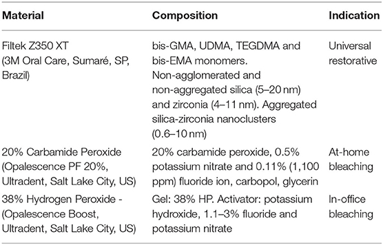
Table 1. Restorative and dental bleaching materials used.
After thermal cycling, specimens (n = 30) were immersed in red wine (Susana Balbo – Gran Reserva – 2012 – Red Blend – Mendonza, Argentina, 13.9%, pH 3.6), coffee (Nescafé Original Extra-forte, powder dissolved in 200 mL water, pH 5.1), and artificial saliva [1.5 mM CaCl2, 0.9 mM Na3PO4, 0.15 mM KCl – (35), pH = 7.0]. Specimens were kept in a container and immersed with the corresponding staining solution (200 mL) for 48 h at 37°C (36). After staining, specimens were washed thoroughly with distilled water and stored at 37°C in relative humidity for 24 h before the bleaching protocols.
After thermal cycling aging and staining, each group was subdivided and submitted to one of the three bleaching protocols (n = 10). The first protocol was a negative control, because the corresponding specimens were left unbleached and stored in AS throughout the experiment. The at-home bleaching protocol was performed using 20% CP (Opalescence PF 20%, Ultradent, Salt Lake City, UT, United States). The CP gel application regimen simulated a 15-day clinical treatment, but the interval times between each session was reduced in this in vitro study. Therefore, three 4-h applications were performed in a day, at 4-h intervals, for 5 days. The in-office bleaching procedures used 38% HP gel (Opalescence Boost, Ultradent). Four sessions were repeated at 24-h intervals, in which the gel was applied for 45-min, but the gel was refreshed every 15 min. The bleaching gels (0.01 g) were applied only on the top surface of the specimens, which was identified because the bottom was marked. The lateral area of the cylindric specimens was left unbleached. At the intervals, the specimens were stored in AS, renewed every 2 days. At the end of bleaching, specimens were stored for 24 h in AS before roughness measurements and colorimetric evaluation.
Surface roughness average was measured (Surf-Corder – SE 1700 - Kosakalab, Tokyo, Japan) at T0, TS, and TB in three different directions (every 45° angle rotation) in each specimen. The Ra (μm) of each specimen was obtained, with a 0.25 mm cut-off and 0.2 mm/s speed (18).
A hand spectrophotometer (Vita Easyshade, Vita-Zahnfabrik, Germany) measured the color of the specimens at T0, Ts, and TB. The tip of the device was standardized and placed on the center of each specimen. Three readings per specimen were performed and the average of the parameters L* (0: black and 100: white), a* (+a*: reed; –a*: green), and b* (+b*: yellow and –b*: blue) were obtained. ΔE00 was determined according to the formula ΔE00 = [(ΔL'/KLSL)2 + (ΔC'/KCSC)2 + (ΔH'/KHSH)2 + (ΔC'/KCSC)*(ΔH'/KHSH)]1/2, in which H represents hue and C, chroma (37). ΔWID was calculated based on the whiteness index for dentistry (WID) = 0.511L* – 2.324a* – 1.100b* (38). Delta values were calculated considering the time interval: (1) T0 and TS; (2) TS and TB. ΔE00 values of 0.81 and 1.77 units were adopted as 50:50% perceptibility and acceptability thresholds, respectively. ΔWID thresholds were 0.61 (PT) and 2.90 units (AT). These thresholds for changes in the color of restorative materials were previously determined in the literature (33, 34).
The obtained data were submitted to exploratory analysis of normality (Shapiro-Wilk, p > 0.05), and Ra and ΔE00 values were transformed to Log10 and square root, respectively. Ra and L* values were analyzed using general mixed model repeated measures and Tukey-Kramer tests. ΔE00 was submitted to two-way ANOVA and Tukey tests. The values of a* and b* parameters did not attend the normality even after transformation and were tested using a general linear model for repeated measures. Data obtained were submitted to Tukey-Kramer, except for ΔWID, which was analyzed by Kruskal-Wallis and Dunn's tests. The significance level was set at 5%.
Table 2 illustrates the Ra average of the nanofilled composite at different evaluation times. No statistical differences were detected among the groups (p > 0.05) at T0, but a significant decrease in Ra was observed for all groups (p < 0.05) at TS. Bleaching with 20% CP or 38% HP significantly increased the roughness of the composites that were aged and stained with RW (p < 0.05) but did not change the roughness of the composite previously aged and stained with CF or immersed in AS (p > 0.05). The surface roughness of the unbleached nanofilled composite aged and stained with RW increased at TB (p < 0.05). Besides, the surface roughness of the RW stained composite at TB was statistically similar to T0 (p > 0.05).
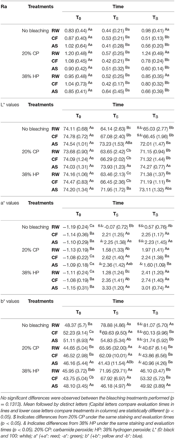
Table 2. Means and standard deviations of surface roughness (Ra, μm) and color parameters (L*, a*, b*) of the nanofilled composite at baseline (T0), after thermal cycling and staining (TS) and after bleaching (TB).
The luminosity (Table 2) of all groups significantly decreased after aging and staining (p < 0.05), except for unbleached composite immersed in AS or AS-group submitted to 20% CP. Bleaching with 20% CP or 38% HP significantly increased the luminosity of RW and CF stained composites (p < 0. 05). RW and CF stained composites exhibited lower luminosity values compared to the composite that remained immersed in AS, regardless of the bleaching treatment performed (p < 0.05). None of the groups were able to reach the baseline luminosity values, except for the composite that remained in AS and was submitted to 38% HP bleaching.
All groups exhibited a significant increase in the a* values (Table 2) after aging and staining. The a* mean values of RW- stained composites increased after 38% HP bleaching or remained unaltered after 20% CP bleaching. The a* values of CF stained composite decreased after 20% CP or remained unaltered after 38% HP bleaching.
The b* mean values of groups (Table 2) significantly increased after aging and staining with either RW or CF (p < 0.05). After bleaching with 20% CP or 38% HP, the b* values of the stained groups decreased compared to TS and were statistically lower than the stained groups not submitted to bleaching. The composite stored in artificial saliva and bleached exhibited no changes in the b* values, regardless of the thermal cycling aging or bleaching treatment. The unbleached stained composites exhibited higher b* values than the unbleached AS group (p < 0.05).
Figure 1 illustrates the ΔE00 of the nanofilled composite at different evaluation times. Overall, after aging and staining (TS – T0) and after bleaching (TB – TS), RW and CF exhibited higher color change than immersion in AS (p < 0.05). After bleaching, RW stained composites still displayed the highest ΔE00 followed by CF and AS (p < 0.05). The thermal cycling aging and staining of the composites induced color changes that were above the PT and AT thresholds, regardless of the staining protocols. Yet, the color changes promoted by bleaching (20% CP and 38% HP) of the stained composites (RW and CF) were above the PT and AT thresholds.
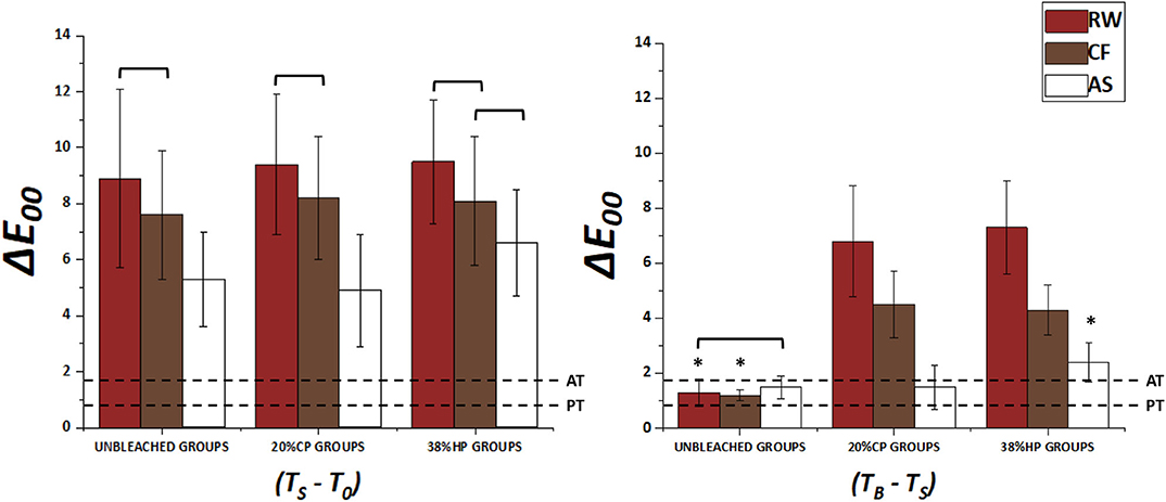
Figure 1. Mean values and standard deviations of ΔE00 of the two time-intervals (T0 and TS /TS and TB). Bars connected by the bracket did not differ statistically within the same level of bleaching agent factor (x-axis). Asterisk symbol differs, at a 5% significance level, within the same staining types between different bleaching agents. PT and AT lines represent the perceptibility (0.81) and acceptability (1.77) thresholds, respectively.
All groups presented negative ΔWID values after aging and staining (TS - T0), which were below PT and AT thresholds (Figure 2). The composite submitted to RW and CF-staining exhibited significantly lower ΔWID than AS, but bleaching with 20% CP or 38% HP was able to reverse the negative values and the whiteness index differences (TB - TS) were above the PT and AT thresholds. Also, RW and CF-bleached composites exhibited significantly higher ΔWID than the unbleached composites (p < .05).
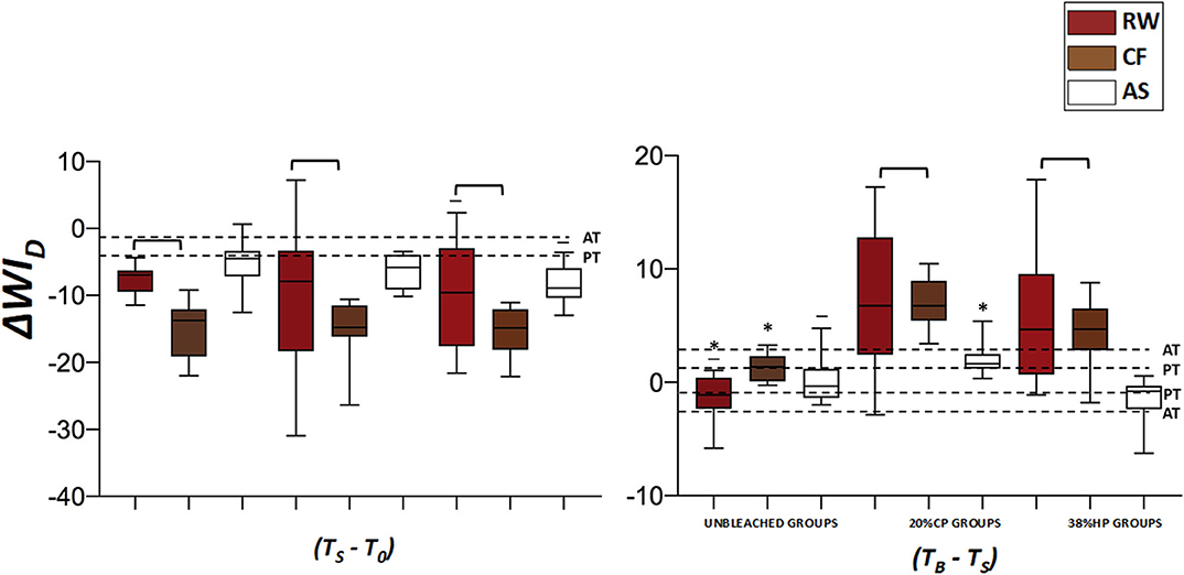
Figure 2. Median and minimum/maximum values of ΔWID between the two time-intervals (T0 and TS /TS and TB). Bars connected by the bracket, or the underline above the error bar, did not differ statistically within the same level of bleaching agent factor (x-axis). Asterisk symbol differs, at a 5% significance level, within the same staining types between different bleaching agents. WPT and WAT lines represent the perceptibility (0.61) and acceptability (2.90) thresholds, respectively.
This study performed thermal cycling to age the composite, simulating a preexisting restoration in the oral cavity, in which the aged polymer-based material could be more susceptible to staining and the effects of bleaching agents. The findings showed that thermal cycling aging decreased the Ra parameter of the nanofilled resin composite, regardless of the staining protocol used. However, bleaching increased surface roughness of the RW stained nanofilled composite, and changed the color of the aged and stained composites, therefore, rejecting the first and the second null hypotheses.
The surface roughness decrease occurred due to the 5,000 thermal cycles that all specimens were submitted to, regardless of exposing the composite to artificial staining. Contrarily to our results, Santos et al. (18) observed that 3,000 thermal cycles increased the roughness of microhybrid, microfilled, and nanofilled composites at different extents. However, compared to our results, after 10,000 thermal cycles, the surface roughness of the nanofilled composite decreased (18). The surface roughness increase observed after 3,000 thermal cycles were possibly due to the inorganic fillers dislodgment promoted by the thermal stress that may lead to microfractures and pores in the organic matrix or at the filler interface and the matrix (39). Additionally, water exposure triggers the hydrolytic degradation of the silane or causes swelling of the organic compound (18). As a consequence of the degradation process, inorganic fillers are exposed and surface roughness may increase. This process is also influenced by the presence of hydrophilic monomers as TEGDMA/TEGMA, which are more susceptible to degradation as water could penetrate more easily due to the hydrophobicity of the organic matrix (18).
The nanofilled composite herein used (Filtek Z350 XT) is composed of minor amounts of TEGDMA, which might be challenging to hydrolytic degradation and inorganic fillers exposure compared to composites with higher amounts of TEGDMA/TEGMA. Yet, the nanometric size (4–20 nm) and elevated inorganic fillers volume (82%), spherical shape, and distribution of the fillers in non-agglomerated, non-aggregated, and aggregated clusters of silica and zirconia particles (4–20 nm) could have contributed for the roughness surface decrease after thermal cycling. Although the artificial staining protocols did not influence the surface roughness of the composite at TS, it has probably accelerated the degradation since the roughness decrease was observed after 5,000 thermal cycles instead of 10,000, as reported by those authors (18).
The bleaching protocols increased the surface roughness of the composite stained with RW, regardless of the bleaching agent concentration (20% CP or 38% HP). Acidic solutions such as red wine (pH = 3.6) generate hydrolysis of the ester group present in the resin matrix and this hydrolysis, in turn, forms carboxylic groups, which are acid and decrease the pH inside the polymeric matrix (40). The speed of the degradation, influenced by the pH, changes the microstructure of the composite resin, creating pores in the resin mass. Therefore, if the thermal cycling initiated degradation, red wine probably exacerbated due to the wine acidity. Besides pH, the presence of ethanol in the RW will soften the polymer matrix (15). The consequence of the RW exposure could be observed in the nanofilled groups that remained unbleached, as this group exhibited a significant surface roughness increase at TB.
This study used a red wine containing 13.9% volume concentration of ethanol. The ethanol acts as a plasticizing of the polymeric matrix, smoothing, and dislodging filler particles, increasing considerably roughness and erosion of the composite (16). The susceptibility of the BIS-GMA and UDMA-based polymers to ethanol elution (41) could contribute to the softening effect of ethanol on the composite surface, and its concentration also influenced the surface integrity of the material. Tanthanuch et al. (15) observed that red wine promoted higher surface roughness and composite erosion than white wine, probably due to the higher ethanol concentration in red - 13.5% vol - than in the white whine - 12.5% vol. These results agree with previous observations on the effect the RW in microhybrid and nanofilled composites (9, 17, 42).
The composite submitted to red wine or coffee exhibited lower luminosity values (L*) than groups immersed in artificial saliva, compromising the composite luminosity after staining. According to Tan et al. (43), red wine, coffee, and tea cause more staining in the nanofilled composite than soft drink (Coke), orange juice, vodka, or distilled water. Red wine can stain due to the presence of flavonoids, and staining is possibly aggravated by the ethanol presence (36), as elution enables staining (44).
Carbamide or hydrogen peroxide bleaching procedures were effective in increasing the composite luminosity (L*) of RW and CF-groups, indicating that the reactive oxygen species could oxidize the staining molecules and this action was sufficient to increase luminosity even after extrinsic staining exposure (45). However, it should be noted that bleaching with 20% CP and 38% HP was unable to reach the baseline L* values of the stained composites. At baseline, the nanofilled composite exhibited negative a* values (–a* = green), however, shifted to positive (+a* = red), particularly after staining, matching the reddish stain of the protocol used. The b* parameter remained positive (+b* = yellow), but RW and CF - staining increased the yellow appearance, which was later decreased by bleaching. Additionally, the composite submitted to the staining protocols exhibited higher yellow appearance than the composite immersed in artificial saliva. Although the yellow (b*) appearance of the stained composite decreased after bleaching, reaching the baseline values, the same was not noticed for the L* values, which might compromise the composite final color outcome. These results agree with those of Poggio et al. (46) who detected significant changes in L*, a*, and b* coordinates and demonstrated the susceptibility of nanofilled composites to staining with red wine and coffee.
Color changes according to the CIEDE 2000 equation were evaluated instead of the CIELAB formula, due to significant corrections made on hue and chroma (37), while the whiteness index for dentistry is formulated based in the L*, a*, b* parameters and provides accurate and reliable data to inform the whiteness level of tooth and restorative-related materials (38). The 50:50% perceptibility and acceptability thresholds adopted in this study are based on perception and acceptance of color and whiteness changes by nonprofessional or professional volunteers investigated in multicenter prospective studies (33, 47).
Thermal cycling aging promoted ΔE00 on nanofilled composites that would be perceptible to patients (above 50:50% PT > 0.81, Figure 1), but not clinically acceptable (AT > 1.77), because thermal cycling and staining darkened the composite, regardless of the staining protocol, as shown by the negative whiteness index values (Figure 2). Therefore, the last null hypothesis was accepted. On the other hand, bleaching was able to reverse darkening of the RW and CF stained composite (as shown by the positive ΔWID values, Figure 2) which would be clinically perceptible to patients (above 50:50% PT > 0.81 and WPT > 0.61), but still not clinically acceptable (above 50:50% AT > 1.44, WAT > 2.90). Therefore, the analysis of the thresholds could be translated into the possible necessity of restoration replacement after bleaching, to overcome the unacceptable color match. Also, a further study could investigate the polishing of the restorations as an alternative to their replacement. A previous study by Rodrigues et al. (48) demonstrated that immediate repolishing after the last bleaching session improved the color stability of micro- and nanohybrid composite resins prior to immersion in red wine solution.
The composite not stained and submitted to 20% CP presented ΔE00 and ΔWID above the 50:50% PT and WPT, but below the 50:50% AT and WAT. The same scenario was observed for composite left unstained and unbleached. This means that color changes of unstained, unbleached resin or submitted to at-home bleaching, were clinically perceptible but acceptable to patients. Della Bona et al. (31) showed that bleaching an aged nanofilled composite (Filtek Z350XT) led to perceptible and acceptable ΔE00 and ΔWID, considering the corresponding 50:50% thresholds. However, this conclusion was drawn for both at-home and in-office bleaching. In this study, 38%HP bleaching of unstained composite still exhibited unacceptable ΔE00 and ΔWID. The higher frequency of exposure and the prolonged contact of the 20% CP agent on the composite surface rendered a more acceptable final color result (49). Based on the colorimetric evaluation, bleaching was able to change the color of aged-stained composites, however, it was unable to reverse all the color parameters (L*, a*, b*) back to the baseline values. Therefore, although the color change was perceptible, it was not clinically acceptable.
The unstained and unbleached resin (control group) also exhibited colorimetric changes (ΔE00 and ΔWID), even when not submitted to any staining or bleaching protocol. Recent studies showed that polymer-based materials stored in distilled water for either 10 or 30 days exhibited perceptible color changes (50, 51). According to the authors, this outcome could be the result of intrinsic changes of the materials probably due to water sorption. Moreover, since the illumination conditions were standardized (room luminosity, background, calibration of the spectrophotometer and measurements at the same direction), such alterations could be a result from the specimens' hydration promoted by the storing solution.
This study presents the inherent limitation of an in vitro evaluation, as other factors could clinically induce surface roughness or color changes of the aged nanofilled composite. As an example, thermal alterations and staining exposure in an oral environment could be influenced by factors such as the presence of saliva and individual oral hygiene habits (such as toothbrushing). However, it should be noted that the surface roughness increase in the composite resin could clinically lead to biofilm formation (20, 21). In this context, further studies could evaluate the impact of toothbrushing on color change and surface roughness, since brushing could influence the deposition of extrinsic stains on the restoration surface (4).
Also, it is important to highlight that the intervals of bleaching applications must follow the manufacturer's instructions and the literature evidence. This is particularly important when bleaching is performed clinically, on the dental surface (not isolated on the composite surface, as it was in this in vitro study), to avoid adverse effects such as exacerbated tooth sensitivity and possible consequences to the pulpal tissues, especially in the presence of adhesive restorations (22, 52).
In the present study, the surface roughness of the nanofilled composite was influenced by thermal cycling aging and staining, combined or not with bleaching. However, the surface roughness decreased after thermal cycling minimizing the deleterious effect of the bleaching agents, as surface roughness after bleaching as similar to baseline. On the other hand, bleaching was not able to reverse the staining promoted by red wine and coffee on the nanofilled composite, which could suggest the need for replacement of a preexisting restoration. Future studies are required to properly access the prolonged thermal cycling aging effect on the nanofilled composite and explore different bleaching protocols and their effects on stained composites.
Within the limitations of the present study, it can be concluded that at-home and in-office bleaching increased the surface roughness of aged nanofilled composite stained with red wine. Besides, although at-home and in-office bleaching changed the color of the aged and stained composite, bleaching was not able to reverse the staining promoted by red wine and coffee on the nanofilled composite.
The original contributions presented in the study are included in the article/supplementary material, further inquiries can be directed to the corresponding author/s.
GS and CP performed the laboratorial procedures and analyses. MK and GS wrote the initial draft of the manuscript. VC, FL, and CP design the study, supervised the students, and edited the writing. All authors contributed to the article and approved the submitted version.
This study was supported in part by Coordenação de Aperfeiçoamento de Pessoal do Nível Superior (CAPES) – 001.
The authors declare that the research was conducted in the absence of any commercial or financial relationships that could be construed as a potential conflict of interest.
1. Blumer L, Schmidli F, Weiger R, and Fischer J. A systematic approach to standardize artificial aging of resin composite cements. Dent Mater. (2015) 31:855–63. doi: 10.1016/j.dental.2015.04.015
2. Szczesio-Wlodarczyk A, Sokolowski J, Kleczewska J, and Bociong K. Ageing of dental composites based on methacrylate resins-a critical review of the causes and method of assessment. Polyck. (2020) 12:882. doi: 10.3390/polym12040882
3. Ferracane JL. Resin-based composite performance: are there some things we can't predict? Dent Mater. (2013) 29:51–8. doi: 10.1016/j.dental.2012.06.013
4. Heintze SD, Reichl FX, and Hickel R. Wear of dental materials: clinical significance and laboratory wear simulation methods -a review. Dent Mater J. (2019) 38:343–53. doi: 10.4012/dmj.2018-140
5. Schulze KA, Marshall SJ, Gansky SA, and Marshall GW. Color stability and hardness in dental composites after accelerated aging. Dent Mater. (2003) 19:612–9. doi: 10.1016/S0109-5641(03)00003-4
6. Çelik EU, Aladag A, Türkün LS, and Yilmaz G. Color changes of dental resin composites before and after polymerization and storage in water. J Esthet Restor Dent. (2011) 23: 179–88. doi: 10.1111/j.1708-8240.2011.00421.x
7. Lee YK. Comparison of CIELAB DeltaE(*) and CIEDE2000 color-differences after polymerization and thermocycling of resin composites. Dent Mater. (2005) 21:678–82. doi: 10.1016/j.dental.2004.09.005
8. Paravina RD, Kimura M, and Powers JM. Evaluation of polymerization-dependent changes in color and translucency of resin composites using two formulae. Odontology. (2005) 93:46–51. doi: 10.1007/s10266-005-0048-7
9. Bansal K, Acharya SR, and Saraswathi V. Effect of alcoholic and non-alcoholic beverages on color stability and surface roughness of resin composites: an in vitro study. J Conserv Dent. (2012) 15:283–8. doi: 10.4103/0972-0707.97961
10. Pruthi G, Jain V, Kandpal HC, Mathur VP, and Shah N. Effect of bleaching on color change and surface topography of composite restorations. Int J Dent. (2010) 2010:695748. doi: 10.1155/2010/695748
11. Malekipour MR, Sharafi A, Kazemi S, Khazaei S, and Shirani F. Comparison of color stability of a composite resin in different color media. Dent Res J. (2012) 9:441−6.
12. Llena C, Fernández S, and Forner L. Color stability of nanohybrid resin-based composites, ormocers and compomers. Clin Oral Investig. (2017) 21:1071–7. doi: 10.1007/s00784-016-1850-z
13. Topcu FT, Sahinkesen G, Yamanel K, Erdemir U, Oktay EA, and Ersahan S. Influence of different drinks on the colour stability of dental resin composites. Eur J Dent. (2009) 3:50–6. doi: 10.1055/s-0039-1697405
14. Ardu S, Braut V, Gutemberg D, Krejci I, Dietschi D, and Feilzer AJ. A long-term laboratory test on staining susceptibility of esthetic composite resin materials. Quintessence Int. (2010) 41:695–702.
15. Tanthanuch S, Kukiattrakoon B, Peerasukprasert T, Chanmanee N, Chaisomboonphun P, and Rodklai A. The effect of red and white wine on color changes of nanofilled and nanohybrid resin composites. Restor Dent Endod. (2016) 41:130–6. doi: 10.5395/rde.2016.41.2.130
16. Sarrett DC, Coletti DP, and Peluso AR. The effects of alcoholic beverages on composite wear. Dent Mater. (2000) 16:62–7. doi: 10.1016/S0109-5641(99)00088-3
17. de Alencar ML, da Cunha Medeiros E, Silva FD, Meireles SS, Duarte RM, and Andrade AK. The effect of drinks on color stability and surface roughness of nanocomposites. Eur J Dent. (2014) 8:330–6. doi: 10.4103/1305-7456.137640
18. Santos PH, Catelan A, Albuquerque Guedes AP, Umeda Suzuki TY, de Lima Godas AG, Fraga Briso AL, et al. Effect of thermocycling on roughness of nanofill, microfill and microhybrid composites. Acta Odontol Scand. (2015) 73:176–81. doi: 10.3109/00016357.2014.971868
19. Karatas O, Gul P, Gündogdu M, and Iskenderoglu DT. An evaluation of surface roughness after staining of different composite resins using atomic force microscopy and a profilometer. Microsc Res Tech. (2020) 83:1251–9. doi: 10.1002/jemt.23519
20. Park JW, Song CW, Jung JH, Ahn SJ, and Ferracane JL. The effects of surface roughness of composite resin on biofilm formation of Streptococcus mutans in the presence of saliva. Oper Dent. (2012) 37:532–9. doi: 10.2341/11-371-L
21. Cazzaniga G, Ottobelli M, Ionescu A, Garcia-Godoy F, and Brambilla E. Surface properties of resin-based composite materials and biofilm formation: a review of the current literature. Am J Dent. (2015) 28:311–20.
22. Kwon SR, and Wertz PW. Review of the mechanism of tooth whitening. J Esthet Restor Dent. (2015) 27:240–57. doi: 10.1111/jerd.12152
23. Wang L, Francisconi LF, Atta MT, Dos Santos JR, Del Padre NC, Gonini Junior A, et al. Effect of bleaching gels on surface roughness of nanofilled composite resins. Eur J Dent. (2011) 5:173–9. doi: 10.1055/s-0039-1698876
24. Andrade ICGB, Basting RT, Rodrigues JA, Amaral FLB, Turssi CP, and França FMG. Microhardness and color monitoring of nanofilled resin composite after bleaching and staining. Eur J Dent. (2014) 8:160–5. doi: 10.4103/1305-7456.130586
25. Öztürk C, Çelik E, and Özden AN. Influence of bleaching agents on the color change and translucency of resin matrix ceramics. J Esthet Restor Dent. (2020) 32:530–5. doi: 10.1111/jerd.12580
26. Attin T, Hannig C, Wiegand A, and Attin R. Effect of bleaching on restorative materials and restorations–a systematic review. Dent Mater. (2004) 20:852–61. doi: 10.1016/j.dental.2004.04.002
27. Torres C, Ribeiro C, Bresciani E, and Borges A. Influence of hydrogen peroxide bleaching gels on color, opacity, and fluorescence of composite resins. Oper Dent. (2012) 37:526–31. doi: 10.2341/11-189-L
28. Markovic L, Jordan RA, Glasser MC, Arnold WH, Nebel J, Tillmann W, et al. Effects of bleaching agents on surface roughness of filling materials. Dent Mater J. (2014) 33:59–63. doi: 10.4012/dmj.2012-217
29. Alzraikat H, Burrow MF, Maghaireh GA, and Taha NA. Nanofilled resin composite properties and clinical performance: a review. Oper Dent. (2018) 43:E173–90. doi: 10.2341/17-208-T
30. Paravina RD, Pérez MM, and Ghinea R. Acceptability and perceptibility thresholds in dentistry: a comprehensive review of clinical and research applications. J Esthet Restor Dent. (2019) 31:103–12. doi: 10.1111/jerd.12465
31. Della Bona A, Pecho OE, Ghinea R, Cardona JC, Paravina RD, and Perez MM. Influence of bleaching and aging procedures on color and whiteness of dental composites. Oper Dent. (2019) 44:648–58. doi: 10.2341/18-209-L
32. Kury M, Perches C, da Silva DP, André CB, Tabchoury CPM, Giannini M, et al. Color change, diffusion of hydrogen peroxide, and enamel morphology after in-office bleaching with violet light or nonthermal atmospheric plasma: An in vitro study. J Esthet Restor Dent. (2020) 32:102–12. doi: 10.1111/jerd.12556
33. Paravina RD, Ghinea R, Herrera LJ, et al. Color difference thresholds in dentistry. J Esthet Restor Dent. (2015) 27:S1–9. doi: 10.1111/jerd.12149
34. Pérez MM, Herrera LJ, Carrillo F, Pecho OE, Dudea D, Gasparik C, et al. Whiteness difference thresholds in dentistry. Dent Mater. (2019) 35:292–7. doi: 10.1016/j.dental.2018.11.022
35. Shinkai RS, Cury AA, and Cury JA. In vitro evaluation of secondary caries development in enamel and root dentin around luted metallic restoration. Oper Dent. (2001) 26:52–9.
36. Berger SB, Coelho AS, Oliveira VAP, Cavalli V, and Giannini M. Enamel susceptibility to red wine staining after 35% hydrogen peroxide bleaching. J Appl Oral Sci. (2008) 16:201–4. doi: 10.1590/S1678-77572008000300007
37. Sharma G, Wu W, and Dalal EN. The CIEDE2000 color-difference formula: implementation notes, supplementary test data, and mathematical observations. Color Res Appl. (2005) 30:21–30. doi: 10.1002/col.20070
38. Pérez Mdel M, Ghinea R, Rivas MJ, Yebra A, Ionescu AM, Paravina RD, et al. Development of a customized whiteness index for dentistry based on CIELAB color space. Dent Mater. (2016) 32:461–7. doi: 10.1016/j.dental.2015.12.008
39. Rinastiti M, Ozcan M, Siswomihardjo W, and Busscher HJ. Effects of surface conditioning on repair bond strengths of non aged and aged microhybrid, nanohybrid, and nanofilled composite resins. Clin Oral Investig. (2011) 15:625–33. doi: 10.1007/s00784-010-0426-6
40. Prakki A, Cilli R, Mondelli RF, Kalachandra S, and Pereira JC. Influence of pH environment on polymer-based dental material properties. J Dent. (2005) 33:91–8. doi: 10.1016/j.jdent.2004.08.004
41. Benetti AR, Ribeiro de Jesus VC, Martinelli NL, Pascotto RC, and Poli-Frederico RC. Colour stability, staining and roughness of silorane after prolonged chemical challenges. J Dent. (2013) 41:1229–35. doi: 10.1016/j.jdent.2013.10.004
42. Lepri CP, and Palma-Dibb RG. Surface roughness and color change of a composite: influence of beverages and brushing. Dent Mat J. (2012) 31:689–96. doi: 10.4012/dmj.2012-063
43. Tan BL, Yap AU, Ma HN, Chew J, and Tan WJ. Effect of beverages on color and translucency of new tooth-colored restoratives. Oper Dent. (2015) 40:E56–65. doi: 10.2341/149027-L
44. Villalta P, Lu H, Okte Z, Garcia-Godoy F, and Powers JM. Effects of staining and bleaching on color change of dental composite resins. J Prosthet Dent. (2006) 95:137–42. doi: 10.1016/j.prosdent.2005.11.019
45. Mohammadi N, Kimyai S, Kahnamoii MA, Chaharom MEE, Sadr A, and Daneshi M. Effect of 15% carbamide peroxide bleaching gel on color stability of giomer and microfilled composite resin: An in vitro comparison. Med Oral Patol Oral Cir Bucal. (2012) 17:e1082–8. doi: 10.4317/medoral.17916
46. Poggio C, Ceci M, Beltrami R, Mirando M, Wassim J, and Colombo M. Color stability of esthetic restorative materials: a spectrophotometric analysis. Acta Biomater Odontol Scand. (2016) 2:95–101. doi: 10.1080/23337931.2016.1217416
47. Pérez MM, Pecho OE, Ghinea R, Pulgar R, and Della Bona A. Recent advances in color and whiteness evaluations in dentistry. Current Dent. (2019) 1:23–9. doi: 10.2174/2542579X01666180719125137
48. Rodrigues CS, Nora BD, Mallmann A, May LG, and Jacques LB. Repolishing resin composites after bleaching treatments: effects on color stability and smoothness. Oper Dent. (2019) 44:54–64. doi: 10.2341/17-107-L
49. Fernandes RA, Strazzi-Sahyon HB, Suzuki TYU, Briso ALF, and Dos Santos PH. Effect of dental bleaching on the microhardness and surface roughness of sealed composite resins. Restor Dent Endod. (2020) 45:e12. doi: 10.5395/rde.2020.45.e12
50. Tinastepe N, Malkondu O, Iscan I, and Kazazoglu E. Effect of home and over the contour bleaching on stainability of CAD/CAM esthetic restorative materials. J Esthet Restor Dent. (2020). doi: 10.1111/jerd.12604. [Epub ahead of print].
51. Aydin N, Karaoglanoglu S, Oktay EA, and Kiliçarslan MA. Investigating the color changes on resin-based CAD/CAM Blocks. J Esthet Restor Dent. (2020) 32:251–6. doi: 10.1111/jerd.12561
Keywords: tooth bleaching, composite resins, carbamide peroxide, hydrogen peroxide, surface
Citation: dos Santos Muniz Mota GM, Kury M, Pereira da Silva Braga Tenório C, Lucisano Botelho do Amaral F, Turssi CP and Cavalli V (2020) Effects of Artificial Staining and Bleaching Protocols on the Surface Roughness, Color, and Whiteness Changes of an Aged Nanofilled Composite. Front. Dent. Med. 1:610586. doi: 10.3389/fdmed.2020.610586
Received: 26 September 2020; Accepted: 10 November 2020;
Published: 10 December 2020.
Edited by:
L. Sebnem Turkun, Ege University, TurkeyReviewed by:
Sandrine Bittencourt Berger, Universidade Norte do Paraná, BrazilCopyright © 2020 dos Santos Muniz Mota, Kury, Pereira da Silva Braga Tenório, Lucisano Botelho do Amaral, Turssi and Cavalli. This is an open-access article distributed under the terms of the Creative Commons Attribution License (CC BY). The use, distribution or reproduction in other forums is permitted, provided the original author(s) and the copyright owner(s) are credited and that the original publication in this journal is cited, in accordance with accepted academic practice. No use, distribution or reproduction is permitted which does not comply with these terms.
*Correspondence: Vanessa Cavalli, Y2F2YWxsaUB1bmljYW1wLmJy
Disclaimer: All claims expressed in this article are solely those of the authors and do not necessarily represent those of their affiliated organizations, or those of the publisher, the editors and the reviewers. Any product that may be evaluated in this article or claim that may be made by its manufacturer is not guaranteed or endorsed by the publisher.
Research integrity at Frontiers
Learn more about the work of our research integrity team to safeguard the quality of each article we publish.