 Ekaterina V. Dmitrieva
94% of researchers rate our articles as excellent or good
Learn more about the work of our research integrity team to safeguard the quality of each article we publish.
Find out more
REVIEW article
Front. Chem. , 26 February 2025
Sec. Analytical Chemistry
Volume 13 - 2025 | https://doi.org/10.3389/fchem.2025.1548364
Surface-enhanced Raman spectroscopy is a powerful analytical technique for the determination of analytes with the advantages of sensitivity, portability, and simplicity, able to provide structural information for the identification of compounds. However, when it comes to the analysis of complex samples, matrix components may interfere with the analyte quantification. To overcome this shortcoming, a number of approaches have been proposed, such as extraction techniques. Among them, the coupling of chromatography with surface-enhanced Raman spectroscopy seems to be promising. It allows combining the advantages of both techniques, i.e., high efficiency of chromatographic separation and high sensitivity of surface enhanced Raman scattering detection, and makes possible simultaneous quantification of multiple analytes. The review summarizes the latest achievements in the combination of these techniques.
Despite the rapid development of analytical methods, most of them still suffer from insufficient sensitivity and selectivity, high cost, and inapplicability for on-site analysis. Therefore, there exists a need to develop techniques devoid of these shortcomings. One such technique is surface enhanced Raman scattering (SERS), discovered in the 1970s (Ay et al., 2024; Jeanmaire and Van Duyne, 1977; Wang et al., 2022), with the advantages of portability, high sensitivity, rapid and powerful non-destructive detection (Han et al., 2023; Zhang et al., 2017), which is very sensitive to both chemical and biological species. It is a molecular vibration spectroscopy technique, resulting from an inelastic scattering process, able to provide structural information of a molecule (Zhang et al., 2017). Two mechanisms of signal enhancement in SERS have been proposed, namely, electromagnetic enhancement and charge-transfer or chemical enhancement (Han et al., 2023; McNay et al., 2011; Zhang et al., 2017). Chemical enhancement is generally limited to two orders of magnitude, while electromagnetic–up to 14 orders of magnitude (Han et al., 2023; Zhang et al., 2017). Historically, these two mechanisms were thought to be quite different (McNay et al., 2011). To date, numerous studies have revealed that these two mechanisms work together; however, their contribution varies (Han et al., 2023; Jensen et al., 2008). For both mechanisms, the analyte should be first adsorbed on a SERS active substrate followed by substrate irradiation by monochromatic radiation usually from a laser (McNay et al., 2011).
Electromagnetic enhancement is caused by localized surface plasmon resonance (LSPR) of compounds at the nanostructured surface of noble metals, e.g., Ag, Au, or bi/trimetallic noble metal alloys (Duy Vu et al., 2024), and depends on the roughness of the metal surface (Han et al., 2023). For the enhancement to occur, the molecule and surface of substrate should be in very close vicinity, i.e., less than 10 nm (Freye et al., 2013; Gao et al., 2015). Chemical enhancement is considered to involve electron transfer between the analyte and nanostructured surface with the formation of a charge transfer complex (Gao et al., 2015; McNay et al., 2011), which alters the molecule polarization (Ay et al., 2024; Duy Vu et al., 2024; Zhang et al., 2017). The morphology and size of the substrates play an essential role in the enhancement of electromagnetic field (Sha et al., 2022). Even slight variations of such parameters of colloidal solutions as cluster sizes and shapes produce changes in the enhancement factors by several orders of magnitude. Optimum enhancement can be observed for metallic substrates with the diameters between 10 and 150 nm (Sackmann and Materny, 2006). To achieve better performance, structures with different shapes, e.g., nanospheres, nanorods, nanostars, etc., were synthesized (Sha et al., 2022). In practice, a substrate should provide reasonable enhancement, be reproducible, stable, and robust (Freye et al., 2013).
SERS is highly efficient in the analysis of samples with a simple matrix. However, it is not a separation technique (Zhang et al., 2017), and when it comes to real sample analysis, matrix components may interfere with the determination of target analytes (Dai and He, 2023; Han et al., 2023; Lee et al., 2018; Minh et al., 2019), e.g., due to high fluorescence of natural compounds or interactions with irrelevant components present in the samples (Minh et al., 2023; Minh et al., 2024; Zhang et al., 2017). The most apparent solution of this issue is a preliminary separation of a complex mixture. For this, several approaches can be applied, such as liquid-liquid extraction (Alves et al., 2020), solid-phase extraction (Radu et al., 2016), dispersive liquid-liquid extraction (Santhoshkumar et al., 2025), cloud-point extraction (Zhang et al., 2021), or utilization of filter membranes (Fateixa et al., 2018; Qu et al., 2017; Yu and White, 2012; Wigginton and Vikesland, 2010). These approaches provide analyte purification from matrix components along with its concentration. However, analyte losses during extraction can occur, and these techniques can be applied for a limited number of compounds. For the simultaneous determination of multiple analytes, chromatography is the most powerful method. This method is suitable for analysis of complex samples and allows the separation of analytes into narrow zones depending on their physicochemical properties. One of the most informative detection methods coupled with chromatography is mass spectrometry, which allows identification of molecules based on their mass-to-charge ratios (m/z) and fragmentation patterns. However, the instruments are expensive, hardly portable, and require highly qualified personnel. At the same time, surface-enhanced Raman spectroscopy, capable of analytes identification based on their fingerprint spectra (Wang et al., 2021), is devoid of these shortcomings. In this regard, hyphenation of Raman spectroscopy with chromatography has been proposed and is widely applied nowadays. The review summarizes the results of studies utilizing chromatographic separation with subsequent SERS detection for the analysis of real samples and discusses future perspectives of the method.
Chromatography is based on the analyte distribution between two phases, i.e., mobile and stationary, followed by detection of separated analytes applying various physicochemical principles. The chromatographic methods are divided into planar and column. Paper chromatography and thin-layer chromatography (TLC) are related to planar chromatography. In these methods, the separation of analytes is carried out on plane surfaces and is driven by capillary forces. In gas chromatography (GC) and liquid chromatography (LC), the separation is conducted in columns. The schematic of this classification is given in Figure 1. Nowadays, all these methods were combined with surface-enhanced Raman spectroscopy. Among them, the coupling of SERS with planar chromatography is easier to implement; as a result, it has been applied more widely.
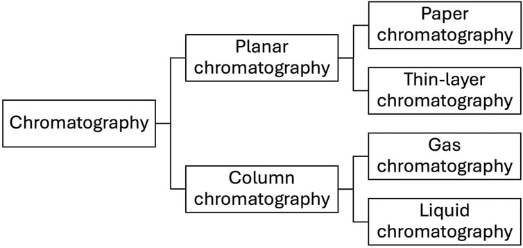
Figure 1. Classification of chromatographic methods coupled with SERS detection.
Compared to column chromatography, thin-layer chromatography with SERS detection has the advantages of rapid on-site detection with low cost, decreased sample pretreatment, and ability to simultaneously analyze several samples (Zhu et al., 2019). In thin layer chromatography, chromatographic plates with a sorbent layer are utilized as the stationary phase. After spiking the sample at the starting line, the plates are placed in a separation chamber containing the mobile phase for the development. The analytes with the eluent pass through the sorbent by capillary forces and are separated on the plate according to their affinity to the stationary phase. The parameter used for analyte identification is the retention factor (Rf), i.e., the ratio of the distance passed by the analyte to the distance passed by the eluent (solvent front). After compounds separation, the analytes can be detected by means of several methods, for example, densitometry or fluorescence spectroscopy. TLC provides fast and efficient analyte separation, but the detection methods lack sensitivity and selectivity; hence, SERS was proposed as an alternative due to their high compatibility (Han et al., 2023). The combination of TLC and SERS was first described in 1977 (Henzel, 1977; Pozzi et al., 2012) and nowadays has found numerous applications in various fields from food safety to environmental protection. Other essential advantages of the method include the lack of cross-contamination due to the use of disposable TLC plates, ability to simultaneously analyze several samples, and its portability. As a result, it can be used for on-site analysis of complex samples. Another interesting application of TLC-SERS is on-site quantitative monitoring of chemical reactions. The reaction mixture is spiked on a TLC plate; after the plate development, SERS is applied for detection, which enables to distinguish even incompletely separated compounds according to their characteristic Raman bands without loss of chemicals. In addition, it can be applied to the detection of compounds unstable under ultraviolet radiation and compounds present at low levels (Chen et al., 2015a; Fukunaga et al., 2023; Zhang et al., 2014).
There exist several approaches for TLC-SERS assay. The first one is based on the analyte separation on commercially available TLC plates with conventional stationary phases followed by the addition of metal nanoparticle (NP) colloids on the developed plate for the sensitive SERS detection (Figure 2); these substrates have random morphology (Freye et al., 2013; Zhao et al., 2019). The other is based on the stationary phase modification to obtain a SERS-active surface with subsequent analyte separation and detection (Figure 3) (Chen et al., 2012; Han et al., 2023; Minh et al., 2019); the substrates have deterministic morphology and were termed as engineered substrates (Freye et al., 2013).
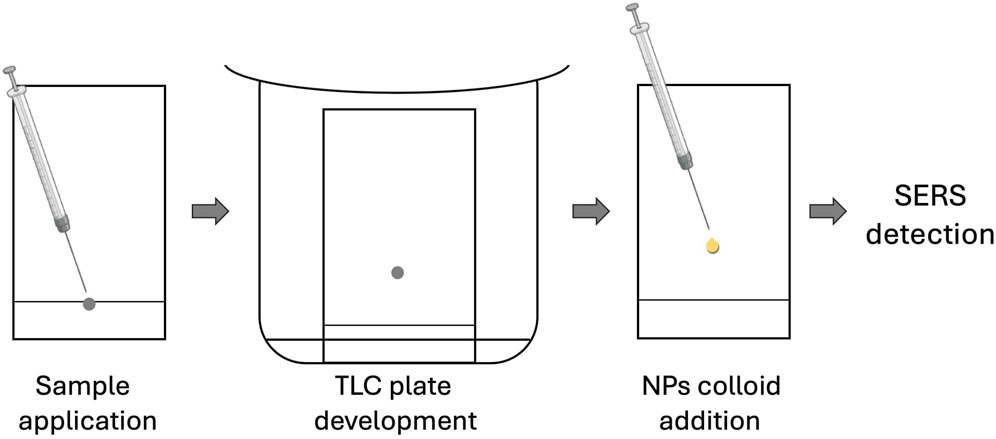
Figure 2. Schematic of TLC-SERS utilizing conventional TLC plates.
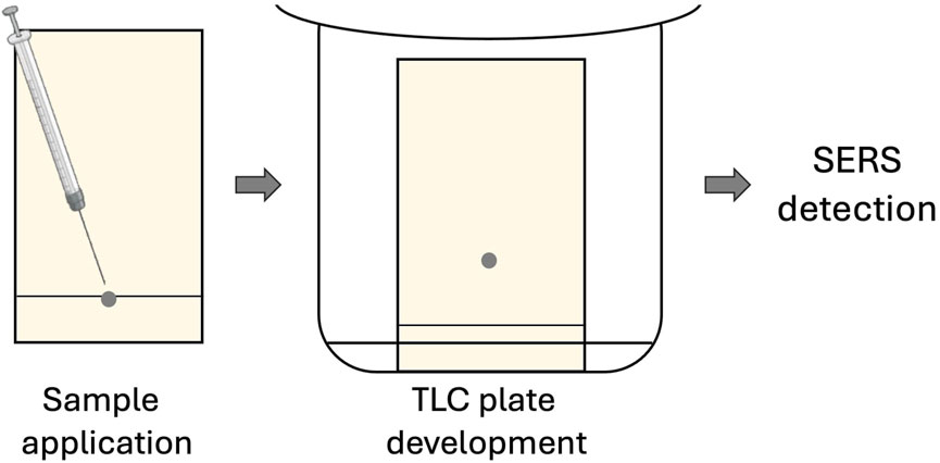
Figure 3. Schematic of TLC-SERS utilizing modified TLC plates.
In the former approach, commercially available TLC plates are mostly used to separate target analytes. Nowadays, there exists a wide range of TLC plates allowing to separate analytes with various physicochemical properties. Even though TLC plates are generally weak Raman scatters (Cimpoiu et al., 2005; Orinak et al., 2008), several studies have revealed that they are able to provide pronounced background Raman scattering from the stationary phase (Horváth et al., 2000; István et al., 2003; Zhu et al., 2014), which should be considered during optimization of conditions. Among the stationary phases, the most commonly used is silica gel. Although it does not provide significant background to the SERS signal, the interactions between the Si−OH groups of the TLC plate and the analyte can result in hydrogen bonding leading to a shift in the obtained spectrum (Freye et al., 2013; White, 1985). What is more, the solvents for the mobile phase should be carefully selected; for example, acetonitrile was reported to adsorb readily to the silver surface and disturb SERS measurements at low analyte concentrations (Trachta et al., 2004). To achieve the signal enhancement by SERS detection, valuable metal colloids are introduced to the separated spots of analytes. The spots of analytes can be easily visualized, since most TLC plates contain a fluorescent indicator. To obtain a strong SERS signal, the choice of NPs material is crucial (Yao et al., 2013). For obtaining the best results, nanoparticles preparation procedure, concentration, and spiking volume are optimized (Orinak et al., 2008; Yao et al., 2013). The most used metals for the preparation of colloid solutions are gold and silver due to the simplicity of their preparation by a chemical reduction method, relatively low cost, reasonable stability, and narrow particle size distribution (Han et al., 2023; McNay et al., 2011). Even though gold is more inert in comparison to silver, it quite strongly absorbs at around 500 nm. Therefore, silver is more suitable in this wavelength range. At the same time, silver has higher chemical reactivity reducing it lifetime (McNay et al., 2011; Murugan et al., 2021).
Application of conventional TLC plates with subsequent addition of noble metal colloids for SERS detection has been described in various fields, i.e., dietary supplements analysis, food quality control, analysis of environmental samples, or showing the method capability on model solutions (Table 1), for the quantification of antitussive and antiasthmatic drugs (Fang et al., 2016), antidiabetic drugs (Minh et al., 2023; Zhu et al., 2014), lipid-lowering agents (Zhu et al., 2017), β-carboline alkaloids (Pozzi et al., 2012), aflatoxins (Qu et al., 2018), pesticides (Kang et al., 2019; Yao et al., 2013), substituted aromatic pollutants (Li et al., 2011), synthetic dyes (Cañamares et al., 2014; Ferretti et al., 2024), etc. Solid samples were mostly prepared for analysis by homogenization followed by their extraction, centrifugation, and spotting supernatant on the starting line of a TLC plate for separation. Liquid samples were diluted with the solvent and centrifuged to reduce matrix effects. In addition to introducing noble metal colloids after TLC separation, immersing TLC plate in silver ion solution was proposed. This approach was applied to the assay of binary mixtures of cresyl violet, bixine, crystal violet, and Cu(II) complex of 4-(2-pyridylazo)resorcinol. Prior to analyte separation, a silica gel 60 TLC plate was immersed in silver nitrate solution. Then, the separated analytes were detected by SERS (Herman et al., 2013). Another implementation of TLC-SERS combination was shown in (Li et al., 2021) for the quantification of 14 citrus flavonoids in various matrices. After their separation on a normal-phase TLC plate, the analytes from spots were extracted in methanol. The supernatant was then analyzed by SERS. A similar approach was applied to the quantification of cotinine and trans-3′-hydroxycotinine, metabolites of nicotine and indicators of smoking status, in urine (Huang et al., 2013). However, non-uniform adsorption of metal colloids on the stationary phase owing to its porosity may occur, and the process of dispensing colloids may disrupt the separated target molecules due to the spot partial solubilization thus affecting accuracy and reproducibility of results (Dai and He, 2023; Han et al., 2023; Sciutto et al., 2017; Takei et al., 2015; Zhao et al., 2019). The phenomenon of the formation of a ring-shape spot after the addition of nanoparticles caused by their aggregation was termed as the coffee ring effect (CRE): single CRE if nanoparticles aggregate alone and double CRE if nanoparticles aggregate with the analyte (Minh et al., 2023). The migration of the nanoparticles and analyte due to the CRE can also affect the repeatability of results (Yao et al., 2022). Another important observation is based on the time of SERS signal measurement since Ag or Au colloid spiking to the analyte spot. The nanoparticles were reported to generate hot spots during solvent volatilization, and solvent volatilization-induced analyte concentration occurred. The SERS signal was the strongest when the droplet tended to become dry. However, the reproducibility of the results was poor even if continuous measurements during solvent evaporation were carried out (Kang et al., 2019). The signal enhancement through modification of the TLC plate surface by adding a thin film of water was also noted in (István et al., 2003; Szabo and Winefordner, 1997). To prevent local drying effects from the laser, a cover glass was used (István et al., 2003). The control of nanoparticles colloid solution stability is also quite challenging, since they are prone to spontaneous aggregation, which may also affect reproducibility of results (Han et al., 2023; Lee et al., 1998).
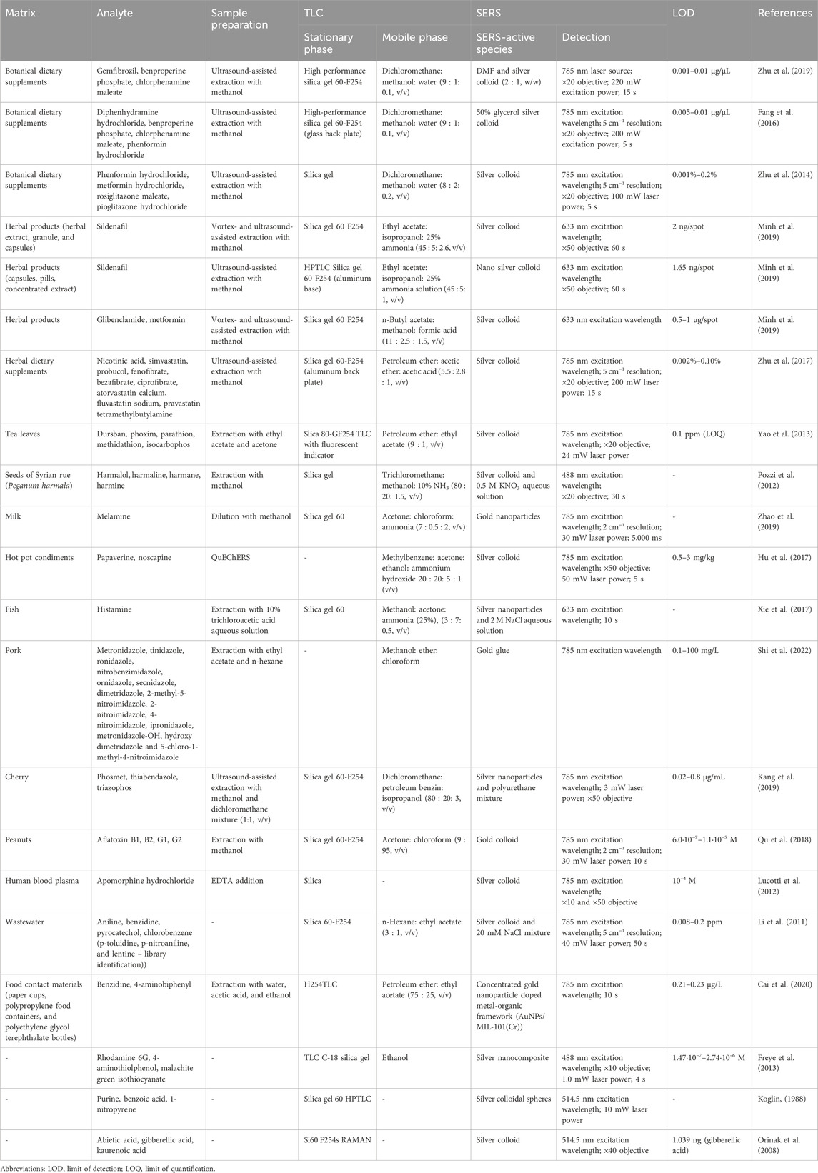
Table 1. TLC-SERS using conventional TLC plates.
The latter strategy consists in the development of TLC plates with a built-in SERS structure (Table 2). This allows to avoid uneven distribution of metal nanoparticles on the TLC surface and obtain desirable SERS signal. The method was applied for the quantification of polyaromatic hydrocarbons (Wang et al., 2022), pesticides (Fang et al., 2024; Shen et al., 2021), antibiotics (Lu et al., 2024), synthetic dyes (Sciutto et al., 2017), and other compounds. In most cases, sample preparation was simple and fast, which is an apparent advantage of TLC. However, sophisticated equipment is required to prepare such substrates resulting in their high cost (Dai and He, 2023; Yao et al., 2022). Silver nanorods have been described as efficient substrates for TLC-SERS experiments. At the same time, their efficiency for the separation of different compounds is to be studied (Freye et al., 2013). Also, metal-organic frameworks (MOF) decorated with metal nanoparticles were proposed as SERS substrates due to their high ability to adsorb compounds owing to porous structure and ultra-high surface area. These structures are also not prone to external interference and undesirable aggregation, but the attempts to provide the metallic structures with stable NPs location and orientation failed. The strength of the SERS signal was found to be inversely dependent on the distance from the noble metal surface, making the thickness of the MOF a key element for optimization during the development of SERS substrates (Jiang et al., 2024). Alternatively, the fabrication of chips has been described (Chen et al., 2015b) for simple and fast SERS detection of polycyclic aromatic hydrocarbons in cooking oil.
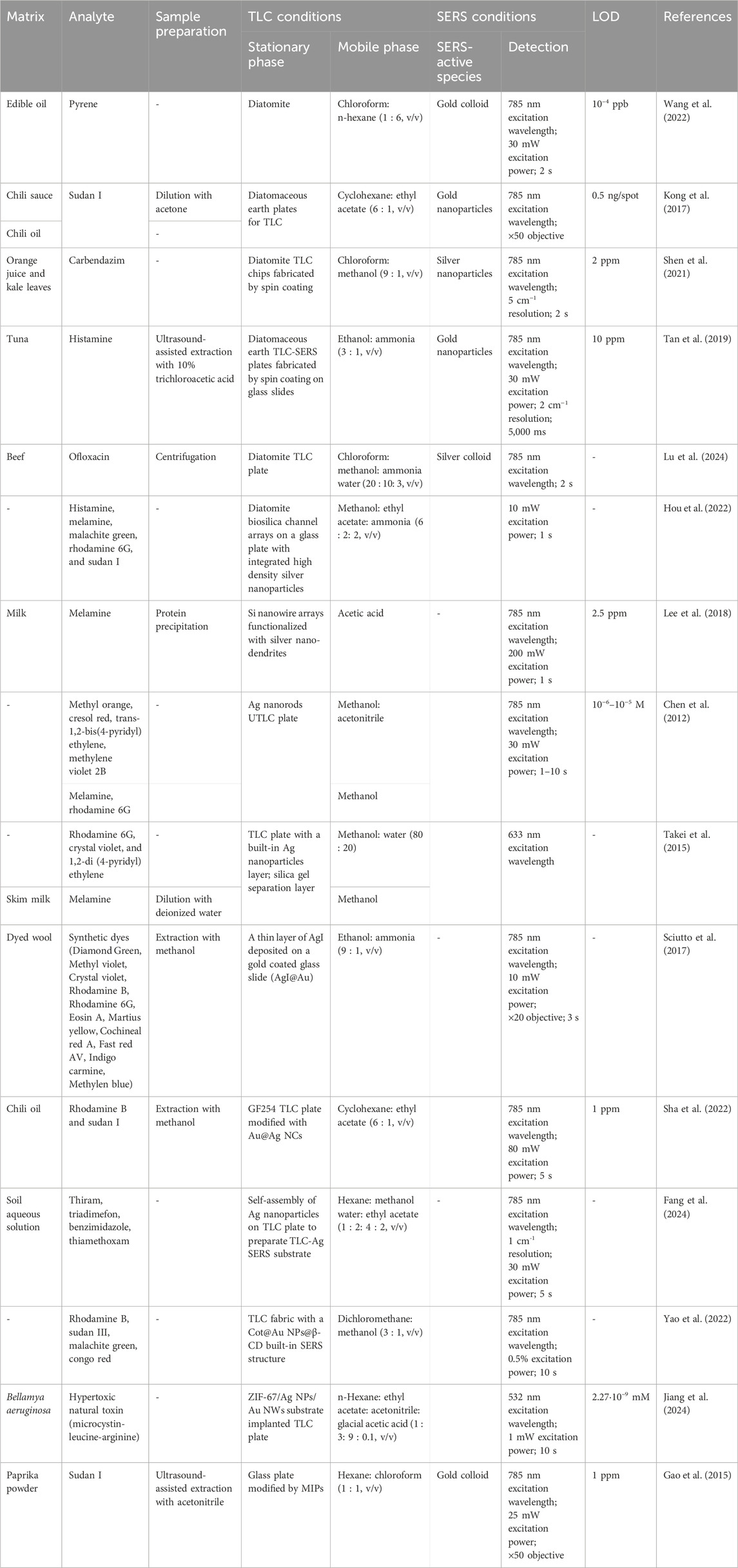
Table 2. TLC-SERS using modified TLC plates.
To improve the results of quantification by TLC-SERS, machine learning algorithms were also applied, i.e., principal component analysis (PCA)-back propagation neural network (Lu et al., 2024) and PCA with machine learning analysis based on support vector regression (SVR) (Tan et al., 2019), which resulted in elimination of interferences from complex samples.
Paper chromatography is a type of planar chromatography utilizing a cellulose paper as the stationary phase. Despite its simplicity, low cost, and portability, the separation process is often slower, less accurate, and spots tend to diffuse higher in comparison to TLC (Wilson, 2000). Therefore, it is less commonly used than TLC. The combination of paper chromatography with SERS detection was applied for the quantification of dyes, i.e., crystal violet, malachite green, and basic fuchsin (Tran, 1984). A conventional chromatographic paper was utilized to separate the analytes, and the developed chromatograms were sprayed with silver colloidal hydrosols prior to SERS detection. Another approach consisted in the paper strip modification for the quantification of rhodamine-6G. For this, a paper strip was fabricated with Au nanodendrite on nickel foam structure (Duy Vu et al., 2024). Also, an inkjet-printed SERS substrate was fabricated by using paper and polymer membranes for sample cleanup and analyte separation with SERS detection. This approach was applied for the determination of melamine (Yu and White, 2013).
In the analysis of complex samples containing compounds with similar structures or polarities, efficiency of TLC separation can be insufficient. Column chromatography, especially, high-performance liquid chromatography, has a significantly higher potential for the resolution of complex samples. Despite this, unique identification of compound peaks is still challenging [Trachta et al., 2004; Zhang et al., 2017). Coupling of HPLC with mass spectrometry significantly improved the capabilities of both methods and allowed to overcome this limitation. However, these instruments are expensive and require highly qualified personnel. SERS can be a suitable alternative allowing sensitive detection of analytes, because chromatographic separation of complex samples allows producing individual SERS signals (Zhang et al., 2017). The combination of HPLC and SERS was first described in 1988 for the determination of pararosaniline hydrochloride, an organic dye (Freeman et al., 1988; Khrushchev et al., 2022). In this method, an effluent from the chromatograph was mixed with Ag sol in a post-column mixing coil followed by SERS detection. Since that time, several studies utilizing HPLC-SERS detection have been reported, which can be divided into at-line and on-line modes. In the at-line approach, after the separation of analytes in the analytical column, the eluate fractions are collected for SERS analysis (Cowcher et al., 2014; Khrushchev et al., 2022; Seifar et al., 1999). This approach is easier to implement, and SERS conditions can be optimized for each fraction independently (Cowcher et al., 2014); however, the analysis time is increased in comparison to on-line approach. In contrast, the on-line approach requires the mixing unit of the eluate with nanoparticles solution and its passing through a Raman flow cell (Cabalin et al., 1993; Freeman et al., 1988; Sheng et al., 1991), but it has a disadvantage that a continued supply of nanoparticles is required (Goodacre et al., 2018). Alternatively, a capillary with SERS-active substrates can be connected to the chromatograph for on-line analysis (Nguyen and Schultz, 2016; Wang et al., 2015). However, such approach can be prone to memory effects, i.e., a subsequent analyte signal is disturbed by a previous compound residue (Cowcher et al., 2014; Zhang et al., 2017). Mobile phase composition can also cause Raman spectral interferences and affect SERS activity of the sol (Carrillo-Carrión et al., 2012). In this way, acetonitrile was noted to increase a background spectrum, therefore it is often replaced by methanol to increase sensitivity due to the reduced background signal (Trachta et al., 2004; Zhang et al., 2017).
For the sensitive determination of purine bases, both at-line and on-line approaches were applied. The former approach was implemented for the quantification of main purine and pyrimidine bases (adenine, guanine, thymine, and cytosine) and two of their most common degradation products (xanthine and hypoxanthine) using a novel SERS substrate based on ZnS/CdSe silver quantum dots for detection (Carrillo-Carrión et al., 2011). In the latter approach, eluate containing four separated purine bases (adenine, guanine, hypoxanthine, and xanthine) was mixed with Ag sol and passed through a Raman flow cell (Sheng et al., 1991). HPLC-SERS was also applied to the quantification of a folate antagonist methotrexate and its metabolites (Subaihi et al., 2017), amoxicillin in milk (Khrushchev et al., 2022), pesticides (Carrillo-Carrión et al., 2012; Wang et al., 2015), illicit drugs (Sägmüller et al., 2003; Trachta et al., 2004), etc. Also, the potential of on-line LC-SERS was demonstrated for untargeted tumor metabolomics. The method was applied to the determination of metabolites from cell lysate samples of tumors and showed results comparable with LC-MS (Xiao et al., 2020). SERS favorably compares to other detection methods due to the ability to identify certain class members of compounds owing to characteristic band patterns (Trachta et al., 2004).
The coupling of gas chromatography with SERS detection is less common, mostly due to the advantages in mass spectrometric (MS) detection (Heaps and Griffiths, 2005), where the direct coupling of a GC with a mass spectrometer is easy to implement. Moreover, for GC-MS method, there exist mass spectral libraries, allowing the identification of compounds, which hinders the benefits of SERS. As a results, even though the combination of GC and SERS in principle is possible (Roth and Kiefer, 1994), the general acceptance is low (Heaps and Griffiths, 2005).
Several attempts were made to combine GC and SERS, e.g., trapping GC eluate either in liquid silver sol (“flow-cell” method) or on TLC plates coated with silver colloidal solution (“mobile-phase elimination” method) followed by SERS quantification of pyridine (Roth and Kiefer, 1994). Alternatively, an off-line approach was proposed for the quantification of caffeine and p-nitrothiophenol by condensing the analytes on a liquid-nitrogen-cooled silver surface that had been formed on the surface of a ZnSe plate by physical vapor deposition (Heaps and Griffiths, 2005).
Even though a huge progress in coupling chromatographic separation with SERS detection has been achieved, there still exist opportunities and challenges in the application of these techniques, especially in substrate stability and reproducibility of results. Noble metal nanoparticles are widely used as SERS substrates; however, the sensitivity of detection is largely dependent on nano-substrate properties. To achieve more accurate and sensitive detection, the shape and formation of the nanoparticles of noble metals should be improved further (Zhang et al., 2017). To improve sensitivity and reproducibility of results, the development of SERS-active surfaces seems to be perspective. For obtaining higher accuracy, there is a great potential of applying machine-learning techniques, especially in the case of coeluting compounds or interferences from matrix components. Also, the application of noble metals, e.g., gold, silver, as SERS substrates hinders biocompatibility and poses other issues (Han et al., 2023). Another important aspect aimed at improving biocompatibility is reusability of the developed SERS-active surfaces. Since highly sensitive and accurate quantification of analytes in diverse complex matrices is required, there is a need to the reveal the potential of the SERS combined techniques on a wider range of target analytes and matrices. Preferably, the developed techniques should be simple, convenient, and inexpensive.
ED: Conceptualization, Methodology, Writing–original draft. OK: Conceptualization, Methodology, Writing–review and editing. SL: Formal Analysis, Writing–review and editing. OS: Writing–review and editing. IV: Conceptualization, Methodology, Project administration, Writing–review and editing.
The author(s) declare that financial support was received for the research, authorship, and/or publication of this article. This research was funded by a grant from the Ministry of Science and Higher Education of the Russian Federation, grant number 075-15-2024-646.
The authors declare that the research was conducted in the absence of any commercial or financial relationships that could be construed as a potential conflict of interest.
The author(s) declare that no Generative AI was used in the creation of this manuscript.
All claims expressed in this article are solely those of the authors and do not necessarily represent those of their affiliated organizations, or those of the publisher, the editors and the reviewers. Any product that may be evaluated in this article, or claim that may be made by its manufacturer, is not guaranteed or endorsed by the publisher.
Alves, I. M., Melo, N. O., Marinho, P. A., and Almeida, M. R. (2020). Liquid–liquid extraction-assisted SERS-based detection of clonazepam in spiked drinks. Vib. Spectrosc. 110, 103112. doi:10.1016/j.vibspec.2020.103112
Ay, K. O., Dikmen, G., and Koyuncu, O. (2024). Application of high sensitive silver nanocubes SERS substrate for detection of metribuzin. J. Mol. Struct. 12975, 136869. doi:10.1016/j.molstruc.2023.136869
Cabalin, L. M., Ruperez, A., and Laserna, J. J. (1993). Surface-enhanced Raman spectrometry for detection in liquid chromatography using a windowless flow cell. Talanta 40 (11), 1741–1747. doi:10.1016/0039-9140(93)80092-6
Cai, G., Ge, K., Ouyang, X., Hu, Y., and Li, G. (2020). Thin-layer chromatography combined with surface-enhanced Raman scattering for rapid detection of benzidine and 4-aminobiphenyl in migration from food contact materials based on gold nanoparticle doped metal-organic framework. J. Sep. Sci. 43 (14), 2834–2841. doi:10.1002/jssc.202000145
Cañamares, M. V., Reagan, D. A., Lombardi, J. R., and Leona, M. (2014). TLC-SERS of mauve, the first synthetic dye. J. Raman Spectrosc. 45, 1147–1152. doi:10.1002/jrs.4508
Carrillo-Carrión, C., Armenta, S., Simonet, B. M., Valcárcel, M., and Lendl, B. (2011). Determination of pyrimidine and purine bases by reversed-phase capillary liquid chromatography with at-line surface-enhanced Raman spectroscopic detection employing a novel SERS substrate based on ZnS/CdSe silver-quantum dots. Anal. Chem. 83 (24), 9391–9398. doi:10.1021/ac201821q
Carrillo-Carrión, C., Simonet, B. M., Valcárcel, M., and Lendl, B. (2012). Determination of pesticides by capillary chromatography and SERS detection using a novel silver-quantum dots “sponge” nanocomposite. J. Chromatogr. A 1225, 55–61. doi:10.1016/j.chroma.2011.12.002
Chen, D., Han, X., Jin, W., and Zhang, B. (2015a). Metal nanoparticle catalyzed cyclobutane cleavage reaction. RSC Adv. 5, 100722–100724. doi:10.1039/C5RA21225A
Chen, J., Abell, J., Huang, Y. W., and Zhao, Y. (2012). On-chip ultra-thin layer chromatography and surface enhanced Raman spectroscopy. Lab. Chip 12 (17), 3096–3102. doi:10.1039/c2lc40221a
Chen, J., Huang, Y. W., and Zhao, Y. (2015b). Detection of polycyclic aromatic hydrocarbons from cooking oil using ultra-thin layer chromatography and surface enhanced Raman spectroscopy. J. Mater. Chem. B 3 (9), 1898–1906. doi:10.1039/c4tb01632g
Cimpoiu, C., Casoni, D., Hosu, A., Miclaus, V., Hodisan, T., and Damian, G. (2005). Separation and identification of eight hydrophilic vitamins using a new TLC method and Raman spectroscopy. J. Liq. Chromatogr. Relat. Technol. 28 (16), 2551–2559. doi:10.1080/10826070500189737
Cowcher, D. P., Jarvis, R., and Goodacre, R. (2014). Quantitative online liquid chromatography-surface-enhanced Raman scattering of purine bases. Anal. Chem. 86 (19), 9977–9984. doi:10.1021/ac5029159
Dai, H., and He, L. (2023). Mirror-Stamping: a new TLC-SERS method for color authentication. Talanta Open 7, 100237. doi:10.1016/j.talo.2023.100237
Duy Vu, T., Thang Nguyen, D., Yen Thi Nguyen, H., Hoang Do, H., Duc Pham, T., Thanh Le, S., et al. (2024). A facile paper-based chromatography coupled Au nanodendrite on nickel foam for application in separation and SERS measurement. Spectrochim. Acta A Mol. Biomol. Spectrosc. 313, 124137. doi:10.1016/j.saa.2024.124137
Fang, F., Qi, Y., Lu, F., and Yang, L. (2016). Highly sensitive on-site detection of drugs adulterated in botanical dietary supplements using thin layer chromatography combined with dynamic surface enhanced Raman spectroscopy. Talanta 146, 351–357. doi:10.1016/j.talanta.2015.08.067
Fang, G., Hasi, W., Lin, X., and Han, S. (2024). Automated identification of pesticide mixtures via machine learning analysis of TLC-SERS spectra. J. Hazard. Mater. 474, 134814. doi:10.1016/j.jhazmat.2024.134814
Fateixa, S., Raposo, M., Nogueira, H. I. S., and Trindade, T. (2018). A general strategy to prepare SERS active filter membranes for extraction and detection of pesticides in water. Talanta 182, 558–566. doi:10.1016/j.talanta.2018.02.014
Ferretti, A., Floris, E., Campanella, B., Degano, I., and Legnaioli, S. (2024). “Welcome to the jungle”: TLC-SERS to wade through real complex mixtures of synthetic dyes. Microchem. J. 206, 111439. doi:10.1016/j.microc.2024.111439
Freeman, R. D., Hammaker, R. M., Meloan, C. E., and Fateley, W. G. (1988). A detector for liquid chromatography and flow injection analysis using surface-enhanced Raman spectroscopy. Appl. Spectrosc. 42 (3), 456–460. doi:10.1366/0003702884427997
Freye, C. E., Crane, N. A., Kirchner, T. B., and Sepaniak, M. J. (2013). Surface enhanced Raman scattering imaging of developed thin-layer chromatography plates. Anal. Chem. 85 (8), 3991–3998. doi:10.1021/ac303710q
Fukunaga, Y., Ogawa, R., Homma, A., and Okada, T. (2023). Thin layer chromatography-freeze surface-enhanced Raman spectroscopy: a powerful tool for monitoring synthetic reactions. Chem. Eur. J. 29, e202300829. doi:10.1002/chem.202300829
Gao, F., Hu, Y., Chen, D., Li-Chan, E. C. Y., Grant, E., and Lu, X. (2015). Determination of Sudan I in paprika powder by molecularly imprinted polymers-thin layer chromatography-surface enhanced Raman spectroscopic biosensor. Talanta 143, 344–352. doi:10.1016/j.talanta.2015.05.003
Goodacre, R., Graham, D., and Faulds, K. (2018). Recent developments in quantitative SERS: moving towards absolute quantification. Trends Anal. Chem. 102, 359–368. doi:10.1016/j.trac.2018.03.005
Han, C., Wang, Q., Yao, Y., Zhang, Q., Huang, J., Zhang, H., et al. (2023). Thin layer chromatography coupled with surface enhanced Raman scattering for rapid separation and on-site detection of multi-components. J. Chromatogr. A 1706, 464217. doi:10.1016/j.chroma.2023.464217
Heaps, D. A., and Griffiths, P. R. (2005). Off-line direct deposition gas chromatography/surface-enhanced Raman scattering and the ramifications for on-line measurements. Appl. Spectrosc. 59 (11), 1305–1309. doi:10.1366/000370205774783160
Henzel, U. B. (1977). Journal of chromatography library. Editors A. Zlatkis,, and R. E. Kaiser (Amsterdam: Elsevier), 9. chapter 8.
Herman, K., Mircescu, N. E., Szabo, L. L., Leopold, F., Chiş, V., and Leopold, N. (2013). In situ silver spot preparation and on-plate surface-enhanced Raman scattering detection in thin layer chromatography separation. J. Appl. Spectrosc. 80, 311–314. doi:10.1007/s10812-013-9765-9
Horváth, E., Kátay, G., Tyihák, E., Kristóf, J., and Rédey, Á. (2000). Critical evaluation of experimental conditions influencing the surface-enhanced Raman spectroscopic (SERS) detection of substances separated by layer liquid chromatographic techniques. Chromatographia 51, S297–S301. doi:10.1007/BF02492821
Hou, X., Sivashanmugan, K., Zhao, Y., Zhang, B., and Wang, A. X. (2022). Multiplex sensing of complex mixtures by machine vision analysis of TLC-SERS images. Sens. Actuators B Chem. 357, 131355. doi:10.1016/j.snb.2021.131355
Hu, X., Fang, G., Han, A., Liu, J., and Wang, S. (2017). Rapid detection of Pericarpium papaveris in hot pot condiments using thin-layer chromatography and surface enhanced Raman spectroscopy combined with a support vector machine. Anal. Methods. 9, 2177–2182. doi:10.1039/C7AY00151G
Huang, R., Han, S., and Li, X. S. (2013). Detection of tobacco-related biomarkers in urine samples by surface-enhanced Raman spectroscopy coupled with thin-layer chromatography. Anal. Bioanal. Chem. 405 (21), 6815–6822. doi:10.1007/s00216-013-7107-7
István, K., Keresztury, G., and Szép, A. (2003). Normal Raman and surface enhanced Raman spectroscopic experiments with thin layer chromatography spots of essential amino acids using different laser excitation sources. Spectrochim. Acta A Mol. Biomol. Spectrosc. 59 (8), 1709–1723. doi:10.1016/s1386-1425(02)00408-0
Jeanmaire, D. L., and Van Duyne, R. P. (1977). Surface Raman spectroelectrochemistry: Part I. Heterocyclic, aromatic, and aliphatic amines adsorbed on the anodized silver electrode. J. Electroanal. Chem. 84 (1), 1–20. doi:10.1016/s0022-0728(77)80224-6
Jensen, L., Aikens, C. M., and Schatz, G. C. (2008). Electronic structure methods for studying surface-enhanced Raman scattering. Chem. Soc. Rev. 37 (5), 1061–1073. doi:10.1039/b706023h
Jiang, J., Liu, M., Xu, D., Jiang, T., and Zhang, J. (2024). Quantitative detection of microcystin-LR in Bellamya aeruginosa by thin-layer chromatography coupled with surface-enhanced Raman spectroscopy based on in-situ ZIF-67/Ag NPs/Au NWs composite substrate. Food Chem. 452, 139481. doi:10.1016/j.foodchem.2024.139481
Kang, Y., Wu, T., Chen, W., Li, L., and Du, Y. (2019). A novel metastable state nanoparticle-enhanced Raman spectroscopy coupled with thin layer chromatography for determination of multiple pesticides. Food Chem. 270, 494–501. doi:10.1016/j.foodchem.2018.07.070
Khrushchev, A.Yu., Akmaev, E. R., Kis, I. V., Gulyaeva, A.Yu., and Bondarenko, V. O. (2022). Combination of HPLC and SERS detection applied to the analysis of the trace content of amoxicillin in milk. Vib. Spectrosc. 123, 103473. doi:10.1016/j.vibspec.2022.103473
Koglin, E. (1988). Combining surface enhanced Raman scattering (SERS) and high-performance thin-layer chromatography (HPTLC). J. Mol. Struct. 173, 369–376. doi:10.1016/0022-2860(88)80068-1
Kong, X., Squire, K., Chong, X., and Wang, A. X. (2017). Ultra-sensitive lab-on-a-chip detection of Sudan I in food using plasmonics-enhanced diatomaceous thin film. Food control. 79, 258–265. doi:10.1016/j.foodcont.2017.04.007
Lee, B. S., Lin, D. Z., Huang, C. H., and Yen, T. J. (2018). A high-performance multifunctional substrate of ultrathin-layer chromatography (UTLC) and surface-enhanced Raman scattering (SERS) for rapid biochemical mixture screening. J. Raman Spectrosc. 49, 1920–1927. doi:10.1002/jrs.5477
Lee, Y.-H., Dai, S., and Young, J. P. (1998). Silver-doped sol–gel films as the substrate for surface-enhanced Raman scattering. J. Raman Spectrosc. 28, 635–639. doi:10.1002/(SICI)1097-4555(199708)28:8<635::AID-JRS152>3.0.CO;2-0
Li, D., Qu, L., Zhai, W., Xue, J., Fossey, J. S., and Long, Y. (2011). Facile on-site detection of substituted aromatic pollutants in water using thin layer chromatography combined with surface-enhanced Raman spectroscopy. Environ. Sci. Technol. 45 (9), 4046–4052. doi:10.1021/es104155r
Li, Y., Zhao, C., Lu, C., Zhou, S., Tian, G., He, L., et al. (2021). Simultaneous determination of 14 bioactive citrus flavonoids using thin-layer chromatography combined with surface enhanced Raman spectroscopy. Food Chem. 338, 128115. doi:10.1016/j.foodchem.2020.128115
Lu, X., Ma, Y., Jiang, S., Wang, Z., Yu, Q., Ji, C., et al. (2024). Quantitative monitoring ofloxacin in beef by TLC-SERS combined with machine learning analysis. Spectrochim. Acta A Mol. Biomol. Spectrosc. 308, 123790. doi:10.1016/j.saa.2023.123790
Lucotti, A., Tommasini, M., Casella, M., Morganti, A., Gramatica, F., and Zerbi, G. (2012). TLC–surface enhanced Raman scattering of apomorphine in human plasma. Vib. Spectrosc. 62, 286–291. doi:10.1016/j.vibspec.2012.07.009
McNay, G., Eustace, D., Smith, W. E., Faulds, K., and Graham, D. (2011). Surface-enhanced Raman scattering (SERS) and surface-enhanced resonance Raman scattering (SERRS): a review of applications. Appl. Spectrosc. 5 (8), 825–837. doi:10.1366/11-06365
Minh, D. T. C., Nhu, N. T. Q., Thi, L. A., Vu, L. V., Lan, D. T. N., Anh, N. T. K., et al. (2024). HPTLC sequentially coupled to UV and SERS: a cost-effective tool for confirmative identification and quantitation of sildenafil in falsified herbal products. J. Pharm. Biomed. Anal. 251, 116392. doi:10.1016/j.jpba.2024.116392
Minh, D. T. C., Thi, L. A., Huyen, N. T. T., Van Vu, L., Anh, N. T. K., and Ha, P. T. T. (2019). Detection of sildenafil adulterated in herbal products using thin layer chromatography combined with surface enhanced Raman spectroscopy: “Double coffee-ring effect” based enhancement. J. Pharm. Biomed. Anal. 174, 340–347. doi:10.1016/j.jpba.2019.05.043
Minh, D. T. C., Tram, L. T. B., Phong, N. H., Huong, H. T. L., Vu, L. V., Thi, L. A., et al. (2023). Single versus double coffee-ring effect patterns in thin-layer chromatography coupled with surface-enhanced Raman spectroscopic analysis of anti-diabetic drugs adulterated in herbal products. Molecules 28 (14), 5492. doi:10.3390/molecules28145492
Murugan, E., Santhoshkumar, S., Govindaraju, S., and Palanichamy, M. (2021). Silver nanoparticles decorated g-C3N4: an efficient SERS substrate for monitoring catalytic reduction and selective Hg2+ ions detection. Spectrochim. Acta A Mol. Biomol. Spectrosc. 246, 119036. doi:10.1016/j.saa.2020.119036
Nguyen, A., and Schultz, Z. D. (2016). Quantitative online sheath-flow surface enhanced Raman spectroscopy detection for liquid chromatography. Analyst 141, 3630–3635. doi:10.1039/c6an00155f
Orinak, A., Talian, I., Efremov, E. V., Ariese, F., and Orinakova, R. (2008). Diterpenoic acids analysis using a coupled TLC-surface-enhanced Raman spectroscopy system. Chromatographia 67 (5–6), 501–319. doi:10.1365/s10337-008-0566-x
Pozzi, F., Shibayama, N., Leona, M., and Lombardi, J. R. (2012). TLC-SERS study of Syrian rue (Peganum harmala) and its main alkaloid constituents. J. Raman Spectrosc. 44 (1), 102–107. doi:10.1002/jrs.4140
Qu, L., Wang, N., Xu, H., Wang, W., Liu, Y., Kuo, L., et al. (2017). Gold nanoparticles and g-C3N4-intercalated graphene oxide membrane for recyclable surface enhanced Raman scattering. Adv. Funct. Mater. 27, 1701714. doi:10.1002/adfm.201701714
Qu, L. L., Jia, Q., Liu, C., Wang, W., Duan, L., Yang, G., et al. (2018). Thin layer chromatography combined with surface-enhanced Raman spectroscopy for rapid sensing aflatoxins. J. Chromatogr. A 1579, 115–120. doi:10.1016/j.chroma.2018.10.024
Radu, A. I., Kuellmer, M., Giese, B., Huebner, U., Weber, K., Cialla-May, D., et al. (2016). Surface-enhanced Raman spectroscopy (SERS) in food analytics: detection of vitamins B2 and B12 in cereals. Talanta 160, 289–297. doi:10.1016/j.talanta.2016.07.027
Roth, E., and Kiefer, W. (1994). Surface-enhanced Raman spectroscopy as a detection method in gas chromatography. Appl. Spectrosc. 48 (10), 1193–1195. doi:10.1366/0003702944027444
Sackmann, M., and Materny, A. (2006). Surface enhanced Raman scattering (SERS) – a quantitative analytical tool? J. Raman Spectrosc. 37, 305–310. doi:10.1002/jrs.1443
Sägmüller, B., Schwarze, B., Brehm, G., Trachta, G., and Schneider, S. (2003). Identification of illicit drugs by a combination of liquid chromatography and surface-enhanced Raman scattering spectroscopy. J. Mol. Struct. 661 (662), 279–290. doi:10.1016/S0022-2860(03)00507-6
Santhoshkumar, S., Wei, S. W., Kuo, C. C., Madhu, M., Kumar, A., Tseng, S. K., et al. (2025). Engineering of phase composition in molybdenum disulfide nanoflowers to amplify SERS activity: sensitivity enhancement through dispersive liquid-liquid microextraction integration. Sens. Actuators B Chem. 422, 136577. doi:10.1016/j.snb.2024.136577
Sciutto, G., Prati, S., Bonacini, I., Litti, L., Meneghetti, M., and Mazzeo, R. (2017). A new integrated TLC/MU-ATR/SERS advanced approach for the identification of trace amounts of dyes in mixtures. Anal. Chim. Acta. 991, 104–112. doi:10.1016/j.aca.2017.08.020
Seifar, R. M., Dijkstra, R. J., Brinkman, U. A. Th., and Gooijer, C. (1999). At-line coupling of surface-enhanced resonance Raman spectroscopy and reversed-phase ion-pair chromatography. Anal. Commun. 36, 273–276. doi:10.1039/A903254A
Sha, X., Han, S., Fang, G., Li, N., Lin, D., and Hasi, W. (2022). A novel suitable TLC-SERS assembly strategy for detection of Rhodamine B and Sudan I in chili oil. Food control. 138, 109040. doi:10.1016/j.foodcont.2022.109040
Shen, Z., Fan, Q., Yu, Q., Wang, R., Wang, H., and Kong, X. (2021). Facile detection of carbendazim in food using TLC-SERS on diatomite thin layer chromatography. Spectrochim. Acta A Mol. Biomol. Spectrosc. 247, 119037. doi:10.1016/j.saa.2020.119037
Sheng, R., Ni, F., and Cotton, T. M. (1991). Determination of purine bases by reversed-phase high-performance liquid chromatography using real-time surface-enhanced Raman spectroscopy. Anal. Chem. 63, 437–442. doi:10.1021/ac00005a010
Shi, S., Yu, H., Yang, F., Yao, W., and Xie, Y. (2022). Simultaneous determination of 14 nitroimidazoles using thin-layer chromatography combined with surface-enhanced Raman spectroscopy (TLC-SERS). Food Biosci. 48, 101755. doi:10.1016/j.fbio.2022.101755
Subaihi, A., Trivedi, D. K., Hollywood, K. A., Bluett, J., Xu, Y., Muhamadali, H., et al. (2017). Quantitative online liquid chromatography-surface-enhanced Raman scattering (LC-SERS) of methotrexate and its major metabolites. Anal. Chem. 89 (12), 6702–6709. doi:10.1021/acs.analchem.7b00916
Szabo, N. J., and Winefordner, J. D. (1997). Evaluation of two commercially available TLC materials as SER substrates. Appl. Spectrosc. 51 (7), 965–975. doi:10.1366/0003702971941629
Takei, H., Saito, J., Kato, K., Vieker, H., Beyer, A., and Gölzhäuser, A. (2015). TLC-SERS plates with a built-in SERS layer consisting of cap-shaped noble metal nanoparticles intended for environmental monitoring and food safety assurance. J. Nanomater. 2015, 316189. doi:10.1155/2015/316189
Tan, A., Zhao, Y., Sivashanmugan, K., Squire, K., and Wang, A. X. (2019). Quantitative TLC-SERS detection of histamine in seafood with support vector machine analysis. Food control. 103, 111–118. doi:10.1016/j.foodcont.2019.03.032
Trachta, G., Schwarze, B., Sägmüller, B., Brehm, G., and Schneider, S. (2004). Combination of high-performance liquid chromatography and SERS detection applied to the analysis of drugs in human blood and urine. J. Mol. Struct. 693 (1–3), 175–185. doi:10.1016/j.molstruc.2004.02.034
Tran, C. D. (1984). In situ identification of paper chromatogram spots by surface enhanced Raman scattering. J. Chromatogr. A 292 (2), 432–438. doi:10.1016/S0021-9673(01)83624-4
Wang, P., Sun, Y., Li, X., Shan, J., Xu, Y., and Li, G. (2021). One-step chemical reaction triggered surface enhanced Raman scattering signal conversion strategy for highly sensitive detection of nitrite. Vib. Spectrosc. 113, 103221. doi:10.1016/j.vibspec.2021.103221
Wang, S., Yu, Q., Guo, J., Yuan, C., and Kong, X. (2022). On-site detection of pyrene from mixture with ppb level sensitivity by plasmonic TLC-DSERS. Spectrochim. Acta A Mol. Biomol. Spectrosc. 280, 121547. doi:10.1016/j.saa.2022.121547
Wang, W., Xu, M., Guo, Q., Yuan, Y., Gua, R., and Yao, J. (2015). Rapid separation and on-line detection by coupling high performance liquid chromatography with surface-enhanced Raman spectroscopy. RSC Adv. 5, 47640–47646. doi:10.1039/C5RA05562H
White, R. L. (1985). Analysis of thin-layer chromatographic adsorbates by Fourier transform infrared photoacoustic spectroscopy. Anal. Chem. 57, 1819–1822. doi:10.1021/ac00286a008
Wigginton, K. R., and Vikesland, P. J. (2010). Gold-coated polycarbonate membrane filter for pathogen concentration and SERS-based detection. Analyst. 135, 1320–1326. doi:10.1039/B919270K
Wilson, I. D. (2000). “Chromatography. Paper chromatography,” in Encyclopedia of separation science. Editor I. D. Wilson (Academic Press), 397–404.
Xiao, L., Wang, C., Dai, C., Littlepage, L. E., Li, J., and Schultz, Z. D. (2020). Untargeted tumor metabolomics with liquid chromatography-surface-enhanced Raman spectroscopy. Angew. Chem. Int. Ed. Engl. 59 (9), 3439–3443. doi:10.1002/anie.201912387
Xie, Z., Wang, Y., Chen, Y., Xu, X., Jin, Z., Ding, Y., et al. (2017). Tuneable surface enhanced Raman spectroscopy hyphenated to chemically derivatized thin-layer chromatography plates for screening histamine in fish. Food Chem. 230, 547–552. doi:10.1016/j.foodchem.2017.03.081
Yao, C., Cheng, F., Wang, C., Wang, Y., Guo, X., Gong, Z., et al. (2013). Separation, identification and fast determination of organophosphate pesticide methidathion in tea leaves by thin layer chromatography–surface-enhanced Raman scattering. Anal. Methods. 5, 5560–5564. doi:10.1039/C3AY41152D
Yao, H., Dong, X., Xiong, H., Liu, J., Zhou, J., and Ye, Y. (2022). Functional cotton fabric-based TLC-SERS matrix for rapid and sensitive detection of mixed dyes. Spectrochim. Acta A Mol. Biomol. Spectrosc. 280, 121464. doi:10.1016/j.saa.2022.121464
Yu, W. W., and White, I. M. (2012). A simple filter-based approach to surface enhanced Raman spectroscopy for trace chemical detection. Analyst 137 (5), 1168–1173. doi:10.1039/c2an15947c
Yu, W. W., and White, I. M. (2013). Chromatographic separation and detection of target analytes from complex samples using inkjet printed SERS substrates. Analyst 138 (13), 3679–3686. doi:10.1039/c3an00673e
Zhang, Y., Zhao, S., He, L., and Zheng, J. (2017). Surface-enhanced Raman spectroscopy (SERS) combined techniques for high-performance detection and characterization. Trends Anal. Chem. 90, 1–13. doi:10.1016/j.trac.2017.02.006
Zhang, Z., Liu, R., and Liu, J. (2021). Simultaneously preconcentration of malachite green and construction of SERS substrate in water based on cloud point extraction. Microchem. J. 169, 106572. doi:10.1016/j.microc.2021.106572
Zhang, Z. M., Liu, J. F., Liu, R., Sun, J. F., and Wei, G. H. (2014). Thin layer chromatography coupled with surface-enhanced Raman scattering as a facile method for on-site quantitative monitoring of chemical reactions. Anal. Chem. 86 (15), 7286–7292. doi:10.1021/ac5017387
Zhao, Y., Tan, A., Squire, K., Sivashanmugan, K., and Wang, A. X. (2019). Quaternion-based parallel feature extraction: extending the horizon of quantitative analysis using TLC-SERS sensing. Sens. Actuators B Chem. 299, 126902. doi:10.1016/j.snb.2019.126902
Zhu, Q., Cao, Y., Cao, Y., Chai, Y., and Lu, F. (2014). Rapid on-site TLC-SERS detection of four antidiabetes drugs used as adulterants in botanical dietary supplements. Anal. Bioanal. Chem. 406 (7), 1877–1884. doi:10.1007/s00216-013-7605-7
Zhu, Q., Cao, Y., Li, D., Fang, F., Lu, F., and Yuan, Y. (2019). A fast response TLC-SERS substrate for on-site detection of hydrophilic and hydrophobic adulterants in botanical dietary supplements. New J. Chem. 43, 13873–13880. doi:10.1039/C9NJ02489A
Keywords: surface-enhanced Raman spectroscopy, chromatography, TLC-SERS, HPLC-SERS, method development
Citation: Dmitrieva EV, Kapitanova OO, Lv S, Sinyashin OG and Veselova IA (2025) Coupling of chromatography with surface-enhanced Raman spectroscopy: trends and prospects. Front. Chem. 13:1548364. doi: 10.3389/fchem.2025.1548364
Received: 19 December 2024; Accepted: 04 February 2025;
Published: 26 February 2025.
Edited by:
Serban Moldoveanu, Independent Researcher, Greensboro, NC, United StatesReviewed by:
Santhoshkumar Sampath Kumar, National Sun Yat-sen University, TaiwanCopyright © 2025 Dmitrieva, Kapitanova, Lv, Sinyashin and Veselova. This is an open-access article distributed under the terms of the Creative Commons Attribution License (CC BY). The use, distribution or reproduction in other forums is permitted, provided the original author(s) and the copyright owner(s) are credited and that the original publication in this journal is cited, in accordance with accepted academic practice. No use, distribution or reproduction is permitted which does not comply with these terms.
*Correspondence: Ekaterina V. Dmitrieva, dmitrieva@smbu.edu.cn
Disclaimer: All claims expressed in this article are solely those of the authors and do not necessarily represent those of their affiliated organizations, or those of the publisher, the editors and the reviewers. Any product that may be evaluated in this article or claim that may be made by its manufacturer is not guaranteed or endorsed by the publisher.
Research integrity at Frontiers
Learn more about the work of our research integrity team to safeguard the quality of each article we publish.