
95% of researchers rate our articles as excellent or good
Learn more about the work of our research integrity team to safeguard the quality of each article we publish.
Find out more
ORIGINAL RESEARCH article
Front. Chem. , 25 October 2023
Sec. Polymer Chemistry
Volume 11 - 2023 | https://doi.org/10.3389/fchem.2023.1287870
 Khursheed Muzammil1Mazin Hadi Kzar2Faraj Mohammed3Zahraa Ibrahim Mohammed4Sarah A. Hamood5Talib Kh. Hussein6Saheb Jubeir Hanoon7Maytham T. Qasim8Ahmed Hussien Alawadi9,10,11
 Ali Alsalamy12*
In this study, aqueous, ethanol, methanol, and hexane extracts from Iraqi Kurdistan Region Daphne mucronata were prepared due to the numerous applications and development of nanofibers in biological and medical fields, including food packaging, enzyme stabilization, and wound dressing. In the initial evaluation of the extracts, the antioxidant properties against DPPH, antimicrobial properties against 3-gram-positive bacterial species, 3-gram negative bacterial species, 3-common bacterial species between aquatic and human, and 3-fungal species, and anticancer properties against breast cancer cells were performed. The results proved that the methanol extract has the highest antimicrobial, antifungal, antioxidant, and anticancer properties. After identifying the compounds of prepared methanol extract using GC/MS, polyvinylpyrrolidone nanofibers containing methanol extract of Daphne mucronata were prepared. The structure and characteristics of prepared nanofibers were confirmed and determined using FTIR, TGA, BET, SEM, flexural strength, compressive strength, and hydrophilicity. Synthesized polyvinylpyrrolidone nanofibers containing methanol extract of D. mucronata were subjected to antimicrobial properties on the strains studied in methanol extract of D. mucronata. The antimicrobial properties of synthesized polyvinylpyrrolidone nanofibers containing methanol extract of D. mucronata were compared. The results showed that synthesized polyvinylpyrrolidone nanofibers containing methanol extract of D. mucronata have the potential to introduction bioactive natural synthesis nanoparticles.
Today, using natural compounds and extracts has found a special place in treating diseases. By examining the previous texts, we realize that the extracts of different plants have many and varied biological properties. The most crucial property of plant extracts is their antioxidant activity. This activity can be attributed to the presence of flavonoids in most plants. Oxidants are the cause of cancer, and in a way, anti-cancer properties can be attributed to plants with high antioxidant properties. However, there have been reports of the anticancer properties of extracts that have been directly subjected to anticancer tests. For example, the anticancer properties of Adenosma bracteosum extract were reported in 2020. In addition, other biological properties such as anti-bacterial activity, anti-inflammatory activity, anti-mutagenic activity, immunostimulatory activity, and anti-tumoral activity have been reported from plant extracts (Mostafa et al., 2018; Dirar et al., 2019; Manandhar et al., 2019; Tanase et al., 2019; Nguyen et al., 2020; Dias et al., 2021).
Different species of the genus Daphne are distributed in many countries, especially in South America, Asia, and Europe and around the Mediterranean Sea. Daphne giraldii and Daphne mucronata are two important species of Daphne with unique biological properties. Anticancer properties against liver cancer cells, antimalarial, hypnotic and sedative, anti-nociceptive, anti-inflammatory, and immunomodulating properties of D. giraldii have been reported (Sun et al., 2017; Han et al., 2020). Biological properties such as inhibition of STAT3 and Smad3/4 cancer, nephrotoxicity and hepatotoxicity, antibacterial activity, and antioxidant activity have been reported for D. mucronata from different regions (Shah et al., 2018; Lutfullah et al., 2019; Ghanadian et al., 2020; Nazir et al., 2021).
The use of environmentally friendly plant and polymer extracts and the preparation of new nanoparticles in the form of nanofibers has been the focus of scientists in recent years. Numerous nanofibers containing plant extracts or other nano compounds can be synthesized using electrospinning technology. The performance and properties of nanofiber extracts can be improved by using this method. Nanofibers increase the activity of the extracts for two crucial reasons, including the nanosizing of the compounds and the increase in the specific active surface area that leads to contact with the desired agents. Natural nanofibers, mainly composed of biodegradable and environmentally friendly polymer compounds such as polyvinylalcohol and polyvinylpyrrolidone, have many biological properties and industrial applications. Applications of nanofibers in biological and medical fields include food packaging, enzyme stabilization, and wound dressing (Park et al., 2019; Kurakula and Rao, 2020; Mehrali et al., 2020; Osanloo et al., 2020; Yadav et al., 2020; Dodero et al., 2021; Ghasemian Lemraski et al., 2021; Li et al., 2022; Wu et al., 2022; Kişla et al., 2023; Wu et al., 2023).
For developing and reporting nanofibers with biological properties and synthesis of new nanofiber compounds, aqueous, ethanol, methanol, and hexane extracts of D. mucronata from the Iraqi Kurdistan Region were prepared. After identifying the active compounds in the extract, biological evaluations such as its antibacterial, antioxidant, and anticancer properties were investigated and compared. Nanofibers that contained Iraqi Kurdistan Region Daphne mucronata and polyvinylpyrrolidone were synthesized using the electrospinning method. After characterizing and confirming the structure, the antimicrobial properties of the synthesized nanofibers were also evaluated, and the results obtained were compared with the extract.
Agilent GC/MS-3223 with HP5 capillary column 30 m long was used to identify the compounds of the extract. In microbiology and antioxidant tests, the Unico S2150 spectrophotometer was used to prepare the concentration of strains and measure absorbance, respectively. BIOTEK ELX800TS Elisa Reader was used in the anticancer trials of the extracts.
Daphne mucronata was collected from the mountains of the Iraqi Kurdistan Region. After that, the leaves were washed with distilled water and dried for a week at room temperature without sunlight. Then, it was made into powder by an electric mill. To prepare an aqueous, ethanol, methanol, and hexane D. mucronata extracts, 1 to 10 wt% powder to solvent was stirred for 48 h, at ambient temperature and in the dark. Finally, condensation took place at a temperature of 37°C (Fazal et al., 2020).
GC/MS technique was used to identify the compounds of the extract. The method used includes a thermal program from 50°C to 280°C with an increase in temperature of 4°C per minute; helium carrier gas with a flow of 1.5 mL per minute; the temperature of the injection chamber and the temperature of the detector is 270°C; and the split ratio was 10:1. The mass spectrum of the compounds was recorded at 70 eV with a mass range of 50–470 amu. Electronic integration was used to obtain information from the surface under the peaks. Identifying the extract’s compounds was achieved by comparing the obtained mass spectra and inhibition indices with those of the standard compounds (Popiel et al., 2014; Rhazi et al., 2022).
In microbiology tests, the guidelines and standards of the Clinical and Laboratory Standards Institute for measuring Minimum Inhibitory Concentration value, Minimum Bactericidal Concentration value, Minimum Fungicidal Concentration value, and Inhibition Zone Diameter value were used (Etemadi et al., 2016; Ahani et al., 2018; Hosseinzadegan et al., 2020).
Previous studies were used to measure the antioxidant properties of the extracts using the DPPH method at a concentration range of 5–20 g/mL. The equation presented in Section 3.3 was utilized to calculate inhibition (%) (Beyzaei et al., 2018; Moghaddam-manesh et al., 2021).
MTT assay protocol or Thiazolyl Blue method was used to evaluate the anticancer activity of the extracts on breast cancer cells (MCF-7). For this purpose, previous reports were used (Akhavan-Sigari et al., 2022; Moghaddam-manesh et al., 2022).
To prepare polyvinylpyrrolidone nanofibers containing methanol extract of D. mucronata, the first polymer solution of polyvinylpyrrolidone and methanolic extract of D. mucronata was prepared. For this purpose, a polymer solution with a 12% w/w of polyvinylpyrrolidone in acetic acid was prepared based on previous studies. Then 2 mg of the D. mucronata extract was added to 10 mg of the prepared polyvinylpyrrolidone polymer. It was stirred for 2 h at room temperature and in the dark for uniformity. Then, electrospinning was done under a voltage of 25 kV, a flow rate of 0.4 mL/h, and a spinning distance of 20 cm (Edikresnha et al., 2019).
Aqueous, ethanol, methanol, and hexane extracts of D. mucronata were made in this study. Antimicrobial and antioxidant tests were performed on the prepared extracts. Section 2.3 and Section 3.3 detail the biological results of the prepared extracts.
In general, it was found that the methanol extract has the most biological activity. The compounds in the methanol extract were obtained using GC/MS and the method presented in Section 2.3. The GC/MS results identified 17 compounds in this extract. The highest percentage of compounds were related to Gallic acid, Catechins, and Hesperidin, respectively.
The structure of the main compounds identified, the amount, and retention time are given in Table 1.
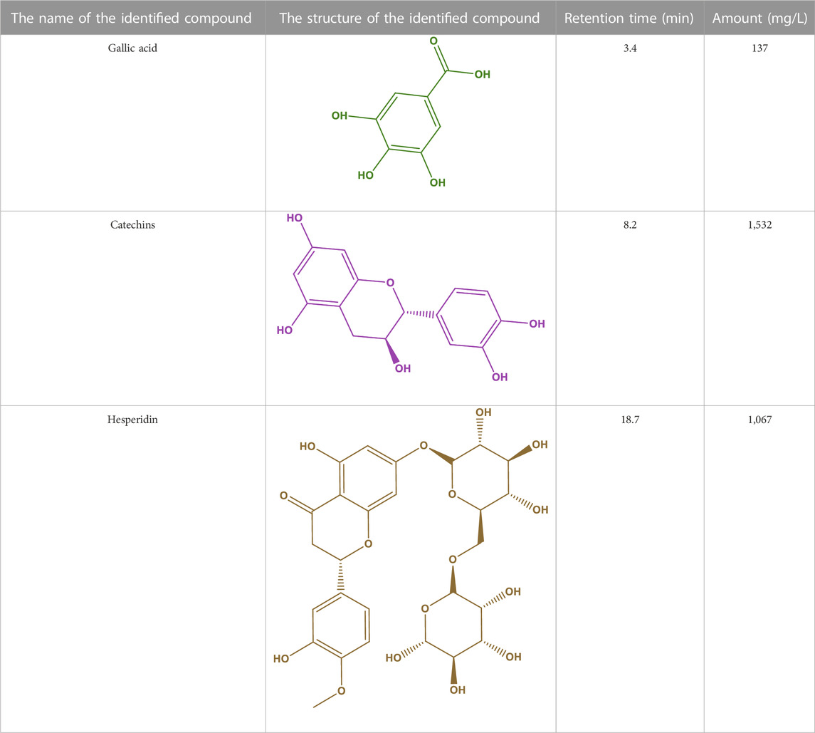
TABLE 1. The main compounds identified in the methanol extract of Daphne mucronata.
Antimicrobial evolution of aqueous, ethanol, methanol, and hexane D. mucronata extracts on 3-gram-positive bacterial species, 3-gram negative bacterial species, 3-common bacterial species between aquatic and human, and 3-fungal species was performed. The results of the tests are given in Table 2. The studied gram positive bacterial, gram negative bacterial, common bacterial between aquatic and human and fungal species are Bacillus cereus (ATCC 11778), Rhodococcus equi (ATCC 25729), Staphylococcus aureus (ATCC 29213); Klebsiella pneumoniae (ATCC 13883), Yersinia enterocolitica (ATCC 9610), Acinetobacter baumannii (ATCC 19606); Yersinia ruckeri (ATCC 29473), Loctococcus garvieae (ATCC 43921), Streptococcus iniae (ATCC 29178); and Fusarium oxysporum (ATCC 7601), Candida albicans (ATCC 10231), Aspergillus fumigatus Fresenius (ATCC 1022), respectively.
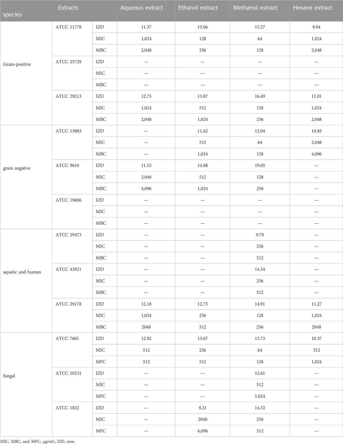
TABLE 2. Antimicrobial activity of aqueous, ethanol, methanol, and hexane Daphne mucronata extracts.
Based on the results obtained and presented in Table 2, the methanol extract of D. mucronata was most effective on the studied bacterial and fungal species. In this extract, MIC between 64 μg/mL- 256 μg/mL, MBC between 128 μg/mL- 512 μg/mL and IZD between 9 mm and 19 mm on studied bacterial species were observed, and MIC between 64 μg/mL- 512 μg/mL, MFC between 128 μg/mL- 1,024 μg/mL and IZD between 12 mm and 15 mm on the studied fungal species were observed.
The review of the literature proves that Gallic acid and Catechins have antimicrobial properties, including strong antibacterial and antifungal properties (Górniak et al., 2019; Ma et al., 2019; Rajamanickam et al., 2019; Panda and Duarte-Sierra, 2022; Shahryari et al., 2022) the effectiveness on the studied strains can be attributed to the high amount of their in the extract.
The DPPH method was used to specifically investigate the antioxidant properties of aqueous, ethanol, methanol, and hexane D. mucronata extracts.
In the studies, ascorbic acid was used as a known natural antioxidant compound to compare the antioxidant properties of the extracts.
The results obtained from antioxidant tests are given in Table 3.
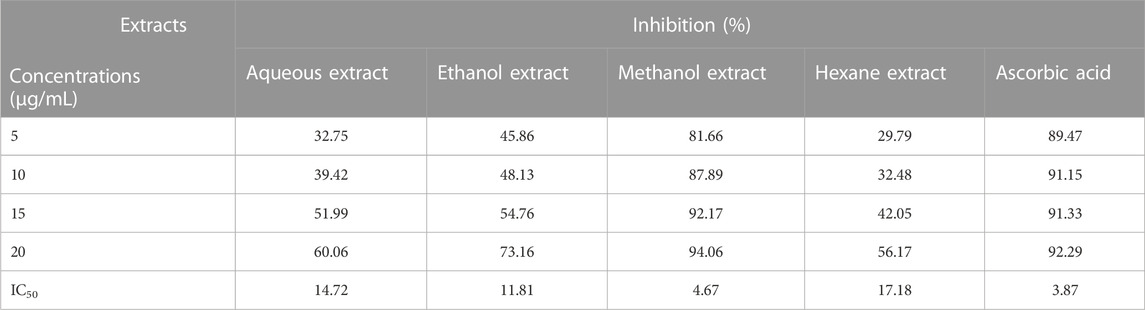
TABLE 3. Antioxidant activity of aqueous, ethanol, methanol, and hexane Daphne mucronata extracts.
In tests, adsorption of DPPH solution and DPPH solution containing extracts were measured, and inhibition (%) and IC50 were calculated. To calculate inhibition (%), Eq. 1 was used.
To calculate IC50, the curves of inhibition (%), and the concentration IC50 were calculated. IC50 for aqueous, ethanol, methanol, and hexane D. mucronata extracts were obtained as 14.72 μg/mL, 11.81 μg/mL, 4.67 μg/mL, and 17.18 μg/mL, respectively.
The antioxidant property of methanol extracts of D. mucronata was very close to ascorbic acid (3.84 μg/mL). High amounts of Gallic acid polyphenol and Hesperidin bioflavonoid, which are known as compounds with potent antioxidant properties (Alavi Rafiee et al., 2018; Binkowska, 2020; Lee et al., 2020; Zahrani et al., 2020), can cause high antioxidant activity of the extract.
The anticancer activity of aqueous, ethanol, methanol, and hexane D. mucronata extracts against breast cancer cells was measured at 48 h at different concentrations (6.25 μg/mL- 50 μg/mL). Cell proliferation and viability of the extracts in various concentrations are given in the diagram of Figure 1.
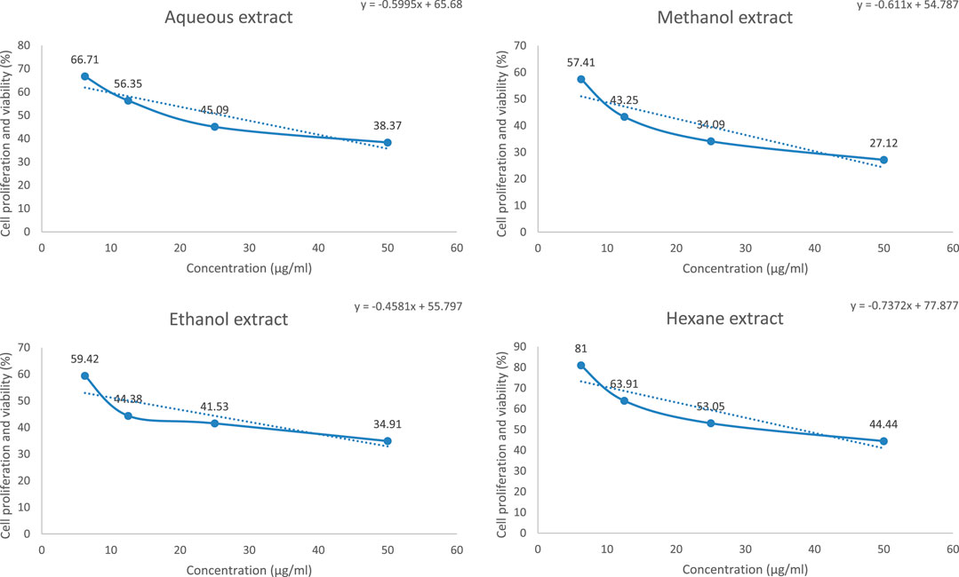
FIGURE 1. Cell proliferation and viability/concentration of aqueous, ethanol, methanol, and hexane Daphne mucronata extracts against anti-breast cancer cells (MCF-7) (n = 3).
Based on the results, it was proved that the lowest cell proliferation and viability are related to the methanol extract.
Using the concentration and cell proliferation and viability graph, the amount of IC50 was measured for the extracts, the results of which are given in Table 4.
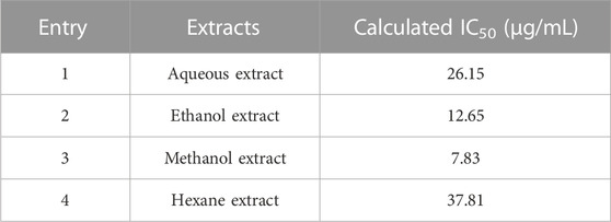
TABLE 4. IC50 values of aqueous, ethanol, methanol, and hexane Daphne mucronata extracts in anti-breast cancer cells (MCF-7) activity.
The lowest IC50 value was observed for methanol extract at 7.83 μg/mL. According to previous studies, the IC50 of abemaciclib, a breast cancer drug, has been reported using the MTT method of 7.29 μg/mL (Anwer et al., 2022). The IC50 value observed for the methanol extract is very close to the Abemaciclib.
In general, as the results of Section 3.2 and Section 3.3 showed, the antimicrobial, antioxidant, and anticancer properties of methanol extract of D. mucronata were more than other extracts. Therefore, in continuation of our studies on D. mucronata extract, polyvinylpyrrolidone nanofibers containing methanol extract of D. mucronata were synthesized by electrospinning method.
The carbonyl oxygen of Polyvinylpyrrolidone can establish hydrogen bonds with alcoholic hydrogens of Gallic acid, Catechins, and Hesperidin, and the structure of Figure 2 can be proposed for the synthesized polyvinylpyrrolidone nanofibers containing methanol extract of D. mucronata.
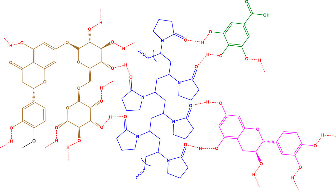
FIGURE 2. Proposed structure for polyvinylpyrrolidone nanofibers containing methanol extract of Daphne mucronata.
The structure and characteristics of prepared nanofibers were confirmed and determined using FTIR, TGA, BET, SEM, flexural strength, compressive strength, and hydrophilicity.
FT-IR of polyvinylpyrrolidone nanofibers (I) and polyvinylpyrrolidone nanofibers containing methanol extract of D. mucronata (II) dose is shown in Figure 3A. As can be seen in the spectrum of the final product, peaks related to O-H groups in the region of 3,300–3,500 cm−1, C-H groups in the area of 2,800–3,000 cm−1, and C=O groups in the region of 1,600–1725 cm−1 are visible. From the comparison of the two spectra in the mentioned areas, it can be concluded that the proposed product of Figure 1 is formed. For example, in the spectrum of the final product, two broad peaks can be observed in the region of 3,300 cm−1 to 3,500 cm−1, one of which can be attributed to the alcoholic hydroxyl groups and the second one can be attributed to the carboxylic acid group of Gallic acid. Other spectra differences in the area below 3,000 cm−1 and in the region 1,600 cm−1 to 1725 cm−1, can be attributed to C-H groups related to Catechins and carbonyls related to Hesperidin.
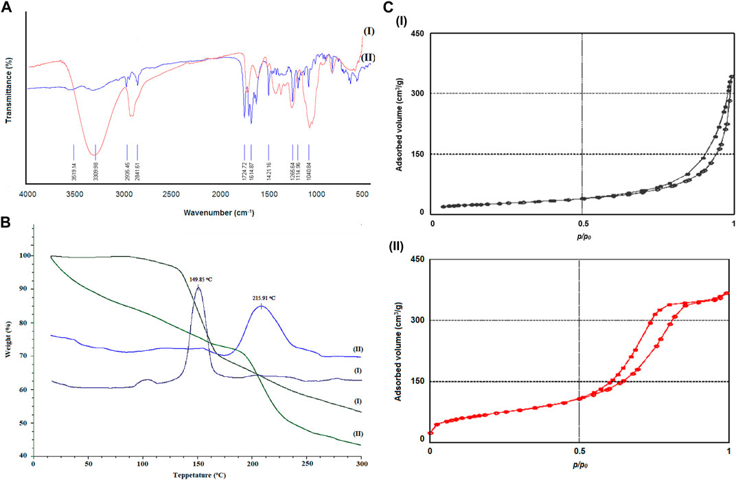
FIGURE 3. FT-IR (A), TGA/DTG curves (B), and N2 adsorption/desorption isotherms (C) of polyvinylpyrrolidone nanofibers (I) and polyvinylpyrrolidone nanofibers containing methanol extract of Daphne mucronata (II).
The TGA/DTG Curves, which show the thermal stability of compounds polyvinylpyrrolidone nanofibers (I) and polyvinylpyrrolidone nanofibers containing methanol extract of D. mucronata (II), are shown in Figure 3B. As can be seen from the comparison of the curves, polyvinylpyrrolidone nanofibers containing methanol extract of D. mucronata have more thermal stability than polyvinylpyrrolidone. From examining of the breaks of 3-B-II, it can be suggested that the weight loss up to the temperature of 150°C is related to methanol and acetic acid molecules trapped in the final composition. Weight loss in areas above 200°C can be attributed to the destruction of organic compounds in synthesized polyvinylpyrrolidone nanofibers containing methanol extract of D. mucronata.
The specific surface area of polyvinylpyrrolidone nanofibers (I) and polyvinylpyrrolidone nanofibers containing methanol extract of D. mucronata (II) was obtained using the N2 adsorption/desorption isotherm presented in Figure 3C. The specific surface area of polyvinylpyrrolidone nanofibers and polyvinylpyrrolidone nanofibers containing methanol extract of D. mucronata was obtained as 780 m2/g and 1,025 m2/g, respectively (Table 5). Nanoparticles’ unique properties, applications, and capabilities are caused by the specific surface area, as we know (Baig et al., 2021; Naseem and Durrani, 2021). In this study, the high specific surface area has led to an increase in the microbiological properties of polyvinylpyrrolidone nanofibers containing the methanol extract of D. mucronata compared to the methanol extract of D. mucronata, which is discussed in detail in Section 3.6.
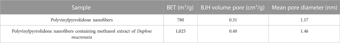
TABLE 5. BET, BJH volume pore and Mean pore diameter of Polyvinylpyrrolidone nanofibers and Polyvinylpyrrolidone nanofibers containing methanol extract of Daphne mucronata.
Choosing the synthesis conditions of nanoparticles is very effective in their morphology and size. The SEM image of the synthesized polyvinylpyrrolidone nanofibers containing methanol extract of D. mucronata shows the same morphology of the composition (Figure 4). In addition, it can be concluded from the image that the synthesized polyvinylpyrrolidone nanofibers containing methanol extract of D. mucronata are in the range of nanostructures. Therefore, suitable conditions have been used in electrospinning to synthesize the desired product.
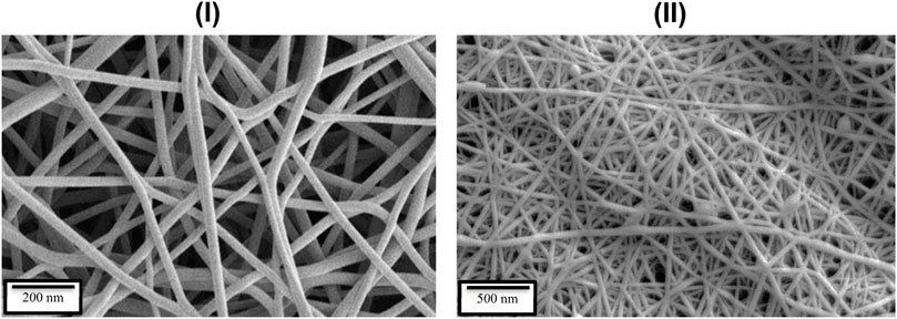
FIGURE 4. SEM image of polyvinylpyrrolidone nanofibers (I) and polyvinylpyrrolidone nanofibers containing methanol extract of Daphne mucronata (II).
Flexural strength and compressive strength are two other vital parameters in the properties of polymers and nanofibers. Flexural strength and compressive strength also depend on the synthesis method of the desired compound. Figure 5A displays the results of the flexural strength test for polyvinylpyrrolidone nanofibers (I) and polyvinylpyrrolidone nanofibers containing methanol extract of D. mucronata (II). The results show that the flexural strength of polyvinylpyrrolidone nanofibers containing methanol extract of D. mucronata (15.8 N/mm2) is higher than that of polyvinylpyrrolidone nanofibers (10.8 N/mm2). A lliterature review showed that the synthesized compound has a higher flexural strength than some of the previously synthesized similar compounds (Ngadiman et al., 2015; Su et al., 2022).
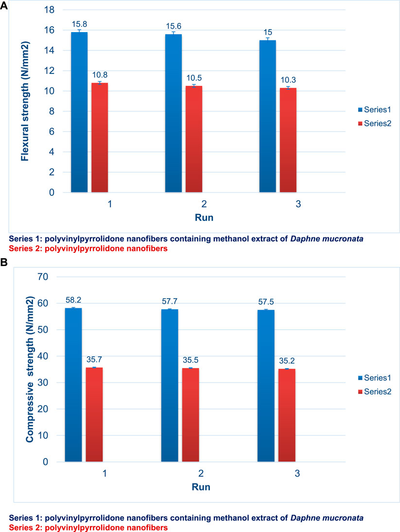
FIGURE 5. Flexural strength (A) and compressive strength (B) of polyvinylpyrrolidone nanofibers (Series 2) and polyvinylpyrrolidone nanofibers containing methanol extract of Daphne mucronata (Series 1).
Figure 5B shows the compressive strength of polyvinylpyrrolidone nanofibers (I) and polyvinylpyrrolidone nanofibers containing methanol extract of D. mucronata (II). Here, too, polyvinylpyrrolidone nanofibers containing methanol extract of D. mucronata with 58.2 65 N/mm2 have a higher compressive strength than polyvinylpyrrolidone nanofibers with 35.7 65 N/mm2.
The high flexural strength and compressive strength of polyvinylpyrrolidone nanofibers containing methanol extract of D. mucronata compared to polyvinylpyrrolidone nanofibers can be attributed to the strong hydrogen bonds created in the final product based on the proposed structure.
The contact angle used for hydrophilicity showed 52o for polyvinylpyrrolidone nanofibers and 38o for polyvinylpyrrolidone nanofibers containing methanol extract of D. mucronata. A decrease in contact angle indicates an increase in hydrophilicity. Here too, according to the proposed structure for the final product, the decline in contact angle can be attributed to the many alcohol groups present in the final product, which have a high ability to perform hydrogen bonding with water molecules (Drelich et al., 2019; Hassan et al., 2019).
As mentioned in the previous sections, the methanol extract of D. mucronata had high biological properties such as antimicrobial, antioxidant, and anticancer activity, and using it and polyvinylpyrrolidone, polyvinylpyrrolidone nanofibers containing methanol extract of D. mucronata were synthesized.
After confirming the structure of polyvinylpyrrolidone nanofibers containing methanol extract of D. mucronata, the synthesized nanofiber and polyvinylpyrrolidone nanofibers were subjected to antimicrobial tests on the previously studied strains. The results of the tests are given in Table 6.
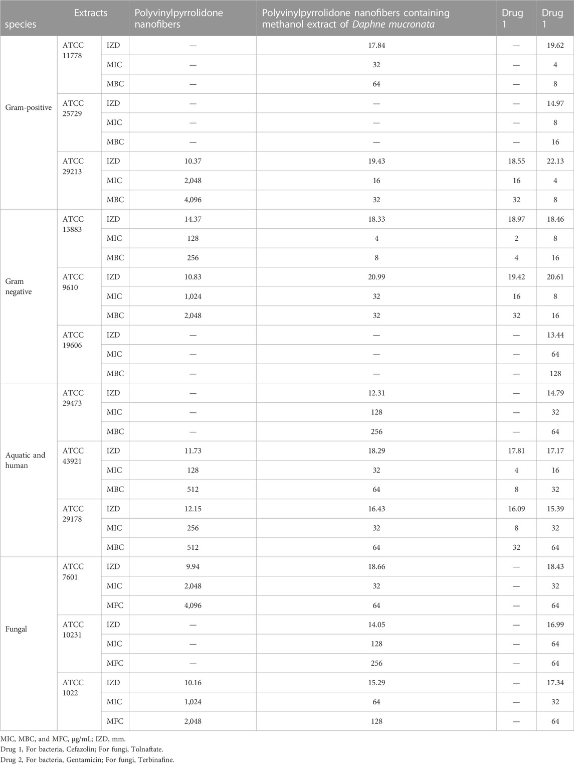
TABLE 6. Antimicrobial activity of Polyvinylpyrrolidone nanofibers and Polyvinylpyrrolidone nanofibers containing methanol extract of Daphne mucronata.
The results showed that the presence of Gallic acid and Catechins in synthesized polyvinylpyrrolidone nanofibers containing methanol extract of D. mucronata increased its antimicrobial properties compared to polyvinylpyrrolidone nanofibers.
Here, Cefazolin, Gentamicin, Tolnaftate, and Terbinafine, which are well-known antibiotics in the market, were used to compare the antimicrobial activity of synthesized polyvinylpyrrolidone nanofibers containing methanol extract of D. mucronata.
The results proved that the synthesized nanofiber is more effective than commercial drugs on some strains. It is possible to mention bacterial and fungal strains such as B. cereus (ATCC 11778), Y. ruckeri (ATCC 29473), F. oxysporum (ATCC 7601), C. albicans (ATCC 10231), and A. fumigatus Fresenius (ATCC 1022), which synthesized nanofiber are effective on them, but Cefazolin and Tolnaftate as commercial drugs were not effective.
As can be seen from the results of Table 6, the effectiveness of polyvinylpyrrolidone nanofibers containing methanol extract of D. mucronata on the studied strains was more than the methanol extract of D. mucronata. The comparison of the antimicrobial activity of extract and nanofiber on the studied strains is given in Table 7.
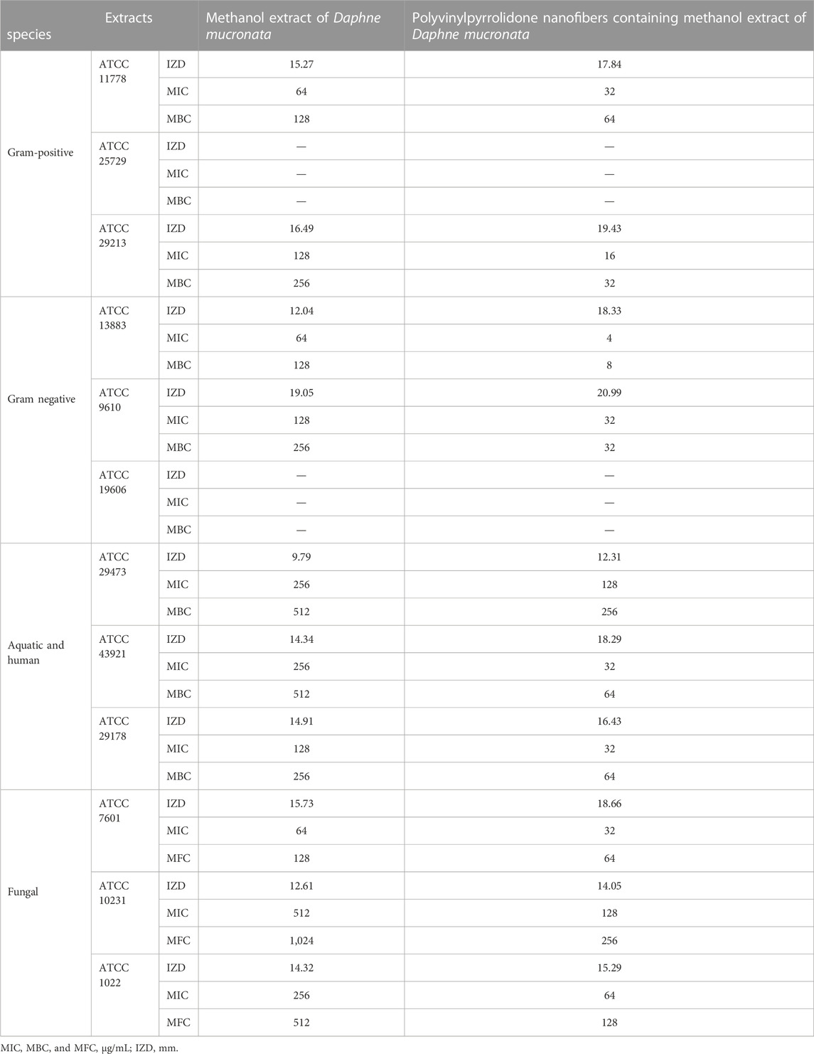
TABLE 7. Comparison of antimicrobial activity of methanol extract of Daphne mucronata and Polyvinylpyrrolidone nanofibers containing methanol extract of Daphne mucronata.
As the final result of the comparison of the antimicrobial activity of the methanol extract of D. mucronata and nanofibers containing methanol extract of D. mucronata, it can be stated that the nanostructure with the high specific surface area has increased the antimicrobial activity of the nanofiber. The high specific surface area of fibrosis results in an increase in the contact surface of molecules with the studied strains and a rise in antimicrobial properties (Lam et al., 2021; Mammari et al., 2022).
The Iraqi Kurdistan Region D. mucronata was used in this study to prepare extracts of aqueous, ethanol, methanol, and hexane. Another goal of this study was the synthesis of nanofibers containing Iraqi Kurdistan Region D. mucronata with high biological properties. The extracts were investigated for their antibacterial, antifungal, antioxidant, and anticancer properties and compared for this purpose. The evaluations and comparisons showed that the highest effectiveness is related to the methanolic extract. So the results for methanolic extract in antimicrobial activity, MIC between 64 and 1,024 μg/mL, in antioxidant activity, IC50 4.67 μg/mL, and in anticancer activity, IC50 7.83 μg/mL were observed. Therefore, by using D. mucronata methanolic extract and polyvinylpyrrolidone with the electrospinning method, the desired nanofibers were synthesized. Characterizing and confirming the structure of the synthesized nanofibers was done using FTIR, TGA, BET, SEM, compressive strength, flexural strength, and hydrophilicity. Higher thermal stability (215.92°C), higher specific surface area (1,025 m2/g), higher flexural strength (15.8 N/mm2) and compressive strength (58.2 65 N/mm2) and higher hydrophilicity than polyvinylpyrrolidone nanofibers were among the characteristics of synthesized polyvinylpyrrolidone nanofibers containing methanol extract of D. mucronata. Which can be attributed to the creation of hydrogen bonds between the extract compounds and polyvinylpyrrolidone. Finally, the antimicrobial properties of nanofibers were also tested, and the results were compared with the antimicrobial properties of the extract. The comparison of the results showed that the nanofibers had higher and more significant properties than the extract due to the unique properties mentioned above, such as high specific surface area. Therefore, nanofiber containing the Iraqi Kurdistan Region D. mucronata and polyvinylpyrrolidone can be introduced as a nanofiber with potential biological properties.
The original contributions presented in the study are included in the article/Supplementary Material, further inquiries can be directed to the corresponding author.
KM: Data curation, Writing–original draft. MK: Software, Writing–original draft. FM: Investigation, Writing–original draft. ZM: Supervision, Writing–review and editing. SAH: Writing–original draft. TH: Resources, Writing–original draft. SJH: Methodology, Writing–review and editing. MQ: Project administration, Writing–original draft. AHA: Writing–original draft, Writing–review and editing. AA: Conceptualization, Visualization, Writing–review and editing.
The author(s) declare financial support was received for the research, authorship, and/or publication of this article. The authors extend their appreciation to the Deanship of Scientific Research at King Khalid University, KSA, for funding this work through a large research group program under grant number RGP.2/350 /44.
The authors declare that the research was conducted in the absence of any commercial or financial relationships that could be construed as a potential conflict of interest.
All claims expressed in this article are solely those of the authors and do not necessarily represent those of their affiliated organizations, or those of the publisher, the editors and the reviewers. Any product that may be evaluated in this article, or claim that may be made by its manufacturer, is not guaranteed or endorsed by the publisher.
The Supplementary Material for this article can be found online at: https://www.frontiersin.org/articles/10.3389/fchem.2023.1287870/full#supplementary-material
Ahani, Z., Nikbin, M., Maghsoodlou, M.-T., Farhadi-Ghalati, F., Valizadeh, J., Beyzaei, H., et al. (2018). Semi-synthesis, antibacterial and antifungal activities of three novel thiazolidin-4-one by essential oil of Anethum graveolens seeds as starting material. J. Iran. Chem. Soc. 15, 2423–2430. doi:10.1007/s13738-018-1431-y
Akhavan-Sigari, R., Zeraati, M., Moghaddam-Manesh, M., Kazemzadeh, P., Hosseinzadegan, S., Chauhan, N. P. S., et al. (2022). Porous Cu-MOF nanostructures with anticancer properties prepared by a controllable ultrasound-assisted reverse micelle synthesis of Cu-MOF. BMC Chem. 16, 10. doi:10.1186/s13065-022-00804-2
Alavi Rafiee, S., Farhoosh, R., and Sharif, A. (2018). Antioxidant activity of gallic acid as affected by an extra carboxyl group than pyrogallol in various oxidative environments. Eur. J. Lipid Sci. Technol. 120, 1800319. doi:10.1002/ejlt.201800319
Anwer, M. K., Fatima, F., Ahmed, M. M., Aldawsari, M. F., Alali, A. S., Kalam, M. A., et al. (2022). Abemaciclib-loaded ethylcellulose based nanosponges for sustained cytotoxicity against MCF-7 and MDA-MB-231 human breast cancer cells lines. Saudi Pharm. J. 30, 726–734. doi:10.1016/j.jsps.2022.03.019
Baig, N., Kammakakam, I., and Falath, W. (2021). Nanomaterials: a review of synthesis methods, properties, recent progress, and challenges. Mater. Adv. 2, 1821–1871. doi:10.1039/d0ma00807a
Beyzaei, H., Deljoo, M. K., Aryan, R., Ghasemi, B., Zahedi, M. M., and Moghaddam-Manesh, M. (2018). Green multicomponent synthesis, antimicrobial and antioxidant evaluation of novel 5-amino-isoxazole-4-carbonitriles. Chem. Central J. 12, 1–8. doi:10.1186/s13065-018-0488-0
Binkowska, I. (2020). Hesperidin: synthesis and characterization of bioflavonoid complex. SN Appl. Sci. 2, 445. doi:10.1007/s42452-020-2256-8
Dias, M. C., Pinto, D. C., and Silva, A. M. (2021). Plant flavonoids: chemical characteristics and biological activity. Molecules 26, 5377. doi:10.3390/molecules26175377
Dirar, A., Alsaadi, D., Wada, M., Mohamed, M., Watanabe, T., and Devkota, H. (2019). Effects of extraction solvents on total phenolic and flavonoid contents and biological activities of extracts from Sudanese medicinal plants. South Afr. J. Bot. 120, 261–267. doi:10.1016/j.sajb.2018.07.003
Dodero, A., Alberti, S., Gaggero, G., Ferretti, M., Botter, R., Vicini, S., et al. (2021). An up-to-date review on alginate nanoparticles and nanofibers for biomedical and pharmaceutical applications. Adv. Mater. Interfaces 8, 2100809. doi:10.1002/admi.202100809
Drelich, J. W., Boinovich, L., Chibowski, E., Della Volpe, C., Hołysz, L., Marmur, A., et al. (2019). Contact angles: history of over 200 years of open questions. Surf. Innov. 8, 3–27. doi:10.1680/jsuin.19.00007
Edikresnha, D., Suciati, T., Munir, M. M., and Khairurrijal, K. (2019). Polyvinylpyrrolidone/cellulose acetate electrospun composite nanofibres loaded by glycerine and garlic extract with in vitro antibacterial activity and release behaviour test. RSC Adv. 9, 26351–26363. doi:10.1039/c9ra04072b
Etemadi, Y., Shiri, A., Eshghi, H., Akbarzadeh, M., Saadat, K., Mozafari, S., et al. (2016). Synthesis, characterisation, and in vitro antibacterial evaluation of a new class of 2-substituted-4-methyl-7, 8-dihydro-5H-pyrimido [4, 5-d] thiazolo [3, 2-a] pyrimidines. J. Chem. Res. 40, 600–603. doi:10.3184/174751916x14737838285904
Fazal, N., Khawaja, H., Naseer, N., Khan, A. J., and Latief, N. (2020). Daphne mucronata enhances cell proliferation and protects human adipose stem cells against monosodium iodoacetate induced oxidative stress in vitro. Adipocyte 9, 495–508. doi:10.1080/21623945.2020.1812242
Ghanadian, M., Ali, Z., Khan, I. A., Balachandran, P., Nikahd, M., Aghaei, M., et al. (2020). A new sesquiterpenoid from the shoots of Iranian Daphne mucronata Royle with selective inhibition of STAT3 and Smad3/4 cancer-related signaling pathways. DARU J. Pharm. Sci. 28, 253–262. doi:10.1007/s40199-020-00336-x
Ghasemian Lemraski, E., Jahangirian, H., Dashti, M., Khajehali, E., Sharafinia, S., Rafiee-Moghaddam, R., et al. (2021). Antimicrobial double-layer wound dressing based on chitosan/polyvinyl alcohol/copper: in vitro and in vivo assessment. Int. J. nanomedicine 16, 223–235. doi:10.2147/ijn.s266692
Górniak, I., Bartoszewski, R., and Króliczewski, J. (2019). Comprehensive review of antimicrobial activities of plant flavonoids. Phytochem. Rev. 18, 241–272. doi:10.1007/s11101-018-9591-z
Han, S., Li, L.-Z., and Song, S.-J. (2020). Daphne giraldii Nitsche (Thymelaeaceae): phytochemistry, pharmacology and medicinal uses. Phytochemistry 171, 112231. doi:10.1016/j.phytochem.2019.112231
Hassan, E. A., Yang, L., Elagib, T. H., Ge, D., Lv, X., Zhou, J., et al. (2019). Synergistic effect of hydrogen bonding and π-π stacking in interface of CF/PEEK composites. Compos. Part B Eng. 171, 70–77. doi:10.1016/j.compositesb.2019.04.015
Hosseinzadegan, S., Hazeri, N., Maghsoodlou, M. T., Moghaddam-Manesh, M., and Shirzaei, M. (2020). Synthesis and evaluation of biological activity of novel chromeno [4, 3-b] quinolin-6-one derivatives by SO 3 H-tryptamine supported on Fe 3 O 4@ SiO 2@ CPS as recyclable and bioactive magnetic nanocatalyst. J. Iran. Chem. Soc. 17, 3271–3284. doi:10.1007/s13738-020-01990-3
Kişla, D., Gökmen, G. G., Evrendilek, G., Akan, T., Vlčko, T., Kulawik, P., et al. (2023). Recent developments in antimicrobial surface coatings: various deposition techniques with nanosized particles, their application and environmental concerns. Trends Food Sci. Technol. 135, 144–172. doi:10.1016/j.tifs.2023.03.019
Kurakula, M., and Rao, G. K. (2020). Moving polyvinyl pyrrolidone electrospun nanofibers and bioprinted scaffolds toward multidisciplinary biomedical applications. Eur. Polym. J. 136, 109919. doi:10.1016/j.eurpolymj.2020.109919
Lam, M., Migonney, V., and Falentin-Daudre, C. (2021). Review of silicone surface modification techniques and coatings for antibacterial/antimicrobial applications to improve breast implant surfaces. Acta Biomater. 121, 68–88. doi:10.1016/j.actbio.2020.11.020
Lee, B. K., Hyun, S.-W., and Jung, Y.-S. (2020). Yuzu and hesperidin ameliorate blood-brain barrier disruption during hypoxia via antioxidant activity. Antioxidants 9, 843. doi:10.3390/antiox9090843
Li, Y., Zhu, G., Xu, X., Chen, L., Lu, T., Hill, J. P., et al. (2022). Embedding metal–organic frameworks for the design of flexible hybrid supercapacitors by electrospinning: synthesis of highly graphitized carbon nanofibers containing metal oxide nanoparticles. Small Struct. 3, 2200015. doi:10.1002/sstr.202200015
Lutfullah, G., Shah, A., Ahmad, K., and Haider, J. (2019). Phytochemical screening, antioxidant and antibacterial properties of daphne mucronata. J. Traditional Chin. Med. 39, 764–771. doi:10.19852/j.cnki.jtcm.2019.06.002
Ma, Y., Ding, S., Fei, Y., Liu, G., Jang, H., and Fang, J. (2019). Antimicrobial activity of anthocyanins and catechins against foodborne pathogens Escherichia coli and Salmonella. Food control. 106, 106712. doi:10.1016/j.foodcont.2019.106712
Mammari, N., Lamouroux, E., Boudier, A., and Duval, R. E. (2022). Current knowledge on the oxidative-stress-mediated antimicrobial properties of metal-based nanoparticles. Microorganisms 10, 437. doi:10.3390/microorganisms10020437
Manandhar, S., Luitel, S., and Dahal, R. K. (2019). In vitro antimicrobial activity of some medicinal plants against human pathogenic bacteria. J. Trop. Med. 2019, 1–5. doi:10.1155/2019/1895340
Mehrali, F., Ziyadi, H., Hekmati, M., Faridi-Majidi, R., and Qomi, M. (2020). Kefiran/poly (vinyl alcohol)/poly (vinyl pyrrolidone) composite nanofibers: fabrication, characterization and consideration of effective parameters in electrospinning. SN Appl. Sci. 2, 895. doi:10.1007/s42452-020-2714-3
Moghaddam-Manesh, M., Beyzaei, H., Heidari Majd, M., Hosseinzadegan, S., and Ghazvini, K. (2021). Investigation and comparison of biological effects of regioselectively synthesized thiazole derivatives. J. Heterocycl. Chem. 58, 1525–1530. doi:10.1002/jhet.4278
Moghaddam-Manesh, M., Sargazi, G., Roohani, M., Zanjani, N. G., Khaleghi, M., and Hosseinzadegan, S. (2022). Synthesis of PVA/Fe3O4@ SiO2@ CPS@ SID@ Ni as novel magnetic fibrous composite polymer nanostructures and evaluation of anti-cancer and antimicrobial activity. Polym. Bull. 80, 11919–11930. doi:10.1007/s00289-022-04584-6
Mostafa, A. A., Al-Askar, A. A., Almaary, K. S., Dawoud, T. M., Sholkamy, E. N., and Bakri, M. M. (2018). Antimicrobial activity of some plant extracts against bacterial strains causing food poisoning diseases. Saudi J. Biol. Sci. 25, 361–366. doi:10.1016/j.sjbs.2017.02.004
Naseem, T., and Durrani, T. (2021). The role of some important metal oxide nanoparticles for wastewater and antibacterial applications: a review. Environ. Chem. Ecotoxicol. 3, 59–75. doi:10.1016/j.enceco.2020.12.001
Nazir, N., Muhammad, J., Ghaffar, R., Nisar, M., Zahoor, M., Uddin, F., et al. (2021). Phytochemical profiling and antioxidant potential of Daphne mucronata Royle and action against paracetamol-induced hepatotoxicity and nephrotoxicity in rabbits. Saudi J. Biol. Sci. 28, 5290–5301. doi:10.1016/j.sjbs.2021.05.051
Ngadiman, N. H. A., Noordin, M., Idris, A., Shakir, A. S. A., and Kurniawan, D. (2015). Influence of polyvinyl alcohol molecular weight on the electrospun nanofiber mechanical properties. Procedia Manuf. 2, 568–572. doi:10.1016/j.promfg.2015.07.098
Nguyen, N. H., Ta, Q. T. H., Pham, Q. T., Luong, T. N. H., Phung, V. T., Duong, T.-H., et al. (2020). Anticancer activity of novel plant extracts and compounds from Adenosma bracteosum (Bonati) in human lung and liver cancer cells. Molecules 25, 2912. doi:10.3390/molecules25122912
Osanloo, M., Arish, J., and Sereshti, H. (2020). Developed methods for the preparation of electrospun nanofibers containing plant-derived oil or essential oil: a systematic review. Polym. Bull. 77, 6085–6104. doi:10.1007/s00289-019-03042-0
Panda, L., and Duarte-Sierra, A. (2022). Recent advancements in enhancing antimicrobial activity of plant-derived polyphenols by biochemical means. Horticulturae 8, 401. doi:10.3390/horticulturae8050401
Park, J.-A., Lee, S.-C., and Kim, S.-B. (2019). Synthesis of dual-functionalized poly (vinyl alcohol)/poly (acrylic acid) electrospun nanofibers with enzyme and copper ion for enhancing anti-biofouling activities. J. Mater. Sci. 54, 9969–9982. doi:10.1007/s10853-019-03578-6
Popiel, S., Nawała, J., Dziedzic, D., SoDerstroM, M., and Vanninen, P. (2014). Determination of mustard gas hydrolysis products thiodiglycol and thiodiglycol sulfoxide by gas chromatography-tandem mass spectrometry after trifluoroacetylation. Anal. Chem. 86, 5865–5872. doi:10.1021/ac500656g
Rajamanickam, K., Yang, J., and Sakharkar, M. K. (2019). Gallic acid potentiates the antimicrobial activity of tulathromycin against two key bovine respiratory disease (BRD) causing-pathogens. Front. Pharmacol. 9, 1486. doi:10.3389/fphar.2018.01486
Rhazi, L., Depeint, F., and Ayerdi Gotor, A. (2022). Loss in the intrinsic quality and the antioxidant activity of sunflower (Helianthus annuus L.) oil during an industrial refining process. Molecules 27, 916. doi:10.3390/molecules27030916
Shah, A., Lutfullah, G., Ahmad, K., Khalil, A. T., and Maaza, M. (2018). Daphne mucronata-mediated phytosynthesis of silver nanoparticles and their novel biological applications, compatibility and toxicity studies. Green Chem. Lett. Rev. 11, 318–333. doi:10.1080/17518253.2018.1502365
Shahryari, T., Alizadeh, V., Kazemzadeh, P., Jadoun, S., Chauhan, N. P. S., and Sargazi, G. (2022). A controllable procedure for removing Navicula algae from drinking water using an ultrasonic-assisted electrospun method for highly efficient synthesis of Co-MOF/PVA polymeric network. Appl. Phys. A 128, 396. doi:10.1007/s00339-022-05524-x
Su, S., Hu, S., and Liu, Q. (2022). Application of polypyrrole cellulose nanocrystalline composite conductive material in garment design. Adv. Mater. Sci. Eng. 2022, 1–11. doi:10.1155/2022/4187826
Sun, Q., Yao, G.-D., Song, X.-Y., Qi, X.-L., Xi, Y.-F., Li, L.-Z., et al. (2017). Autophagy antagonizes apoptosis induced by flavan enantiomers from Daphne giraldii in hepatic carcinoma cells in vitro. Eur. J. Med. Chem. 133, 1–10. doi:10.1016/j.ejmech.2017.03.072
Tanase, C., Coșarcă, S., and Muntean, D.-L. (2019). A critical review of phenolic compounds extracted from the bark of woody vascular plants and their potential biological activity. Molecules 24, 1182. doi:10.3390/molecules24061182
Wu, S., Li, K., Shi, W., and Cai, J. (2022). Preparation and performance evaluation of chitosan/polyvinylpyrrolidone/polyvinyl alcohol electrospun nanofiber membrane for heavy metal ions and organic pollutants removal. Int. J. Biol. Macromol. 210, 76–84. doi:10.1016/j.ijbiomac.2022.05.017
Wu, S., Shi, W., Li, K., Cai, J., Xu, C., Gao, L., et al. (2023). Chitosan-based hollow nanofiber membranes with polyvinylpyrrolidone and polyvinyl alcohol for efficient removal and filtration of organic dyes and heavy metals. Int. J. Biol. Macromol. 239, 124264. doi:10.1016/j.ijbiomac.2023.124264
Yadav, D., Amini, F., and Ehrmann, A. (2020). Recent advances in carbon nanofibers and their applications–a review. Eur. Polym. J. 138, 109963. doi:10.1016/j.eurpolymj.2020.109963
Keywords: methanolic extract, Iraqi Kurdistan Region Daphne mucronata, polyvinylpyrrolidone nanofibers, antioxidant activity, antimicrobial activity, anticancer activity
Citation: Muzammil K, Kzar MH, Mohammed F, Mohammed ZI, Hamood SA, Hussein TK, Hanoon SJ, Qasim MT, Hussien Alawadi A and Alsalamy A (2023) Methanol extract of Iraqi Kurdistan Region Daphne mucronata as a potent source of antioxidant, antimicrobial, and anticancer agents for the synthesis of novel and bioactive polyvinylpyrrolidone nanofibers. Front. Chem. 11:1287870. doi: 10.3389/fchem.2023.1287870
Received: 02 September 2023; Accepted: 02 October 2023;
Published: 25 October 2023.
Edited by:
Sanjib Banerjee, Indian Institute of Technology Bhilai, IndiaReviewed by:
Luqman Jameel Rather, Southwest University, ChinaCopyright © 2023 Muzammil, Kzar, Mohammed, Mohammed, Hamood, Hussein, Hanoon, Qasim, Hussien Alawadi and Alsalamy. This is an open-access article distributed under the terms of the Creative Commons Attribution License (CC BY). The use, distribution or reproduction in other forums is permitted, provided the original author(s) and the copyright owner(s) are credited and that the original publication in this journal is cited, in accordance with accepted academic practice. No use, distribution or reproduction is permitted which does not comply with these terms.
*Correspondence: Ali Alsalamy, YWxpaGFzaGltYWxzYWxhbXk3OEBnbWFpbC5jb20=
Disclaimer: All claims expressed in this article are solely those of the authors and do not necessarily represent those of their affiliated organizations, or those of the publisher, the editors and the reviewers. Any product that may be evaluated in this article or claim that may be made by its manufacturer is not guaranteed or endorsed by the publisher.
Research integrity at Frontiers
Learn more about the work of our research integrity team to safeguard the quality of each article we publish.