 Ali Abedini1
 Mojtaba Rostami2,3Hamid Reza Banafshe4
 Mehdi Rahimi-Nasrabadi5,6,7Ali SobhaniNasab8,9*Mohammad Reza Ganjali10,11
- 1Young Researchers and Elite club, Central Tehran Branch, Islamic Azad University, Tehran, Iran
- 2School of Chemistry, College of Science, University of Tehran, Tehran, Iran
- 3International Iberian Nanotechnology Laboratory (INL), Braga, Portugal
- 4Department of Pharmacology, School of Medicine, Kashan University of Medical Sciences, Kashan, Iran
- 5Chemical Injuries Research Center, Systems Biology and Poisonings Institute, Baqiyatallah University of Medical Sciences, Tehran, Iran
- 6Faculty of Pharmacy, Baqiyatallah University of Medical Sciences, Tehran, Iran
- 7Institute of Electronic and Sensor Materials, TU Bergakademie Freiberg, Freiberg, Germany
- 8Physiology Research Center, Institute for Basic Sciences, Kashan University of Medical Sciences, Kashan, Iran
- 9Core Research Lab, Kashan University of Medical Sciences, Kashan, Iran
- 10Center of Excellence in Electrochemistry, School of Chemistry, College of Science, University of Tehran, Tehran, Iran
- 11National Institute of Genetic Engineering and Biotechnology (NIGEB), Tehran, Iran
Nanotechnology mainly deals with the production and application of compounds with dimensions in nanoscale. Given their dimensions, these materials have considerable surface/volume ratios, and hence, specific characteristics. Nowadays, environmentally friendly procedures are being proposed for fabrication of Fe nanoparticles because a large amount of poisonous chemicals and unfavorable conditions are needed to prepare them. This work includes an inclusive overview on the economical and green procedures for the preparation of such nanoparticles (flower, fruits, tea, carbohydrates, and leaves). Pure and bimetallic iron nanoparticles, for instance, offer a high bandwidth and excitation binding energy and are applicable in different areas ranging from antibacterial, anticancer, and bioimaging agents to drug delivery systems. Preparation of nano-sized particles, such as those of Fe, requires the application of high quantities of toxic materials and harsh conditions, and naturally, there is a tendency to develop more facile and even green pathways (Sultana, Journal of Materials Science & Technology, 2013, 29, 795–800; Bushra et al., Journal of hazardous materials, 2014, 264, 481–489; Khan et al., Ind. Eng. Chem. Res., 2015, 54, 76–82). This article tends to provide an overview on the reports describing green and biological methods for the synthesis of Fe nanoparticles. The present review mainly highlights selenium nanoparticles in the biomedical domain. Specifically, this review will present detailed information on drug delivery, bioimaging, antibacterial, and anticancer activity. It will also focus on procedures for their green synthesis methods and properties that make them potential candidates for various biomedical applications. Finally, we provide a detailed future outlook.
1 Introduction
Preparation of magnetic or non-magnetic nanomaterials through reduction of metal salts, thermal decomposition, and metal evaporation has been a hot topic during the recent years, due to their unique physical/chemical properties as opposed to the corresponding bulk forms. Consequently, a wide range of applications of such materials in bioseparation and biomedical procedures, biosensors, cytotoxicity, antibacterial, catalysts, magnetic resonance imaging (MRI), and environmental remediation are increasing alarmingly (Rahimi-Nasrabadi et al., 2015; Rahimi-Nasrabadi et al., 2016; Abamor et al., 2017; Naderi et al., 2017; Rostami, 2017; Sobhani-Nasab et al., 2017; Eghbali-Arani et al., 2018a; Amani et al., 2018; Eghbali-Arani et al., 2018b; Padash et al., 2018; Pourmasoud et al., 2018; Sedighi et al., 2018; Gandomi et al., 2019; Sobhani-Nasab et al., 2019; Nayak et al., 2022; Rostami et al., 2022; Wang et al., 2022).
Various research teams have reported the synthesis of mono- and bimetallic nanoparticles of iron through chemical reduction reactions using diverse polymers and organic solvents (Wang and Zhang, 1997; Ponder et al., 2000; Sun et al., 2000; Saleh et al., 2005; Peng et al., 2006). Also, a wide range of plant extracts, carbohydrates, peptides, proteins, organic solvents, oligonucleotides, lipids, dendrimers, phospholipids, surfactants, and polymers have been reportedly used as stabilizers to control the shape of noble metal nanoparticles (Murphy et al., 2005; Khullar et al., 2012; Bashir et al., 2014; Murray et al., 2016). Lin et al. (2001) reported a reverse micelle procedure based on the application of 1-butanol, octane, and cetyltrimethylammonium bromide to prepare iron nanoparticles coated with a gold film. In another work, He and Zhao (2005) used solutions of starch in water for stabilizing the potentials in a procedure for preparation of iron nanoparticles using a mixed solution of Fe2+ and Fe3+ in water using sodium borohydride (NaBH4). There is a general trend toward green environmentally friendly chemical technologies, in the light of the global environmental crises. This concern is reflected by the publication of various green chemistry books in the past decade (Ahluwalia, 2009; Anastas and Li, 2012) covering general and specialized procedures like ultrasound and microwave approaches as well as green synthesis, analysis, tribology, polymerization, engineering, and manufacturing. Furthermore, environmentally friendly techniques for production of food, textiles, hydrogen and syngas, biocomposites, particle technology, biomass, wastewater treatment, and biofuels have been covered in different references (Allen and Shonnard, 2001; Chen et al., 2011; Mathers and Meier, 2011; Mulvaney, 2011; Puigjaner, 2011; Boye and Arcand, 2012; de la Guardia and Garrigues, 2012; Kan, 2012; Lee and Henthorn, 2012; Lee and Shah, 2012; Lofrano, 2012; Nosonovsky and Bhushan, 2012; Schiebl, 2012; Siyamak, 2012; Debalina and Pike, 2017). Chemists all over the world prescribe the 12 principles of green chemistry in their works (Anastas and Warner, 1998; Singh et al., 2021). It is a fact that various methods, such as aerosol technologies, UV irradiation, laser ablation, lithography, photochemical reduction, and ultrasonic procedures, have proven to be applicable for production of nanoparticles. The economic and environmental costs of these techniques are still high, and therefore an ongoing interest exists to develop sustainable eco-friendly procedures for the purpose mentioned previously (Narayanan and Sakthivel, 2010).
On the other hand, evaluation of the negative effects of nanomaterials is still a relatively hot topic with lots of unanswered questions, and it is believed that the preparation of these materials through using eco-friendly and biocompatible reagents can lessen the known and unknown adverse effects of these materials on humans and the environment (Varma, 2012). Non-toxic solvents (ideally water), sealed reactors, eco-friendly procedures not requiring contacting reaction media and air (microwave, ultrasound, biological methods, magnetic, and hydrothermal, among others), and low-temperature processes are among the solutions. This review also focuses on the eco-friendly methods for preparation of nanoparticles and nanomaterials using green reducing and capping agents like herbal extracts (Gan and Li, 2012). Furthermore, natural materials like vitamins, sugars, biodegradable polymers and microorganisms and herbal extracts, among the rest of these reducing and capping agents, have proven to be suitable for large-scale applications (Iravani, 2011).
The main chemical agent in some reactions has been found to be polyphenols. These methods are simple, cost-effective, and rather reproducible and produce stable products (Kalaiarasi et al., 2010). It is also possible to use microorganisms for production of nanoparticles. Yet, the methods are rather slow and produce limited particle sizes and shapes as compared with the method based on herbal extracts. Fungi extracts are currently finding worldwide popularity as tools for use in the synthesis of nanoparticles (Dhillon et al., 2012). Biomaterials generally act as eco-friendly through eliminating the need for using toxic chemical agents or harsh conditions (Parsons et al., 2007). The mechanisms of the corresponding bioreduction reactions (Xie et al., 2007a; Zhou et al., 2010; Huang et al., 2011) and those of the catalytic reactions of the products of these reactions have been evaluated (Vilchis-Nestor et al., 2009; Du et al., 2011; Zhan et al., 2012).
It is noteworthy that although the application of these green routes is gaining momentum, it is still not as widespread as classical wet-chemistry procedures (Kharissova and Kharisov, 2008; Kharisov et al., 2012a), while they have important advantages like very near to zero contaminations. Even when wastes are left or produced (except for the case of microbial reactions), they are compatible with the environment and living creatures due to their origin. Another advantage of these methods is the rather low-cost precursors (Abdullah et al., 2020). These can change the methods to interest substitutes in many conventional techniques. In this review, the major focus is laid on the biomedical domain of iron nanoparticles by covering their utilities in cancer therapy, drug delivery, bioimaging, and antibacterial applications. Figure 1 shows the schematic representation of the methods for the green synthesis of Fe nanoparticles and their biomedical applications.
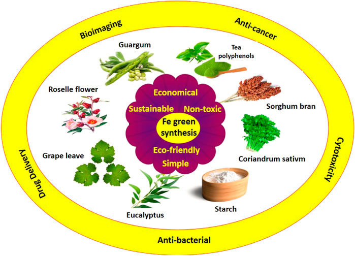
FIGURE 1. Green capping agent used for the preparation of Fe nanoparticles.
2 Preparation of Fe Nanoparticles and Its Biometallic Through Plants
Reduction of metal ions and the stability of their complexes during the nucleation and growth phases, leads to the formation of metal oxide nanoparticles. Extracts of seeds, flowers, leaves, and green plants, are known to contain high quantities of proteins, amino acids, polyphenols, and reducing sugars (Nejati et al., 2022). These materials can act as reduction and capping agents (Besbes et al., 2004; Xie et al., 2007b; Khan et al., 2012; Hussain et al., 2014). Njagi et al. prepared Fe and Ag nanoparticles using vitamin aqueous sorghum bran extracts (Njagi et al., 2010; Abdullah et al., 2020). Verma et al., used tea polyphenols (Camellia sinensis) to prepare metallic Fe nanoparticles at an ambient temperature without using surfactants or polymers as stabilizers (Hoag et al., 2009; Nadagouda et al., 2010). Khan and Al-Thabaiti (2018) reported a biomimetic method for formation of Fe nanoparticles using FeCl3 and Hibiscus sabdariffa, Roselle flower aqueous extract (HBS), as capping and reducing agents. The morphology of green synthesized Fe nanoparticles was found to depend on the extract of Fe3+ concentrations and also the reaction time. The average particle diameter was found to be 18 nm. Fang Luo and et al. reported the synthesis of bimetallic Fe/Pd nanoparticles via the green method. This single-step approach was based on the application of an aqueous extract of grape leaves for cost-effective and large-scale preparation of Fe/Pd nanoparticles for field remediation. Some herbal extracts have also been used for the synthesis of bimetallic nanoparticles containing Fe (He and Zhao, 2005; Smuleac et al., 2011). The synthesis of iron nanoparticles and their biometallic composites with plants is environmentally friendly, but controlling the morphology and size of nanoparticles can be difficult. For instance, Fe/Pd nanoparticles were prepared using green tea extracts rather than the application of NaBH4 which is a common reducing agent. A summary of the several bio-based template media employed in the preparation of Fe nanoparticles is shown in Table 1.
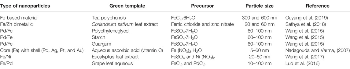
TABLE 1. Biological and biogenic syntheses for preparing Fe NPs.
3 Pure and Bimetallic Iron Particles
During the past 3 decades, nanoparticles of metallic iron have been widely used in remediation and treatment of polluted water containing organic or inorganic species, due to their favorable surface properties (Zhang et al., 2011). The various morphologies of iron nanoparticles are significantly attractive for water disinfection and remediation of heavy metals from soils (Kharisov et al., 2012b). The main methods used for the preparation of these particles are based on the application of tea extracts on polyphenol or other herbal extracts (Victor, n.d.). Thus, Fe nanoparticles are prepared using tea extracts (Nadagouda et al., 2010).
Iron nanoparticles prepared using green tea leaf extracts may contain oxohydroxide and iron oxide (Shahwan et al., 2011). These particles have been reportedly used as a Fenton-like catalyst for removal of methyl orange (MO) and methylene blue (MB) dyes from water and is found to have fast removal kinetics, with a second-order behavior for MB and close first-order behavior in the case of MO (Rostami et al., 2018; Rostami, 2019; Akbari et al., 2021). The removal efficiency was found to be complete for both dyes over a wide concentration window of 10–200 mg/l. The same method has been used for preparation of nanoparticles of iron alloys. For instance, polymer membranes [e.g., polyacrylic acid-coated polyvinylidene fluoride membrane] containing immobilized reactive nanoparticles (of Fe/Pd and Fe) have been using tea extracts (Smuleac et al., 2011) and are used for the destruction of trichloroethylene (TCE) as a common contaminant.
4 Biomedical Applications
Then, in this review, we have focused mainly on the latest studies of applications, including cancer therapy, drug delivery, and antibacterial, of Fe NPs. Table 2 illustrates several metal-based nanomaterials that are utilized in various domains for enormous applications.
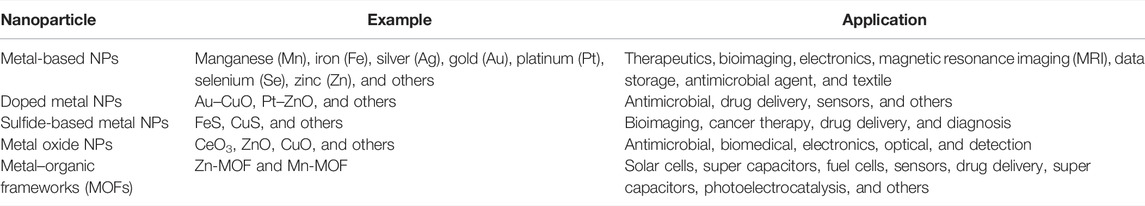
TABLE 2. Various types of metal-based NPs that are utilized in various fields (Singh et al., 2021).
4.1 Anticancer Effects
The application of nanotechnology in treating cancer involves engineering, pharmaceutical science, and molecule-based imaging.
Given the unique optical, magnetic, or structural characteristics of semiconducting nanoparticles and nanocrystals, they do not offer target neoplasm antigens (biomarkers), when interacting with neoplasm-targeting ligands (e.g., peptides or antibodies). Nanoparticles with the size of 1–100 nm have very high active surface areas to interact with various detection agents, such as radio-isotopic, optical or magnetic, and medicinal (e.g., anticancer), which creates the chances for genetic and molecular bio-marking opportunities specifically for personalized cancer treatment (Nie et al., 2007). Some research have proven the anticancer effects of magnetic nanoparticles (Peymani-Motlagh et al., 2019a; Peymani-Motlagh et al., 2019b). G.F. Goya and et al. studied and experimented internalizing magnetic nanostructures into dendritic cells to determine the location of the particles and the viability of the cultured cells (Rostami et al., 2021a). Fe nanostructures coated with carbon proved to have toxic effects on dendritic cells and left no effects on their viability (Rostami et al., 2021b).
The observations indicated that dendritic cells can incorporate 10-ca magnetic nanoparticles. Magnetic nanoparticles after 1 day incubation were observed to be 200 nm. Our results suggest that loading dendritic cells with properly functionalized magnetic nanostructures could be a promising strategy for improvement of vectorization in cancer diagnosis and treatment (Goya et al., 2008).
Abdullaeva et al. (2012) prepared onion-like carbon-encapsulated iron nanoparticles. The iron particles were face-centered cubic crystals of 20–30 nm size and with 2–10 nm layered onion-like coatings. The cytotoxicity evaluations involved exposing human lung epithelial A549 cells to these particles for 24 h. Nanoparticles of 10, 20, 40, 80, and 160 lg/ml suspensions were used for this purpose, and the cytotoxic effects were evaluated using MTT and XTT assays, and it was found that higher cell viability was possible under the same conditions, i.e., 95% for the Fe–C nanoparticles. The results showed that the viability of the Fe–C sample was 8% more than that reported in the case of Fe–C prepared through arc-discharge by Kratschmer–Huffmann (Goya et al., 2008). Also, it was reported that Fe nanoparticles held the potentials for use as effective chemotherapeutic and bacteriostatic agents for the fast distinction of various cancers (Naderi et al., 2018). The synthesis of iron nanoparticles and their biomedical composites with eco-friendly plants is a great way to eradicate cancer.
4.1.1 Toxicity Mechanism
Metal nanoparticles have toxic effects mainly due to the oxidative stress they cause in the intercellular space or through their oxidation leading to formation of free ions in the solution. Fe can pass through cell membranes via calcium transport proteins, and nanoparticles find their way to the cytoplasm through endocytosis. The cell toxicity of the nanoparticles is presented in Figure 2.
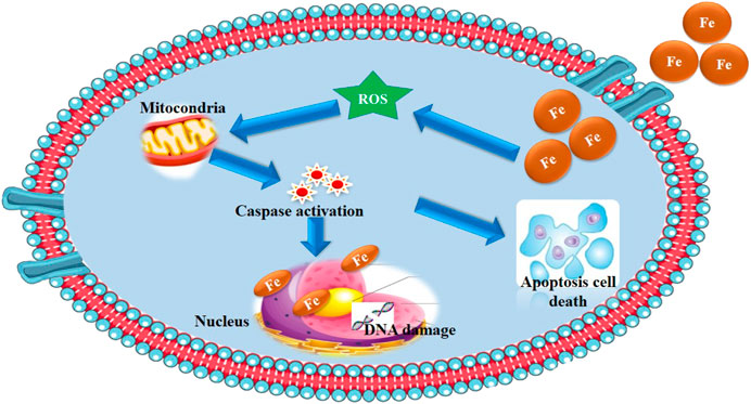
FIGURE 2. Schematic representation of the mechanism for the cytotoxicity activity of Fe NPs.
4.1.2 Oxidative Stress
The main reasons for the toxic effect of nanoparticles are the formation of ROS and the oxidative stress (Yang et al., 2009). It has been reported that the presence of small amounts of metallic species in a cell can give rise to the formation of large amounts of O2−, OH., and H2O2 (ROS) (Dudev and Lim, 2008) through interference with the cellular electron transport chain (Xia et al., 2008).
The main source of intracellular ROS is mitochondria (Xia et al., 2007). It is known that OH radicals (hydroxyl radicals) are the most reactive ROS and oxidize nearly all organelle species (Yamakoshi et al., 2003). The ROS formed in the cell lead to the formation of O2− through surface electron capture by the nanoparticles (De Berardis et al., 2010). Free radicals create a degree of imbalance in the oxidant/anti-oxidant phenomena of the cells through oxidizing lipids, denaturing proteins and nucleic acids, and stimulating an anti-oxidant defense system (Yang et al., 2009).
Nanoparticles can deactivate lysosomes and destroy their membranes if the pH of lysosomes is acidic (Nohl and Gille, 2005), leading to the transfer of nanoparticles’ nuclei and mitochondria (Wang et al., 2012). In the latter case, the deposition of iron nanoparticles onto the membrane enhances the membrane depolarization to disturb transduction of electrons in the inner membrane. This, in turn, leads to the permeability of membranes and formation of reactive oxygen species (Gabbay et al., 2006; Xia et al., 2007).
The presence of reactive oxygen species in lysosomes can ruin DNA double helix or lead to DNA point mutations (Singh et al., 2009), but in mitochondria, the ROS change mitochondrial respiration and apoptosis, damage cellular redox disequilibria, and per-oxidize the cell membrane lipids (Xia et al., 2007). H2O2 generated in the cytoplasm easily diffuses the mitochondrial membranes and undergoes the Fenton reaction to form OH damaging DNA and leading to cellular death (Xia et al., 2007).
High amounts of intracellular ROS enhance the gene expression of the death receptor nanoparticles (Yang et al., 2009) as well as redox-sensitive signaling paths under average oxidative stress (Nel et al., 2006; He et al., 2007). Inflammatory responses which could be caused by death and cellular fibrosis would initiate as a result of activation of mitogen-activated protein kinase and the signaling cascades of nuclear factor NF-kB (Nel et al., 2006). Nuclear pores which are larger than 50 nm can allow quantum dots (QDs) to enter the nucleus. This can also happen through direct physical injuries of the nucleus membrane. As a result, the quantum dots can directly interact with the DNA of the nucleus (Wang et al., 2012).
4.1.3 Dissolution of Nanoparticles
The presence of free ions through the dissolution of nanoparticles is the second reason for the high toxicity of metallic nanoparticles (Gabbay et al., 2006). Fe NPs’ high concentrations in suspensions or cellular media (Gunawan et al., 2011) catalyze the Fenton reactions leading to the formation of high quantities of OH, which in turn harms lipids, proteins, and nucleic acids (Hartwig, 2013). Nanoparticles which are present in cells can also move in acidic organelle (e.g., lysosomes) or react with acidic materials (e.g., nucleic acids), freeing more Fe2+, and hence, enhancing damages through oxidation (Cuillel et al., 2014).
Evidently, the dissolution of particles depends on their dimensions, surface area, composition, and other physical/chemical properties as well as conditions such as temperature and pH.
The fact that free orbitals of the cation are able to interact with free electron pairs of atoms like N and O present in chelates allows them to interact and deactivate biomolecules and disrupt their normal functions. Fine nanoparticles in the cell can find their way into nuclei through nuclear pores or have access to the nucleus available due to cellular division, inhibiting the transcription and translation phenomena, and damaging the genetic material through interacting proteins of DNA or DNA (Singh et al., 2009). Furthermore, interactions with cellular signal species can further activate signaling cascades (Miller et al., 2010). Dissolved cations can also destabilize or degrade mRNA through interaction with its proteins (Soenen et al., 2010). These free ions play an important role in the homeostasis of cells (Cuillel et al., 2014), and if their concentration exceeds a certain level, this effect can reverse. Dissolution of nanoparticles can increase the local concentration of ions (Xia et al., 2008), increasing the tendency for the influx of ions through calcium ion channels present in the endoplasmic reticulum–plasma membrane (Hoyal et al., 1998). As mentioned, the ions participate in a series of cellular processes like activating transcription factors (e.g., NF-kB) (Dolmetsch et al., 1998), producing superoxide anions, and secreting proteins and nitric oxide. These can, in turn, disrupt mitochondria harming the cell (Raddassi et al., 1994; Berridge et al., 1998; Xia et al., 2008). Figure 2 shows the schematic representation of the brief cellular toxicity that stemmed from Fe nanoparticles and their bimetallic nanocomposites.
4.2 Bioimaging
It is of great value to be able to observe sole molecules in living cells (Pinaud et al., 2010). This is, as it may sound, naturally a hard target, especially in the case of a drug molecule, since the process can inversely affect the molecule’s selectivity or activity due to interactions with the tracer or probe species. Therefore, choosing the functional group which leads to minimum disruptive effects on the functions of the molecules could be critical to be able to use covalent conjugation (Byrne et al., 2007). In few recent cases, the pharmaceutical molecules were conjugated to fluorophores (Uddin et al., 2010), yet this is neither always possible nor free from disadvantages (e.g. photobleaching) (Resch-Genger et al., 2008).
Semiconducting nanoparticles can be used in this case, due to their distinctive optical properties (e.g. long fluorescence lifetimes and controlled sizes) in comparison to common dyes (Michalet et al., 2005; Rosenthal et al., 2011). Surface engineering and bio-functionalization of nanoparticles have added to the potentials of these particles for use as cellular probes applicable in the case of biomolecules (Biju et al., 2010; Zrazhevskiy et al., 2010). The fluorescence signal of nanoparticles can be monitored, in vivo, for the determination of properties such as specific targeting and drug release rates.
Zhuo et al. (2019) suggested a new procedure for preparation of iron-doped carbon quantum dots (Fe-CQDs) as bioimaging agents. The procedure constituted hydrothermal carbonization, and the reagents used were ethylenediamine tetraacetic acid (EDTA) salts and ferric nitrate. These quantum dots contain dopamine-bonding Fe sites and luminescent carbon quantum dots (fluorophores). Cell-imaging studies revealed high photostability and low cytotoxicity on the part of the quantum dots, indicating their fitness for biological applications.
4.3 Antibacterial Activity
Non-toxic nanoparticles of ZnO, Cu and Fe have been prepared and evaluated as an antibacterial material (Table 3). It was found that several factors influence the sensitivity or resistance of bacteria to these nanoparticles. The influences of nanoparticles on pathogenic gene expression hrpE were evaluated through real-time PCR. It was observed that the Xanthomonas campestris strain exposed to metallic iron nanoparticles increased the growth rate, and this trend continued with increasing the concentration of these particles. However, increasing the amount of the copper nanoparticles lowered the growth percentage of Xanthomonas campestris. The results showed that the expression levels of the pathogenic gene expression hrpE in the case of copper and iron nanoparticles increased 9- and 3-fold (Moradian et al., 2018). The effect of iron nanoparticles on bacteria is a strong function of ROS. Fe nanoparticles are known to sterilize bacteria through exudation, absorption, and complexation. In general, smaller particles have higher antibacterial activity due to their larger surface area. The superoxide radicals, hydrogen peroxide, react to the ROS, damaging deoxyribonucleic acid and cellular proteins leading to the death of the cell. Fe nanoparticles show an antimicrobial activity through generation of ROS under radiation, which in turn kills cells through aerophilous stress on the microorganism. The ROS formed include hydrogen peroxide, superoxide anion radical, and hydroxyl radical. Superoxide anion radicals (.O2−) undergo reactions with proton ions forming HO2− and then with electrons to form HO2−, which next react with protons to form H2O2. The formed hydrogen peroxide radicals through this multistep reaction reach the deoxyribonucleic acid, and through cell membranes and cellular proteins kill microorganisms. Smaller crystallites form more ROS, and hence nanoparticles are more efficient antibacterial and antimicrobial agents. A second ground for this antibacterial activity is due to the release of Fe2+ from the surface of Fe nanoparticles and its interaction with the negatively charged membranes of the microorganism and penetration through the semi-permeable membranes. Based on the results, the nanomaterials directly damage the cell membranes of pathogenic bacteria. A summary of the antibacterial activity by Fe nanoparticles is represented in Figure 3.
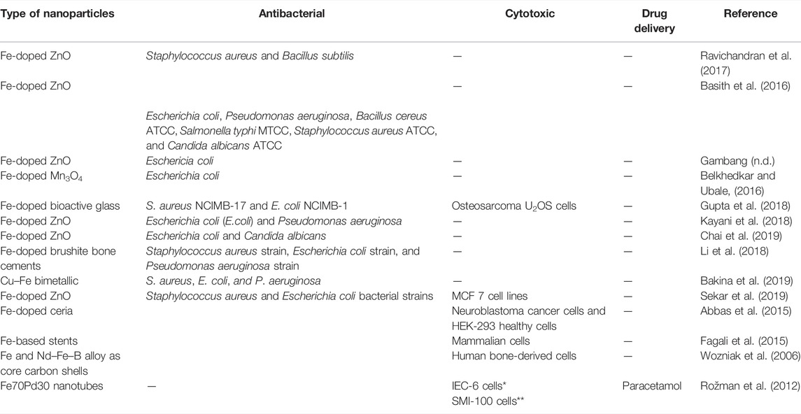
TABLE 3. Some antibacterial, anticancer, and drug delivery applications of nanoparticles.
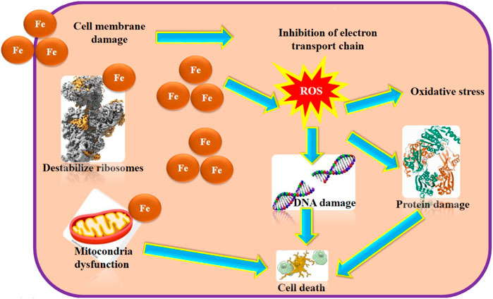
FIGURE 3. Schematic mechanism for the antibacterial activity by Fe nanoparticles.
4.4 Drug Delivery
One of the most interactive applications of nanotechnology which is related to cancer treatment is the delivery of pharmaceutical structures to suitable targets (Li et al., 2014). Iron nanoparticles are the major materials which could be employed in drug delivery (Rostami et al., 2021c; Rostami et al., 2021d; Hafezi et al., 2021). Iron nanoparticles have a strong affinity to generate ROS with the photo-Fenton process which helps eradicate tumor tissue. Also, they have been used in gene treatments such as tumor cells. Their distinctive properties such as multi-functionality, considerable volume to surface ration, and the possibility of surface tailoring of iron nanoparticles have led to extensive applications in nanotechnology (Katz and Willner, 2004; Kong et al., 2011).
Since Fe nanomaterials have considerable optical properties, they could be used in optical switches, bio-labels, and chemical sensors as well as display systems. Rozman K. Z et al. (Andrade et al., 2020) reported Fe–Pd-based tubular nanostructures. Tests on the magnetic properties of Fe70Pd30 nanotubes indicated these nanostructures to be ferromagnetic species with a magnetization saturation value of 170 emu g−1. This was suggested to make them proper candidates for drug delivery. Li et al. (2019) devised a novel magnetic carrier CNT (Fe)/HA nanocomposite [carbon nanotube (CNT) and hydroxyapatite (HA)] for the targeted delivery of doxorubicin (DOX), using carriers based on a through in situ synthesis of carbon nanotubes in the nanoscale hydroxyapatite powder using iron catalysts, followed by chemical modification using folic acid (FA) and chitosan (CS). The synthesis involved the in situ self-assembly of Fe, HA, and CNTs into a composite structure followed with acid treatment, which makes the CNTs shorter and homogeneously dispersed. Furthermore, the acid treatment opens the tips of the CNTs and grafts oxygen-containing groups onto them. After functional modification by coating the surface of the tubes with chitosan and folic acid, the composite can be loaded with DOX as a result of π–π stacking and electrostatic adsorption up to an average of 130 wt%. Using a phosphate-buffered saline (PBS) at pH = 5.5, FA-CS-CNT (Fe)/HA released a large quantity of DOX at an average rate of 52 wt% after 72 h, while this value reached only 8 wt% in PBS at pH = 7.4. The respective values of remanence/saturation magnetization, saturation magnetization, and coercive force for the composite were 0.44, 0.88 emu/g, and 668.96 Oe, which reflect the potentials of this composite for drug transport and hence delivery in a strong external magnetic field. Figure 4 shows a summary of the drug delivery caused by Fe nanoparticles schematically.
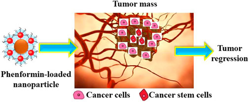
FIGURE 4. Schematic mechanism for the drug delivery by Fe NPs.
5 Conclusion and Perspectives
Nanobiotechnology is a combination of nanotechnology and biology, which produces eco-friendly products. Today, this field has evolved in the biomedical domain. Fe NPs are a basic element in the body and are an essential nutrient, which play a vital role in catalytic enzymatic reactions inside the cell. Fe NPs are a good strategy for cancer treatment and delivery of pharmaceutical structures to suitable targets. There are various green methods for preparation of Fe NPs using herbal extracts as capping, reducing, and stabilizing agents. The preparation methods can greatly impact the physicochemical and biological characters, and hence, the applications of the products. Single and bimetallic nanoparticles of iron have been used in drug delivery, topic formulations, dressings, and coating of textiles. One of the main reasons for applications of these NPs is due to their antimicrobial ability, which allows for their application in various products ranging from disinfecting agents for different surfaces and medical devices to wound dressings, textiles, and different coatings.
To improve the biomedical applications, researchers are evaluating various approaches for minimizing their toxicity while enhancing their diagnostic and even therapeutic efficiencies. Evaluating the exposure conditions to Fe nanoparticles is not enough. The toxic effects can be due to particle dissolution, agglomeration, and precipitation. These highly depend on size of particles and agglomerates, surface properties, and even exposure mechanisms. Consequently, toxicity assessments should provide a detailed description of exposure conditions as an essential background for the validation of the comparisons. Furthermore, there are not enough toxicity data on low exposure levels, long-term effects, and chronic stress. It is hence critical to concentrate on the long-term effects of chronic exposure to Fe nanoparticles even at low concentrations, which is much closer to real life conditions. Lastly, dissolved Fe2+ has a key role in toxicity, and there is need for techniques to distinguish the toxicity effects induced by solid Fe nanoparticles and dissolved Fe2+.
Author Contributions
AS and AA drafted the green method for the preparation of Fe nanostructures. HB and MR-N drafted the evaluation of drug delivery, anticancer potential, and antibacterial potential of Fe nanomaterials. MR-N and MG proposed the research and critically edited the manuscript.
Conflict of Interest
The authors declare that the research was conducted in the absence of any commercial or financial relationships that could be construed as a potential conflict of interest.
Publisher’s Note
All claims expressed in this article are solely those of the authors and do not necessarily represent those of their affiliated organizations, or those of the publisher, the editors, and the reviewers. Any product that may be evaluated in this article, or claim that may be made by its manufacturer, is not guaranteed or endorsed by the publisher.
References
Abamor, E. S., Allahverdiyev, A. M., Bagirova, M., and Rafailovich, M. (2017). Meglumine Antımoniate-TiO2@Ag Nanoparticle Combinations Reduce Toxicity of the Drug while Enhancing its Antileishmanial Effect. Acta trop. 169, 30–42. doi:10.1016/j.actatropica.2017.01.005
Abbas, F., Jan, T., Iqbal, J., and Naqvi, M. S. H. (2015). Fe Doping Induced Enhancement in Room Temperature Ferromagnetism and Selective Cytotoxicity of CeO2 Nanoparticles. Curr. Appl. Phys. 15, 1428–1434. doi:10.1016/j.cap.2015.08.007
Abdullaeva, Z., Omurzak, E., Iwamoto, C., Ganapathy, H. S., Sulaimankulova, S., Liliang, C., et al. (2012). Onion-like Carbon-Encapsulated Co, Ni, and Fe Magnetic Nanoparticles with Low Cytotoxicity Synthesized by a Pulsed Plasma in a Liquid. Carbon 50, 1776–1785. doi:10.1016/j.carbon.2011.12.025
Abdullah, J. A. A., Eddine, L. S., Abderrhmane, B., Alonso-González, M., Guerrero, A., and Romero, A. (2020). Green Synthesis and Characterization of Iron Oxide Nanoparticles by Pheonix Dactylifera Leaf Extract and Evaluation of Their Antioxidant Activity. Sustain. Chem. Pharm. 17, 100280. doi:10.1016/j.scp.2020.100280
Akbari, M., Jafari, H., Rostami, M., Mahdavinia, G. R., nasab, A. S., Tsurkan, D., et al. (2021). Adsorption of Cationic Dyes on a Magnetic 3D Spongin Scaffold with Nano-Sized Fe3O4 Cores. Mar. Drugs 19, 512. doi:10.3390/md19090512
Allen, D. T., and Shonnard, D. R. (2001). Green Engineering: Environmentally Conscious Design of Chemical Processes and Products. AIChE J. 47, 1906–1910. doi:10.1002/aic.690470902
Amani, J., Maleki, M., Khoshroo, A., Sobhani-Nasab, A., and Rahimi-Nasrabadi, M. (2018). An Electrochemical Immunosensor Based on Poly P-Phenylenediamine and Graphene Nanocomposite for Detection of Neuron-specific Enolase via Electrochemically Amplified Detection. Anal. Biochem. 548, 53–59. doi:10.1016/j.ab.2018.02.024
Anastas, P. T., and Warner, J. C. (1998). Principles of Green Chemistry. Green Chem. Theory Pract., 29–56.
Andrade, R. G. D., Veloso, S. R. S., and Castanheira, E. M. S. (2020). Shape Anisotropic Iron Oxide-Based Magnetic Nanoparticles: Synthesis and Biomedical Applications. Ijms 21, 2455. doi:10.3390/ijms21072455
Bakina, O. V., Glazkova, E. A., Svarovskaya, N. V., Rodkevich, N. G., and Lerner, M. I. (2019). «Janus»-like Cu-Fe Bimetallic Nanoparticles with High Antibacterial Activity. Mater. Lett. 242, 187–190. doi:10.1016/j.matlet.2019.01.105
Bashir, O., Hussain, S., Khan, Z., and Al-Thabaiti, S. A. (2014). Encapsulation of Silver Nanocomposites and Effects of Stabilizers. Carbohydr. Polym. 107, 167–173. doi:10.1016/j.carbpol.2014.02.055
Basith, N. M., Vijaya, J. J., Kennedy, L. J., Bououdina, M., Shenbhagaraman, R., and Jayavel, R. (2016). Influence of Fe-Doping on the Structural, Morphological, Optical, Magnetic and Antibacterial Effect of ZnO Nanostructures. J. Nanosci. Nanotechnol. 16, 1567–1577. doi:10.1166/jnn.2016.10756
Belkhedkar, M. R., and Ubale, A. U. (2016). Physical Properties of Fe Doped Mn 3 O 4 Thin Films Synthesized by SILAR Method and Their Antibacterial Performance against E. coli. J. Saudi Chem. Soc. 20, 553–560. doi:10.1016/j.jscs.2014.11.004
Berridge, M. J., Bootman, M. D., and Lipp, P. (1998). Calcium - a Life and Death Signal. Nature 395, 645–648. doi:10.1038/27094
Besbes, S., Blecker, C., Deroanne, C., Drira, N.-E., and Attia, H. (2004). Date Seeds: Chemical Composition and Characteristic Profiles of the Lipid Fraction. Food Chem. 84, 577–584. doi:10.1016/s0308-8146(03)00281-4
Biju, V., Itoh, T., and Ishikawa, M. (2010). Delivering Quantum Dots to Cells: Bioconjugated Quantum Dots for Targeted and Nonspecific Extracellular and Intracellular Imaging. Chem. Soc. Rev. 39, 3031–3056. doi:10.1039/b926512k
Boye, J., and Arcand, Y. (2012). Green Technologies in Food Production and Processing. Springer Science & Business Media.
Bushra, R., Shahadat, M., Ahmad, A., Nabi, S. A., Umar, K., Oves, M., et al. (2014). Synthesis, Characterization, Antimicrobial Activity and Applications of polyanilineTi(IV)arsenophosphate Adsorbent for the Analysis of Organic and Inorganic Pollutants. J. Hazard. Mater. 264, 481–489. doi:10.1016/j.jhazmat.2013.09.044
Byrne, S. J., le Bon, B., Corr, S. A., Stefanko, M., O'Connor, C., Gun'ko, Y. K., et al. (2007). Synthesis, Characterisation, and Biological Studies of CdTe Quantum Dot-Naproxen Conjugates. ChemMedChem 2, 183–186. doi:10.1002/cmdc.200600232
Chai, H.-Y., Lam, S.-M., and Sin, J.-C. (2019). Green Synthesis of Magnetic Fe-Doped ZnO Nanoparticles via Hibiscus Rosa-Sinensis Leaf Extracts for Boosted Photocatalytic, Antibacterial and Antifungal Activities. Mater. Lett. 242, 103–106. doi:10.1016/j.matlet.2019.01.116
Chen, D., Sharma, S. K., and Mudhoo, A. (2011). Handbook on Applications of Ultrasound: Sonochemistry for Sustainability. CRC Press.
Cuillel, M., Chevallet, M., Charbonnier, P., Fauquant, C., Pignot-Paintrand, I., Arnaud, J., et al. (2014). Interference of CuO Nanoparticles with Metal Homeostasis in Hepatocytes under Sub-toxic Conditions. Nanoscale 6, 1707–1715. doi:10.1039/c3nr05041f
De Berardis, B., Civitelli, G., Condello, M., Lista, P., Pozzi, R., Arancia, G., et al. (2010). Exposure to ZnO Nanoparticles Induces Oxidative Stress and Cytotoxicity in Human Colon Carcinoma Cells. Toxicol. Appl. Pharmacol. 246, 116–127. doi:10.1016/j.taap.2010.04.012
de la Guardia, M., and Garrigues, S. (2012). Handbook of Green Analytical Chemistry. Wiley Online Library.
Debalina, S., and Pike, R. (2017). Chemicals from Biomass: Integrating Bioprocesses into Chemical Production Complexes for Sustainable Development, Chemicals from Biomass: Integrating Bioprocesses into Chemical Production Complexes for Sustainable Development.
Dhillon, G. S., Brar, S. K., Kaur, S., and Verma, M. (2012). Green Approach for Nanoparticle Biosynthesis by Fungi: Current Trends and Applications. Crit. Rev. Biotechnol. 32, 49–73. doi:10.3109/07388551.2010.550568
Dolmetsch, R. E., Xu, K., and Lewis, R. S. (1998). Calcium Oscillations Increase the Efficiency and Specificity of Gene Expression. Nature 392, 933–936. doi:10.1038/31960
Du, M., Zhan, G., Yang, X., Wang, H., Lin, W., Zhou, Y., et al. (2011). Ionic Liquid-Enhanced Immobilization of Biosynthesized Au Nanoparticles on TS-1 toward Efficient Catalysts for Propylene Epoxidation. J. Catal. 283, 192–201. doi:10.1016/j.jcat.2011.08.011
Dudev, T., and Lim, C. (2008). Metal Binding Affinity and Selectivity in Metalloproteins: Insights from Computational Studies. Annu. Rev. Biophys. 37, 97–116. doi:10.1146/annurev.biophys.37.032807.125811
Eghbali-Arani, M., Sobhani-Nasab, A., Rahimi-Nasrabadi, M., Ahmadi, F., and Pourmasoud, S. (2018). Ultrasound-assisted Synthesis of YbVO4 Nanostructure and YbVO4/CuWO4 Nanocomposites for Enhanced Photocatalytic Degradation of Organic Dyes under Visible Light. Ultrason. sonochemistry 43, 120–135. doi:10.1016/j.ultsonch.2017.11.040
Eghbali-Arani, M., Sobhani-Nasab, A., Rahimi-Nasrabadi, M., and Pourmasoud, S. (2018). Green Synthesis and Characterization of SmVO 4 Nanoparticles in the Presence of Carbohydrates as Capping Agents with Investigation of Visible-Light Photocatalytic Properties. J. Electron. Mater. 47 (7), 3757–3769. doi:10.1007/s11664-018-6236-3
Fagali, N. S., Grillo, C. A., Puntarulo, S., and de Mele, M. A. F. L. (2015). Cytotoxicity of Corrosion Products of Degradable Fe-Based Stents: Relevance of pH and Insoluble Products. Colloids Surfaces B Biointerfaces 128, 480–488. doi:10.1016/j.colsurfb.2015.02.047
Gabbay, J., Borkow, G., Mishal, J., Magen, E., Zatcoff, R., and Shemer-Avni, Y. (2006). Copper Oxide Impregnated Textiles with Potent Biocidal Activities. J. Industrial Text. 35, 323–335. doi:10.1177/1528083706060785
Gan, P. P., and Li, S. F. Y. (2012). Potential of Plant as a Biological Factory to Synthesize Gold and Silver Nanoparticles and Their Applications. Rev. Environ. Sci. Biotechnol. 11, 169–206. doi:10.1007/s11157-012-9278-7
Gandomi, F., Peymani-Motlagh, S. M., Rostami, M., Sobhani-Nasab, A., Fasihi-Ramandi, M., Eghbali-Arani, M., et al. (2019). Simple Synthesis and Characterization of Li0.5Fe2.5O4, LiMg0.5Fe2O4 and LiNi0.5Fe2O4, and Investigation of Their Photocatalytic and Anticancer Properties on Hela Cells Line. J. Mater Sci. Mater Electron 30, 19691–19702. doi:10.1007/s10854-019-02320-x
Goya, G. F., Marcos-Campos, I., Fernández-Pacheco, R., Sáez, B., Godino, J., Asín, L., et al. (2008). Dendritic Cell Uptake of Iron-Based Magnetic Nanoparticles. Cell Biol. Int. 32, 1001–1005. doi:10.1016/j.cellbi.2008.04.001
Gunawan, C., Teoh, W. Y., Marquis, C. P., and Amal, R. (2011). Cytotoxic Origin of Copper(II) Oxide Nanoparticles: Comparative Studies with Micron-Sized Particles, Leachate, and Metal Salts. ACS Nano 5, 7214–7225. doi:10.1021/nn2020248
Gupta, N., Santhiya, D., Murugavel, S., Kumar, A., Aditya, A., Ganguli, M., et al. (2018). Effects of Transition Metal Ion Dopants (Ag, Cu and Fe) on the Structural, Mechanical and Antibacterial Properties of Bioactive Glass. Colloids Surfaces A Physicochem. Eng. Aspects 538, 393–403. doi:10.1016/j.colsurfa.2017.11.023
Hafezi, M., Rostami, M., Hosseini, A., Rahimi-Nasrabadi, M., Fasihi-Ramandi, M., Badiei, A., et al. (2021). Cur-loaded ZnFe2O4@mZnO@N-GQDs Biocompatible Nano-Carriers for Smart and Controlled Targeted Drug Delivery with pH-Triggered and Ultrasound Irradiation. J. Mol. Liq. 322, 114875. doi:10.1016/j.molliq.2020.114875
Hartwig, A. (2013). Metal Interaction with Redox Regulation: An Integrating Concept in Metal Carcinogenesis? Free Radic. Biol. Med. 55, 63–72. doi:10.1016/j.freeradbiomed.2012.11.009
He, F., and Zhao, D. (2005). Preparation and Characterization of a New Class of Starch-Stabilized Bimetallic Nanoparticles for Degradation of Chlorinated Hydrocarbons in Water. Environ. Sci. Technol. 39, 3314–3320. doi:10.1021/es048743y
He, X., Lin, G. X., Chen, M. G., Zhang, J. X., and Ma, Q. (2007). Protection against Chromium (VI)-Induced Oxidative Stress and Apoptosis by Nrf2. Recruiting Nrf2 into the Nucleus and Disrupting the Nuclear Nrf2/Keap1 Association. Toxicol. Sci. 98, 298–309. doi:10.1093/toxsci/kfm081
Hoag, G. E., Collins, J. B., Holcomb, J. L., Hoag, J. R., Nadagouda, M. N., and Varma, R. S. (2009). Degradation of Bromothymol Blue by 'greener' Nano-Scale Zero-Valent Iron Synthesized Using Tea Polyphenols. J. Mat. Chem. 19, 8671–8677. doi:10.1039/b909148c
Hoyal, C. R., Giron‐Calle, J., and Forman, H. J. (1998). The Alveolar Macrophage as a Model of Calcium Signaling in Oxidative Stress. J. Toxicol. Environ. Health, Part B 1, 117–134. doi:10.1080/10937409809524547
Huang, J., Zhan, G., Zheng, B., Sun, D., Lu, F., Lin, Y., et al. (2011). Biogenic Silver Nanoparticles by Cacumen Platycladi Extract: Synthesis, Formation Mechanism, and Antibacterial Activity. Ind. Eng. Chem. Res. 50, 9095–9106. doi:10.1021/ie200858y
Hussain, S., Akrema, , Rahisuddin, , and Khan, Z. (2014). Extracellular Biosynthesis of Silver Nanoparticles: Effects of Shape-Directing Cetyltrimethylammonium Bromide, pH, Sunlight and Additives. Bioprocess Biosyst. Eng. 37, 953–964. doi:10.1007/s00449-013-1067-3
Iravani, S. (2011). Green Synthesis of Metal Nanoparticles Using Plants. Green Chem. 13, 2638–2650. doi:10.1039/c1gc15386b
Kalaiarasi, R., Jayallakshmi, N., and Venkatachalam, P. (2010). Phytosynthesis of Nanoparticles and its Applications. Plant Cell Biotechnol. Mol. Biol. 11, 1–16.
Kan, C. (2012). A Novel Green Treatment for Textiles: Plasma Treatment as a Sustainable Technology (Sustainability). CRC Press.
Katz, E., and Willner, I. (2004). Integrated Nanoparticle-Biomolecule Hybrid Systems: Synthesis, Properties, and Applications. Angew. Chem. Int. Ed. 43, 6042–6108. doi:10.1002/anie.200400651
Kayani, Z. N., Abbas, E., Saddiqe, Z., Riaz, S., and Naseem, S. (2018). Photocatalytic, Antibacterial, Optical and Magnetic Properties of Fe-Doped ZnO Nano-Particles Prepared by Sol-Gel. Mater. Sci. Semicond. Process. 88, 109–119. doi:10.1016/j.mssp.2018.08.003
Khan, M. A., Ahmad, A., Umar, K., and Nabi, S. A. (2015). Synthesis, Characterization, and Biological Applications of Nanocomposites for the Removal of Heavy Metals and Dyes. Ind. Eng. Chem. Res. 54, 76–82. doi:10.1021/ie504148k
Khan, Z., and Al-Thabaiti, S. A. (2018). Green Synthesis of Zero-Valent Fe-Nanoparticles: Catalytic Degradation of Rhodamine B, Interactions with Bovine Serum Albumin and Their Enhanced Antimicrobial Activities. J. Photochem. Photobiol. B Biol. 180, 259–267. doi:10.1016/j.jphotobiol.2018.02.017
Khan, Z., Bashir, O., Hussain, J. I., Kumar, S., and Ahmad, R. (2012). Effects of Ionic Surfactants on the Morphology of Silver Nanoparticles Using Paan (Piper Betel) Leaf Petiole Extract. Colloids Surfaces B Biointerfaces 98, 85–90. doi:10.1016/j.colsurfb.2012.04.033
Kharisov, B. I., Dias, H. V. R., Kharissova, O. V., Jiménez-Pérez, V. M., Pérez, B. O., and Flores, B. M. (2012). Iron-containing Nanomaterials: Synthesis, Properties, and Environmental Applications. RSC Adv. 2, 9325–9358. doi:10.1039/c2ra20812a
Kharisov, B. I., Kharissova, O. V., and Ortiz-Mendez, U. (2012). Handbook of Less-Common Nanostructures. CRC Press.
Kharissova, O., and Kharisov, B. (2008). Synthetic Techniques and Applications of Activated Nanostructurized Metals: Highlights up to 2008. Nanotec 2, 103–119. doi:10.2174/187221008784534505
Khullar, P., Singh, V., Mahal, A., Dave, P. N., Thakur, S., Kaur, G., et al. (2012). Bovine Serum Albumin Bioconjugated Gold Nanoparticles: Synthesis, Hemolysis, and Cytotoxicity toward Cancer Cell Lines. J. Phys. Chem. C 116, 8834–8843. doi:10.1021/jp300585d
Kong, R., Chen, Z., Ye, M., Zhang, X., and Tan, W. (2011). Cell-SELEX-based Aptamer-Conjugated Nanomaterials for Enhanced Targeting of Cancer Cells. Sci. China Chem. 54, 1218–1226. doi:10.1007/s11426-011-4336-5
Li, G., Zhang, N., Zhao, S., Zhang, K., Li, X., Jing, A., et al. (2018). Fe-doped Brushite Bone Cements with Antibacterial Property. Mater. Lett. 215, 27–30. doi:10.1016/j.matlet.2017.12.054
Li, H., Sun, X., Li, Y., Li, B., Liang, C., and Wang, H. (2019). Preparation and Properties of Carbon Nanotube (Fe)/hydroxyapatite Composite as Magnetic Targeted Drug Delivery Carrier. Mater. Sci. Eng. C 97, 222–229. doi:10.1016/j.msec.2018.11.042
Li, J., Shi, L., Shao, Y., Selke, M., Chen, B., Jiang, H., et al. (2014). The Cellular Labeling and pH-Sensitive Responsive-Drug Release of Celastrol in Cancer Cells Based on Cys-CdTe QDs. Sci. China Chem. 57, 833–841. doi:10.1007/s11426-014-5092-0
Lin, J., Zhou, W., Kumbhar, A., Wiemann, J., Fang, J., Carpenter, E. E., et al. (2001). Gold-Coated Iron (Fe@Au) Nanoparticles: Synthesis, Characterization, and Magnetic Field-Induced Self-Assembly. J. Solid State Chem. 159, 26–31. doi:10.1006/jssc.2001.9117
Lofrano, G. (2012). Green Technologies for Wastewater Treatment: Energy Recovery and Emerging Compounds Removal. Springer Science & Business Media.
Luo, F., Yang, D., Chen, Z., Megharaj, M., and Naidu, R. (2016). One-step Green Synthesis of Bimetallic Fe/Pd Nanoparticles Used to Degrade Orange II. J. Hazard. Mater. 303, 145–153. doi:10.1016/j.jhazmat.2015.10.034
Mathers, R. T., and Meier, M. A. (2011). Green Polymerization Methods: Renewable Starting Materials, Catalysis and Waste Reduction. John Wiley & Sons.
Michalet, X., Pinaud, F. F., Bentolila, L. A., Tsay, J. M., Doose, S., Li, J. J., et al. (2005). Quantum Dots for Live Cells, In Vivo Imaging, and Diagnostics. science 307, 538–544. doi:10.1126/science.1104274
Miller, I. S., Lynch, I., Dowling, D., Dawson, K. A., and Gallagher, W. M. (2010). Surface-induced Cell Signaling Events Control Actin Rearrangements and Motility. J. Biomed. Mater Res. A 93, 493–504. doi:10.1002/jbm.a.32530
Moradian, F., Ghorbani, R., and Biparva, P. (2018). Assessment of Different Antibacterial Effects of Fe and Cu Nanoparticles on Xanthomonas Campestris Growth and Expression of its Pathogenic Gene hrpE. J. Agric. Sci. Technol. 20, 1059–1070.
Murphy, C. J., Sau, T. K., Gole, A. M., Orendorff, C. J., Gao, J., Gou, L., et al. (2005). Anisotropic Metal Nanoparticles: Synthesis, Assembly, and Optical Applications. ACS Publications.
Murray, I. A., Nichols, R. G., Zhang, L., Patterson, A. D., and Perdew, G. H. (2016). Expression of the Aryl Hydrocarbon Receptor Contributes to the Establishment of Intestinal Microbial Community Structure in Mice. Sci. Rep. 6, 33969. doi:10.1038/srep33969
Nadagouda, M. N., Castle, A. B., Murdock, R. C., Hussain, S. M., and Varma, R. S. (2010). In Vitro biocompatibility of Nanoscale Zerovalent Iron Particles (NZVI) Synthesized Using Tea Polyphenols. Green Chem. 12, 114–122. doi:10.1039/b921203p
Nadagouda, M. N., and Varma, R. S. (2007). A Greener Synthesis of Core (Fe, Cu)-Shell (Au, Pt, Pd, and Ag) Nanocrystals Using Aqueous Vitamin C. Cryst. Growth Des. 7, 2582–2587. doi:10.1021/cg070554e
Naderi, H. R., Sobhani-Nasab, A., Rahimi-Nasrabadi, M., and Ganjali, M. R. (2017). Decoration of Nitrogen-Doped Reduced Graphene Oxide with Cobalt Tungstate Nanoparticles for Use in High-Performance Supercapacitors. Appl. Surf. Sci. 423, 1025–1034. doi:10.1016/j.apsusc.2017.06.239
Naderi, S., Zare, H., Taghavinia, N., Irajizad, A., Aghaei, M., and Panjehpour, M. (2018). Cadmium Telluride Quantum Dots Induce Apoptosis in Human Breast Cancer Cell Lines. Toxicol. Ind. Health 34, 339–352. doi:10.1177/0748233718763517
Narayanan, K. B., and Sakthivel, N. (2010). Biological Synthesis of Metal Nanoparticles by Microbes. Adv. colloid interface Sci. 156, 1–13. doi:10.1016/j.cis.2010.02.001
Nayak, V., Singh, K. R., Verma, R., Pandey, M. D., Singh, J., and Singh, R. P. (2022). Recent Advancements of Biogenic Iron Nanoparticles in Cancer Theranostics. Mater. Lett. 313, 131769. doi:10.1016/j.matlet.2022.131769
Nejati, M., Rostami, M., Mirzaei, H., Rahimi-Nasrabadi, M., Vosoughifar, M., Nasab, A. S., et al. (2022). Green Methods for the Preparation of MgO Nanomaterials and Their Drug Delivery, Anti-cancer and Anti-bacterial Potentials: A Review. Inorg. Chem. Commun. 136, 109107. doi:10.1016/j.inoche.2021.109107
Nel, A., Xia, T., Mädler, L., and Li, N. (2006). Toxic Potential of Materials at the Nanolevel. science 311, 622–627. doi:10.1126/science.1114397
Nie, S., Xing, Y., Kim, G. J., and Simons, J. W. (2007). Nanotechnology Applications in Cancer. Annu. Rev. Biomed. Eng. 9, 257–288. doi:10.1146/annurev.bioeng.9.060906.152025
Njagi, E. C., Huang, H., Stafford, L., Genuino, H., Galindo, H. M., Collins, J. B., et al. (2010). Biosynthesis of Iron and Silver Nanoparticles at Room Temperature Using Aqueous Sorghum Bran Extracts. Langmuir 27, 264–271. doi:10.1021/la103190n
Nohl, H., and Gille, L. (2005). Lysosomal ROS Formation. Redox Rep. 10, 199–205. doi:10.1179/135100005x70170
Nosonovsky, M., and Bhushan, B. (2012). Green Tribology: Biomimetics, Energy Conservation and Sustainability. Springer.
Ouyang, Q., Kou, F., Tsang, P. E., Lian, J., Xian, J., Fang, J., et al. (2019). Green Synthesis of Fe-Based Material Using Tea Polyphenols and its Application as a Heterogeneous Fenton-like Catalyst for the Degradation of Lincomycin. J. Clean. Prod. 232, 1492–1498. doi:10.1016/j.jclepro.2019.06.043
Padash, R., Sobhani-Nasab, A., Rahimi-Nasrabadi, M., Mirmotahari, M., Ehrlich, H., Rad, A. S., et al. (2018). Is it Possible to Use X12Y12 (X = Al, B, and Y = N, P) Nanocages for Drug-Delivery Systems? A DFT Study on the Adsorption Property of 4-aminopyridine Drug. Appl. Phys. A 124, 582. doi:10.1007/s00339-018-1965-y
Parsons, J. G., Peralta-Videa, J. R., and Gardea-Torresdey, J. L. (2007). Chapter 21 Use of Plants in Biotechnology: Synthesis of Metal Nanoparticles by Inactivated Plant Tissues, Plant Extracts, and Living Plants. Dev. Environ. Sci. 5, 463–485. doi:10.1016/s1474-8177(07)05021-8
Peng, S., Wang, C., Xie, J., and Sun, S. (2006). Synthesis and Stabilization of Monodisperse Fe Nanoparticles. J. Am. Chem. Soc. 128, 10676–10677. doi:10.1021/ja063969h
Peymani-Motlagh, S. M., Moeinian, N., Rostami, M., Fasihi-Ramandi, M., Sobhani-Nasab, A., Rahimi-Nasrabadi, M., et al. (2019). Effect of Gd3+-, Pr3+- or Sm3+-Substituted Cobalt-Zinc Ferrite on Photodegradation of Methyl Orange and Cytotoxicity Tests. J. Rare Earths 37, 1288–1295. doi:10.1016/j.jre.2019.04.010
Peymani-Motlagh, S. M., Sobhani-Nasab, A., Rostami, M., Sobati, H., Eghbali-Arani, M., Fasihi-Ramandi, M., et al. (2019). Assessing the Magnetic, Cytotoxic and Photocatalytic Influence of Incorporating Yb3+ or Pr3+ Ions in Cobalt-Nickel Ferrite. J. Mater Sci. Mater Electron 30, 6902–6909. doi:10.1007/s10854-019-01005-9
Pinaud, F., Clarke, S., Sittner, A., and Dahan, M. (2010). Probing Cellular Events, One Quantum Dot at a Time. Nat. Methods 7, 275–285. doi:10.1038/nmeth.1444
Ponder, S. M., Darab, J. G., and Mallouk, T. E. (2000). Remediation of Cr(VI) and Pb(II) Aqueous Solutions Using Supported, Nanoscale Zero-Valent Iron. Environ. Sci. Technol. 34, 2564–2569. doi:10.1021/es9911420
Pourmasoud, S., Sobhani-Nasab, A., Behpour, M., Rahimi-Nasrabadi, M., and Ahmadi, F. (2018). Investigation of Optical Properties and the Photocatalytic Activity of Synthesized YbYO4 Nanoparticles and YbVO4/NiWO4 Nanocomposites by Polymeric Capping Agents. J. Mol. Struct. 1157, 607–615. doi:10.1016/j.molstruc.2017.12.077
Raddassi, K., Berthon, B., Petit, J.-F., and Lemaire, G. (1994). Role of Calcium in the Activation of Mouse Peritoneal Macrophages: Induction of NO Synthase by Calcium Ionophores and Thapsigargin. Cell. Immunol. 153, 443–455. doi:10.1006/cimm.1994.1041
Rahimi-Nasrabadi, M., Behpour, M., Sobhani-Nasab, A., and Hosseinpour-Mashkani, S. M. (2015). ZnFe2−xLaxO4 Nanostructure: Synthesis, Characterization, and its Magnetic Properties. J. Mater Sci. Mater Electron 26, 9776–9781. doi:10.1007/s10854-015-3648-1
Rahimi-Nasrabadi, M., Behpour, M., Sobhani-Nasab, A., and Jeddy, M. R. (2016). Nanocrystalline Ce-Doped Copper Ferrite: Synthesis, Characterization, and its Photocatalyst Application. J. Mater Sci. Mater Electron 27, 11691–11697. doi:10.1007/s10854-016-5305-8
Ravichandran, A. T., Karthick, R., Pushpa, K. C. S., Ravichandran, K., and Chandramohan, R. (2017). Uniform and Well-Dispersed ZnO:Fe Nanoparticles with High Photoluminescence and Antibacterial Properties Prepared by Soft Chemical Route. J. Inorg. Organomet. Polym. 27, 1084–1089. doi:10.1007/s10904-017-0558-0
Resch-Genger, U., Grabolle, M., Cavaliere-Jaricot, S., Nitschke, R., and Nann, T. (2008). Quantum Dots versus Organic Dyes as Fluorescent Labels. Nat. Methods 5, 763–775. doi:10.1038/nmeth.1248
Rosenthal, S. J., Chang, J. C., Kovtun, O., McBride, J. R., and Tomlinson, I. D. (2011). Biocompatible Quantum Dots for Biological Applications. Chem. Biol. 18, 10–24. doi:10.1016/j.chembiol.2010.11.013
Rostami, M., Badiei, A., Sorouri, A. M., Fasihi-Ramandi, M., Ganjali, M. R., Rahimi-Nasrabadi, M., et al. (2022). Cur-loaded Magnetic ZnFe2O4@L-Cysteine - Ox, N-Rich Mesoporous -gC3N4 Nanocarriers as a Targeted Sonodynamic Chemotherapeutic Agent for Enhanced Tumor Eradication. Surfaces Interfaces 30, 101900. doi:10.1016/j.surfin.2022.101900
Rostami, M. (2017). Construction of La-Doped TiO2@La-Doped ZnO-B-Doped Reduced Graphene Oxide Ternary Nanocomposites for Improved Visible Light Photocatalytic Activity. RSC Adv. 7, 43424–43431. doi:10.1039/c7ra06767d
Rostami, M., Nasab, A. S., Fasihi-Ramandi, M., Badiei, A., Ganjali, M. R., Rahimi-Nasrabadi, M., et al. (2021). Cur-loaded Magnetic ZnFe2O4@mZnO-Ox-P-G-C3n4 Composites as Dual pH- and Ultrasound Responsive Nano-Carriers for Controlled and Targeted Cancer Chemotherapy. Mater. Chem. Phys. 271, 124863. doi:10.1016/j.matchemphys.2021.124863
Rostami, M., Nasab, A. S., Fasihi-Ramandi, M., Badiei, A., Rahimi-Nasrabadi, M., and Ahmadi, F. (2021). The ZnFe2O4@mZnO-N/RGO Nano-Composite as a Carrier and an Intelligent Releaser Drug with Dual pH- and Ultrasound-Triggered Control. New J. Chem. 45, 4280–4291. doi:10.1039/d0nj04758a
Rostami, M., Nayebossadr, S., Mozaffari, S., Sobhani-Nasab, A., Rahimi-Nasrabadi, M., Fasihi-Ramandi, M., et al. (2021). Heterojunction of N/B/RGO and G-C3n4 Anchored Magnetic ZnFe2O4@ZnO for Promoting UV/Vis-induced Photo-Catalysis and In Vitro Toxicity Studies. Environ. Sci. Pollut. Res. 28, 11430–11443. doi:10.1007/s11356-020-10572-y
Rostami, M. (2019). Photodecomposition and Adsorption of Hazardous Organic Pollutants by Ce-Doped ZnO@Ce-Doped TiO2-N/s-Dual Doped RGO Ternary Nano-Composites Photocatalyst for Water Remediation. J. Mol. Struct. 1185, 191–199. doi:10.1016/j.molstruc.2019.02.094
Rostami, M., Sharafi, P., Mozaffari, S., Adib, K., Sobhani-Nasab, A., Rahimi-Nasrabadi, M., et al. (2021). A Facile Preparation of ZnFe2O4-CuO-N/B/RGO and ZnFe2O4-CuO-C3n4 Ternary Heterojunction Nanophotocatalyst: Characterization, Biocompatibility, photo-Fenton-like Degradation of MO and Magnetic Properties. J. Mater Sci. Mater Electron 32, 5457–5472. doi:10.1007/s10854-021-05268-z
Rostami, M., Zamani, R. M., Aghajanzadeh, K. M., and Danafar, H. (2018). Sol-gel Synthesis and Characterization of Zinc Ferrite-Graphene Nano-Hybrids for Photo-Catalytic Degradation of the Paracetamol. J. Pharm. Investig. 48, 657–664. doi:10.1007/s40005-017-0362-4
Rožman, K. Ž., Pečko, D., Šturm, S., Maver, U., Nadrah, P., Bele, M., et al. (2012). Electrochemical Synthesis and Characterization of Fe70Pd30 Nanotubes for Drug-Delivery Applications. Mater. Chem. Phys. 133, 218–224.
Saleh, N., Phenrat, T., Sirk, K., Dufour, B., Ok, J., Sarbu, T., et al. (2005). Adsorbed Triblock Copolymers Deliver Reactive Iron Nanoparticles to the Oil/water Interface. Nano Lett. 5, 2489–2494. doi:10.1021/nl0518268
Sathya, K., Saravanathamizhan, R., and Baskar, G. (2018). Ultrasonic Assisted Green Synthesis of Fe and Fe/Zn Bimetallic Nanoparticles for Invitro Cytotoxicity Study against HeLa Cancer Cell Line. Mol. Biol. Rep. 45, 1397–1404. doi:10.1007/s11033-018-4302-9
Schiebl, M. (2012). Green Chemical Space Propulsion: Auto Ignition Conditions in a Micro Rocket Combustion Chamber for a Hydrogen Peroxide Based Micro Propulsion System. AV Akademikerverlag.
Sedighi, F., Esmaeili-Zare, M., Sobhani-Nasab, A., and Behpour, M. (2018). Synthesis and Characterization of CuWO4 Nanoparticle and CuWO4/NiO Nanocomposite Using Co-precipitation Method; Application in Photodegradation of Organic Dye in Water. J. Mater Sci. Mater Electron 29, 13737–13745. doi:10.1007/s10854-018-9504-3
Sekar, A. D., Kumar, V., Muthukumar, H., Gopinath, P., and Matheswaran, M. (2019). Electrospinning of Fe-Doped ZnO Nanoparticles Incorporated Polyvinyl Alcohol Nanofibers for its Antibacterial Treatment and Cytotoxic Studies. Eur. Polym. J. 118, 27–35. doi:10.1016/j.eurpolymj.2019.05.038
Shahwan, T., Sirriah, S. A., Nairat, M., Boyacı, E., Eroğlu, A. E., Scott, T. B., et al. (2011). Green Synthesis of Iron Nanoparticles and Their Application as a Fenton-like Catalyst for the Degradation of Aqueous Cationic and Anionic Dyes. Chem. Eng. J. 172, 258–266. doi:10.1016/j.cej.2011.05.103
Singh, K. R., Nayak, V., Singh, J., Singh, A. K., and Singh, R. P. (2021). Potentialities of Bioinspired Metal and Metal Oxide Nanoparticles in Biomedical Sciences. RSC Adv. 11, 24722–24746. doi:10.1039/d1ra04273d
Singh, N., Manshian, B., Jenkins, G. J. S., Griffiths, S. M., Williams, P. M., Maffeis, T. G. G., et al. (2009). NanoGenotoxicology: The DNA Damaging Potential of Engineered Nanomaterials. Biomaterials 30, 3891–3914. doi:10.1016/j.biomaterials.2009.04.009
Siyamak, S. (2012). Preparation and Characterisation of Green Composites Chemicals' Effects on Fundamental Properties of Poly (Butylene Adipate-Co-Terephtalate)/oil Palm EFB Fibre Biocomposites.
Smuleac, V., Varma, R., Sikdar, S., and Bhattacharyya, D. (2011). Green Synthesis of Fe and Fe/Pd Bimetallic Nanoparticles in Membranes for Reductive Degradation of Chlorinated Organics. J. Membr. Sci. 379, 131–137. doi:10.1016/j.memsci.2011.05.054
Sobhani-Nasab, A., Pourmasoud, S., Ahmadi, F., Wysokowski, M., Jesionowski, T., Ehrlich, H., et al. (2019). Synthesis and Characterization of MnWO4/TmVO4 Ternary Nano-Hybrids by an Ultrasonic Method for Enhanced Photocatalytic Activity in the Degradation of Organic Dyes. Mater. Lett. 238, 159–162. doi:10.1016/j.matlet.2018.11.175
Sobhani-Nasab, A., Ziarati, A., Rahimi-Nasrabadi, M., Ganjali, M. R., and Badiei, A. (2017). Five-component Domino Synthesis of Tetrahydropyridines Using Hexagonal PbCr X Fe12−x O19 as Efficient Magnetic Nanocatalyst. Res. Chem. Intermed. 43, 6155–6165. doi:10.1007/s11164-017-2982-8
Soenen, S. J. H., Himmelreich, U., Nuytten, N., Pisanic, T. R., Ferrari, A., and De Cuyper, M. (2010). Intracellular Nanoparticle Coating Stability Determines Nanoparticle Diagnostics Efficacy and Cell Functionality. Small 6, 2136–2145. doi:10.1002/smll.201000763
Sultana, S., Khan, M. Z., Umar, K., and Muneer, M. (2013). Electrical, Thermal, Photocatalytic and Antibacterial Studies of Metallic Oxide Nanocomposite Doped Polyaniline. J. Mater. Sci. Technol. 29, 795–800. doi:10.1016/j.jmst.2013.06.001
Sun, S., Murray, C. B., Weller, D., Folks, L., and Moser, A. (2000). Monodisperse FePt Nanoparticles and Ferromagnetic FePt Nanocrystal Superlattices. science 287, 1989–1992. doi:10.1126/science.287.5460.1989
Uddin, M. J., Crews, B. C., Blobaum, A. L., Kingsley, P. J., Gorden, D. L., McIntyre, J. O., et al. (2010). Selective Visualization of Cyclooxygenase-2 in Inflammation and Cancer by Targeted Fluorescent Imaging Agents. Cancer Res. 70, 3618–3627. doi:10.1158/0008-5472.can-09-2664
Varma, R. S. (2012). Greener Approach to Nanomaterials and Their Sustainable Applications. Curr. Opin. Chem. Eng. 1, 123–128. doi:10.1016/j.coche.2011.12.002
Vilchis-Nestor, A. R., Avalos-Borja, M., Gómez, S. A., Hernández, J. A., Olivas, A., and Zepeda, T. A. (2009). Alternative Bio-Reduction Synthesis Method for the Preparation of Au(AgAu)/SiO2-Al2O3 Catalysts: Oxidation and Hydrogenation of CO. Appl. Catal. B Environ. 90, 64–73. doi:10.1016/j.apcatb.2009.02.016
Wang, C.-B., and Zhang, W.-X. (1997). Synthesizing Nanoscale Iron Particles for Rapid and Complete Dechlorination of TCE and PCBs. Environ. Sci. Technol. 31, 2154–2156. doi:10.1021/es970039c
Wang, G., Zhang, L., and Zhang, J. (2012). A Review of Electrode Materials for Electrochemical Supercapacitors. Chem. Soc. Rev. 41, 797–828. doi:10.1039/c1cs15060j
Wang, M., Jiang, H., Liu, X., and Wang, X. (2022). Biophysics Involved in the Process of Tumor Immune Escape. iScience 25, 104124. doi:10.1016/j.isci.2022.104124
Wang, X., Le, L., Alvarez, P. J. J., Li, F., and Liu, K. (2015). Synthesis and Characterization of Green Agents Coated Pd/Fe Bimetallic Nanoparticles. J. Taiwan Inst. Chem. Eng. 50, 297–305. doi:10.1016/j.jtice.2014.12.030
Weng, X., Guo, M., Luo, F., and Chen, Z. (2017). One-step Green Synthesis of Bimetallic Fe/Ni Nanoparticles by eucalyptus Leaf Extract: Biomolecules Identification, Characterization and Catalytic Activity. Chem. Eng. J. 308, 904–911. doi:10.1016/j.cej.2016.09.134
Wozniak, M. J., Wozniak, P., Bystrzejewski, M., Cudzilo, S., Huczko, A., Jelen, P., et al. (2006). Magnetic Nanoparticles of Fe and Nd-Fe-B Alloy Encapsulated in Carbon Shells for Drug Delivery Systems: Study of the Structure and Interaction with the Living Cells. J. alloys Compd. 423, 87–91. doi:10.1016/j.jallcom.2005.12.028
Xia, T., Kovochich, M., Liong, M., Mädler, L., Gilbert, B., Shi, H., et al. (2008). Comparison of the Mechanism of Toxicity of Zinc Oxide and Cerium Oxide Nanoparticles Based on Dissolution and Oxidative Stress Properties. ACS Nano 2, 2121–2134. doi:10.1021/nn800511k
Xia, T., Kovochich, M., and Nel, A. E. (2007). Impairment of Mitochondrial Function by Particulate Matter (PM) and Their Toxic Components: Implications for PM-Induced Cardiovascular and Lung Disease. Front. Biosci. 12, 1238. doi:10.2741/2142
Xie, J., Lee, J. Y., Wang, D. I. C., and Ting, Y. P. (2007). Identification of Active Biomolecules in the High-Yield Synthesis of Single-Crystalline Gold Nanoplates in Algal Solutions. small 3, 672–682. doi:10.1002/smll.200600612
Xie, J., Lee, J. Y., Wang, D. I. C., and Ting, Y. P. (2007). Silver Nanoplates: From Biological to Biomimetic Synthesis. ACS Nano 1, 429–439. doi:10.1021/nn7000883
Yamakoshi, Y., Umezawa, N., Ryu, A., Arakane, K., Miyata, N., Goda, Y., et al. (2003). Active Oxygen Species Generated from Photoexcited Fullerene (C60) as Potential Medicines: O2- versus 1O2. J. Am. Chem. Soc. 125, 12803–12809. doi:10.1021/ja0355574
Yang, H., Liu, C., Yang, D., Zhang, H., and Xi, Z. (2009). Comparative Study of Cytotoxicity, Oxidative Stress and Genotoxicity Induced by Four Typical Nanomaterials: The Role of Particle Size, Shape and Composition. J. Appl. Toxicol. 29, 69–78. doi:10.1002/jat.1385
Zhan, G., Huang, J., Du, M., Sun, D., Abdul-Rauf, I., Lin, W., et al. (2012). Liquid Phase Oxidation of Benzyl Alcohol to Benzaldehyde with Novel Uncalcined Bioreduction Au Catalysts: High Activity and Durability. Chem. Eng. J. 187, 232–238. doi:10.1016/j.cej.2012.01.051
Zhang, X., Lin, S., Chen, Z., Megharaj, M., and Naidu, R. (2011). Kaolinite-supported Nanoscale Zero-Valent Iron for Removal of Pb2+ from Aqueous Solution: Reactivity, Characterization and Mechanism. Water Res. 45, 3481–3488. doi:10.1016/j.watres.2011.04.010
Zhou, Y., Lin, W., Huang, J., Wang, W., Gao, Y., Lin, L., et al. (2010). Biosynthesis of Gold Nanoparticles by Foliar Broths: Roles of Biocompounds and Other Attributes of the Extracts. Nanoscale Res. Lett. 5, 1351–1359. doi:10.1007/s11671-010-9652-8
Zhuo, S., Guan, Y., Li, H., Fang, J., Zhang, P., Du, J., et al. (2019). Facile Fabrication of Fluorescent Fe-Doped Carbon Quantum Dots for Dopamine Sensing and Bioimaging Application. Analyst 144, 656–662. doi:10.1039/c8an01741g
Keywords: green method, Fe nanoparticles, drug delivery, antibacterial, anticancer
Citation: Abedini A, Rostami M, Banafshe HR, Rahimi-Nasrabadi M, SobhaniNasab A and Ganjali MR (2022) Utility of Biogenic Iron and Its Bimetallic Nanocomposites for Biomedical Applications: A Review. Front. Chem. 10:893793. doi: 10.3389/fchem.2022.893793
Received: 10 March 2022; Accepted: 06 May 2022;
Published: 01 July 2022.
Edited by:
Luís D. Carlos, University of Aveiro, PortugalReviewed by:
Ravindra Pratap Singh, Indira Gandhi National Tribal University, IndiaKhalid Umar, Universiti Sains Malaysia (USM), Malaysia
Ramchander Merugu, Mahatma Gandhi University, Nalgonda, India
Copyright © 2022 Abedini, Rostami, Banafshe, Rahimi-Nasrabadi, SobhaniNasab and Ganjali. This is an open-access article distributed under the terms of the Creative Commons Attribution License (CC BY). The use, distribution or reproduction in other forums is permitted, provided the original author(s) and the copyright owner(s) are credited and that the original publication in this journal is cited, in accordance with accepted academic practice. No use, distribution or reproduction is permitted which does not comply with these terms.
*Correspondence: Ali SobhaniNasab, QWxpLnNvYmhhbmluYXNhYkBnbWFpbC5jb20=