- Institut de Neurociències, Departament de Bioquímica i Biologia Molecular, Centro de Investigación Biomédica en Red Enfermedades Neurodegenerativas (CIBERNED), Universitat Autònoma de Barcelona, Barcelona, Spain
Alzheimer’s disease (AD) is a neurodegenerative disorder characterized by abnormal accumulation of β-amyloid and tau and synapse dysfunction in memory-related neural circuits. Pathological and functional changes in the medial temporal lobe, a region essential for explicit memory encoding, contribute to cognitive decline in AD. Surprisingly, functional imaging studies show increased activity of the hippocampus and associated cortical regions during memory tasks in presymptomatic and early AD stages, whereas brain activity declines as the disease progresses. These findings suggest an emerging scenario where early pathogenic events might increase neuronal excitability leading to enhanced brain activity before clinical manifestations of the disease, a stage that is followed by decreased brain activity as neurodegeneration progresses. The mechanisms linking pathology with synaptic excitability and plasticity changes leading to memory loss in AD remain largely unclear. Recent studies suggest that increased brain activity parallels enhanced expression of genes involved in synaptic transmission and plasticity in preclinical stages, whereas expression of synaptic and activity-dependent genes are reduced by the onset of pathological and cognitive symptoms. Here, we review recent evidences indicating a relationship between transcriptional deregulation of synaptic genes and neuronal activity and memory loss in AD and mouse models. These findings provide the basis for potential clinical applications of memory-related transcriptional programs and their regulatory mechanisms as novel biomarkers and therapeutic targets to restore brain function in AD and other cognitive disorders.
Introduction
The rise of life expectancy has profoundly increased the aging population, and hence the prevalence of age-related cognitive disorders, including Alzheimer’s disease (AD). AD is a devastating neurological disorder characterized by early episodic memory deficits that progresses with cognitive impairments and neuropsychiatric symptoms and finally ends with general disabling dementia. The disease is preceded by a presymptomatic or preclinical stage that can last for years during which the clinical symptoms do not manifest but the pathological process starts (Sperling et al., 2014). In a subsequent prodromal stage named mild cognitive impairment (MCI), the disease is characterized by impairment of memory (amnesia) and other cognitive functions. The majority of subjects with MCI, which represent 10–20% of population older than 65 years, suffer from the disease in the following years (Petersen, 2011).
Memory decline is accompanied by pathological features in the brain of AD patients, including accumulation of extracellular amyloid plaques composed of β-amyloid (Aβ) peptides and intracellular neurofibrillary tangles (NFTs) formed by aggregated hyperphosphorylated microtubule-associated protein tau. These pathological lesions accumulate in brain regions essential for memory encoding and storage, such as the medial temporal lobe (MTL) and related cortical areas (Spires-Jones and Hyman, 2014). Tau pathology starts in the entorhinal cortex (EC) and then spreads to the hippocampal formation and limbic and association cortices (Braak and Braak, 1991). Progression of NFTs correlates well with cognitive decline and neuron loss, whereas amyloid plaques are abundant in neocortical regions but they do not correlate with the degree of memory loss (Arriagada et al., 1992; Gomez-Isla et al., 1997).
It is becoming clear that specific memory circuits are affected by changes in synaptic function and plasticity during the course of the disease. Indeed, synapse dysfunction and loss is an early pathological feature that correlates closely with cognitive impairment (Terry, 2000; Scheff et al., 2007). Recent functional imaging studies reveal decreased activity of the MTL in AD patients, whereas function of cortical and temporal lobe regions, particularly the hippocampus, are increased during memory tasks in preclinical and early stages of the disease (i.e., MCI; for review see Sperling et al., 2010). This enhancement of brain activity may represent a compensatory mechanism resulting from reduced neuronal connectivity that can maintain memory encoding at the beginning of the disease process. Notably, enhanced neuronal activity parallels increased expression of genes involved in synaptic transmission and plasticity at presymptomatic or very early AD stages, whereas deregulation of synaptic gene programs occurs at early and late pathological stages. Here, we summarize pathological as well as functional features occurring in the brain of human and AD mouse models during aging, and discuss recent evidences suggesting a relationship between gene expression changes and neuronal activity and memory disturbances during the progression of AD.
Hippocampal Pathology and Activity in AD
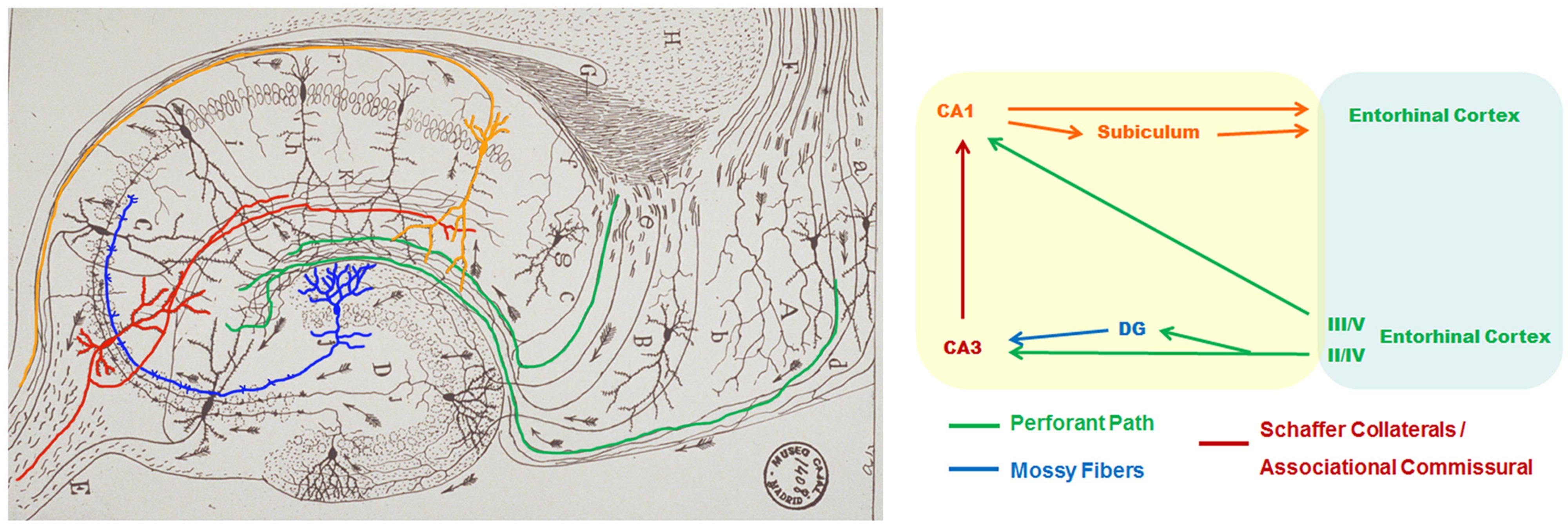
Declarative episodic memories of live facts and events depend on the MTL and connected cortical regions. The MTL includes the hippocampal formation (CA fields, dentate gyrus and subiculum), amygdala and adjacent cortical regions (entorhinal, perirhinal, and parahippocampal cortices; Squirre and Zola-Morgan, 1991). The EC receives cortical sensory information and projects excitatory inputs directly to CA1 pyramidal neurons or to the dentate gyrus and CA3 hippocampus through the perforant pathway (Van Hoesen and Pandya, 1975). CA3 neurons project Schaffer collaterals to CA1 pyramidal neurons, which finally project to the subiculum and deep EC layers IV, V and VI (EC-IV-VI; Figure 1). The MTL undergoes atrophy and hypometabolism not only in AD but also in MCI stages (Press et al., 1989; Mosconi et al., 2005; La Joie et al., 2013), an effect observed at least 4 years in advance to cognitive symptoms (Tondelli et al., 2012). Indeed, disruption of the hippocampus, a critical component of this memory circuit, is sufficient to produce anterograde amnesia (Zola-Morgan et al., 1986).

FIGURE 1. Hippocampal circuitry in the mouse brain. Modified original drawing of Santiago Ramón y Cajal’s (1911, left) and schematic diagram (right) of the rodent hippocampal circuitry. The picture shows the flux of excitatory projections (arrows) from entorhinal cortex (EC) neurons (green) directly to CA1 (orange) or CA3 (red) hippocampal pyramidal neurons or indirectly through projections to the dentate gyrus (DG, blue) through the perforant pathway. DG granule neurons project along the mossy fibers to CA3 pyramidal neurons. CA3 axons project through the Schaffer collaterals to CA1 pyramidal neurons, which finally project to the subiculum and deep EC IV- VI layers.
The EC is severely affected by pathological events and neurodegeneration early in AD, likely contributing to memory impairment. AD brains are characterized by a specific pattern of degenerating neurons in EC-II/IV layers and subiculum (Hyman et al., 1984). Cholinergic neurons are particularly vulnerable in AD, and therefore classical therapeutic treatments are based on acetylcholinesterase inhibition. One of the earliest pathological features linked to AD progression is accumulation of NFTs, which occurrs in at least 70% of brains of healthy individuals at sixties (Nelson et al., 2012). Tau pathology starts in the EC and continues to CA1/subiculum field and amygdala prior to clinical symptoms (Braak stages I–II; Hyman et al., 1986; Arriagada et al., 1992). As AD progresses, tau pathology propagates in a sequential regional fashion to limbic and association cortices (Braak stages III–VI) apparently through an aggregation spreading mechanism (Braak and Braak, 1991; Clavaguera et al., 2013). Indeed, NFTs and amyloid plaques are abundant in the terminal sites of the EC projections such as the dentate gyrus (Hyman et al., 1990). Collectively, progressive accumulation and spreading of pathological hallmarks in the MTL suggests that disruption of this neural circuit may contribute to memory decline during the progression of the disease.
Functional magnetic resonance imaging (fMRI) studies show decreased activity and connectivity of the hippocampus, and temporal and prefrontal cortices during episodic memory tasks in AD patients [(Press et al., 1989; Small et al., 1999; Sperling et al., 2003; Pariente et al., 2005; Bai et al., 2009), for review see (Dickerson and Sperling, 2009)]. By contrast, MCI subjects show abnormal activation of the hippocampus and EC during face-name, visual object and verbal associative memory tasks (Dickerson et al., 2005; Hamalainen et al., 2007; Kircher et al., 2007). Compared with healthy aged controls, asymptomatic subjects at risk for AD, including presenilin-1 (PSEN1) C410Y and E280A carriers, show higher activation of the hippocampus and frontal and temporal cortices during associative memory encoding years before clinical symptoms (Bassett et al., 2006; Mondadori et al., 2006; Yassa et al., 2008; Reiman et al., 2012). This increase of brain activity seems to reflect a compensatory mechanism to overcome neural dysfunction in preclinical stages, a process that may be necessary for appropriate memory encoding and retrieval (Kircher et al., 2007; O’Brien et al., 2010; Sperling et al., 2010). Taken together, these studies demonstrate increased activity of the MTL early during presymptomatic AD stages followed by decreased activity as the disease progresses.
Cortical Default Network in AD
Memory encoding and retrieval are affected by interconnected neocortical regions known as the default mode network, which is active at wakeful rest and deactivates during memory encoding (Buckner et al., 2008). The default mode network is connected to the MTL and includes the medial prefrontal cortex, the posteromedial cingulate cortex, the adjacent ventral precuneus, and the medial, lateral and inferior parietal cortices (Kobayashi and Amaral, 2007).
Specific regions of the cortical default network are functionally disrupted in AD and subjects at risk for AD (i.e., MCI; for a review, see Sperling et al., 2010). Hyperactivation of parietal and prefrontal cortices during memory encoding is accompanied by hippocampal hypoactivation in AD patients (Grady et al., 2003; Pariente et al., 2005). Reduced default network connectivity at rest, but increased activity during performance of attentional and associative memory tasks is observed in MCI and mild AD stages (Koch et al., 2014). Similar to AD patients, older cognitively normal subjects with brain amyloid deposition show significant reduced connectivity of the default network to the hippocampus in resting state (Hedden et al., 2009; Sheline et al., 2010). More striking, asymptomatic subjects with AD-linked autosomal dominant PSEN1 mutations show decreased activity of the precuneus/posterior cingulate and parietal cortex in resting state (Chhatwal et al., 2013), but increased activity of the frontal, parietal and prefrontal cortex during memory encoding (Wishart et al., 2006; Reiman et al., 2012). These results indicate disruption of the default mode network years before cognitive or behavioral symptoms, which suggests that early AD-associated pathology exerts deleterious functional effects on distinct memory circuits prior to memory impairment.
Effect of Aβ on Hippocampal Activity and Memory in AD Mouse Models
Mice expressing human β-amyloid precursor protein (APP), TAU and/or PSEN1 genes harboring familial AD-linked mutations develop AD pathological hallmarks, neuroinflamation and memory impairments (McGowan et al., 2006). APP and APP/PS1 transgenic mice develop age-dependent amyloid deposits and memory impairments in the absence of tau inclusions (Hsiao et al., 1996; Chapman et al., 1999; Koistinaho et al., 2001). APP transgenic mice show spatial and contextual memory impairments tightly associated with changes in long-term potentiation (LTP), a form of synaptic plasticity thought to be the cellular basis of learning and memory. Tg2576 (Swedish: APP KM670/671NL), APPSw,Ind (J20) and APP V717I transgenic mice that develop amyloid plaques display impaired hippocampal synaptic plasticity and memory deficits (Chapman et al., 1999; Dewachter et al., 2002; Saura et al., 2005). In APP transgenic mice, including PDAPP (Indiana: APP V717F), Tg2576, APPLd2 (London: V642I), APP23 (Swedish), APPSw,Ind (J20), APP/PS1 and 3xTg-AD (APP Swedish, Tau P301L, PS1 M146V), altered hippocampal synaptic plasticity and memory deficits precede amyloid plaque pathology suggesting that disruption of memory neural circuits is independent of plaque deposition (Dodart et al., 1999; Hsia et al., 1999; Koistinaho et al., 2001; Kelly et al., 2003; Van Dam et al., 2003; Dominguez-del-Toro et al., 2004; Billings et al., 2005; Saura et al., 2005; Jacobsen et al., 2006; Gruart et al., 2008). Notably, hippocampal-dependent synaptic plasticity and memory deficits in 3xTg-AD, APPSw,Ind and ArcAβ transgenic mice are associated with the presence of intraneuronal Aβ accumulation, which precedes amyloid plaques (Billings et al., 2005; España et al., 2010a).
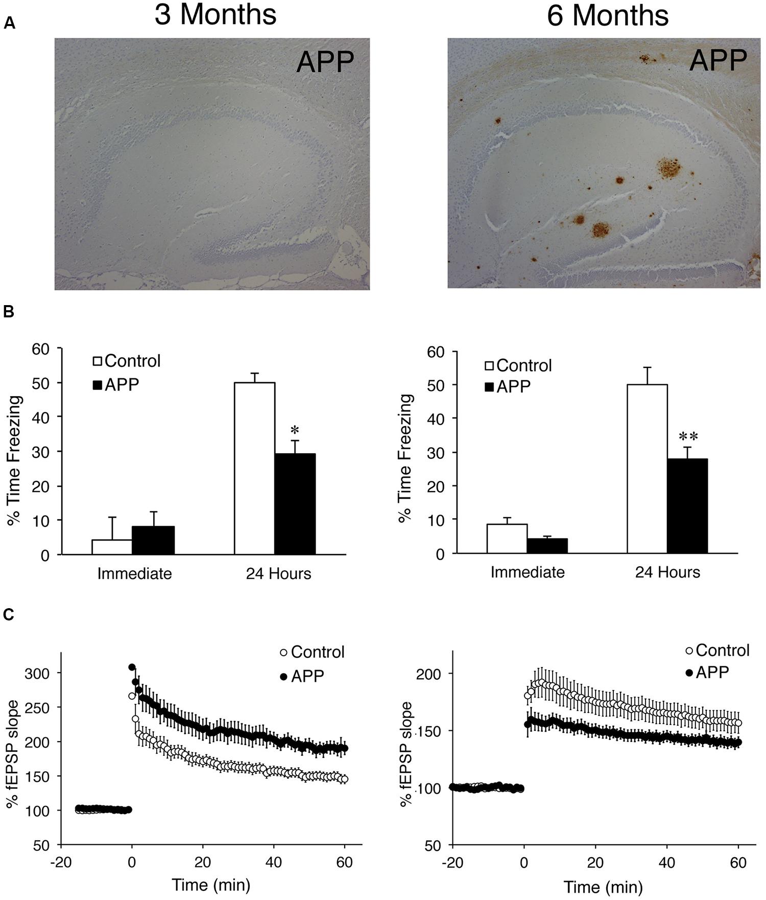
It has been recently postulated that synaptic excitability changes may alter memory networks leading to cognitive disturbances in AD (Santos et al., 2010, for review). APPSw,Ind (J20) mice show prior to amyloid plaque deposition enhanced synaptic plasticity in the Schaffer collateral pathway coinciding with early hippocampal-dependent memory deficits (Saura et al., 2005). During aging, APPSw,Ind mice develop associative memory deficits accompanied by amyloid plaque accumulation and LTP impairments in the hippocampus (Figure 2). Similarly, young free-plaque TgCRND8 and 3xTg-AD mice show increased synaptic plasticity caused by enhancement of synaptic excitability in the hippocampus, a phenotype associated with episodic memory impairments (Jolas et al., 2002; Davis et al., 2014). Several studies have also shown increased neuronal hyperactivity and excitability in the cortex of young APP transgenic mice before or when the first amyloid plaques appear (Palop et al., 2007; Busche et al., 2008; Minkeviciene et al., 2009; Gurevicius et al., 2013). This increased excitability is likely responsible for spontaneous epileptic seizures and premature death of APP mice (Palop et al., 2007; Minkeviciene et al., 2009). Enhancement of neuronal activity associated with early pathological and memory changes in AD mouse models resembles the clinical symptoms of MCI subjects (see above).

FIGURE 2. Age-dependent pathological, synaptic plasticity and associative memory changes in APP transgenic mice. (A) Brain sections of 3–6 months-old APPSw,Ind (J20) transgenic mice (APP) stained with an anti-Aβ antibody revealing the presence of amyloid plaques in the hippocampus at 6 months. (B) Contextual associative memory in one-shock contextual fear conditioning task. Three and six month-old APP mice exhibit significantly reduced levels of freezing at 24 h indicating disruption of long-term associative memory. Data represent the mean ± SEM. ∗P < 0.05, ∗∗P < 0.0001. (C), Time course of LTP induction at the CA1 Schaffer collaterals after theta burst stimulation (TBS) in 3- and 6-months old non-transgenic (control) and APP mice (n = 5–7). Notice the differential LTP in APP mice at 3 months (up) and 6 months (down) compared to the respective non-transgenic (control) mice. fEPSP, field excitatory postsynaptic potentials. Images are adapted from Saura et al. (2005).
The mechanism by which Aβ induces neuronal hyperexcitability is mediated by an increase of spontaneous action potential firing likely due to an impairment of inhibitory interneuron activity and/or increase of presynaptic vesicle release (Palop et al., 2007; Minkeviciene et al., 2009; Fogel et al., 2014). In this regard, hyperactivity of CA1 hippocampal neurons caused by loss of somatostatin inhibitory interneurons results in memory disturbances in APP transgenic mice (Perez-Cruz et al., 2011). By contrast, increasing the inhibitory activity of parvalbumin interneurons by restoring the voltage-gated sodium channel subunit Nav1.1 improves memory in APPSw,Ind mice (Verret et al., 2012). Finally, Aβ contributes to emotional psychiatric disturbances by disrupting glutamatergic excitatory/GABAergic inhibitory neurotransmission in the basolateral amygdala (España et al., 2010a). Based on these results, it is plausible that early Aβ accumulation affects the inhibitory/excitatory neuronal balance of specific memory-related neural circuits. This will result in increased neuronal excitability leading to excitotoxicity and synapse and neuronal loss at later pathological stages, when plaque load, synaptic plasticity deficits and memory loss are prominent (Figure 3).

FIGURE 3. Hypothetical model linking expression of synaptic genes and neuronal and memory network activities during AD progression. In healthy state, gene transcription controls expression of synaptic genes to maintain neuronal activity and synaptic plasticity in active memory circuits. In prodromal and very early AD stages, pathological changes increase expression of synaptic genes contributing to inhibitory/excitatory imbalance resulting in enhancement of synaptic excitability and plasticity in memory circuits. At intermediate and severe AD stages, sustained neuronal dysfunction causes transcriptional deregulation of synaptic genes resulting in synapse dysfunction and plasticity impairments, which contributes to memory network disruption and neurodegeneration.
Synapse Dysfunction in AD
Synapse dysfunction is a common pathological feature of several dementing disorders being the major correlate of cognitive impairment in AD (Terry et al., 1991). Synapse loss affects different neuronal populations and neurotransmitter systems in brains of AD subjects (Masliah et al., 1990; Scheff et al., 1990, 2007). Individuals with amnestic MCI and AD have significantly fewer synapses and synaptic proteins in CA1 hippocampus and inferior temporal and posterior cingulate gyrus (Scheff et al., 2007). Accumulation of soluble toxic forms of tau and Aβ at synapses may be a crucial event leading to synapse loss and neurodegeneration (Spires-Jones and Hyman, 2014). Thus, loss of dendritic spines in cortical pyramidal neurons parallels tau phosphorylation during aging (Merino-Serrais et al., 2013), whereas soluble Aβ peptides and oligomers induce synapse loss in mice, rats and non-human primates (Forny-Germano et al., 2014). In APP transgenic, synapse loss and morphology changes are common features that precede amyloid deposition (Lanz et al., 2003; Wu et al., 2004; Rutten et al., 2005; Jacobsen et al., 2006; Wilke et al., 2014). Interestingly, reduced spine density in hippocampal neurons is associated with synaptic plasticity and memory deficits in Tg2576 mice (Jacobsen et al., 2006; Rocher et al., 2008; D’Amelio et al., 2011; Perez-Cruz et al., 2011; Ricobaraza et al., 2012).
The molecular mechanisms leading to synapse dysfunction and loss in AD are largely unclear. Aβ oligomers impair glutamatergic neurotransmission in an activity-dependent manner (Lacor et al., 2004; Deshpande et al., 2009) and cause synapse loss by postsynaptic mechanisms involving deregulation, removal and/or mistargeting of extrasynaptic NMDA and synaptic α-amino-3-hydroxy-5-methylisoxazole-4-propionic acid (AMPA) glutamate receptors (Shankar et al., 2007; D’Amelio et al., 2011; Miñano-Molina et al., 2011). For instance, reduced phosphorylated and surface expression of GluA1 is associated with early spatial memory deficits in APP transgenic mice (Miñano-Molina et al., 2011). Pharmacological treatments that inhibit aberrant extrasynaptic NMDA receptors or activate cAMP/PKA/CREB signaling reverse Aβ-induced dendritic spine loss and memory deficits (Smith et al., 2009; Talantova et al., 2013).
On the other hand, both Aβ and APP modulate excitatory presynaptic vesicle release in an activity-dependent manner (Abramov et al., 2009; Fogel et al., 2014), whereas neuronal activity modulates generation and deposition of Aβ in vivo (Bero et al., 2011), suggesting that neuronal hyperactivity can contribute to Aβ generation and accumulation. Taken together, these results point toward a bidirectional regulation between Aβ and neuronal activity through presynaptic and postsynaptic mechanisms.
Differential Brain Gene Expression in Presymptomatic and Pathological AD Stages
Cognitive decline is associated with changes of gene expression in the brain during aging and AD. Transcriptome profile studies indicate that genes related to synaptic function, energy metabolism and protein synthesis are downregulated in the brain during aging, while expression of inflammatory genes increases especially in the sixth to seventh decades of life (Berchtold et al., 2008; Cribbs et al., 2012; Kumar et al., 2013). Transcription of genes associated with neuron loss, glial activation and lipid metabolism increases with aging while inflammatory cytokines and microglial genes are activated early in AD (Podtelezhnikov et al., 2011), which corroborates an early inflammatory response in AD (Parachikova et al., 2007). Systems biology analysis identified two relevant pathways related to mitochondrial/energy metabolism and synaptic plasticity conserved between AD and aging (Miller et al., 2008). Interestingly, APP and tau transcripts are upregulated and regulators of APP metabolism (BACE1, PSEN1, PSEN2) and tau phosphorylation (MARK1/3/4, CDK5, PINK1) are downregulated in memory-related brain regions in individuals with moderate and clinical diagnosis of AD (Liang et al., 2010).
Altered expression of genes related to synapse, energy metabolism and transcriptional regulation processes exacerbate in the brain during the progression of AD pathology contributing likely to cognitive dysfunction (Yao et al., 2003; Blalock et al., 2004; Liang et al., 2008; Miller et al., 2008; Tan et al., 2010; Silva et al., 2012; Berchtold et al., 2013). Remarkably, downregulation of synaptic gene transcripts in CA1 hippocampal neurons of MCI/AD brains correlates with pathological cognitive status (Ginsberg et al., 2012; Counts et al., 2014). By contrast, genes changes related to metabolic/mitochondrial function occur in neurons and astrocytes in AD brain (Liang et al., 2008, 2010; Sekar et al., 2015). In astrocytes, deregulation of genes associated with cytoskeleton, proliferation, apoptosis, and ubiquitin-mediated degradation occur at early Braak stages, while deregulation of intracellular signaling pathways (PI3K/Akt, MAP, insulin) are associated with late pathological stages (Simpson et al., 2011).
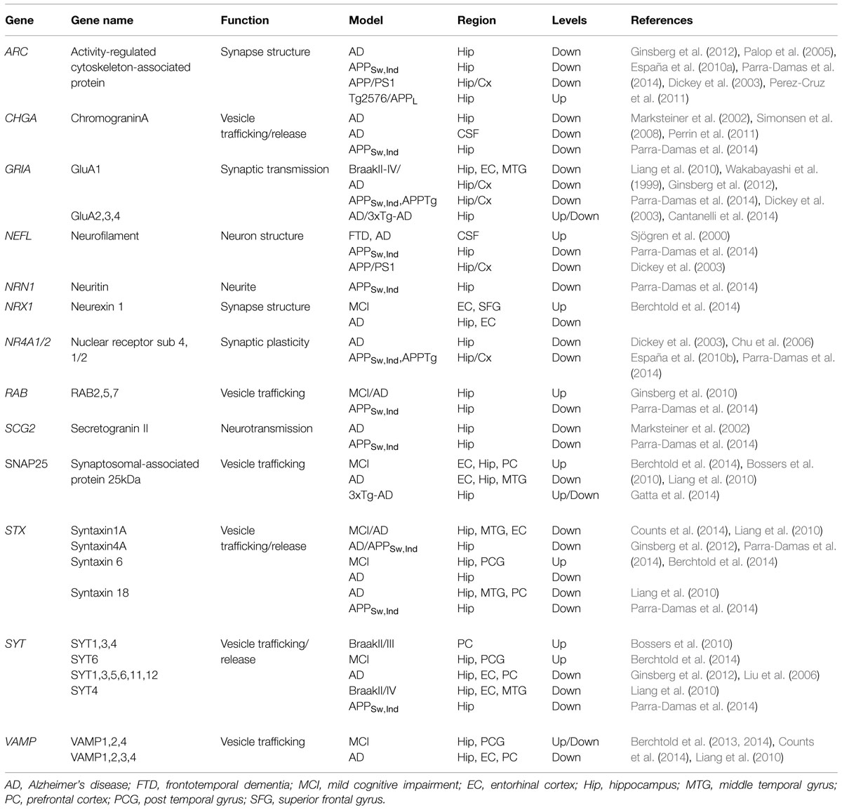
Microarrays comparison analysis of synaptic genes in control and AD brains at different ages (20–99 years) revealed significant expression changes in genes regulating vesicle trafficking/release, neurotransmitter receptors, postsynaptic density, cell adhesion and neuromodulation in normal aging and AD, suggesting that similar synaptic genes are vulnerable to aging and AD (Berchtold et al., 2013). Indeed, expression of genes associated with synaptic signaling and structure, protein biosynthesis and mitochondrial/energy metabolism is predominantly increased in the hippocampus, EC and/or temporal gyrus in MCI and decline in AD (Berchtold et al., 2014). Genes that regulate vesicle and synapse function, including those encoding different isoforms of synaptophysin (SYP), SNAP25, synapsin (SYN), synaptogyrin (SYNGR1), synaptobrevin (VAMP), synaptotagmin (SYT), syntaxin-1 (STX1), synaptopodin (SYNPO) and PSD-95 are downregulated in the hippocampus and EC at moderate and severe AD stages (Liang et al., 2010; Ginsberg et al., 2012; Counts et al., 2014). Indeed, altered expression of genes related to synapse function and plasticity correlates better with AD pathology and clinical severity (Gomez Ravetti et al., 2010; Berchtold et al., 2014). Table 1 summarizes changes of expression of some synaptic genes in AD brain and mouse models. It should be noticed that transcript changes in AD brain could reflect the loss of neurons and synapses in advanced disease stages, a possibility not generally considered in the majority of these studies.
In the prefrontal cortex, transcriptome changes affecting cell signaling, metabolic, inflammation and neurotransmission pathways occur at early pathological stages coinciding with the presence of intraneuronal Aβ (Bossers et al., 2010). Two patterns of gene regulation can be detected: (1) genes related to synaptic function, ATP synthesis and RNA increase in early pathological stages (Braak 0–III) and decline later (Braak IV–VI), and (2) genes related to cell differentiation/proliferation, metal ion binding, antigen processing and transcriptional regulation decrease early and then increase in late Braak stages (Bossers et al., 2010). Synaptic genes upregulated at early pathological stages include potassium voltage-gated channels (KCNS3, KCNB1, KCNA1, and KCNAB1), GABA receptors (GABRA1, GABRD, GABRG2), vesicle exocitosis (SNAP25, CPLX1, VAMP7, SYT1, SYT3, SYT4, NAPB, and SV2C) and vesicle endocytosis (clathrin, CLTC, PACSIN1) proteins.
These above findings indicate a close relationship between transcriptional deregulation and AD-associated neuropathology in memory-related neural circuits. We therefore hypothesize that increased expression of synaptic genes resulting from excitatory/inhibitory imbalance can enhance neural excitability and circuit activity during pre-symptomatic and very early disease stages of AD. In turn, this leads to global gene deregulation, synaptic dysfunction and degeneration and memory loss during the progression of the disease (Figure 3).
Synaptic Gene Expression Changes in AD Mouse Models
Transcriptome profile studies in AD mouse models have revealed altered expression of genes related to mitochondrial function, metabolism, insulin signaling, calcium homeostasis, inflammation, and synaptic plasticity during AD-like pathological progression (see Table 1, for synaptic genes). 3xTg-AD mice shows early hippocampal deregulation of genes linked to mitochondrial morphology and function, neuroinflammation, calcium homeostasis, neurotransmission, neuronal loss, and cell cycle (Gatta et al., 2014). 3xTg-AD mice show age-dependent expression changes on AMPA receptor subunits, with marked reduction of Gria2 and Gria3 in the hippocampus at 12 months. Interestingly, levels of Gria2, Gria3, and Gria4 transcripts are increased in the hippocampus of young 3xTg-AD animals suggesting a compensatory mechanism against AD-related synaptic dysfunction (Cantanelli et al., 2014). Several synaptic plasticity genes, including Arc, early growth response 1 (Egr1), NR2B, Gria1, Homer-1 and Nr4a1/Nur77, are significantly reduced in the hippocampus of 18 months-old APP/PS1 transgenic mice coinciding with amyloid plaques and memory deficits (Dickey et al., 2003). Interestingly, expression of genes directly implicated in learning/memory and plasticity is increased in the hippocampus of environmental enriched APP/PS1 (Lazarov et al., 2005).
Recently, comparison of different lines of APP and tau transgenic mice revealed that elevation of immune system genes is associated with appearance of amyloid plaques, whereas reduced expression of synaptic genes and increased cell death genes correlate with cortical and hippocampal tau pathology (Matarin et al., 2015). This result agrees with previous reports showing reduced expression of genes related to glutamatergic (Arc, Gria1, Gria2, Grik4, Psd95), or GABAergic (Gad67, Gabarap-11) neurotransmission and vesicle trafficking (Syn3, Syb, Synj, Snap29, Syp, Stx4a, Stx7) in hTau mice (Alldred et al., 2012), and elevation of inflammatory genes in brain regions containing amyloid deposits in APP mice (Dickey et al., 2003; Landel et al., 2014). In summary, transcriptome analysis demonstrates deregulation of common cellular pathways in several AD transgenic mouse models during AD-associated pathology.
Activity-Dependent Gene Expression and Memory Deficits in AD Mouse Models
Activity-dependent gene expression is a fundamental mechanism mediating structural changes at synapses during memory formation. Cognitive deficits in human and mice are associated with dysregulation of activity-dependent genes and transcription factors (West and Greenberg, 2011). Downregulation of activity-dependent genes involved in synaptic plasticity and memory, including the activity-regulated cytoskeleton-associated protein (Arc), c-fos and Bdnf, are associated with learning and memory deficits in AD and APP transgenic mice (Phillips et al., 1991; Dewachter et al., 2009; España et al., 2010b). Notably, ARC transcripts are significantly reduced at early and advanced AD pathological stages (Ginsberg et al., 2012; Parra-Damas et al., 2014). Similarly, Arc expression is markedly decreased in the hippocampus and visual cortex of APP transgenic mice after experience and memory training (Palop et al., 2005; Rudinskiy et al., 2012; Parra-Damas et al., 2014). Paradoxicaly, Arc is increased in individual cortical neurons close to amyloid plaques and CA1 pyramidal neurons in APP mice, an effect attributed to neuronal hyperactivity caused by decreased synaptic inhibition (Perez-Cruz et al., 2011; Rudinskiy et al., 2012). Despite the established disruption of activity-dependent gene expression in AD, the regulatory transcriptional mechanisms underlying gene changes causing memory loss in this disease are largely unknown. Understanding these mechanisms may offer new opportunities for therapeutic intervention in cognitive disorders.
To discern transcriptional mechanisms related to memory impairment in AD, we recently performed genome-wide transcriptome analyses in naïve and memory trained non-transgenic and APPSw,Ind (J9) mice. Gene-annotation analysis revealed a gene cluster of 164 transcripts deregulated in the hippocampus of 6 months-old APPSw,Ind mice compared to non-transgenic mice after memory training. The biological pathways associated with these genes are learning/memory, neurotransmission, synaptic plasticity, glutamatergic and GABAergic neurotransmission, oxidative phosphorylation and AD (Parra-Damas et al., 2014). Coinciding with initial intraneuronal Aβ accumulation and memory deficits, APPSw,Ind mice show deregulation of a transcriptional program dependent on the cAMP-response element binding protein (CREB)-regulated transcription coactivator-1 (CRTC1), which includes genes involved in neurotransmission (Scg2, Syt4, Stx4, Stx18, Rab2a, Gria1, Chga), synaptic plasticity/memory (Arc, c-fos, Nr4a1, Nr4a2, Bdnf) and neuritogenesis (Nefl, Nrn1) (Parra-Damas et al., 2014) (Table 1). This result is consistent with a decline of synaptic gene transcripts coinciding with the presence of intraneuronal Aβ and preceding synapse loss in human prefrontal cortex at intermediate pathological stages (Bossers et al., 2010).
Genetic and pharmacological studies have shown that disruption of CREB signaling mediates synaptic plasticity and memory impairments in AD (Saura and Valero, 2011). Accordingly, CREB activation ameliorates synaptic and memory deficits in APP transgenic mice (Smith et al., 2009; Yiu et al., 2011), whereas CRTC1 gene therapy reverses early transcriptional changes and memory impairments in AD mice (Parra-Damas et al., 2014). In summary, disruption of CREB/CRTC1-dependent transcription underlies early memory deficits whereas its activation ameliorates AD-related synaptic and memory impairments, which provides evidence that targeting this pathway may be therapeutically beneficial in AD.
Pathogenic and Therapeutic Implications of Gene Deregulation in AD
AD is the most common form of dementia in the aging population but, unfortunately, current therapies are not effective to ameliorate or reverse the clinical symptoms. Classical pharmacological treatments based on inhibition of acetylcholinesterase (e.g., donepezil, rivastigmine) or excitotoxicity (memantine) slow the disease progression but do not prevent or stop the neurodegeneration process. Alternatively, anti-amyloid immunotherapy treatments that efficiently reduce amyloid plaque burden fail to improve cognitive performance in mild-to moderate AD patients (Doody et al., 2014; Salloway et al., 2014). The discouraging failures of anti-amyloid clinical trials have raised doubts about the contribution of Aβ as the initiating factor in AD pathophysiology (Herrup, 2015). Alternatively, several molecular, genetic and cellular events affected by aging, the main risk factor of the disease, may contribute to neuronal dysfunction and degeneration leading to dementia in AD.
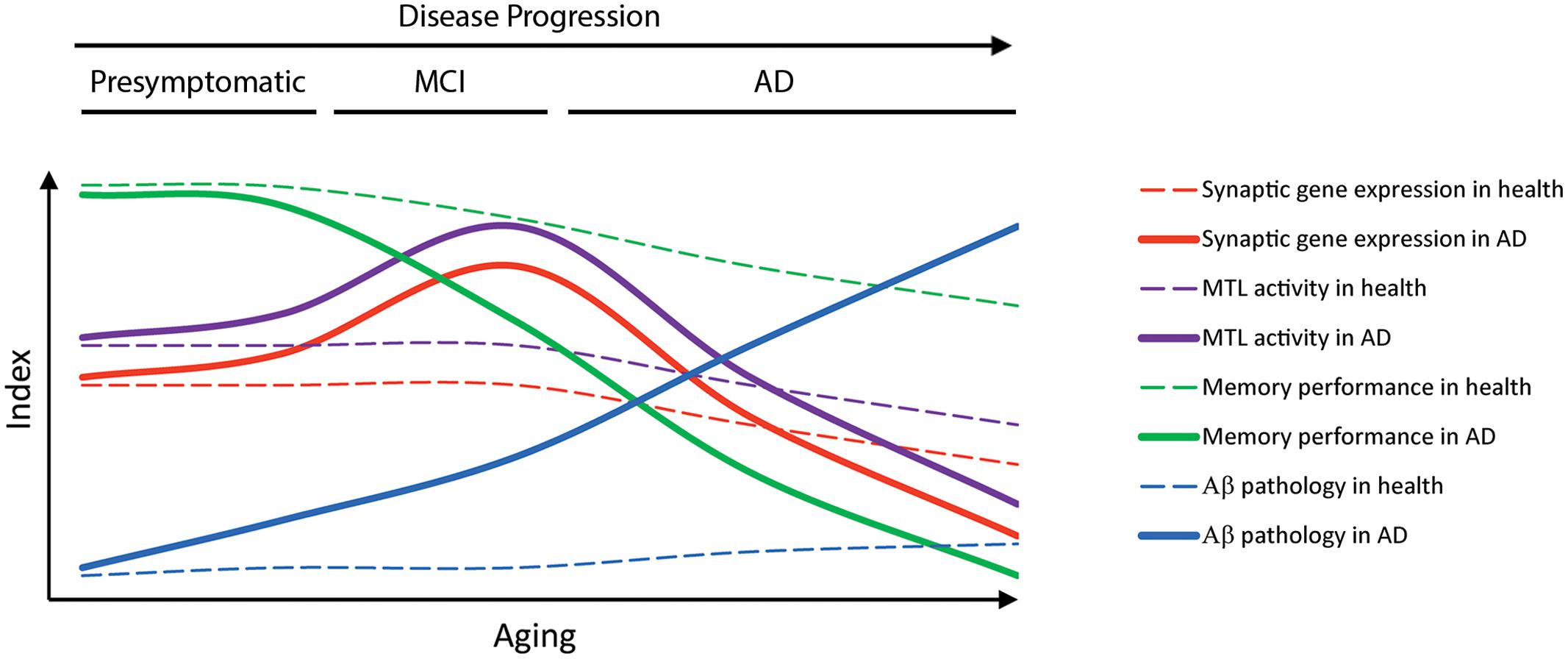
In this context, deregulation of genes involved in pathological pathways, including oxidative stress, mitochondrial/energy metabolism, synapse dysfunction and inflammation, may be crucial in the etiology of AD. Thus, despite massive gene expression changes in the brain, few vulnerable biological pathways, including energy metabolism, synaptic function/plasticity and inflammation are generally altered in normal aging and AD (Berchtold et al., 2008, 2013; Cribbs et al., 2012). Gene expression deregulation occurs already in presymptomatic or early diseases phases (Bossers et al., 2010; Liang et al., 2010; Berchtold et al., 2014). Thus, synaptic and energy metabolism gene clusters are upregulated early during the disease process declining later at intermediate/severe pathological stages. Based on these results, we hypothesize that upregulation of synaptic genes contributes to increased neural excitability and memory circuit activity at presymptomatic or very early disease stages, which could then trigger gene deregulation, synaptic dysfunction, degeneration and memory loss during the progression of the disease (Figure 3). Indeed, changes in expression of synaptic genes parallel altered activity of memory circuits indicating a close relationship between neuropathology, transcriptional deregulation and activity of susceptible memory circuits in AD (Figure 4).

FIGURE 4. Temporal progression of pathological and transcriptional changes in aging and AD. The diagram represents the hypothetical temporal progression of memory deficits, expression of synaptic genes, medial temporal lobe (MTL) activity and Aβ pathology during aging and AD. Enhanced brain activity at prodromal AD stages is associated with increased expression of synaptic genes, whereas decreased brain activity parallels reduction of synaptic genes as the disease progresses.
How changes of synaptic gene programs contribute to neuronal activity and connectivity alterations leading to memory loss in cognitive disorders is starting to be elucidated. One suggested possibility is that Aβ enhances intrinsic neuronal excitability leading to memory network changes and clinical manifestations of AD (Palop et al., 2007). Therapeutic approaches aimed at reducing neuronal hyperactivity may be beneficial to stabilize synaptic function and improve cognitive abilities at early disease stages. In support of this idea, a recent clinical trial indicates that levetiracetam, an antiepileptic drug that reduces hippocampal hyperactivity, improves cognition in amnestic MCI subjects (Bakker et al., 2012). Levetiracetam treatment also reverses synaptic gene changes as well as functional abnormalities and cognitive deficits in APP transgenic mice (Sanchez et al., 2012), whereas decreasing oxidative stress, excitotoxicity and hyperexcitability without interfering with amyloid or tau pathologies prevents AD-related memory deficits (Isopi et al., 2014). Likewise, the antiepileptic drug valproate ameliorates memory deficits and neuropsychiatric symptoms in APP transgenic mice (España et al., 2010a; Yao et al., 2014), but fails to slow cognitive decline and behavioral symptoms at late diseases stages (Fleisher et al., 2011). The efficacy of anti-hyperactivity drugs for treating AD will ultimately depend on the drug type, dosage and disease stage.
Gene profiling and transcriptional regulatory mechanisms involved in memory loss could offer clinical applications as diagnostic tools, novel biomarkers and therapeutic targets in AD. First, it is conceivable that meta-analysis of transcriptomic data from large population-based cohorts of sporadic AD patients may reveal differentially altered pathways related to specific pathogenic mechanisms, opening new venues to design personalized therapeutic strategies. Second, individual or particular set of genes altered in AD brain could be applied as novel early biomarkers to predict the progression of the disease and to monitor therapeutic effects in personalized medicine. Of relevance, synaptic genes identified by wide-genome profile analysis in mouse models are deregulated in AD brain or biological fluids (Table 1). Several of these synaptic proteins, such as BDNF, secretogranin II, synaptotagmin, chromogranin A and SNAP25, have been proposed as novel biomarkers for AD (Simonsen et al., 2008; Li et al., 2009; Perrin et al., 2011; Brinkmalm et al., 2014). Since testing brain tissue could be of limited application in population-based screenings, the use of CSF and blood samples may represent a valuable non-invasive tool for biomarker analysis. Indeed, deregulation of genes and microRNAs (miRNAs) in CSF and/or blood (plasma and blood cells) occurs in AD patients (Chen et al., 2011; Bekris et al., 2013; Roed et al., 2013), and a gene signature in blood related to inflammation, transcription and cell death was recently used for diagnosis and prediction of MCI to AD conversion (Roed et al., 2013). Interestingly, the abundant changes of transcripts of genes related to synaptic plasticity/transmission, neuritogenesis and neurological diseases (AD, Parkinsons’disease, and mental retardation) in blood cells of AD patients suggest a strong link between blood and brain transcriptional profiles in AD (Naughton et al., 2015). Future identification of relevant biomarkers in biological fluids may be useful for early and accurate diagnosis of AD.
An important point is that gene expression is regulated by multiple mechanisms including transcription, translation and posttranscriptional or posttranslational mechanisms. Among these, epigenetic regulation has been the intense focus of research in neurodegenerative diseases in recent years. Epigenetic chromatin remodeling and DNA modifications regulate gene expression during memory formation, whereas epigenetic dysregulation is associated with aging and cognitive disorders (Graff and Mansuy, 2009). Thus, global reduction of DNA methylation and hydroxymethylation occur in the hippocampus of AD patients at early pathological stages (Chouliaras et al., 2013; Sanchez-Mut et al., 2013; De Jager et al., 2014). Other epigenetic factors such as non-coding RNA, in particular miRNAs and long non-coding RNAs, have also received increasing attention in neurodegenerative diseases due to their role in modulating gene expression. A set of miRNAs and long non-coding RNAs are deregulated in brain, blood, and CSF of AD patients (Dorval et al., 2013; Lau et al., 2013), which raises the possibility that non-coding RNAs may play a key role in gene expression deregulation during the course of the disease. Nonetheless, specific miRNAs and long non-coding RNAs affect expression of genes involved in AD pathology including gene regulating APP processing, tau, inflammation and apoptosis (Goodall et al., 2013). The diversity of miRNAs and their potential to target gene expression of multiple pathways offer alternative applications of these molecules as novel biomarkers and therapeutic targets for AD and other neurodegenerative diseases.
Based on the above studies, epigenetic therapeutic approaches have been applied in AD mouse models. As an example, histone deacetylase inhibition increases expression of plasticity genes and ameliorates synaptic pathology and cognitive deficits in APP transgenic mice (Ricobaraza et al., 2012). Long-term systemic treatment with epigenetic drugs may, however, cause broad and deleterious effects on brain function. Alternatively, targeting molecules or pathways regulating specific gene expression programs in vulnerable memory circuits may represent potential therapeutic targets for AD. Interestingly, a recent report demonstrates that a gene therapy approach targeting CRTC1 to enhance expression of specific synaptic genes prevents memory impairments in an AD mouse model (Parra-Damas et al., 2014). A future scientific challenge will be the identification of transcriptome signatures in the brain or biological fluids for early diagnosis and prediction of the disease. In parallel, a better understanding of the expression regulatory mechanisms of genes involved in synaptic dysfunction and neurodegeneration will be crucial to develop efficient therapeutic treatments for AD and other cognitive disorders.
Conflict of Interest Statement
The authors declare that the research was conducted in the absence of any commercial or financial relationships that could be construed as a potential conflict of interest.
Acknowledgments
We thank current and past members of the C. Saura’s lab for contributing to the scientific basis of this review. This manuscript was supported by grants from the Ministerio de Economia y Competitividad of Spain (SAF2013-43900 and CIBERNED CB06/05/0042 to CS and BES-2011-044405 to AP-D), BrightFocus Foundation (A2014417S) and Generalitat de Catalunya (2014 SGR 0984).
References
Abramov, E., Dolev, I., Fogel, H., Ciccotosto, G. D., Ruff, E., and Slutsky, I. (2009). Amyloid-β as a positive endogenous regulator of release probability at hippocampal synapses. Nat. Neurosci. 12, 1567–1576. doi: 10.1038/nn.2433
Alldred, M. J., Duff, K. E., and Ginsberg, S. D. (2012). Microarray analysis of CA1 pyramidal neurons in a mouse model of tauopathy reveals progressive synaptic dysfunction. Neurobiol. Dis. 45, 751–762. doi: 10.1016/j.nbd.2011.10.022
Arriagada, P. V., Growdon, J. H., Hedley-Whyte, E. T., and Hyman, B. T. (1992). Neurofibrillary tangles but not senile plaques parallel duration and severity of Alzheimer’s disease. Neurology 42, 631–639. doi: 10.1212/WNL.42.3.631
Bai, F., Zhang, Z., Watson, D. R., Yu, H., Shi, Y., Yuan, Y., et al. (2009). Abnormal functional connectivity of hippocampus during episodic memory retrieval processing network in amnestic mild cognitive impairment. Biol. Psychiatry 65, 951–958. doi: 10.1016/j.biopsych.2008.10.017
Bakker, A., Krauss, G. L., Albert, M. S., Speck, C. L., Jones, L. R., Stark, C. E., et al. (2012). Reduction of hippocampal hyperactivity improves cognition in amnestic mild cognitive impairment. Neuron 74, 467–474. doi: 10.1016/j.neuron.2012.03.023
Bassett, S. S., Yousem, D. M., Cristinzio, C., Kusevic, I., Yassa, M. A., Caffo, B. S., et al. (2006). Familial risk for Alzheimer’s disease alters fMRI activation patterns. Brain 129, 1229–1239. doi: 10.1093/brain/awl089
Bekris, L. M., Lutz, F., Montine, T. J., Yu, C. E., Tsuang, D., Peskind, E. R., et al. (2013). MicroRNA in Alzheimer’s disease: an exploratory study in brain, cerebrospinal fluid and plasma. Biomarkers 18, 455–466. doi: 10.3109/1354750X.2013.814073
Berchtold, N. C., Coleman, P. D., Cribbs, D. H., Rogers, J., Gillen, D. L., and Cotman, C. W. (2013). Synaptic genes are extensively downregulated across multiple brain regions in normal human aging and Alzheimer’s disease. Neurobiol. Aging 34, 1653–1661. doi: 10.1016/j.neurobiolaging.2012.11.024
Berchtold, N. C., Cribbs, D. H., Coleman, P. D., Rogers, J., Head, E., Kim, R., et al. (2008). Gene expression changes in the course of normal brain aging are sexually dimorphic. Proc. Natl. Acad. Sci. U.S.A. 105, 15605–15610. doi: 10.1073/pnas.0806883105
Berchtold, N. C., Sabbagh, M. N., Beach, T. G., Kim, R. C., Cribbs, D. H., and Cotman, C. W. (2014). Brain gene expression patterns differentiate mild cognitive impairment from normal aged and Alzheimer’s disease. Neurobiol. Aging 35, 1961–1972. doi: 10.1016/j.neurobiolaging.2014.03.031
Bero, A. W., Yan, P., Roh, J. H., Cirrito, J. R., Stewart, F. R., Raichle, M. E., et al. (2011). Neuronal activity regulates the regional vulnerability to amyloid-β deposition. Nat. Neurosci. 14, 750–756. doi: 10.1038/nn.2801
Billings, L. M., Oddo, S., Green, K. N., Mcgaugh, J. L., and Laferla, F. M. (2005). Intraneuronal Aβ causes the onset of early Alzheimer’s disease-related cognitive deficits in transgenic mice. Neuron 45, 675–688. doi: 10.1016/j.neuron.2005.01.040
Blalock, E. M., Geddes, J. W., Chen, K. C., Porter, N. M., Markesbery, W. R., and Landfield, P. W. (2004). Incipient Alzheimer’s disease: microarray correlation analyses reveal major transcriptional and tumor suppressor responses. Proc. Natl. Acad. Sci. U.S.A. 101, 2173–2178. doi: 10.1073/pnas.0308512100
Bossers, K., Wirz, K. T., Meerhoff, G. F., Essing, A. H., Van Dongen, J. W., Houba, P., et al. (2010). Concerted changes in transcripts in the prefrontal cortex precede neuropathology in Alzheimer’s disease. Brain 133, 3699–3723. doi: 10.1093/brain/awq258
Braak, H., and Braak, E. (1991). Neuropathological stageing of Alzheimer-related changes. Acta Neuropathol. (Berl.) 82, 239–259. doi: 10.1007/BF00308809
Brinkmalm, A., Brinkmalm, G., Honer, W. G., Frolich, L., Hausner, L., Minthon, L., et al. (2014). SNAP-25 is a promising novel cerebrospinal fluid biomarker for synapse degeneration in Alzheimer’s disease. Mol. Neurodegener. 9, 53. doi: 10.1186/1750-1326-9-53
Buckner, R. L., Andrews-Hanna, J. R., and Schacter, D. L. (2008). The brain’s default network: anatomy, function, and relevance to disease. Ann. N. Y. Acad. Sci. 1124, 1–38. doi: 10.1196/annals.1440.011
Busche, M. A., Eichhoff, G., Adelsberger, H., Abramowski, D., Wiederhold, K. H., Haass, C., et al. (2008). Clusters of hyperactive neurons near amyloid plaques in a mouse model of Alzheimer’s disease. Science 321, 1686–1689. doi: 10.1126/science.1162844
Cantanelli, P., Sperduti, S., Ciavardelli, D., Stuppia, L., Gatta, V., and Sensi, S. L. (2014). Age-dependent modifications of AMPA receptor subunit expression levels and related cognitive effects in 3xTg-AD mice. Front. Aging Neurosci. 6:200. doi: 10.3389/fnagi.2014.00200
Chapman, P., White, G., Jones, M., Cooper-Blacketer, D., Marshall, V., Irizarry, M., et al. (1999). Impaired synaptic plasticity and learning in aged amyloid precursor protein transgenic mice. Nat. Neurosci. 2, 271–276. doi: 10.1038/6374
Chen, K. D., Chang, P. T., Ping, Y. H., Lee, H. C., Yeh, C. W., and Wang, P. N. (2011). Gene expression profiling of peripheral blood leukocytes identifies and validates ABCB1 as a novel biomarker for Alzheimer’s disease. Neurobiol. Dis. 43, 698–705. doi: 10.1016/j.nbd.2011.05.023
Chhatwal, J. P., Schultz, A. P., Johnson, K., Benzinger, T. L., Jack, C. Jr., Ances, B. M., et al. (2013). Impaired default network functional connectivity in autosomal dominant Alzheimer disease. Neurology 81, 736–744. doi: 10.1212/WNL.0b013e3182a1aafe
Chouliaras, L., Mastroeni, D., Delvaux, E., Grover, A., Kenis, G., Hof, P. R., et al. (2013). Consistent decrease in global DNA methylation and hydroxymethylation in the hippocampus of Alzheimer’s disease patients. Neurobiol. Aging 34, 2091–2099. doi: 10.1016/j.neurobiolaging.2013.02.021
Chu, Y., Le, W., Kompoliti, K., Jankovic, J., Mufson. E. J., and Kordower, J. H. (2006). Nurr1 in Parkinson’s disease and related disorders. J. Comp. Neurol. 494, 495–514. doi: 10.1002/cne.20828
Clavaguera, F., Akatsu, H., Fraser, G., Crowther, R. A., Frank, S., Hench, J., et al. (2013). Brain homogenates from human tauopathies induce tau inclusions in mouse brain. Proc. Natl. Acad. Sci. U.S.A. 110, 9535–9540. doi: 10.1073/pnas.1301175110
Counts, S. E., Alldred, M. J., Che, S., Ginsberg, S. D., and Mufson, E. J. (2014). Synaptic gene dysregulation within hippocampal CA1 pyramidal neurons in mild cognitive impairment. Neuropharmacology 79, 172–179. doi: 10.1016/j.neuropharm.2013.10.018
Cribbs, D. H., Berchtold, N. C., Perreau, V., Coleman, P. D., Rogers, J., Tenner, A. J., et al. (2012). Extensive innate immune gene activation accompanies brain aging, increasing vulnerability to cognitive decline and neurodegeneration: a microarray study. J. Neuroinflammation 9, 179. doi: 10.1186/1742-2094-9-179
D’Amelio, M., Cavallucci, V., Middei, S., Marchetti, C., Pacioni, S., Ferri, A., et al. (2011). Caspase-3 triggers early synaptic dysfunction in a mouse model of Alzheimer’s disease. Nat. Neurosci. 14, 69–76. doi: 10.1038/nn.2709
Davis, K. E., Fox, S., and Gigg, J. (2014). Increased hippocampal excitability in the 3xTgAD mouse model for Alzheimer’s disease in vivo. PLoS ONE 9:e91203. doi: 10.1371/journal.pone.0091203
De Jager, P. L., Srivastava, G., Lunnon, K., Burgess, J., Schalkwyk, L. C., Yu, L., et al. (2014). Alzheimer’s disease: early alterations in brain DNA methylation at ANK1, BIN1, RHBDF2 and other loci. Nat. Neurosci. 17, 1156–1163. doi: 10.1038/nn.3786
Deshpande, A., Kawai, H., Metherate, R., Glabe, C. G., and Busciglio, J. (2009). A role for synaptic zinc in activity-dependent Aβ oligomer formation and accumulation at excitatory synapses. J. Neurosci. 29, 4004–4015. doi: 10.1523/JNEUROSCI.5980-08.2009
Dewachter, I., Filipkowski, R. K., Priller, C., Ris, L., Neyton, J., Croes, S., et al. (2009). Deregulation of NMDA-receptor function and down-stream signaling in APP[V717I] transgenic mice. Neurobiol. Aging 30, 241–256. doi: 10.1016/j.neurobiolaging.2007.06.011
Dewachter, I., Reverse, D., Caluwaerts, N., Ris, L., Kuiperi, C., Van Den Haute, C., et al. (2002). Neuronal deficiency of presenilin 1 inhibits amyloid plaque formation and corrects hippocampal long-term potentiation but not a cognitive defect of amyloid precursor protein [V717I] transgenic mice. J. Neurosci. 22, 3445–3453.
Dickerson, B. C., Salat, D. H., Greve, D. N., Chua, E. F., Rand-Giovannetti, E., Rentz, D. M., et al. (2005). Increased hippocampal activation in mild cognitive impairment compared to normal aging and AD. Neurology 65, 404–411. doi: 10.1212/01.wnl.0000171450.97464.49
Dickerson, B. C., and Sperling, R. A. (2009). Large-scale functional brain network abnormalities in Alzheimer’s disease: insights from functional neuroimaging. Behav. Neurol. 21, 63–75. doi: 10.3233/BEN-2009-0227
Dickey, C. A., Loring, J. F., Montgomery, J., Gordon, M. N., Eastman, P. S., and Morgan, D. (2003). Selectively reduced expression of synaptic plasticity-related genes in amyloid precursor protein + presenilin-1 transgenic mice. J. Neurosci. 23, 5219–5226.
Dodart, J. C., Meziane, H., Mathis, C., Bales, K. R., Paul, S. M., and Ungerer, A. (1999). Behavioral disturbances in transgenic mice overexpressing the V717F beta-amyloid precursor protein. Behav. Neurosci. 113, 982–990. doi: 10.1037/0735-7044.113.5.982
Dominguez-del-Toro, E., Rodriguez-Moreno, A., Porras-Garcia, E., Sanchez-Campusano, R., Blanchard, V., Laville, M., et al. (2004). An in vitro and in vivo study of early deficits in associative learning in transgenic mice that over-express a mutant form of human APP associated with Alzheimer’s disease. Eur. J. Neurosci. 20, 1945–1952. doi: 10.1111/j.1460-9568.2004.03643.x
Doody, R. S., Thomas, R. G., Farlow, M., Iwatsubo, T., Vellas, B., Joffe, S., et al. (2014). Phase 3 trials of solanezumab for mild-to-moderate Alzheimer’s disease. N. Engl. J. Med. 370, 311–321. doi: 10.1056/NEJMoa1312889
Dorval, V., Nelson, P. T., and Hebert, S. S. (2013). Circulating microRNAs in Alzheimer’s disease: the search for novel biomarkers. Front. Mol. Neurosci. 6:24. doi: 10.3389/fnmol.2013.00024
España, J., Gimenez-Llort, L., Valero, J., Miñano, A., Rabano, A., Rodriguez-Alvarez, J., et al. (2010a). Intraneuronal β-amyloid accumulation in the amygdala enhances fear and anxiety in Alzheimer’s disease transgenic mice. Biol. Psychiatry 67, 513–521. doi: 10.1016/j.biopsych.2009.06.015
España, J., Valero, J., Miñano-Molina, A. J., Masgrau, R., Martín, E., Guardia-Laguarta, C., et al. (2010b). β-Amyloid disrupts activity-dependent gene transcription required for memory through the CREB coactivator CRTC1. J. Neurosci. 30, 9402–9410. doi: 10.1523/JNEUROSCI.2154-10.2010
Fleisher, A. S., Truran, D., Mai, J. T., Langbaum, J. B., Aisen, P. S., Cummings, J. L., et al. (2011). Chronic divalproex sodium use and brain atrophy in Alzheimer disease. Neurology 77, 1263–1271. doi: 10.1212/WNL.0b013e318230a16c
Fogel, H., Frere, S., Segev, O., Bharill, S., Shapira, I., Gazit, N., et al. (2014). APP homodimers transduce an amyloid-β-mediated increase in release probability at excitatory synapses. Cell Rep. 7, 1560–1576. doi: 10.1016/j.celrep.2014.04.024
Forny-Germano, L., Lyra, E., Silva, N. M., Batista, A. F., Brito-Moreira, J., Gralle, M., et al. (2014). Alzheimer’s disease-like pathology induced by amyloid-β oligomers in nonhuman primates. J. Neurosci. 34, 13629–13643. doi: 10.1523/JNEUROSCI.1353-14.2014
Gatta, V., D’aurora, M., Granzotto, A., Stuppia, L., and Sensi, S. L. (2014). Early and sustained altered expression of aging-related genes in young 3xTg-AD mice. Cell Death Dis. 5, e1054. doi: 10.1038/cddis.2014.11
Ginsberg, S. D., Alldred, M. J., and Che, S. (2012). Gene expression levels assessed by CA1 pyramidal neuron and regional hippocampal dissections in Alzheimer’s disease. Neurobiol. Dis. 45, 99–107. doi: 10.1016/j.nbd.2011.07.013
Ginsberg, S. D., Mufson, E. J., Counts, S. E., Wuu, J., Alldred, M. J., Nixon, R. A., et al. (2010). Regional selectivity of rab5 and rab7 protein upregulation in mild cognitive impairment and Alzheimer’s disease. J. Alzheimers Dis. 22, 631–639. doi: 10.3233/JAD-2010-101080
Gomez Ravetti, M., Rosso, O. A., Berretta, R., and Moscato, P. (2010). Uncovering molecular biomarkers that correlate cognitive decline with the changes of hippocampus’ gene expression profiles in Alzheimer’s disease. PLoS ONE 5:e10153. doi: 10.1371/journal.pone.0010153
Gomez-Isla, T., Hollister, R., West, H., Mui, S., Growdon, J., Peterson, R., et al. (1997). Neuronal loss correlates with but exceeds neurofibrillary tangles in Alzheimer’s disease. Ann. Neurol. 41, 17–24. doi: 10.1002/ana.410410106
Goodall, E. F., Heath, P. R., Bandmann, O., Kirby, J., and Shaw, P. J. (2013). Neuronal dark matter: the emerging role of microRNAs in neurodegeneration. Front. Cell. Neurosci. 7:178. doi: 10.3389/fncel.2013.00178
Grady, C. L., Mcintosh, A. R., Beig, S., Keightley, M. L., Burian, H., and Black, S. E. (2003). Evidence from functional neuroimaging of a compensatory prefrontal network in Alzheimer’s disease. J. Neurosci. 23, 986–993.
Graff, J., and Mansuy, I. M. (2009). Epigenetic dysregulation in cognitive disorders. Eur. J. Neurosci. 30, 1–8. doi: 10.1111/j.1460-9568.2009.06787.x
Gruart, A., Lopez-Ramos, J. C., Munoz, M. D., and Delgado-Garcia, J. M. (2008). Aged wild-type and APP, PS1, and APP + PS1 mice present similar deficits in associative learning and synaptic plasticity independent of amyloid load. Neurobiol. Dis. 30, 439–450. doi: 10.1016/j.nbd.2008.03.001
Gurevicius, K., Lipponen, A., and Tanila, H. (2013). Increased cortical and thalamic excitability in freely moving APPswe/PS1dE9 mice modeling epileptic activity associated with Alzheimer’s disease. Cereb. Cortex 23, 1148–1158. doi: 10.1093/cercor/bhs105
Hamalainen, A., Pihlajamaki, M., Tanila, H., Hanninen, T., Niskanen, E., Tervo, S., et al. (2007). Increased fMRI responses during encoding in mild cognitive impairment. Neurobiol. Aging 28, 1889–1903. doi: 10.1016/j.neurobiolaging.2006.08.008
Hedden, T., Van Dijk, K. R., Becker, J. A., Mehta, A., Sperling, R. A., Johnson, K. A., et al. (2009). Disruption of functional connectivity in clinically normal older adults harboring amyloid burden. J. Neurosci. 29, 12686–12694. doi: 10.1523/JNEUROSCI.3189-09.2009
Herrup, K. (2015). The case for rejecting the amyloid cascade hypothesis. Nat. Neurosci. 18, 794–799. doi: 10.1038/nn.4017
Hsia, A. Y., Masliah, E., Mcconlogue, L., Yu, G. Q., Tatsuno, G., Hu, K., et al. (1999). Plaque-independent disruption of neural circuits in Alzheimer’s disease mouse models. Proc. Natl. Acad. Sci. U.S.A. 96, 3228–3233. doi: 10.1073/pnas.96.6.3228
Hsiao, K., Chapman, P., Nilsen, S., Ekman, C., Harigaya, Y., Younkin, S., et al. (1996). Correlative memory deficits, Aβ elevation, and amyloid plaques in transgenic mice. Science 274, 99–102.
Hyman, B. T., Van Hoesen, G. W., and Damasio, A. R. (1990). Memory-related neural systems in Alzheimer’s disease: an anatomic study. Neurology 40, 1721–1730. doi: 10.1212/WNL.40.11.1721
Hyman, B. T., Van Hoesen, G. W., Damasio, A. R., and Barnes, C. L. (1984). Alzheimer’s disease: cell-specific pathology isolates the hippocampal formation. Science 225, 1168–1170. doi: 10.1126/science.6474172
Hyman, B. T., Van Hoesen, G. W., Kromer, L. J., and Damasio, A. R. (1986). Perforant pathway changes and the memory impairment of Alzheimer’s disease. Ann. Neurol. 20, 472–481. doi: 10.1002/ana.410200406
Isopi, E., Granzotto, A., Corona, C., Bomba, M., Ciavardelli, D., Curcio, M., et al. (2014). Pyruvate prevents the development of age-dependent cognitive deficits in a mouse model of Alzheimer’s disease without reducing amyloid and tau pathology. Neurobiol. Dis. doi: 10.1016/j.nbd.2014.11.013 [Epub ahead of print].
Jacobsen, J. S., Wu, C. C., Redwine, J. M., Comery, T. A., Arias, R., Bowlby, M., et al. (2006). Early-onset behavioral and synaptic deficits in a mouse model of Alzheimer’s disease. Proc. Natl. Acad. Sci. U.S.A. 103, 5161–5166. doi: 10.1073/pnas.0600948103
Jolas, T., Zhang, X. S., Zhang, Q., Wong, G., Del Vecchio, R., Gold, L., et al. (2002). Long-term potentiation is increased in the CA1 area of the hippocampus of APP(swe/ind) CRND8 mice. Neurobiol. Dis. 11, 394–409. doi: 10.1006/nbdi.2002.0557
Kelly, P. H., Bondolfi, L., Hunziker, D., Schlecht, H. P., Carver, K., Maguire, E., et al. (2003). Progressive age-related impairment of cognitive behavior in APP23 transgenic mice. Neurobiol. Aging 24, 365–378. doi: 10.1016/S0197-4580(02)00098-2
Kircher, T. T., Weis, S., Freymann, K., Erb, M., Jessen, F., Grodd, W., et al. (2007). Hippocampal activation in patients with mild cognitive impairment is necessary for successful memory encoding. J. Neurol. Neurosurg. Psychiatry 78, 812–818. doi: 10.1136/jnnp.2006.104877
Kobayashi, Y., and Amaral, D. G. (2007). Macaque monkey retrosplenial cortex: III. Cortical efferents. J. Comp. Neurol. 502, 810–833. doi: 10.1002/cne.21346
Koch, K., Myers, N. E., Gottler, J., Pasquini, L., Grimmer, T., Forster, S., et al. (2014). Disrupted intrinsic networks link amyloid-β pathology and impaired cognition in prodromal Alzheimer’s disease. Cereb. Cortex doi: 10.1093/cercor/bhu151 [Epub ahead of print].
Koistinaho, M., Ort, M., Cimadevilla, J. M., Vondrous, R., Cordell, B., Koistinaho, J., et al. (2001). Specific spatial learning deficits become severe with age in β-amyloid precursor protein transgenic mice that harbor diffuse β-amyloid deposits but do not form plaques. PNAS 98, 14675–14680. doi: 10.1073/pnas.261562998
Kumar, A., Gibbs, J. R., Beilina, A., Dillman, A., Kumaran, R., Trabzuni, D., et al. (2013). Age-associated changes in gene expression in human brain and isolated neurons. Neurobiol. Aging 34, 1199–1209. doi: 10.1016/j.neurobiolaging.2012.10.021
La Joie, R., Perrotin, A., De La Sayette, V., Egret, S., Doeuvre, L., Belliard, S., et al. (2013). Hippocampal subfield volumetry in mild cognitive impairment, Alzheimer’s disease and semantic dementia. Neuroimage Clin. 3, 155–162. doi: 10.1016/j.nicl.2013.08.007
Lacor, P. N., Buniel, M. C., Chang, L., Fernandez, S. J., Gong, Y., Viola, K. L., et al. (2004). Synaptic targeting by Alzheimer’s-related amyloid beta oligomers. J. Neurosci. 24, 10191–10200. doi: 10.1523/JNEUROSCI.3432-04.2004
Landel, V., Baranger, K., Virard, I., Loriod, B., Khrestchatisky, M., Rivera, S., et al. (2014). Temporal gene profiling of the 5XFAD transgenic mouse model highlights the importance of microglial activation in Alzheimer’s disease. Mol. Neurodegener. 9, 33. doi: 10.1186/1750-1326-9-33
Lanz, T. A., Carter, D. B., and Merchant, K. M. (2003). Dendritic spine loss in the hippocampus of young PDAPP and Tg2576 mice and its prevention by the ApoE2 genotype. Neurobiol. Dis. 13, 246–253. doi: 10.1016/S0969-9961(03)00079-2
Lau, P., Bossers, K., Janky, R., Salta, E., Frigerio, C. S., Barbash, S., et al. (2013). Alteration of the microRNA network during the progression of Alzheimer’s disease. EMBO Mol. Med. 5, 1613–1634. doi: 10.1002/emmm.201201974
Lazarov, O., Robinson, J., Tang, Y. P., Hairston, I. S., Korade-Mirnics, Z., Lee, V. M., et al. (2005). Environmental enrichment reduces Aβ levels and amyloid deposition in transgenic mice. Cell 120, 701–713. doi: 10.1016/j.cell.2005.01.015
Li, G., Peskind, E. R., Millard, S. P., Chi, P., Sokal, I., Yu, C. E., et al. (2009). Cerebrospinal fluid concentration of brain-derived neurotrophic factor and cognitive function in non-demented subjects. PLoS ONE 4:e5424. doi: 10.1371/journal.pone.0005424
Liang, W. S., Dunckley, T., Beach, T. G., Grover, A., Mastroeni, D., Ramsey, K., et al. (2010). Neuronal gene expression in non-demented individuals with intermediate Alzheimer’s disease neuropathology. Neurobiol. Aging 31, 549–566. doi: 10.1016/j.neurobiolaging.2008.05.013
Liang, W. S., Reiman, E. M., Valla, J., Dunckley, T., Beach, T. G., Grover, A., et al. (2008). Alzheimer’s disease is associated with reduced expression of energy metabolism genes in posterior cingulate neurons. Proc. Natl. Acad. Sci. U.S.A. 105, 4441–4446. doi: 10.1073/pnas.0709259105
Liu, Q. Y., Sooknanan, R. R., Malek, L. T., Ribecco-Lutkiewicz, M., Lei, J. X., Shen, H., et al. (2006). Novel subtractive transcription-based amplification of mRNA (STAR) method and its application in search of rare and differentially expressed genes in AD brains. BMC Genomics 7:286. doi: 10.1186/1471-2164-7-286
Marksteiner, J., Kaufmann, W. A., Gurka, P., and Humpel, C. (2002). Synaptic proteins in Alzheimer’s disease. J. Mol. Neurosci. 18, 53–63. doi: 10.1385/JMN:18:1-2:53
Masliah, E., Terry, R. D., and Mallory, M. (1990). Diffuse plaques do not accentuate synapse loss in Alzheimer disease. Am. J. Path. 137, 1293–1297.
Matarin, M., Salih, D. A., Yasvoina, M., Cummings, D. M., Guelfi, S., Liu, W., et al. (2015). A genome-wide gene-expression analysis and database in transgenic mice during development of amyloid or tau pathology. Cell Rep. 10, 633–644. doi: 10.1016/j.celrep.2014.12.041
McGowan, E., Eriksen, J., and Hutton, M. (2006). A decade of modeling Alzheimer’s disease in transgenic mice. Trends Genet. 22, 281–289. doi: 10.1016/j.tig.2006.03.007
Merino-Serrais, P., Benavides-Piccione, R., Blazquez-Llorca, L., Kastanauskaite, A., Rabano, A., Avila, J., et al. (2013). The influence of phospho-tau on dendritic spines of cortical pyramidal neurons in patients with Alzheimer’s disease. Brain 136, 1913–1928. doi: 10.1093/brain/awt088
Miller, J. A., Oldham, M. C., and Geschwind, D. H. (2008). A systems level analysis of transcriptional changes in Alzheimer’s disease and normal aging. J. Neurosci. 28, 1410–1420. doi: 10.1523/JNEUROSCI.4098-07.2008
Miñano-Molina, A. J., Espana, J., Martin, E., Barneda-Zahonero, B., Fado, R., Sole, M., et al. (2011). Soluble oligomers of amyloid-beta peptide disrupt membrane trafficking of alpha-amino-3-hydroxy-5-methylisoxazole-4-propionic acid receptor contributing to early synapse dysfunction. J. Biol. Chem. 286, 27311–27321. doi: 10.1074/jbc.M111.227504
Minkeviciene, R., Rheims, S., Dobszay, M. B., Zilberter, M., Hartikainen, J., Fulop, L., et al. (2009). Amyloid β-induced neuronal hyperexcitability triggers progressive epilepsy. J. Neurosci. 29, 3453–3462. doi: 10.1523/JNEUROSCI.5215-08.2009
Mondadori, C. R., Buchmann, A., Mustovic, H., Schmidt, C. F., Boesiger, P., Nitsch, R. M., et al. (2006). Enhanced brain activity may precede the diagnosis of Alzheimer’s disease by 30 years. Brain 129, 2908–2922. doi: 10.1093/brain/awl266
Mosconi, L., Tsui, W. H., De Santi, S., Li, J., Rusinek, H., Convit, A., et al. (2005). Reduced hippocampal metabolism in MCI and AD: automated FDG-PET image analysis. Neurology 64, 1860–1867. doi: 10.1212/01.WNL.0000163856.13524.08
Naughton, B. J., Duncan, F. J., Murrey, D. A., Meadows, A. S., Newsom, D. E., Stoicea, N., et al. (2015). Blood genome-wide transcriptional profiles reflect broad molecular impairments and strong blood-brain links in Alzheimer’s disease. J. Alzheimers. Dis. 43, 93–108. doi: 10.3233/JAD-140606
Nelson, P. T., Alafuzoff, I., Bigio, E. H., Bouras, C., Braak, H., Cairns, N. J., et al. (2012). Correlation of Alzheimer disease neuropathologic changes with cognitive status: a review of the literature. J. Neuropathol. Exp. Neurol. 71, 362–381. doi: 10.1097/NEN.0b013e31825018f7
O’Brien, J. L., O’keefe, K. M., Laviolette, P. S., Deluca, A. N., Blacker, D., Dickerson, B. C., et al. (2010). Longitudinal fMRI in elderly reveals loss of hippocampal activation with clinical decline. Neurology 74, 1969–1976. doi: 10.1212/WNL.0b013e3181e3966e
Palop, J. J., Chin, J., Bien-Ly, N., Massaro, C., Yeung, B. Z., Yu, G. Q., et al. (2005). Vulnerability of dentate granule cells to disruption of Arc expression in human amyloid precursor protein transgenic mice. J. Neurosci. 25, 9686–9693. doi: 10.1523/JNEUROSCI.2829-05.2005
Palop, J. J., Chin, J., Roberson, E. D., Wang, J., Thwin, M. T., Bien-Ly, N., et al. (2007). Aberrant excitatory neuronal activity and compensatory remodeling of inhibitory hippocampal circuits in mouse models of Alzheimer’s disease. Neuron 55, 697–711. doi: 10.1016/j.neuron.2007.07.025
Parachikova, A., Agadjanyan, M. G., Cribbs, D. H., Blurton-Jones, M., Perreau, V., Rogers, J., et al. (2007). Inflammatory changes parallel the early stages of Alzheimer disease. Neurobiol. Aging 28, 1821–1833. doi: 10.1016/j.neurobiolaging.2006.08.014
Pariente, J., Cole, S., Henson, R., Clare, L., Kennedy, A., Rossor, M., et al. (2005). Alzheimer’s patients engage an alternative network during a memory task. Ann. Neurol. 58, 870–879. doi: 10.1002/ana.20653
Parra-Damas, A., Valero, J., Chen, M., Espana, J., Martin, E., Ferrer, I., et al. (2014). Crtc1 activates a transcriptional program deregulated at early Alzheimer’s disease-related stages. J. Neurosci. 34, 5776–5787. doi: 10.1523/JNEUROSCI.5288-13.2014
Perez-Cruz, C., Nolte, M. W., Van Gaalen, M. M., Rustay, N. R., Termont, A., Tanghe, A., et al. (2011). Reduced spine density in specific regions of CA1 pyramidal neurons in two transgenic mouse models of Alzheimer’s disease. J. Neurosci. 31, 3926–3934. doi: 10.1523/JNEUROSCI.6142-10.2011
Perrin, R. J., Craig-Schapiro, R., Malone, J. P., Shah, A. R., Gilmore, P., Davis, A. E., et al. (2011). Identification and validation of novel cerebrospinal fluid biomarkers for staging early Alzheimer’s disease. PLoS ONE 6:e16032. doi: 10.1371/journal.pone.0016032
Petersen, R. C. (2011). Clinical practice. Mild cognitive impairment. N. Engl. J. Med. 364, 2227–2234. doi: 10.1056/NEJMcp0910237
Phillips, H. S., Hains, J. M., Armanini, M., Laramee, G. R., Johnson, S. A., and Winslow, J. W. (1991). BDNF mRNA is decreased in the hippocampus of individuals with Alzheimer’s disease. Neuron 7, 695–702. doi: 10.1016/0896-6273(91)90273-3
Podtelezhnikov, A. A., Tanis, K. Q., Nebozhyn, M., Ray, W. J., Stone, D. J., and Loboda, A. P. (2011). Molecular insights into the pathogenesis of Alzheimer’s disease and its relationship to normal aging. PLoS ONE 6:e29610. doi: 10.1371/journal.pone.0029610
Press, G. A., Amaral, D. G., and Squire, L. R. (1989). Hippocampal abnormalities in amnesic patients revealed by high-resolution magnetic resonance imaging. Nature 341, 54–57. doi: 10.1038/341054a0
Ramón y Cajal, S. (1911). Histologie du Système Nerveux de l’Homme et des Vertébrés. Vols, 1 and 2. Paris: Maloine.
Reiman, E. M., Quiroz, Y. T., Fleisher, A. S., Chen, K., Velez-Pardo, C., Jimenez-Del-Rio, M., et al. (2012). Brain imaging and fluid biomarker analysis in young adults at genetic risk for autosomal dominant Alzheimer’s disease in the presenilin 1 E280A kindred: a case-control study. Lancet Neurol. 11, 1048–1056. doi: 10.1016/S1474-4422(12)70228-4
Ricobaraza, A., Cuadrado-Tejedor, M., Marco, S., Perez-Otano, I., and Garcia-Osta, A. (2012). Phenylbutyrate rescues dendritic spine loss associated with memory deficits in a mouse model of Alzheimer disease. Hippocampus 22, 1040–1050. doi: 10.1002/hipo.20883
Rocher, A. B., Kinson, M. S., and Luebke, J. I. (2008). Significant structural but not physiological changes in cortical neurons of 12-month-old Tg2576 mice. Neurobiol. Dis. 32, 309–318. doi: 10.1016/j.nbd.2008.07.014
Roed, L., Grave, G., Lindahl, T., Rian, E., Horndalsveen, P. O., Lannfelt, L., et al. (2013). Prediction of mild cognitive impairment that evolves into Alzheimer’s disease dementia within two years using a gene expression signature in blood: a pilot study. J. Alzheimers. Dis. 35, 611–621. doi: 10.3233/JAD-122404
Rudinskiy, N., Hawkes, J. M., Betensky, R. A., Eguchi, M., Yamaguchi, S., Spires-Jones, T. L., et al. (2012). Orchestrated experience-driven Arc responses are disrupted in a mouse model of Alzheimer’s disease. Nat. Neurosci. 15, 1422–1429. doi: 10.1038/nn.3199
Rutten, B. P., Van Der Kolk, N. M., Schafer, S., Van Zandvoort, M. A., Bayer, T. A., Steinbusch, H. W., et al. (2005). Age-related loss of synaptophysin immunoreactive presynaptic boutons within the hippocampus of APP751SL, PS1M146L, and APP751SL/PS1M146L transgenic mice. Am. J. Pathol. 167, 161–173. doi: 10.1016/S0002-9440(10)62963-X
Salloway, S., Sperling, R., and Brashear, H. R. (2014). Phase 3 trials of solanezumab and bapineuzumab for Alzheimer’s disease. N. Engl. J. Med. 370, 1460. doi: 10.1056/NEJMoa1304839
Sanchez, P. E., Zhu, L., Verret, L., Vossel, K. A., Orr, A. G., Cirrito, J. R., et al. (2012). Levetiracetam suppresses neuronal network dysfunction and reverses synaptic and cognitive deficits in an Alzheimer’s disease model. Proc. Natl. Acad. Sci. U.S.A. 109, E2895–E2903. doi: 10.1073/pnas.1121081109
Sanchez-Mut, J. V., Aso, E., Panayotis, N., Lott, I., Dierssen, M., Rabano, A., et al. (2013). DNA methylation map of mouse and human brain identifies target genes in Alzheimer’s disease. Brain 136, 3018–3027. doi: 10.1093/brain/awt237
Santos, S. F., Pierrot, N., and Octave, J. N. (2010). Network excitability dysfunction in Alzheimer’s disease: insights from in vitro and in vivo models. Rev. Neurosci. 21, 153–171.
Saura, C. A., Chen, G., Malkani, S., Choi, S. Y., Takahashi, R. H., Zhang, D., et al. (2005). Conditional inactivation of presenilin-1 prevents amyloid accumulation and temporarily rescues contextual and spatial working memory impairments in amyloid precursor protein transgenic mice. J. Neurosci. 25, 6755–6764. doi: 10.1523/JNEUROSCI.1247-05.2005
Saura, C. A., and Valero, J. (2011). The role of CREB signaling in Alzheimer’s disease and other cognitive disorders. Rev. Neurosci. 22, 153–169. doi: 10.1515/RNS.2011.018
Scheff, S. W., Dekosky, S. T., and Price, D. A. (1990). Quantitative assessment of cortical synaptic density in Alzheimer’s disease. Neurobiol. Aging 11, 29–37. doi: 10.1016/0197-4580(90)90059-9
Scheff, S. W., Price, D. A., Schmitt, F. A., Dekosky, S. T., and Mufson, E. J. (2007). Synaptic alterations in CA1 in mild Alzheimer disease and mild cognitive impairment. Neurology 68, 1501–1508. doi: 10.1212/01.wnl.0000260698.46517.8f
Sekar, S., Mcdonald, J., Cuyugan, L., Aldrich, J., Kurdoglu, A., Adkins, J., et al. (2015). Alzheimer’s disease is associated with altered expression of genes involved in immune response and mitochondrial processes in astrocytes. Neurobiol. Aging 36, 583–591. doi: 10.1016/j.neurobiolaging.2014.09.027
Shankar, G. M., Bloodgood, B. L., Townsend, M., Walsh, D. M., Selkoe, D. J., and Sabatini, B. L. (2007). Natural oligomers of the Alzheimer amyloid-β protein induce reversible synapse loss by modulating an NMDA-type glutamate receptor-dependent signaling pathway. J. Neurosci. 27, 2866–2875. doi: 10.1523/JNEUROSCI.4970-06.2007
Sheline, Y. I., Raichle, M. E., Snyder, A. Z., Morris, J. C., Head, D., Wang, S., et al. (2010). Amyloid plaques disrupt resting state default mode network connectivity in cognitively normal elderly. Biol. Psychiatry 67, 584–587. doi: 10.1016/j.biopsych.2009.08.024
Silva, A. R., Grinberg, L. T., Farfel, J. M., Diniz, B. S., Lima, L. A., Silva, P. J., et al. (2012). Transcriptional alterations related to neuropathology and clinical manifestation of Alzheimer’s disease. PLoS ONE 7:e48751. doi: 10.1371/journal.pone.0048751
Simonsen, A. H., Mcguire, J., Podust, V. N., Davies, H., Minthon, L., Skoog, I., et al. (2008). Identification of a novel panel of cerebrospinal fluid biomarkers for Alzheimer’s disease. Neurobiol. Aging 29, 961–968. doi: 10.1016/j.neurobiolaging.2007.01.011
Simpson, J. E., Ince, P. G., Shaw, P. J., Heath, P. R., Raman, R., Garwood, C. J., et al. (2011). Microarray analysis of the astrocyte transcriptome in the aging brain: relationship to Alzheimer’s pathology and APOE genotype. Neurobiol. Aging 32, 1795–1807. doi: 10.1016/j.neurobiolaging.2011.04.013
Sjögren, M., Rosengren, L., Minthon, L., Davidsson, P., Blennow, K., and Wallin, A. (2000). Cytoskeleton proteins in CSF distinguish frontotemporal dementia from AD. Neurology 54, 1960–1964.
Small, S. A., Perera, G. M., Delapaz, R., Mayeux, R., and Stern, Y. (1999). Differential regional dysfunction of the hippocampal formation among elderly with memory decline and Alzheimer’s disease. Ann. Neurol. 45, 466–472. doi: 10.1002/1531-8249(199904)45:4<466::AID-ANA8>3.0.CO;2-Q
Smith, D. L., Pozueta, J., Gong, B., Arancio, O., and Shelanski, M. (2009). Reversal of long-term dendritic spine alterations in Alzheimer disease models. Proc. Natl. Acad. Sci. U.S.A. 106, 16877–16882. doi: 10.1073/pnas.0908706106
Sperling, R. A., Bates, J. F., Chua, E. F., Cocchiarella, A. J., Rentz, D. M., Rosen, B. R., et al. (2003). fMRI studies of associative encoding in young and elderly controls and mild Alzheimer’s disease. J. Neurol. Neurosurg. Psychiatry 74, 44–50. doi: 10.1136/jnnp.74.1.44
Sperling, R. A., Dickerson, B. C., Pihlajamaki, M., Vannini, P., Laviolette, P. S., Vitolo, O. V., et al. (2010). Functional alterations in memory networks in early Alzheimer’s disease. Neuromolecular Med. 12, 27–43. doi: 10.1007/s12017-009-8109-7
Sperling, R., Mormino, E., and Johnson, K. (2014). The evolution of preclinical Alzheimer’s disease: implications for prevention trials. Neuron 84, 608–622. doi: 10.1016/j.neuron.2014.10.038
Spires-Jones, T. L., and Hyman, B. T. (2014). The intersection of amyloid beta and tau at synapses in Alzheimer’s disease. Neuron 82, 756–771. doi: 10.1016/j.neuron.2014.05.004
Squirre, L. R., and Zola-Morgan, S. (1991). The medial temporal lobe memory system. Science 253, 1380–1386. doi: 10.1126/science.1896849
Talantova, M., Sanz-Blasco, S., Zhang, X., Xia, P., Akhtar, M. W., Okamoto, S., et al. (2013). Aβ induces astrocytic glutamate release, extrasynaptic NMDA receptor activation, and synaptic loss. Proc. Natl. Acad. Sci. U.S.A. 110, E2518–E2527. doi: 10.1073/pnas.1306832110
Tan, M. G., Chua, W. T., Esiri, M. M., Smith, A. D., Vinters, H. V., and Lai, M. K. (2010). Genome wide profiling of altered gene expression in the neocortex of Alzheimer’s disease. J. Neurosci. Res. 88, 1157–1169. doi: 10.1002/jnr.22290
Terry, R. D. (2000). Cell death or synaptic loss in Alzheimer disease. J. Neuropathol. Exp. Neurol. 59, 1118–1119.
Terry, R. D., Masliah, E., Salmon, D. P., Butters, N., Deteresa, R., Hill, R., et al. (1991). Physical basis of cognitive alterations in Alzheimer’s disease: synapse loss is the major correlate of cognitive impairment. Ann. Neurol. 30, 572–580. doi: 10.1002/ana.410300410
Tondelli, M., Wilcock, G. K., Nichelli, P., De Jager, C. A., Jenkinson, M., and Zamboni, G. (2012). Structural MRI changes detectable up to ten years before clinical Alzheimer’s disease. Neurobiol. Aging 33, e825–e836. doi: 10.1016/j.neurobiolaging.2011.05.018
Van Dam, D., D’hooge, R., Staufenbiel, M., Van Ginneken, C., Van Meir, F., and De Deyn, P. P. (2003). Age-dependent cognitive decline in the APP23 model precedes amyloid deposition. Eur. J. Neurosci. 17, 388–396. doi: 10.1046/j.1460-9568.2003.02444.x
Van Hoesen, G. W., and Pandya, D. N. (1975). Some connections of the entorhinal (area 28) and perirhinal (area 35) cortices of the rhesus monkey. III. Efferent connections. Brain Res. 95, 39–59. doi: 10.1016/0006-8993(75)90206-1
Verret, L., Mann, E. O., Hang, G. B., Barth, A. M., Cobos, I., Ho, K., et al. (2012). Inhibitory interneuron deficit links altered network activity and cognitive dysfunction in Alzheimer model. Cell 149, 708–721. doi: 10.1016/j.cell.2012.02.046
Wakabayashi, K., Narisawa-Saito, M., Iwakura, Y., Arai, T., Ikeda, K., Takahashi, H., et al. (1999). Phenotypic down-regulation of glutamate receptor subunit GluR1 in Alzheimer’s disease. Neurobiol. Aging 20, 287–295. doi: 10.1016/S0197-4580(99)00035-4
West, A. E., and Greenberg, M. E. (2011). Neuronal activity-regulated gene transcription in synapse development and cognitive function. Cold Spring Harb. Perspect. Biol. 3, a005744. doi: 10.1101/cshperspect.a005744
Wilke, S. A., Raam, T., Antonios, J. K., Bushong, E. A., Koo, E. H., Ellisman, M. H., et al. (2014). Specific disruption of hippocampal mossy fiber synapses in a mouse model of familial Alzheimer’s disease. PLoS ONE 9:e84349. doi: 10.1371/journal.pone.0084349
Wishart, H. A., Saykin, A. J., Rabin, L. A., Santulli, R. B., Flashman, L. A., Guerin, S. J., et al. (2006). Increased brain activation during working memory in cognitively intact adults with the APOE epsilon4 allele. Am. J. Psychiatry 163, 1603–1610. doi: 10.1176/appi.ajp.163.9.1603
Wu, C. C., Chawla, F., Games, D., Rydel, R. E., Freedman, S., Schenk, D., et al. (2004). Selective vulnerability of dentate granule cells prior to amyloid deposition in PDAPP mice: digital morphometric analyses. Proc. Natl. Acad. Sci. U.S.A. 101, 7141–7146. doi: 10.1073/pnas.0402147101
Yao, P. J., Zhu, M., Pyun, E. I., Brooks, A. I., Therianos, S., Meyers, V. E., et al. (2003). Defects in expression of genes related to synaptic vesicle trafficking in frontal cortex of Alzheimer’s disease. Neurobiol. Dis. 12, 97–109. doi: 10.1016/S0969-9961(02)00009-8
Yao, Z. G., Liang, L., Liu, Y., Zhang, L., Zhu, H., Huang, L., et al. (2014). Valproate improves memory deficits in an Alzheimer’s disease mouse model: investigation of possible mechanisms of action. Cell. Mol. Neurobiol. 34, 805–812. doi: 10.1007/s10571-013-0012-y
Yassa, M. A., Verduzco, G., Cristinzio, C., and Bassett, S. S. (2008). Altered fMRI activation during mental rotation in those at genetic risk for Alzheimer disease. Neurology 70, 1898–1904. doi: 10.1212/01.wnl.0000312288.45119.d1
Yiu, A. P., Rashid, A. J., and Josselyn, S. A. (2011). Increasing CREB function in the CA1 region of dorsal hippocampus rescues the spatial memory deficits in a mouse model of Alzheimer’s disease. Neuropsychopharmacology 36, 2169–2186. doi: 10.1038/npp.2011.107
Keywords: Alzheimer’s disease, memory, transcriptome, neurodegeneration, Aβ, gene expression
Citation: Saura CA, Parra-Damas A and Enriquez-Barreto L (2015) Gene expression parallels synaptic excitability and plasticity changes in Alzheimer’s disease. Front. Cell. Neurosci. 9:318. doi: 10.3389/fncel.2015.00318
Received: 21 May 2015; Accepted: 03 August 2015;
Published: 25 August 2015.
Edited by:
Lydia Jimenez-Diaz, University of Castilla-La Mancha, SpainReviewed by:
Hermona Soreq, The Hebrew University of Jerusalem, IsraelAgnes Gruart, Universidad Pablo de Olavide, Spain
Valentina Gatta, University of Chieti-Pescara, Italy
Copyright © 2015 Saura, Parra-Damas and Enriquez-Barreto. This is an open-access article distributed under the terms of the Creative Commons Attribution License (CC BY). The use, distribution or reproduction in other forums is permitted, provided the original author(s) or licensor are credited and that the original publication in this journal is cited, in accordance with accepted academic practice. No use, distribution or reproduction is permitted which does not comply with these terms.
*Correspondence: Carlos A. Saura, Institut de Neurociències, Departament de Bioquímica i Biologia Molecular, Centro de Investigación Biomédica en Red Enfermedades Neurodegenerativas (CIBERNED), Universitat Autònoma de Barcelona, Barcelona 08193, Spain,Y2FybG9zLnNhdXJhQHVhYi5lcw==
 Carlos A. Saura
 Arnaldo Parra-Damas
 Lilian Enriquez-Barreto