 Manmohan Kumar
 Shagun Sharma
 Shibnath Mazumder
94% of researchers rate our articles as excellent or good
Learn more about the work of our research integrity team to safeguard the quality of each article we publish.
Find out more
REVIEW article
Front. Cell. Infect. Microbiol., 16 May 2023
Sec. Molecular Bacterial Pathogenesis
Volume 13 - 2023 | https://doi.org/10.3389/fcimb.2023.1135203
This article is part of the Research TopicReviews in Molecular Bacterial Pathogenesis and Host Immunity-Pathogens InteractionsView all 6 articles
The immune system of a host contains a group of heterogeneous cells with the prime aim of restraining pathogenic infection and maintaining homeostasis. Recent reports have proved that the various subtypes of immune cells exploit distinct metabolic programs for their functioning. Mitochondria are central signaling organelles regulating a range of cellular activities including metabolic reprogramming and immune homeostasis which eventually decree the immunological fate of the host under pathogenic stress. Emerging evidence suggests that following bacterial infection, innate immune cells undergo profound metabolic switching to restrain and countervail the bacterial pathogens, promote inflammation and restore tissue homeostasis. On the other hand, bacterial pathogens affect mitochondrial structure and functions to evade host immunity and influence their intracellular survival. Mitochondria employ several mechanisms to overcome bacterial stress of which mitochondrial UPR (UPRmt) and mitochondrial dynamics are critical. This review discusses the latest advances in our understanding of the immune functions of mitochondria against bacterial infection, particularly the mechanisms of mitochondrial UPRmt and mitochondrial dynamics and their involvement in host immunity.
Host–pathogen interaction is an ever-emerging and evolving field. Almost every other day, pathogens pose new challenges, and the immune system provides respite and protection to the host. The innate immune system is the first line of host defense against invading pathogens (Medzhitov, 2010). Cells of the innate immune system (monocytes/macrophages, neutrophils, dendritic cells, etc.) are ubiquitous and recognize pathogens or their products with the help of germline-encoded pattern-recognition receptors (PRRs) triggering inflammatory responses, which help in pathogen clearance (Arango Duque and Descoteaux, 2014; Weiss and Schaible, 2015). Recent studies have reported that activation of innate immune cells is closely regulated by shifting in mitochondrial metabolism and physiology such as mitochondrial dynamics and mitochondrial proteastasis, and mitochondria provides a central scaffolding platform for innate immune signaling pathways induced by pattern recognition receptors (PRRs) in response to microbial ligands (Tur et al., 2017; Banoth and Cassel, 2018; Faas and De Vos, 2020).
Over the years, mitochondria were viewed as semi-autonomous cellular organelles required only for the bioenergetics and biosynthesis of macromolecules. Although the regulation of apoptotic signaling by mitochondria has been well appreciated during past years (Wang and Youle, 2009; Tiku et al., 2020), recent studies suggest that mitochondria also participate in several additional innate immune signaling pathways (Banoth and Cassel, 2018; Shen et al., 2022). Hence, mitochondria serve as the metabolic hubs and respond to intrinsic cues and environmental stressors with an implausible degree of plasticity, which enables them to participate in various cellular signaling pathways (Bahat and Gross, 2019; Chakrabarty and Chandel, 2021). However, the interplay between innate immunity and mitochondria goes well beyond the control of death or survival of the host cell. Recent pieces of evidence demonstrated that multitudinous innate immune signaling pathways are regulated by mitochondria, revealing mutual relationships between cellular metabolism and innate immunity. Besides functioning as an innate immune regulon, several studies have also documented the importance of this organelle in shaping adaptive immune responses, thus placing it at the crossroads of adaptive and innate immunity (Sena et al., 2013; Weinberg et al., 2015; Sandoval et al., 2018). Every single mitochondrion harbours trenchant agonists of inflammation (Nakahira et al., 2015), collectively known as danger-associated molecular patterns (DAMPs). Structural and functional damage to mitochondria due to bacterial infections leads to mitochondrial DAMPs causing inflammation, and even autoimmune neurodegenerative disorders. Recent studies have also established mitochondria as a key player in regulating and establishing immune responses against pathogens. Thus, production of anti-microbial proteins by immune cell survival against pathogenic infection depends on maintaining proper mitochondrial function by mitochondrial quality control (MQC). Among the different MQC mechanisms, mitochondrial unfolded protein response (UPRmt), mitophagy, biogenesis, and mitochondrial fusion-fission dynamics are essential. Bacterial infection induces mitochondrial stress which stimulates the mito-nuclear response pathway known as UPRmt for counteracting mitochondrial stress and thus, maintains homeostasis. In addition, mitochondrial dynamics are also altered in response to bacterial infection, this could be a direct effect of virulent strategy adapted by bacterial pathogens or it could be a host cell response to restrain bacterial infection. However, the molecular mechanisms underlying both these events, and the dependency on each other still remains elusive. This review provides an updated perspective of UPRmt and mitochondrial dynamics, the underlying molecular mechanisms, and how they conjure innate immunity to bacterial pathogens.
Mitochondrion plays a crucial role in regulating bacterial pathogenesis. Hence, bacterial pathogens have evolved strategies to subvert mitochondrial functions to promote proliferation and infection (Galmiche and Rassow, 2010; Escoll et al., 2016). For example, pathogenic bacteria manipulate mitochondria via the secretion of pore-forming toxins (Papatheodorou et al., 2006; Stavru et al., 2011; Palframan et al., 2012), manipulate mitochondrial-dependent cell death pathways via type III secretion system to promote their survival (Pallett et al., 2014; Arizmendi et al., 2016), produce electron chain transport inhibitors (Raveh et al., 2013), and iron chelators that perturb mitochondrial functions thereby aiding their survival (Kirienko et al., 2015).
Conversely, mitochondria have evolved several mechanisms to resist bacterial infections, like production of anti-bacterial ROS (West et al., 2011; Kumar et al., 2022b; Kumar et al., 2022a), inflammasome activation (Zhou et al., 2011), xenophagy (Gatica et al., 2018) and consequently apoptosis of infected cells (Deo et al., 2020; Kumar et al., 2022b; Kumar et al., 2022a). To achieve this, immune cells synthesize several factors such as antimicrobial peptides, cytoplasmic and cell-surface surveillance proteins, signaling proteins, and inflammatory cytokines to countervail bacterial infection; hence, proteostasis remains a challenge for immune cells under bacterial assault. Proteostasis depends on the subtle harmony between maintaining protein conformation, refolding of misfolded proteins, and degrading damaged proteins; the process is precisely regulated in the endoplasmic reticulum (ER). The ER and mitochondria are tightly associated with dynamic modules termed mitochondria-associated membranes (MAMs) or mitochondria-ER contact sites (MERCs), which provide an excellent platform for cross-talk between the two organelles (Missiroli et al., 2018; Namgaladze et al., 2019).
Following bacterial infection, damaged or misfolded proteins accumulate in the ER. Recent studies suggest that MAMs/MERCs act as hotspots through which misfolded proteins transit into mitochondria, implicating the role of this organelle in sequestering anomalous proteins, thereby restoring ER functioning (Li et al., 2019; Ruan et al., 2020). The build-up of such proteins in mitochondria triggers proteotoxic stress, thereby activating its protein quality control mechanism, known as mitochondrial unfolded protein response (UPRmt), as a proteostatic mechanism (Haynes and Ron, 2010; Jovaisaite et al., 2014; Tran and Van Aken, 2020).
UPRmt is an evolutionarily conserved response that enhances the transcription of protective factors and chaperones, which are involved in the homeostasis and repair of damaged or stressed mitochondria (Ryan and Hoogenraad, 2007; Quirós et al., 2016; Topf et al., 2016). Thus, besides playing a role in maintaining the mitochondria status quo, UPRmt also aids in animal fitness and host survival under different conditions of stress such as oxidative stress, and proteotoxic stress. Notably, less is known regarding the regulation of the UPRmt in vertebrates, and further studies are needed to understand this. Impaired protein import in the mitochondria has been linked with the activation of UPRmt (Haastrup et al., 2023). And the import of mitochondrial protein is a biologist’s enigma! Around 99% of mitochondrial proteins are encoded in the nuclear genome. The proteins destined for mitochondria contain a signal sequence (or pre-sequences) that direct them to mitochondria from the cytosol. Mitochondrial matrix-targeting sequences are rich in hydrophobic amino acids, positively-charged basic amino acids (arginine and lysine), and hydroxylated amino acids (threonine and serine) and lack negatively-charged acidic residues (aspartate and glutamate). The amphipathic nature and positive charge of the pre-sequences is recognized by protein import receptors at mitochondria rather than a precise amino acid sequence for the translocation of the protein into the mitochondria. Thus, the postal addresses for mitochondrial proteins are ill-defined in terms of amino acid sequence or specific chemical moieties. It has been suggested that physio-chemical traits like pre-sequences’ ability to bind to specific receptors coupled with maintaining thermodynamic equilibrium favouring unfolded proteins facilitate mitochondrial import of the pre-sequences. Mitochondrial precursor proteins aren’t imported into mitochondria in their native state; they are unfolded during translocation and move across the import machinery as linear chains. The internal diameter of TOM (translocase of the outer mitochondrial membrane) is ~22 Å (Schwartz et al., 1999); hence, only small folded proteins can translocate. TIM (translocase of the inner mitochondrial membrane) contains a much narrower channel, and a slight steric hindrance halts translocation into the matrix, suggesting precursor proteins must be unfolded to pass through this channel. The overall mechanisms of mitochondrial import differ depending upon the charge, size, and presence of cysteine moieties (Harbauer et al., 2014), but grossly it has been suggested that the negative charge of the inner mitochondrial membrane attracts positively-charged mitochondrial targeting sequence (MTS) and the difference in membrane potential subsequently drives the positively charged pre-sequence across the inner membrane (Martin et al., 1991; Harbauer et al., 2014). The TOM complex serves as the entry point for most precursors at the outer membrane, and the binding of pre-sequences with TOM is facilitated by hydrophobic interactions (Genge and Mokranjac, 2022). TIM complex facilitates the entry of proteins into the mitochondrial matrix. The interaction between intermembrane space (IMS)-domains of TOM and TIM family members with the pre-sequence of the incoming precursor proteins facilitate the transfer of these proteins across the mitochondrial membrane.
Activating transcription factor associated with stress (ATFS-1), a basic leucine zipper (bZIP) protein, contains both nuclear localization sequence (NLS) and MTS enabling this transcription factor to shuttle between the two organelles and helping mitochondria in establishing communication with the nucleus (Haynes et al., 2010; Nargund et al., 2012) thereby regulating UPRmt. The import of ATFS-1 into the mitochondrial matrix requires several factors, including the members of TOM/TIM complex, ETC, matrix-localized molecular chaperone mtHsp70 (Yoneda et al., 2004; Chacinska et al., 2009; Nargund et al., 2012; Rolland et al., 2019). Under normal conditions, ATFS-1 is imported into mitochondria due to the positively-charged MTS, where it is cleaved, and the protein is degraded by the matrix protease, Lon Protease (LONP) (Melber and Haynes, 2018). Under stress, mitochondrial import is compromised, and ATFS-1 undergoes a topological change and accumulates in the cytosol. Due to the presence of NLS, it translocates into the nucleus, functions with homeobox protein defective proventriculus homolog protein (DVE-1) and ubiquitin-like protein 5 (UBL-5), initiating the transcription of UPRmt genes essential for a myriad of biological functions such as anti-microbial genes, protein folding machinery encoding genes, proteins regulating mitochondrial dynamics and mitochondrial proteases etc (Benedetti et al., 2006; Haynes et al., 2007; Nargund et al., 2012; Tian et al., 2016). (Figure 1). The MTS of ATFS-1 is relatively weak as compared to other mitochondrial proteins (Rolland et al., 2019; Shpilka et al., 2021; Xin et al., 2022) which renders ATFS-1 much more sensitive to the mitochondrial perturbations that in turn reduces import of ATFS-1 into the mitochondria. Thus, impairment in ATFS-1 translocation into the mitochondria serves as a rheostat for UPRmt activation. Work emanating from several laboratories has identified that multiple components are required for UPRmt activation, including sensors of mitochondrial dysfunction, regulators of mitochondrial-nuclear communication, chromatin regulators, and transcription factors collectively suggesting the dynamism in the regulatory mechanisms (Tian et al., 2016; Wang et al., 2022). It was proposed that a mitochondrial protease, ClpP-1, hydrolyses mitochondrial matrix proteins and generates peptides that might be exported into the cytoplasm through HAF-1, a transporter located in the inner membrane of mitochondria (Haynes et al., 2010). CLPP-1 is a protease that digest soluble proteins in an ATP-dependent manner in combination with another enzyme, AAA+ ATPase (Kang et al., 2002; Gottesman, 2003). CLPP-1 is localized at the mitochondrial matrix which implicates a role for CLPP-1-mediated proteolysis in signaling a proximal step in UPRmt activation (Haynes et al., 2007). It has been suggested that the UPRmt regulator HAF-1 plays a role in the mitochondrial import of ATFS-1 (Nargund et al., 2012). CLPP-1 digests proteins into small peptides which are then transported to cytoplasm via ABC transporter (HAF-1), and these peptides are processed into amino acids via cytoplasmic peptidases in cytoplasm. HAF-1 is a 677 amino acid protein with a putative N-terminal mitochondrial import signal, a transmembrane region and a single ATP-binding cassette. Deletion of mitochondrial import signal or ATP-binding cassette of HAF-1 results in the impairment in UPRmt activation (Haynes et al., 2010). Importantly, contribution of HAF-1 in peptide efflux from stressed mitochondria is integrated with the prerequisite for CLPP-1-mediated proteolysis in the UPRmt. Together, these findings suggest efflux of peptides derived from stress-induced proteolysis is important to signaling the UPRmt. Most studies have been conducted in Caenorhabditis elegans where it has been observed that ATFS-1 is the essential protein responsible for UPRmt activation. It was previously reported that GCN-2 (general control nonderepressible 2, a serine/threonine-kinase) phosphorylates translation initiation factor eIF2α upon mitochondrial stress in C. elegans, leading to the inhibition of global translation and restoration of mitochondrial homeostasis (Baker et al., 2012). But recently, a molecular mechanism has been reported (Li et al., 2022) for the activation of ATFS-1 under mitochondrial dysfunction. Mitochondrial stress induces vacuolar H+-ATPase (v-ATPase)/Rhe-dependent activation of TORC1 (target of rapamycin complex 1), which enhances translation of ATFS-1 protein (Shpilka et al., 2021; Li et al., 2022), and also reported that this pathway is independent of GCN-2-mediated signaling pathway, and contradicting the previous report. Moreover, recent study suggested several other organelles, such as lysosomes and ribosomes, are also involved in UPRmt activation (Li et al., 2022). v-ATPase (vacuolar-ATPase) is a large, multi-subunit complex, ATP-dependent proton pumps that function in the acidification of intracellular compartments such as lysosomes (Forgac, 2007), and in addition, it has also been reported in the fusion of endosomal membrane (Peters et al., 2001). Knock-down of multiple subunits of v-ATPase (such as vha-1, vha-4, vha-6, vha-10, vha-12, vha-15, vha-16, vha-19) attenuated cco-1 (cytochrome c oxidase subunit 1) or mrps-5 (mitochondrial ribosomal protein S5) RNAi-induced UPRmt activation in C. elegans (Li et al., 2022). Specifically, inhibition of vha-1 significantly blocked transcription of several UPRmt genes (e.g., hsp-60, hsp-6, clec-4, gpd-2) in response to mrps-5 or cco-1 knockdown (Li et al., 2022). And surprisingly, silencing of vha-1, vha-4, vha-16, vha-19 didn’t have affect activation of ER-stress or cytosolic UPR. v-ATPase also acts as a crucial mediator in mTORC1 signaling pathway (Li et al., 2022). Pharmacological inhibition of TORC1 by Torin1, or silencing of let-363 (mTOR) and rheb-1 (upstream activator of TORC1) attenuated UPRmt activation induced via mrps-5 or cco-1 knockdown, similar to the effects observed in case of v-ATPase RNAi (Li et al., 2022). Consistent with the importance of lysosomes in mTORC1 activation (Lawrence and Zoncu, 2019), inhibition of lysosomal acidification attenuated induction of UPRmt genes, TORC1 activity (Fedele and Proud, 2020; Li et al., 2022) and accumulation of ATFS-1 (Li et al., 2022) which implicates the importance of lysosomes is essential for TORC1 and UPRmt activation.
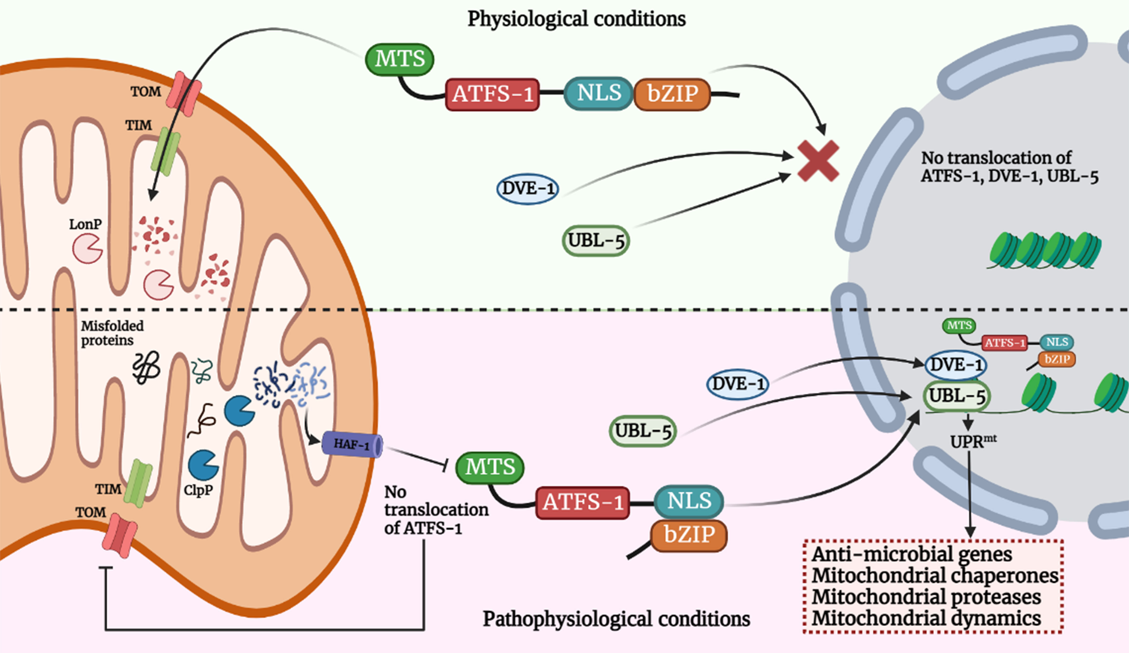
Figure 1 Model for UPRmt signaling pathway. Activation of the UPRmt in cells occurs in response to different stress signals such as mtROS, mutation in mtDNA, accumulation of misfolded mitochondrial proteins in the mitochondrial matrix or in IMS and alteration in mitochondrial membrane potential due to bacterial infection. Such events trigger the translocation of ATFS-1 (mammal homologue - ATF-5) which is bZIP protein, and contains MTS and NLS sequence into the nucleus and along with DVE-1 and UBL-5, it induces a transcriptional up-regulation program known as UPRmt. Under physiological conditions, it is imported in the mitochondria via TOM/TIM machinery and digested in the mitochondrial matrix by mitochondrial protease LonP which prevents activation of UPRmt. Under pathophysiological or stressed conditions, import of ATFS-1 is blocked in mitochondria. The misfolded proteins digested in the mitochondrial matrix via ClpP and efflux of such peptides into the cytoplasm via HAF-1 prevents ATFS-1 translocation into mitochondria, and therefore it moves into the nucleus and induces multiple genes which functions in restoring mitochondrial functions, innate immunity facilitating bacterial clearance. The image was created with the help of BioRender.com.
Furthermore, knockdown of ribosomal subunits, including large subunits (rpl-14, rpl-25.1, rpl-27, rpl-36 and rpl-43) and small subunits (rps-8 and rps-10) individually blocked cco-1 or mrps-5 RNAi-induced UPRmt activation (Li et al., 2022). In addition, significant increase in polysomal mRNA of ATFS-1 was reported in response to cco-1 RNAi, which was attenuated with vha-1 RNAi co-treatment (Li et al., 2022). Knockdown of cco-1 resulted in shifting from polysomes to monosomes, which confirms reduction in cytosolic translation in response to mitochondrial stress (D’Amico et al., 2017; Suhm et al., 2018; Molenaars et al., 2020). Together, these results suggest that increased translation of ATFS-1, mediated by v-ATPase/TORC1 and cytosolic ribosomes, is a key mechanism that leads to the accumulation of ATFS-1 protein for UPRmt activation in response to mitochondrial stress.
Under stressed conditions, mitochondria enhance TORC1 activity via v-ATPase- and Rheb-dependent mechanism. TORC1 has been documented to be essential for UPRmt activity (Shpilka et al., 2021), and activated TORC1 is associated with increased translation of ATFS-1, reliant on the cytosolic ribosomes (Li et al., 2022). Although, how TORC1 is activated in response to mitochondrial stress remains a mystery and highlights an important direction for future work. One possibility could be that the unfolded proteins produced upon mitochondrial stress might be transported from mitochondria to lysosomes, and eventually digested to peptides/amino acids within the lysosomes, which could then lead to TORC1 activation at the lysosomal surface (Zoncu et al., 2011; Shimobayashi and Hall, 2014; Wolfson and Sabatini, 2017; Lawrence and Zoncu, 2019), however, what type of peptides/amino acids led to TORC1 activation still needs an investigation. Thus, mitochondrial stress likely signifies an inimitable intrinsic cue for TORC1 activation through lysosome-derived peptides/amino acids (Hesketh et al., 2020); furthermore, stressed mitochondria might also establish direct contact with lysosomes via mitochondria–lysosome membrane contact sites (Wong et al., 2019). As v-ATPase are involved in fusion of endosomal membranes, hence, it facilitates transportation of mitochondria-derived unfolded proteins to lysosomes, and thus, are crucial in UPRmt activation.
Under pathophysiological conditions, the molecular mechanism that exposes the NLS region of ATFS-1 in the cytosol is not well understood and merits further studies. Based on the studies concerned with functional alteration in ATFS-1 (Nargund et al., 2012; Rolland et al., 2019), it can be suggested that import of ATFS-1 is impaired due to reduction in mitochondrial membrane potential and hence, translocate into the nucleus; furthermore, another hypothetical explanation could be that ATFS-1 senses stress-induced structural-functional alterations of the translocases and other constituents of mitochondrial protein import complex, and translocate to the nucleus. Identifying bacterial virulence proteins-induced alterations in mitochondria that selectively inhibits mitochondrial import of ATFS-1 is vital to understand mitochondrial dysfunction in bacterial infections. Importantly, ATFS-1 per se regulates the induction of several core components of mitochondrial protein import complex core components (Chacinska et al., 2009), suggesting the cross-talk between ATFS-1 and mitochondrial protein import complex essential for the maintenance of mitochondrial health; the failure to import ATFS-1 provides a feedback loop to the nucleus to initiate mitochondrial protein import and restore normalcy.
Besides pathogenic insult, various other factors, such as impairment of ETC, alteration of mitochondrial network dynamics, deletion of mitochondrial DNA (mtDNA), inhibition of mitochondrial chaperones, impaired expression of genes regulating mitochondrial functioning like protein import, OXPHOS, coenzyme and lipid biogenesis also trigger UPRmt in C. elegans (Nargund et al., 2012; Runkel et al., 2013; Qureshi et al., 2017; Voth and Jakob, 2017) which suggests the pluralism behind triggering UPRmt and its impact on different physiological processes and pathologic manifestations.
UPRmt is less well understood in other higher animals and mammals. It has been observed that besides pathogenic stress, other factors like the perturbation of mitochondrial ribosomes, ectopic expression of mutant ornithine transcarbamylase (OTC), and depletion of mtRNA induce UPRmt in mammalian cells (Zhao et al., 2002; Wu et al., 2014; Moullan et al., 2015). Even the presence of cytosolic protein aggregates has been found to initiate UPRmt in several neurodegenerative diseases (Zhu et al., 2021); however, the link between cytosolic, mitochondrial proteostasis and UPRmt is unclear under such settings. In mammals, several orthologous transcription factors (ATF4, ATF5, CHOP and C/EBP-β) have been reported (Fiorese et al., 2016; Quirós et al., 2017; Melber and Haynes, 2018). It has been suggested that both ATF4 and ATF5 are involved in expressing stress-responsive and cytoprotective genes in mammals (Fiorese et al., 2016; Quirós et al., 2017), but their relationship is not known. Additionally, mammalian cells, on sensing dysfunctional mitochondria activate MEK/JNK2 pathway inducing downstream expression of CHOP and C/EBP-β, which in turn promotes the transcription of downstream stress-responsive genes (Wang et al., 2018). Further, it has been noted that CHOP regulate ATF5 expression to upregulate UPRmt genes (Teske et al., 2013; Fiorese et al., 2016) by interacting with SatB2 (mammalian orthologue of DVE-1) and Ubl5 (mammalian orthologue of UBL-5) (Haynes et al., 2007). There are also reports implicating ATF4 plays an indirect role in inducing UPRmt responses by mounting the general cytoprotective integrated stress response (ISR) (Quirós et al., 2017). Furthermore, another type of UPRmt that occurs in the IMS has also been reported in mammals (Papa and Germain, 2011). IMS-UPRmt instigates reactive oxygen species (ROS) production and triggers the phosphorylation of AKT kinase that activates estrogen receptor α (ERα) and upregulates the transcription of the mitochondrial regulator, NRF1 and the IMS protease Omi (HTRA2) alleviating stress (Papa and Germain, 2011). Collectively, these findings lead to an essential question of whether the UPRmt in mammals and C. elegans act similarly, and identifying homologous transcription factors in higher metazoans will shed light on this.
With the role of mitochondria as an innate immune hub being established, and UPRmt central to mitochondrial health and resilience, scientists have long been interested in studying its involvement in immunity. It has only recently been observed that UPRmt supports host tolerance by regulating mitochondrial homeostasis and endorsing host resistance through the transcription of innate immunity genes (Liu et al., 2014; Pellegrino et al., 2014; Jeong et al., 2017).
Report suggested that bacterial toxins which target the mitochondria and host cell trigger UPRmt to counter mitochondrial dysfunction and initiate innate immune responses in C. elegans (Pellegrino et al., 2014). Using the C. elegans model, Pellegrino et al., 2014 studied the relationship between UPRmt and innate immunity, and observed that UPRmt confers long-term protection against Pseudomonas aeruginosa by inducing the expression of innate immune genes (Pellegrino et al., 2014). The involvement of ATFS-1 in upregulation of antibacterial factor-related peptide 2 (Abf-2), caenacin (CNC-4), lysozyme, clec-4 has also been reported in C. elegans (Nargund et al., 2012; Pellegrino et al., 2014; Nargund et al., 2015; Sapkota et al., 2021; Li et al., 2022).
Although UPRmt is not well studied in higher animals, its involvement in inducing immune response genes and improving fitness against pathogens is getting increasingly evident (Wang et al., 2018; Chamseddine et al., 2022). UPRmt components such as ATF5/ATFS-1 and HSP60 are upregulated during pathogenic infection in reef-building coral and the whiteleg shrimp (Chen et al., 2016; Dimos et al., 2019). ATFS-1 induces the expression of antimicrobial peptides required for survival following Vibrio alginolyticus infection in whiteleg shrimp (Chen et al., 2016). Our recent study suggested that infection with Aeromonas hydrophila-induced UPRmt triggered apoptosis of infected fish macrophages by enhancing expression of dnm1l gene which encodes DRP1 protein, therefore results in the fragmentation of the mitochondrial network; and in turn, mitochondrial fission reduced mitochondrial membrane potential (ΔΨm) which prompted release of cyt c in the cytosol leading to the activation of caspase-9 facilitating clearance of the intracellular bacteria (Kumar et al., 2022a). A recent study by reported that UPRmt plays a protective role during enteric pathogenic infection in mammalian host. ATF5-mediated UPRmt activation protects the mice against Salmonella enterica subsp. enterica (Serovar Typhimurium) by promoting intestinal barrier function via stimulation of satiety response and averting excessive glycolytic flux, which in turn prevented microbial and microbial toxins’ infiltration into underlying tissues (Chamseddine et al., 2022). ATF5 prevented intestinal barrier dysfunction by promoting cholecystokinin/leptin-mediated satiety response (Chamseddine et al., 2022). The list of UPRmt-induced genes along with their function in regulating immunity has been depicted in (Table 1). Though it is not evident whether different pathogens employ distinct mechanisms to induce UPRmt, these preliminary findings suggest that UPRmt-mediated regulation of innate immune genes is conserved across metazoans.
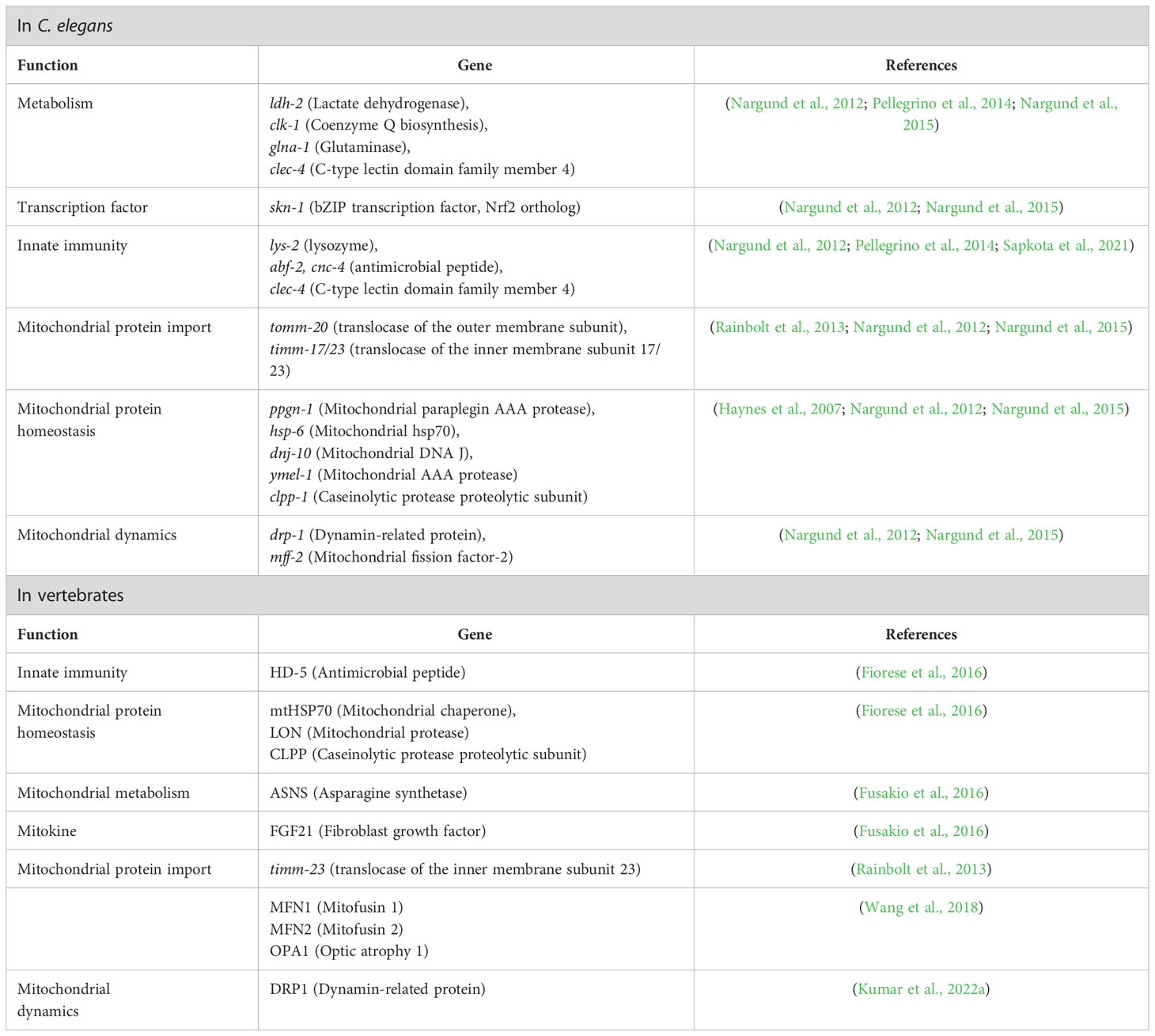
Table 1 UPRmt-induced genes in C. elegans and vertebrates.
Stressed mitochondria release DAMPS in bacterial infection (Jabir et al., 2015; Kumar et al., 2022a), which induce inflammatory responses and are critically involved in the pathogenesis of various diseases. UPRmt may be regarded as an additional intracellular sensing mechanism that helps perceive damage inflicted by bacterial pathogens targeting mitochondrial bauplan and functions, and initiating remedial pathways crucial for restoring mitochondrial function and atoning inflammatory responses to promote cellular survival. Thus, it can be suggested that UPRmt acts as a conduit coupling host resistance and tolerance, safeguarding the host during infection.
Recent reports demonstrated that bacterial pathogen subvert UPRmt using bacterial enzymes (Mahmud et al., 2020) and targeting host factors (Deng et al., 2019) which regulate UPRmt activation. For instance, Acyl-CoA dehydrogenase (FadE2) of P. aeruginosa possesses substrate specificity for the catabolites produced during the breakdown of the branched chain amino acids (valine and leucine), i.e., isobutyryl CoA and isovaleryl CoA respectively. During infection, bacterial FadE2 limits the availability of such catabolites for C. elegans through an unknown mechanism, which subsequently hinders in the activation of the UPRmt (Mahmud et al., 2020). FadE2 impairs several energy pathways such as glycolysis, β-oxidation, and amino acid metabolism in C. elegans, all of which culminate with the TCA cycle. Consequently, loss of FadE2 in P. aeruginosa, results in restoration of energy metabolism due to availability of the catabolites in C. elegans and the ability to activate UPRmt through a mechanism that is yet unknown. However, the exact molecular underpinnings of how FadE2 affects UPRmt activity during infection via changes in valine or leucine levels is currently not resolved. Recently, it was shown that the C. elegans bZIP transcription factor, ZIP-3 is involved with the repression of the UPRmt during P. aeruginosa infection (Deng et al., 2019). During infection, P. aeruginosa exploits the regulative activities of this transcription factor for manoeuvring UPRmt activation. Although, the mechanism of how P. aeruginosa manipulates ZIP-3 are unclear. The overlap observed between the genes negatively regulated by FadE2 and ZIP-3 strongly implicates some link between ZIP-3 and FadE2 in regulating UPRmt. P. aeruginosa negatively impacts both host energy metabolism and the activation of UPRmt by outcompeting the host for specific nutrients. However, the molecular mechanisms linking these catabolites to host energy pathways and mitochondrial stress signaling are still unresolved, and therefore will be an exciting area of future research.
Mitochondria are highly mobile organelles due to two significant processes termed mitochondrial dynamics: fission and fusion (Shaw and Nunnari, 2002; Chen and Chan, 2004). The term ‘mitochondrial dynamics’ refers to variations in shape, size, and localization of mitochondria and the system which controls these processes (Liesa and Shirihai, 2016). Besides assisting in various cellular functions such as oxidative phosphorylation, mitochondrial segregation into daughter cells at the time of cell division, propagation of intra-mitochondrial Ca2+ signal, and refurbishing of defective mitochondria (Chen and Chan, 2005; Twig et al., 2008; Westermann, 2010), the fission-fusion cycle also contributes in aiding the organelle to adapt to different conditions of stress (Westermann, 2012; Youle and van der Bliek, 2012; Ren et al., 2020). Mitochondrial dynamics also influence the kinetics of biochemical reactions, mitochondrial inner membrane topology, and protein super-complex assembly in the mitochondrial cristae (Lizana et al., 2008; Cogliati et al., 2013; Mannella et al., 2013). Most importantly, it has been reported that disruption of mitochondrial dynamics undermines their function and causes several human diseases (Suárez-Rivero et al., 2016; Chan, 2020).
A group of classical and distant dynamin-related GTPases regulates the fusion-fission process. Mitochondrial fusion is a two-step process highlighted by the union of the outer mitochondrial membrane (OMM) followed by the union of the inner mitochondrial membrane (IMM) of two originally distinct mitochondrion. This process is controlled by IMM protein, optic atrophy 1 (OPA1), and OMM proteins-mitofusin 1 and mitofusin 2 (MFN1 and MFN2). The trans-interaction between MFNs on opposing OMMs involves continuous hydrolysis of GTP, whereas the IMM fusion depends on OPA1 assembly stimulating GTPase activity and membrane tubulation (Gao and Hu, 2021) (Figure 2). It has been speculated that OPA1-mediated inner membrane fusion may occur only after receiving a signal from active mitofusins on the outer membrane, thus coordinating the fusion process (Hoppins, 2014). In the case of mitochondrial fission, a classical dynamin-related protein, termed dynamin-related protein 1 (DRP1) in mammals, is the master regulator of mitochondrial fission machinery (Westermann, 2010). It is a soluble protein containing an N-terminal GTPase, a middle domain, and a c-terminal GTPase effector domain involved in self-assembly. The fission cycle starts with the involvement of two partner proteins, mitochondrial fission 1 (FIS1) and mitochondrial fission factor (MFF) (Losón et al., 2013; Kraus et al., 2021). First, FIS1, an OMM-anchored protein, recruits MFF from the cytosol to the OMM, leading to the nucleation of GTP-bound DRP1 oligomers on the OMM (Palmer et al., 2011; Losón et al., 2013). Next, in this line, DRP1-GTP oligomers form spirals that are eventually wrapped around the organelle and sever the mitochondrial membranes following GTP hydrolysis (Mears et al., 2011) (Figure 2). Mitochondrial fusion and fission processes are antagonistic. The right balance between these two must be achieved by tightly controlling the rate of fusion and fission. This regulation is a prerequisite for the maintenance of mitochondrial morphology or for adapting to ever-changing physiological conditions.
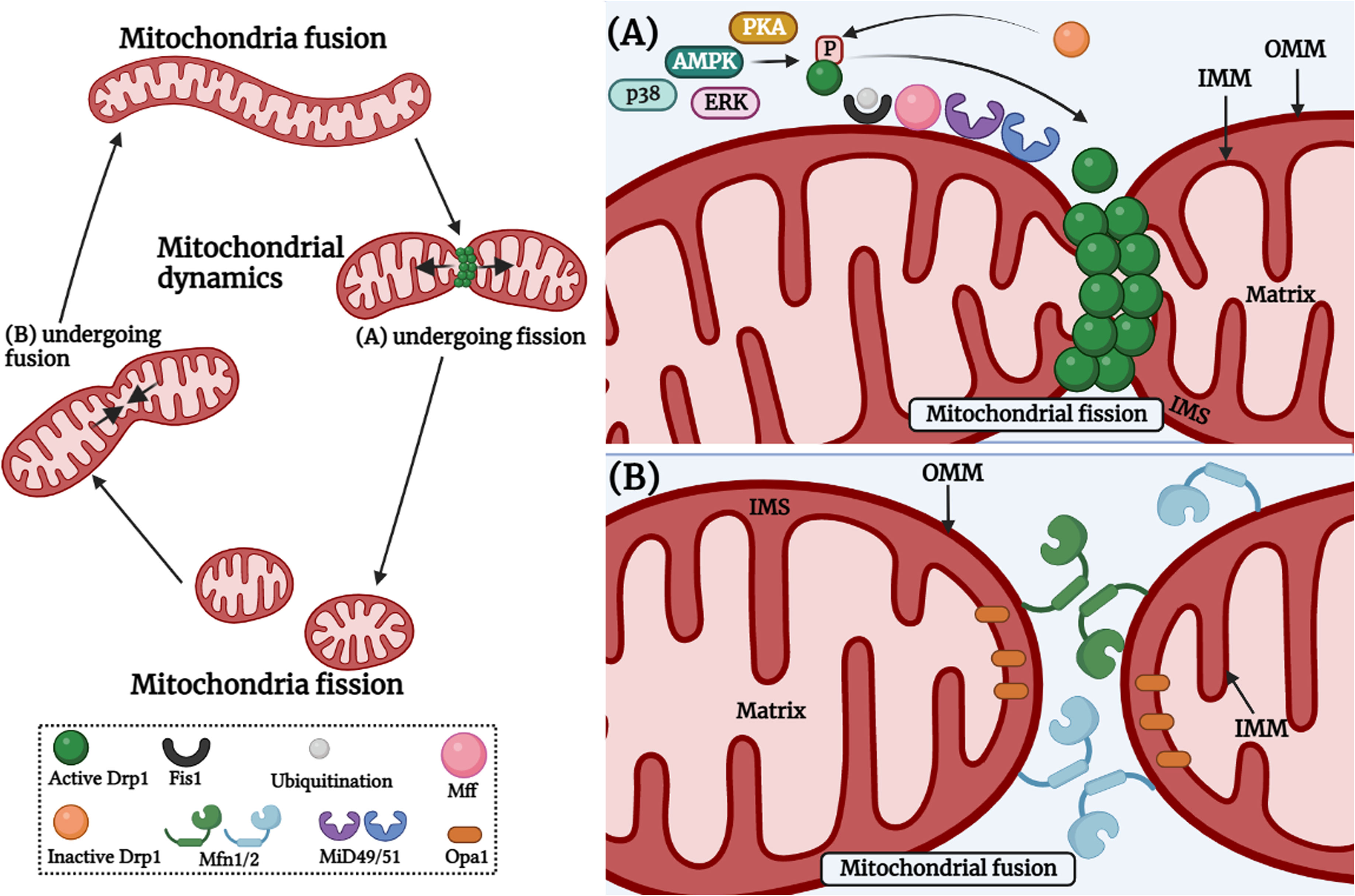
Figure 2 Mitochondrial fission and fusion cycle. Mitochondria are constantly dynamic cellular organelles and morphology of mitochondrial network keeps on changing. Mitochondrial fission involves the contact between endoplasmic reticulum and mitochondria, and the constriction occurs via actin cytoskeleton proteins. (A) Mitochondrial fission is controlled by post-translational modifications of Drp1, which includes S-nitrosylation, phosphorylation, SUMOylation, O-GlcNAcylation, and ubiquitination in response to various cellular stimuli via PKA, AMPK, ERK, p38 etc. After constriction, DRP1 is recruited to the fission site at the mitochondrial outer surface and interacts with adaptor proteins such as FIS1, MFF, MiD49 and MiD51 on the OMM (Right upper panel). (B) Mitochondrial fusion involves fusion of the OMM which occurs via the interaction of MFN1 and MFN2 between two individual mitochondria. Then, fusion of the IMM occurs with the help of OPA1 GTPase which is present at the IMM (Right lower panel). The image was created with the help of BioRender.com.
In the past decade, numerous studies have revealed the importance of mitochondrial metabolism in immunity (Bahat et al., 2021; Timblin et al., 2021) and mitochondrial dynamics are essential for immune responses mediated by various cell types (Mishra and Chan, 2016; Wai and Langer, 2016) and interconnected with MQC (Ježek et al., 2018) such as UPRmt. The disruption of mitochondrial dynamics has detrimental consequences for mitochondrial and cellular homeostasis, and this leads to the activation of the UPRmt (Kim and Sieburth, 2018; Rolland et al., 2019; Haeussler et al., 2020; Haeussler et al., 2021). Loss of intestinal and neuronal fzo-1 (encodes for mitofusin) in C. elegans induces fragmentation of the mitochondrial network, reduces mitochondrial membrane potential (Haeussler et al., 2020; Chen et al., 2021) which in turn triggers UPRmt in intestine (Chen et al., 2021). Chen et al., 2021 further reported that silencing of neuronal fzo-1 triggers non-autonomous UPRmt via neuromodulators such as tyramine, and neurotransmitters such as serotonin (Chen et al., 2021). A recent report highlighted the role of neuronal signals in non-autonomous UPRmt regulation. unc-17 (vesicular acetylcholine transporter), eat-4 (vesicular glutamate transporter), tdc-1 (tyrosine decarboxylase), tph-1 (tryptophan hydroxylase) mutations suppressed UPRmt activation which clearly implicated that acetylcholine, glutamate, tyramine, and serotonin are essential in communicating mitochondrial stress between neurons and intestine. The interruption in the activity of RIM and RIC significantly reduced non-autonomous UPRmt activation, and tyramine plays a crucial role in propagating non-autonomous UPRmt via TYRA-3 receptor. And in addition to this, perturbation of neuronal mitochondrial dynamics activated UPRmt without any involvement of mitochondrial respiration (Chen et al., 2021) which clearly implicates direct involvement of mitochondrial fragmentation in UPRmt activation, and activation of UPRmt in C. elegans confers protection against P. aeruginosa infection (Pellegrino et al., 2014).
In C. elegans, knockdown of fzo-1, eat-3 which regulate mitochondrial fusion or drp-1 which regulates mitochondrial fission induces UPRmt (Kim and Sieburth, 2018; Zhang et al., 2018). Mitochondrial dynamics affects levels of triacylglycerols, and fzo-1 mutants exhibited significant reduction in the number of short-chain length triacylglycerols, and increase in unsaturated triacylglycerols (Haeussler et al., 2020) implicating alteration in the contact sites between mitochondria and triacylglycerols. Disruption in mitochondrial fusion affects transfer of fatty acid from lipid droplets to mitochondria in mammalian cells, leading to heterogeneous distribution of fatty acid among mitochondria (Rambold et al., 2015), and loss of fzo-1 or drp-1 results in disruption of the contact sites between lipid droplets and mitochondria (Haeussler et al., 2020); and such alterations induces metabolic changes. Autophagy suppresses UPRmt. And recent report highlighted the involvement of mitochondrial dynamics in autophagy-mediated UPRmt regulation (Haeussler et al., 2020). Autophagy regulates metabolism of fatty acids, it triggers breakdown of triacylglycerols, and thus ensures a continuous supply of fatty acids to mitochondria for β-oxidation (Singh et al., 2009). And thus, autophagy reverts such changes in the levels of triacylglycerols in fzo-1 mutants (Haeussler et al., 2020). Hence, autophagy triggers enhanced breakdown of triacylglycerols in mutants with defects in mitochondrial dynamics, that are subsequently used to fuel mitochondrial metabolism, and thereby increases mitochondrial membrane potential leading to the suppression of UPRmt. Decrease in mitochondrial membrane potential has recently been shown to be a prerequisite signal for UPRmt induction (Rolland et al., 2019). The first evidence that autophagy can affect UPRmt was the finding by Haynes et al. that knock-down of rheb-1, a known positive regulator of TOR (Honjoh et al., 2009), and autophagy is dependent on ATFS-1 (Nargund et al., 2012; Guo et al., 2014) and induction of autophagy leads to improved mitochondrial function by affecting lipid metabolism and ameliorating cellular homeostasis, thereby suppressing UPRmt in mutants with defects in mitochondrial dynamics. These reports clearly demonstrate the relationship between mitochondrial dynamics and UPRmt. Further, the below mentioned section mainly discusses the role of mitochondrial dynamics in immunity and host-pathogen infections.
Innate immunity is the first line of defence against invading pathogens. It is evolutionarily conserved and shows little variation while responding to pathogens. Neutrophils are essential constituents of the innate immune system. These cells are recruited earliest to the sites of infection and respond by releasing ROS, proinflammatory cytokines, and neutrophil extracellular traps (NETs) (Papayannopoulos, 2018; Schultz et al., 2022). It was observed that silencing of OPA reduced ATP production and NET formation, implicating mitochondrial fission’s role in the process (Cervantes-Silva et al., 2021). Monocytes are phagocytic cells that can differentiate into macrophages and dendritic cells. Under pathogenic stress, circulating blood monocytes are recruited to the sites of inflammation and differentiate into macrophages (Shi and Pamer, 2011). The metabolic status of monocytes alters in response to the environmental stimuli, and the cellular metabolism plays a crucial role in monocyte activation in which mitochondria is a major central player in regulating cellular metabolism. Hence, mitochondrial dynamics is closely associated with monocyte-macrophage differentiation. And mitochondrial dynamics is reported to play a critical role in monocyte-macrophage differentiation (Rambold and Pearce, 2018). Stimulation of human PBMCs by TLR4 agonist i.e., LPS induced differentiation of monocytes into macrophages (Krutzik et al., 2005). The size of the mitochondrial network and mitochondrial footprints, i.e., mitochondrial mass increases in BMDMs as compared to the PBMCs which showed tubular mitochondria (Li et al., 2020), which clearly suggested mitochondria of monocytes undergo biosynthesis and elongation during monocyte-macrophage differentiation. Furthermore, PBMCs mainly relies on the basal level of OXPHOS and glycolysis (Chacko et al., 2013), whereas, BMDMs relies on enhanced OXPHOS activity (Na et al., 2015), and the elongation of mitochondrial network elevates OXPHOS activity (Yao et al., 2019). Pre-treatment of CD14+ human monocytes with LPS triggers their activation which involved reduced expression of MTFP1 (Mitochondrial Fission Process 1), which in turn increase mitochondrial fusion (Duroux-Richard et al., 2016). Similarly, elongation of mitochondrial network is also reported during monocyte to macrophage differentiation in murine cells (Li et al., 2020). TLR-4 senses LPS of Gram-negative bacteria, and TLR-4-LPS axis results in marked inhibition in the expression of mitochondrial fission machinery with a concordant increase in mitochondrial fusion (Duroux-Richard et al., 2016; Cervantes-Silva et al., 2021). However, reports also suggest TLR signaling alters mitochondrial morphology initiating a switch from fusion to fission (Hoppins, 2014; Kumar et al., 2022a). Though contradictory, these findings are proof of the involvement of TLRs in mitochondrial dynamics. Notably, the TLR family contains several members and isoforms thereof. Given the evolutionarily conserved nature of TLR signaling it will be interesting to determine how mitochondria perceive TLR signaling and whether the molecular cross-talk between mitochondria and different TLRs is the same. Both mitochondria and TLR represent keystones of innate immunity, future research addressing how mitochondrial dynamics harness TLR signaling and vice versa will help in therapeutics for innate immune disorders.
Macrophages are terminally differentiated phagocytes broadly classified into pro-inflammatory M1 and anti-inflammatory M2 sub-types (Cervantes-Silva et al., 2021). The M1 phenotype exhibits a fragmented mitochondrial network, and M2 phenotype shows elongated and fused mitochondria (Hinshaw et al., 2021). LPS stimulation triggers the development of the M1 subtype by inhibiting the expression of fusion molecules (MFN1 and MFN2) and phosphorylation of Drp-1 (Ser637) critical for triggering the fission pathway (Gao et al., 2017). Mitochondrial dynamics are crucial in regulating inflammasome activation in macrophages which help host cells against microbial infections. It has been shown by several groups that the expression of mitochondrial fusion proteins and defective mitochondrial fission favour inflammasome formation (Ichinohe et al., 2013; Park et al., 2015). Mast cells are involved in hypersensitivity reactions and innate immunity to several pathogens. They recognize ligands through FcϵRI/II receptors and act by releasing secretory granules, which induce diverse effects depending on the nature of the ligand and anatomical locations. Inhibiting DRP1 activity negatively affects mitochondrial translocation and granule exocytosis suggesting the importance of mitochondrial dynamics in mast cell functioning (Zhang et al., 2011).
Mitochondrial dynamics have been linked with several aspects of B- and T-cell functioning. Studies by several groups have suggested an intimate relationship between mitochondrial dynamics and immunological synapse formation and regulation (Campello and Scorrano, 2010; Quintana and Hoth, 2012). Inhibiting the expression of Drp1 was found to have little effect on T-cell differentiation but hindered T-cell migration and activation following antigenic challenge (Simula et al., 2018). The naive T-cells differentiate into effector (TE) and memory (TM) subtypes upon antigenic exposure. While the naive T-cells have small fragmented mitochondria and are essentially dependent on mitochondrial OXPHOS for energy requirements, TE cells undergo a quantum increase in mitochondrial mass via Drp-1 mediated fission and acquire their energy quotient by aerobic glycolysis (Buck et al., 2016). Drp-1 mediated fission also aids in generating mtROS (Gao et al., 2017) which in turn influences the activation of nuclear factor of activated T cells (NFAT) and subsequent IL-2 production, essential for the functioning of TE cells (Sena et al., 2013). Prolonged mtROS generation is deleterious for cellular health, and how TE cells regulate the Drp-1/mtROS cascade, thereby maintaining a delicate balance between clonal activation and clonal deletion, merits future investigations. Alteration in mitochondrial mass and ensuing morphological changes are reported to play a critical role in the development of TM cells. The TM cells have hyper-fused mitochondria and depend on fatty acids for energetics and recall responses. Inhibition of OPA1 expression reduced TM cell survival, and further reported that pharmacological and genetic ablation/silencing of Drp-1 or endorsing OPA1 expression tilts the balance in favour of TM cell development (Buck et al., 2016). Importantly, TM cells are long-lived and require a low energy budget which also implicates the role of mitochondrial fusion in their longevity. These findings suggest mitochondrial dynamics is essential in shaping different aspects of T-cell biology, but how the fine-tuning between two disparate events, fission, and fusion, is achieved needs to be better understood.
Right from inception in the bone marrow, B-cells undergo cycles of differentiation and activation, each stage posing distinct metabolic demands. Like T-cells, activated B-cells have a higher energy demand, and depend more on anabolic metabolism for their energy requirements than naive B-cells (Sandoval et al., 2018). The replacement of elongated mitochondria in naive B-cells with numerous rounded mitochondria following activation suggests the involvement of mitochondrial fission in B-cell activation (Waters et al., 2018). Though these early findings reflect the intimate relationship between the two molecular events, further studies are needed to cement the role of mitochondrial dynamics on B-cell development, differentiation, and functioning.
Mitochondrial dynamics, the key to mitochondrial health, has been targeted by numerous pathogens for survival and causing infections. Pathogenic bacteria have been shown to modulate mitochondrial dynamics to create a niche ideal for intracellular replication, immune evasion, and persistence (Khan et al., 2020; Tiku et al., 2020). The role of mitochondrial dynamics on host immunity has been studied in detail in Listeria monocytogenes pathogenesis. L. monocytogenes elicits short-term mitochondrial fission and reduction in energy production using the pore-forming toxin listeriolysin O (LLO) (Figure 3A). Mitochondrial respiration restricts the growth of this intracellular pathogen, and it survives by sophisticated manipulation of mitochondrial dynamics (Stavru et al., 2011). During infection, it hijacks the mitochondrial metabolism machinery enforcing the expression of receptors that aid pathogen entry inside cells, suggestive of novel manoeuvring of mitochondrial function by the pathogen (Spier et al., 2021). Additionally, while pathogenic L. monocytogenes fails to infect cells with fragmented mitochondria, the infection was more prevalent in cells with fused mitochondria (Stavru et al., 2011), suggesting the positive correlation between infectibility and L. monocytogenes-induced mitochondrial fission is critical for pathogenesis induced by the bacterium. Interestingly, L. monocytogenes-induced mitochondrial fission is transient and occurs in the early phase of infection (Stavru et al., 2011). Selectively impairing mitochondrial dynamics at the initial stages provides L. monocytogenes a window for successful infection and ensures that the infected cell remains free of DAMPS released by damaged mitochondria, which otherwise induces pro-inflammatory responses counterproductive to the pathogen. How the bacteria sense the extent of mitochondrial damage and precisely manoeuvre the mito-repair machinery need to be studied. Recently, it was reported that Mic10, a critical component of the mitochondrial contact site and cristae organizing system (MICOS) complex, is responsible for L. monocytogenes-induced mitochondrial fission (Carvalho et al., 2020), suggestive of novel mechanisms used by pathogens to circumvent host immunity and it is crucial to know whether other pore-forming bacteria use similar mechanisms to establish infections.
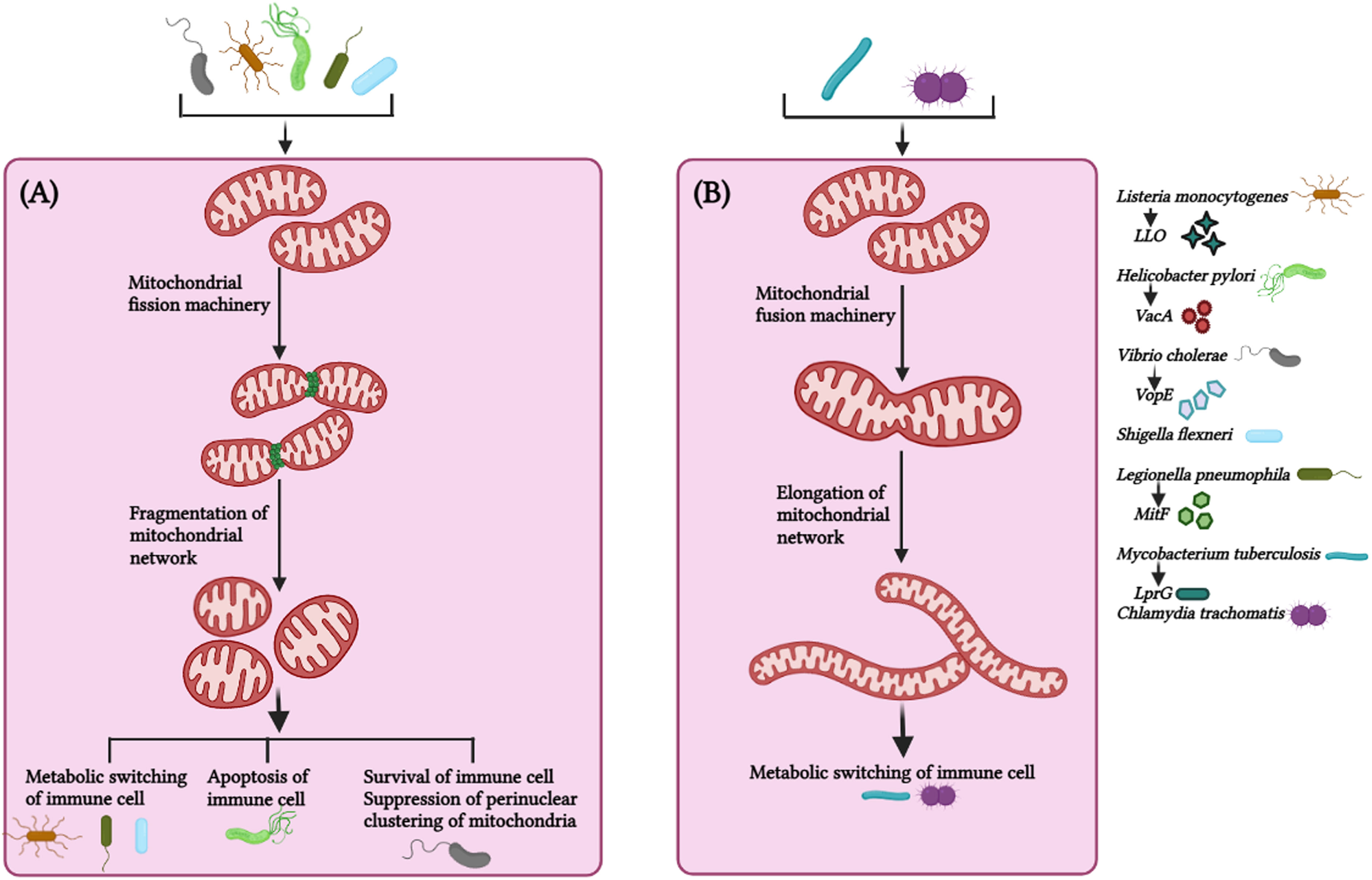
Figure 3 Mitochondrial dynamics in host immunity against bacterial infection. (A) Bacteria such as Listeria monocytogenes, Helicobacter pylori, Vibro cholerae, Shigella flexneri, Legionella pneumophila triggers fragmentation of the mitochondrial network via virulence factors (LLO – Listeriolysin, VacA – Vacuolating cytotoxin A, VopE – mitochondrial targeting T3SS effector protein, MitF – T4SS effector protein, respectively and effector protein in case of Shigella flexneri is not known) to maintain a favorable microenvironment for pathogen survival and thus promote bacterial survival, persistence and metabolic switching of the immune cells. (B) Bacteria such as Mycobacterium tuberculosis and Chlamydia trachomatis circumvent host immune response by manipulating the mitochondrial dynamics and maintains elongated network of mitochondria at initial phase of infection for their survival. Alteration in mitochondrial fission (by L. monocytogenes, H. pylori, V. cholerae, S. flexneri, L. pneumophila) and mitochondrial fusion (M. tuberculosis, C. trachomatis) suppress bacterial replication and facilitate bacterial clearance from the cell. M. tuberculosis utilize LprG (Lipoarabinomannan carrier protein) for mitochondrial fusion. The image was created with the help of BioRender.com.
Helicobactor pylori cause persistent gastric infections leading to peptic ulcers and stomach cancer. The bacteria release vacuolating cytotoxin VacA, which activates Drp1-mediated fragmentation of the mitochondrial network culminating in apoptosis of infected cells and pathogen survival (Jain et al., 2011) (Figure 3A). Inhibiting Drp-1 attenuated the expression of pro-apoptotic Bax and cytochrome C besides helping in pathogen clearance suggesting the cross-talk between mitochondrial dynamics and apoptosis critical for H. pylori pathogenesis (Jain et al., 2011).
The non-invasive pathogen Vibrio cholerae colonizes the small intestine and produces cholera toxin, causing severe secretory diarrhoea. Miro (mitochondrial Rho GTPases) is a mitochondrial outer membrane protein that regulates mitochondrial dynamics by interacting with MFN1, MFN2, OPA1, DRP1, and FIS1 (Chan et al., 2006; Westermann, 2010; Panchal and Tiwari, 2021). V. cholerae triggers fragmentation of the mitochondrial network and restricts perinuclear clustering of the mitochondrial network, which is critical for mitochondria-mediated immune responses (Suzuki et al., 2014) (Figure 3A). The overexpression of EFm mutants of Miro or Mfn 1 mutant (loss-of-function GTPase in activity) significantly occluded NF-κB activation and the clustering of the perinuclear mitochondrial network during infection (Suzuki et al., 2014), implicating the involvement of mitochondrial dynamics in regulating V. cholerae pathogenesis. Shigella flexneri utilizes mitochondrial dynamics for successful infection (Figure 3A). Silencing DRP1 expression revokes mitochondrial fragmentation, reduces the bacterial load, and prevents the cellular spread of S. flexneri (Lum and Morona, 2014).
Legionella pneumophila is a gram-negative bacterium responsible for Legionnaires’ disease. Inhibition of mitochondrial fragmentation or promotion of mitochondrial fusion decreases L. pneumophila replication (Escoll et al., 2017). L. pneumophila-induced alterations in mitochondrial dynamics via MitF allow the repurposing of the metabolism in macrophages to impair oxidative phosphorylation and activate the glycolytic cycle (Figure 3A). This metabolic shift of the infected macrophages strikingly resembles the Warburg effect, which is exploited by L. pneumophila for replication (Escoll and Buchrieser, 2018). Based on these observations, the inhibition of mitochondrial fission halts the onset of Warburg effect and restrains L. pneumophila infection in macrophages.
A Warburg effect-like scenario is observed with Mycobacterium tuberculosis infection, but this is quintessential for host cells to clear the intracellular pathogen (Aguilar-López et al., 2019) (Figure 3B). The probable mechanism behind this M. tuberculosis-induced transient altered mitochondrial dynamics is that mitochondrial fragmentation of the mitochondrial network at early stages provides a suitable micro-environment for effective infection and survival by impairing the metabolism of macrophages. In contrast, mitochondrial fragmentation is attenuated at later stages to reduce the damage to the infected macrophages, thus enabling persistent infection.
Interestingly, some pathogens invoke mitochondrial fusion for survival and spread. Chlamydia trachomatis is responsible for STD in males and females. The bacteria lack several key enzymes for metabolic activities and depend on the host for energy and survival. Chlamydia increases mitochondrial fusion by inhibiting DRP1 activation and maintains the mitochondrial network in the host for meeting energy requirements and its proliferation (Chowdhury et al., 2017; Kurihara et al., 2019) (Figure 3B). It was noted that C. trachomatis induces elongation of mitochondria in the early phase of infection but augments mitochondrial fragmentation during the later phases (Kurihara et al., 2019; Rother et al., 2019), the reason being that C. trachomatis requires ATP from the host cells for enhancing intracellular growth, and elongation of mitochondrial network via cAMP-mediated phosphorylation of Drp1-S637 promote ATP synthesis at the initial stages of infection, and later on, C. trachomatis shifts the metabolism from mitochondrial respiration to aerobic glycolysis in order to meet up the high metabolic demands of rapidly growing intracellular bacteria inside the cells. The Warburg effect supports the rapid proliferation of the intracellular bacteria by increasing cellular biomass, in which mitochondrial respiration is decreased and aerobic glycolysis is enhanced, therefore, C. trachomatis induces mitochondrial fission at the later stages of infection, such strategies by intracellular bacteria implicates the requirement of distinct morphology of mitochondrial network at different stages of infection. Table 2 summarizes the alteration of mitochondrial dynamics in response to diverse pathogenic infection (Table 2).
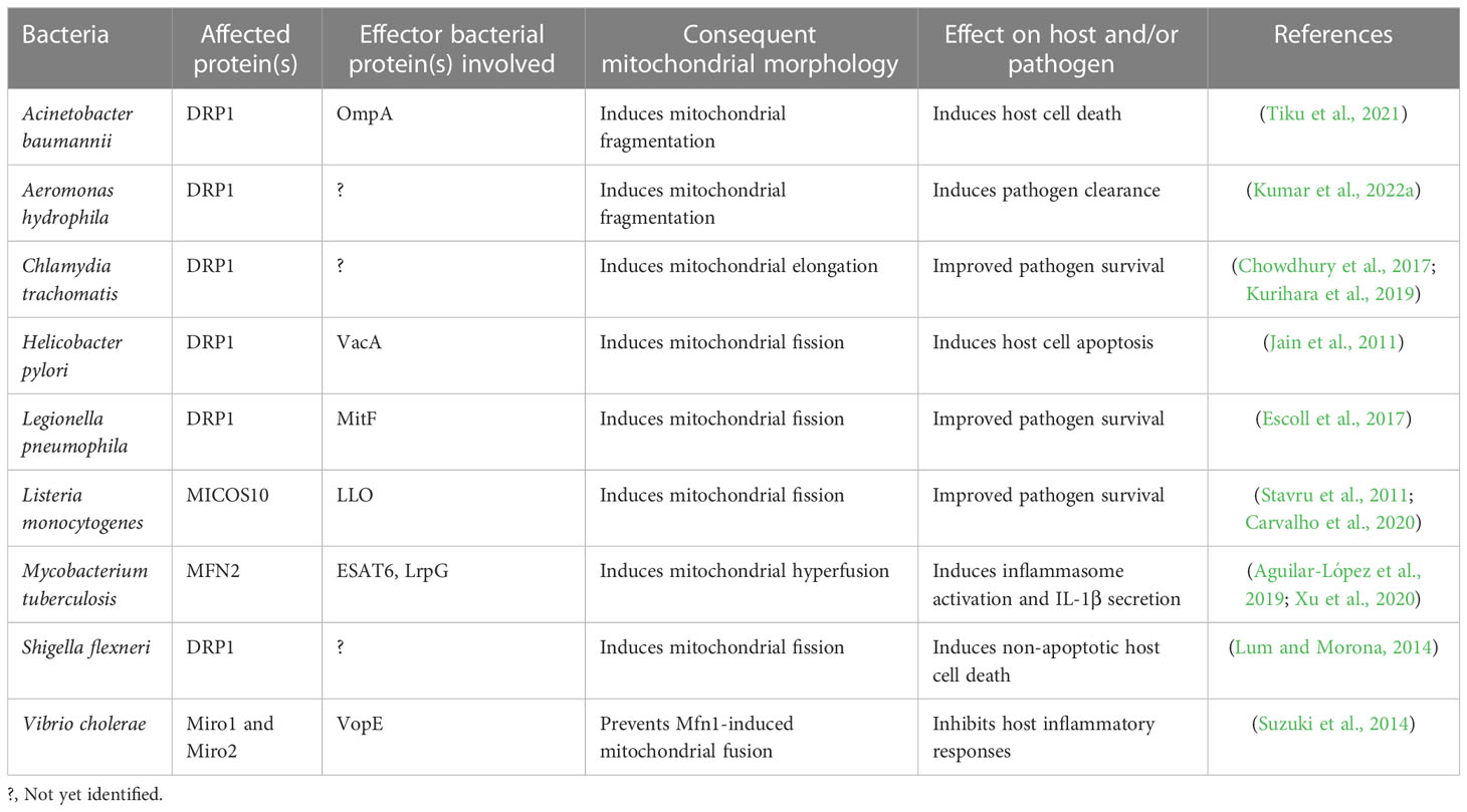
Table 2 Alteration in mitochondrial dynamics in response to various bacteria.
Recently, MFN2 has been reported as a master regulator of the immune response against bacterial infection (Tur et al., 2020). Out of all three mitochondrial fusion proteins, only MFN2 is highly expressed in macrophages predominantly upon immune stress signals such as pro-inflammatory activators (e.g., TLR ligands) (Lloberas et al., 2020), which also suggests that despite being highly homologous, MFN2 differs from MFN1 in certain functions. For example, mtROS production was impaired in MFN2-depleted macrophages, while depletion of MFN1 did not alter mtROS production in response to LPS endotoxemia (Tur et al., 2020). As expected, MFN2-depleted macrophages show highly fragmented mitochondria and a drastic reduction in mitochondrial membrane potential. MFN2-deficient macrophages cannot enhance their respiration rate in response to a metabolic change induced due to LPS endotoxemia (Tur et al., 2020). The depletion of MFN2 in macrophages affects phagocytic activity, as MFN2-/–macrophages showed a reduction in uptake of Aeromonas hydrophila, Staphylococcus aureus, and E. coli (Tur et al., 2020), which highlights the importance of this mitochondrial fusion protein in enhancing phagocytic activity of macrophages. Mice deficient in MFN2 are more susceptible to intracellular bacterial pathogens such as Mycobacterium tuberculosis and Listeria monocytogenes. Furthermore, the survival rate of MFN2-/- mice is strongly reduced in LPS-induced endotoxemia (Tur et al., 2020).
Thus, it can be concluded that the imbalance in mitochondrial dynamics results in diametrically opposite pro-host and pro-microbial outcomes. Although some progress has been made in this field dealing with the perturbation of mitochondrial dynamics at the host-pathogen interface, there are still many unresolved questions, including the mechanism of interaction between mitochondrial dynamic proteins and bacterial virulence factors. Understanding these aspects will improve our understanding of the involvement of mitochondria in regulating bacterial pathogenesis. The functional correlation between mitochondrial dynamics and bacterial pathogenesis should be further elucidated to develop new antibacterial treatment strategies and efficient therapeutics.
Mitochondria acts as a highly versatile player in governing different aspects in immune cells for mounting an efficient host response against bacterial infections, thus, it is understandable that variety of intracellular bacteria have evolved explicit strategies to hijack mitochondrial functions in order to create a viable niche for themselves. Likewise, extracellular bacteria have also developed numerous ways of targeting mitochondria to induce cell death to avail themselves with nutrients. Downstream of a bacterial infection, it is fascinating that a plethora of immune responses, be it against bacteria or bacterial products, are strongly impacted by mitochondrial dynamics and UPRmt (Figure 4). However, the investigations of these two functional aspects of mitochondria in immune response just started to unravel recently therefore, some key queries (see - In need of answers) still need a systematic comprehensive investigation. Thus, it will be exhilarating to extrapolate this current understanding and explore into how these fundamental mechanisms governed by mitochondria can be translated into active therapeutics to foster immunity against bacterial pathogens, and/or to keep unconcealed immune responses under control in the case of inflammatory disorders induced by bacterial infection.
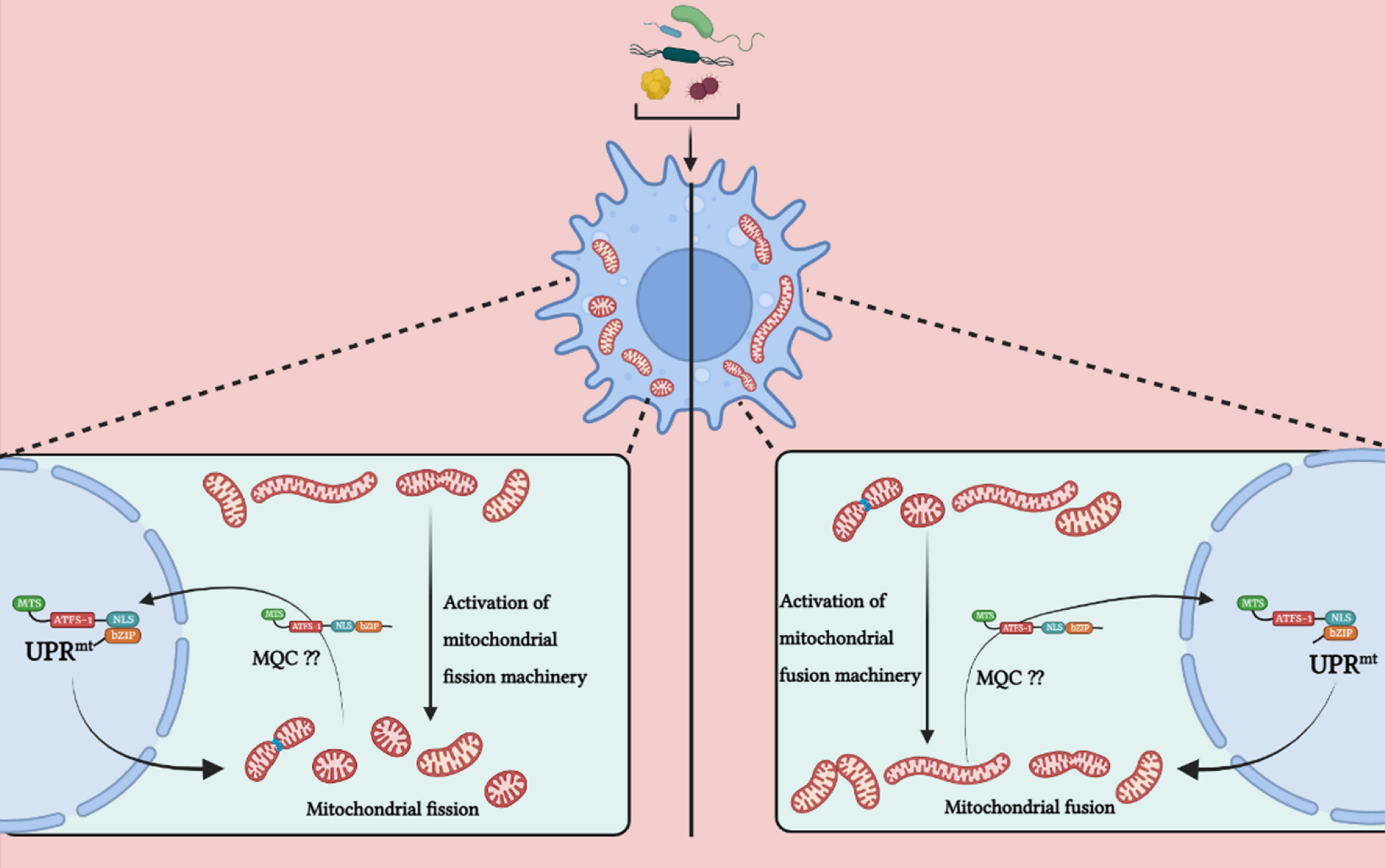
Figure 4 The involvement of mitochondrial fission, fusion and UPRmt in host immunity.
In need of answers
● What sort of peptides are effluxed by HAF-1 into the cytosol that halts ATFS-1 translocation into the mitochondria?
◦ Specifically which peptides are involved in this process, need to be investigated.
◦ Do these peptides change the conformation of ATFS-1 in cytosol, exposing NLS region; and if yes, what are the underlying mechanisms?
● Why does the manipulation of mitochondrial dynamics vary differentially in immune cells by different species of bacterial pathogens?
◦ Is this variation in mitochondrial dynamics in bacterial infection depends upon variety of bacterial virulence factors, or host factors, or interaction of these factors decide the state of mitochondrial dynamics?
● Does the targeting of mitochondrial processes only via therapeutics reinforce immune response against bacterial infection and consequently, restrain their replication and spreading?
MK, SS, and SM designed the theme of the manuscript. MK and SS conducted the literature search and wrote the manuscript. MK and SS drew the schematic diagram and tables. SM critically reviewed and edited the final version of the manuscript. All authors read and approved the final manuscript.
MK and SS were supported by CSIR-UCG NET fellowship (Government of India, India).
The authors are grateful to Prof. K. Muralidhar for helpful discussion and critically analyzing the manuscript. The authors would like to acknowledge all the contributors to the field of mitochondria, and immune response to bacterial infection that could not be included in this review due to space limitation.
The authors declare that the research was conducted in the absence of any commercial or financial relationships that could be construed as a potential conflict of interest.
All claims expressed in this article are solely those of the authors and do not necessarily represent those of their affiliated organizations, or those of the publisher, the editors and the reviewers. Any product that may be evaluated in this article, or claim that may be made by its manufacturer, is not guaranteed or endorsed by the publisher.
Aguilar-López, B. A., Correa, F., Moreno-Altamirano, M. M., Espitia, C., Hernández-Longoria, R., Oliva-Ramírez, J., et al. (2019). LprG and PE_PGRS 33 Mycobacterium tuberculosis virulence factors induce differential mitochondrial dynamics in macrophages. Scand. J. Immunol. 89, e12728. doi: 10.1111/sji.12728
Arango Duque, G., Descoteaux, A. (2014). Macrophage cytokines: involvement in immunity and infectious diseases. Front. Immunol. 5. doi: 10.3389/fimmu.2014.00491
Arizmendi, O., Picking, W. D., Picking, W. L. (2016). Macrophage apoptosis triggered by IpaD from shigella flexneri. Infect. Immun. 84, 1857–1865. doi: 10.1128/IAI.01483-15
Bahat, A., Gross, A. (2019). Mitochondrial plasticity in cell fate regulation. J. Biol. Chem. 294, 13852–13863. doi: 10.1074/jbc.REV118.000828
Bahat, A., MacVicar, T., Langer, T. (2021). Metabolism and innate immunity meet at the mitochondria. Front. Cell Dev. Biol. 2019. doi: 10.3389/fcell.2021.720490
Baker, B. M., Nargund, A. M., Sun, T., Haynes, C. M. (2012). Protective coupling of mitochondrial function and protein synthesis via the eIF2α kinase GCN-2. PloS Genet. 8, e1002760. doi: 10.1371/journal.pgen.1002760
Banoth, B., Cassel, S. L. (2018). Mitochondria in innate immune signaling. Transl. Res. 202, 52–68. doi: 10.1016/j.trsl.2018.07.014
Benedetti, C., Haynes, C. M., Yang, Y., Harding, H. P., Ron, D. (2006). Ubiquitin-like protein 5 positively regulates chaperone gene expression in the mitochondrial unfolded protein response. Genetics 174, 229–239. doi: 10.1534/genetics.106.061580
Buck, M. D., O’Sullivan, D., Geltink, R. I., Curtis, J. D., Chang, C. H., Sanin, D. E., et al. (2016). Mitochondrial dynamics controls T cell fate through metabolic programming. Cell 166, 63–76. doi: 10.1016/j.cell.2016.05.035
Campello, S., Scorrano, L. (2010). Mitochondrial shape changes: orchestrating cell pathophysiology. EMBO Rep. 11, 678–684. doi: 10.1038/embor.2010.115
Carvalho, F., Spier, A., Chaze, T., Matondo, M., Cossart, P., Stavru, F. (2020). Listeria monocytogenes exploits mitochondrial contact site and cristae organizing system complex subunit mic10 to promote mitochondrial fragmentation and cellular infection. MBio 11, e03171–e03119. doi: 10.1128/mBio.03171-19
Cervantes-Silva, M. P., Cox, S. L., Curtis, A. M. (2021). Alterations in mitochondrial morphology as a key driver of immunity and host defence. EMBO Rep. 22, e53086. doi: 10.15252/embr.202153086
Chacinska, A., Koehler, C. M., Milenkovic, D., Lithgow, T., Pfanner, N. (2009). Importing mitochondrial proteins: machineries and mechanisms. Cell 138, 628–644. doi: 10.1016/j.cell.2009.08.005
Chacko, B. K., Kramer, P. A., Ravi, S., Johnson, M. S., Hardy, R. W., Ballinger, S. W. (2013). Methods for defining distinct bioenergetic profiles in platelets, lymphocytes, monocytes, and neutrophils, and the oxidative burst from human blood. Lab. Invest. 93, 690–700. doi: 10.1038/labinvest.2013.53
Chakrabarty, R. P., Chandel, N. S. (2021). Mitochondria as signaling organelles control mammalian stem cell fate. Cell Stem Cell. 28, 394–408. doi: 10.1016/j.stem.2021.02.011
Chamseddine, D., Mahmud, S. A., Westfall, A. K., Castoe, T. A., Berg, R. E., Pellegrino, M. W. (2022). The mitochondrial UPR regulator ATF5 promotes intestinal barrier function via control of the satiety response. Cell Rep. 41, 111789. doi: 10.1016/j.celrep.2022.111789
Chan, D. C. (2020). Mitochondrial dynamics and its involvement in disease. Annu. Rev. Path. Mech. Dis. 15, 235–259. doi: 10.1146/annurev-pathmechdis-012419-032711
Chan, D., Frank, S., Rojo, M. (2006). Mitochondrial dynamics in cell life and death. Cell Death Differ. 13, 680–684. doi: 10.1038/sj.cdd.4401857
Chen, H., Chan, D. C. (2004). Mitochondrial dynamics in mammals. Curr. Top. Dev. Biol. 59, 119–144. doi: 10.1016/S0070-2153(04)59005-1
Chen, H., Chan, D. C. (2005). Emerging functions of mammalian mitochondrial fusion and fission. Hum. Mol. Genet. 14, R283–R289. doi: 10.1093/hmg/ddi270
Chen, L. T., Lin, C. T., Lin, L. Y., Hsu, J. W., Wu, Y. C., Pan, C. L. (2021). Neuronal mitochondrial dynamics coordinate systemic mitochondrial morphology and stress response to confer pathogen resistance in c. elegans. Dev. Cell 56, 1770–1785. doi: 10.1016/j.devcel.2021.04.021
Chen, Y. G., Yue, H. T., Zhang, Z. Z., Yuan, F. H., Bi, H. T., Yuan, K., et al. (2016). Identification and characterization of a mitochondrial unfolded protein response transcription factor ATFS-1 in litopenaeus vannamei. Fish Shellfish Immunol. 54, 144–152. doi: 10.1016/j.fsi.2015.10.019
Chowdhury, S. R., Reimer, A., Sharan, M., Kozjak-Pavlovic, V., Eulalio, A., Prusty, B. K., et al. (2017). Chlamydia preserves the mitochondrial network necessary for replication via microRNA-dependent inhibition of fission. J. Cell Biol. 216, 1071–1089. doi: 10.1083/jcb.201608063
Cogliati, S., Frezza, C., Soriano, M. E., Varanita, T., Quintana-Cabrera, R., Corrado, M., et al. (2013). Mitochondrial cristae shape determines respiratory chain supercomplexes assembly and respiratory efficiency. Cell 155, 160–171. doi: 10.1016/j.cell.2013.08.032
D’Amico, D., Sorrentino, V., Auwerx, J. (2017). Cytosolic proteostasis networks of the mitochondrial stress response. Trends Biochem. Sci. 42, 712–725. doi: 10.1016/j.tibs.2017.05.002
Deng, P., Uma Naresh, N., Du, Y., Lamech, L. T., Yu, J., Zhu, L. J. (2019). Mitochondrial UPR repression during Pseudomonas aeruginosa infection requires the bZIP protein ZIP-3. Proc. Natl. Acad. Sci. U. S. A. 116, 6146–6151. doi: 10.1073/pnas.1817259116
Deo, P., Chow, S. H., Han, M. L., Speir, M., Huang, C., Schittenhelm, R. B., et al. (2020). Mitochondrial dysfunction caused by outer membrane vesicles from gram-negative bacteria activates intrinsic apoptosis and inflammation. Nat. Microbiol. 5, 1418–1427. doi: 10.1038/s41564-020-0773-2
Dimos, B. A., Mahmud, S. A., Fuess, L. E., Mydlarz, L. D., Pellegrino, M. W. (2019). Uncovering a mitochondrial unfolded protein response in corals and its role in adapting to a changing world. Proc. R. Soc B. 286, 20190470. doi: 10.1098/rspb.2019.0470
Duroux-Richard, I., Roubert, C., Ammari, M., Présumey, J., Grün, J. R., Häupl, T., et al. (2016). miR-125b controls monocyte adaptation to inflammation through mitochondrial metabolism and dynamics. Am. J. Hematol. 128, 3125–3136. doi: 10.1182/blood-2016-02-697003
Escoll, P., Buchrieser, C. (2018). Metabolic reprogramming of host cells upon bacterial infection: why shift to a warburg-like metabolism? FEBS J. 285, 2146–2160. doi: 10.1111/febs.14446
Escoll, P., Mondino, S., Rolando, M., Buchrieser, C. (2016). Targeting of host organelles by pathogenic bacteria: a sophisticated subversion strategy. Nat. Rev. Microbiol. 14, 5–19. doi: 10.1038/nrmicro.2015.1
Escoll, P., Song, O. R., Viana, F., Steiner, B., Lagache, T., Olivo-Marin, J. C., et al. (2017). Legionella pneumophila modulates mitochondrial dynamics to trigger metabolic repurposing of infected macrophages. Cell Host Microbe 22, 302–316. doi: 10.1016/j.chom.2017.07.020
Faas, M. M., De Vos, P. (2020). Mitochondrial function in immune cells in health and disease. Biochim. Biophys. Acta Mol. Basis Dis. 1866, 165845. doi: 10.1016/j.bbadis.2020.165845
Fedele, A. O., Proud, C. G. (2020). Chloroquine and bafilomycin a mimic lysosomal storage disorders and impair mTORC1 signalling. Biosci. Rep. 40, BSR20200905. doi: 10.1042/BSR20200905
Fiorese, C. J., Schulz, A. M., Lin, Y. F., Rosin, N., Pellegrino, M. W., Haynes, C. M. (2016). The transcription factor ATF5 mediates a mammalian mitochondrial UPR. Curr. Biol. 26, 2037–2043. doi: 10.1016/j.cub.2016.06.002
Forgac, M. (2007). Vacuolar ATPases: rotary proton pumps in physiology and pathophysiology. Nat. Rev. Mol. Cell Biol. 8, 917–929. doi: 10.1038/nrm2272
Fusakio, M. E., Willy, J. A., Wang, Y., Mirek, E. T., Al Baghdadi, R. J., Adams, C. M., et al. (2016). Transcription factor ATF4 directs basal and stress-induced gene expression in the unfolded protein response and cholesterol metabolism in the liver. Mol. Biol. Cell 27, 1536–1551. doi: 10.1091/mbc.E16-01-0039
Galmiche, A., Rassow, J. (2010). Targeting of Helicobacter pylori VacA to mitochondria. Gut. Microbes 1, 392–395. doi: 10.4161/gmic.1.6.13894
Gao, S., Hu, J. (2021). Mitochondrial fusion: the machineries in and out. Trends Cell Biol. 31, 62–74. doi: 10.1016/j.tcb.2020.09.008
Gao, Z., Li, Y., Wang, F., Huang, T., Fan, K., Zhang, Y., et al. (2017). Mitochondrial dynamics controls anti-tumour innate immunity by regulating CHIP-IRF1 axis stability. Nat. Commun. 8, 1–3. doi: 10.1038/s41467-017-01919-0
Gatica, D., Lahiri, V., Klionsky, D. J. (2018). Cargo recognition and degradation by selective autophagy. Nat. Cell Biol. 20, 233–242. doi: 10.1038/s41556-018-0037-z
Genge, M. G., Mokranjac, D. (2022). Coordinated translocation of presequence-containing precursor proteins across two mitochondrial membranes: knowns and unknowns of how TOM and TIM23 complexes cooperate with each other. Front. Physiol. 12. doi: 10.3389/fphys.2021.806426
Gottesman, S. (2003). Proteolysis in bacterial regulatory circuits. Annu. Rev. Cell Dev. Biol. 19, 565–587. doi: 10.1146/annurev.cellbio.19.110701.153228
Guo, B., Huang, X., Zhang, P., Qi, L., Liang, Q., Zhang, X., et al. (2014). Genome-wide screen identifies signaling pathways that regulate autophagy during Caenorhabditis elegans development. EMBO Rep. 15, 705–713. doi: 10.1002/embr.201338310
Haastrup, M. O., Vikramdeo, K. S., Singh, S., Singh, A. P., Dasgupta, S. (2023). The journey of mitochondrial protein import and roadmap to follow. Int. J. Mol. Sci. 24, 2479. doi: 10.3390/ijms24032479
Haeussler, S., Köhler, F., Witting, M., Premm, M. F., Rolland, S. G., Fischer, C., et al. (2020). Autophagy compensates for defects in mitochondrial dynamics. PloS Genet. 16, e1008638. doi: 10.1371/journal.pgen.1008638
Haeussler, S., Yeroslaviz, A., Rolland, S. G., Luehr, S., Lambie, E. J., Conradt, B. (2021). Genome-wide RNAi screen for regulators of UPRmt in caenorhabditis elegans mutants with defects in mitochondrial fusion. G3 11, jkab095. doi: 10.1093/g3journal/jkab095
Harbauer, A. B., Zahedi, R. P., Sickmann, A., Pfanner, N., Meisinger, C. (2014). The protein import machinery of mitochondria–a regulatory hub in metabolism, stress, and disease. Cell Metab. 19, 357–372. doi: 10.1016/j.cmet.2014.01.010
Haynes, C. M., Petrova, K., Benedetti, C., Yang, Y., Ron, D. (2007). ClpP mediates activation of a mitochondrial unfolded protein response in C. elegans. Dev. Cell 13, 467–480. doi: 10.1016/j.devcel.2007.07.016
Haynes, C. M., Ron, D. (2010). The mitochondrial UPR–protecting organelle protein homeostasis. J. Cell Sci. 123, 3849–3855. doi: 10.1242/jcs.075119
Haynes, C. M., Yang, Y., Blais, S. P., Neubert, T. A., Ron, D. (2010). The matrix peptide exporter HAF-1 signals a mitochondrial UPR by activating the transcription factor ZC376. 7 in C. elegans. Mol. Cell 37, 529–540. doi: 10.1016/j.molcel.2010.01.015
Hesketh, G. G., Papazotos, F., Pawling, J., Rajendran, D., Knight, J. D., Martinez, S., et al. (2020). The GATOR-rag GTPase pathway inhibits mTORC1 activation by lysosome-derived amino acids. Science 370, 351–356. doi: 10.1126/science.aaz0863
Hinshaw, D. C., Hanna, A., Lama-Sherpa, T., Metge, B., Kammerud, S. C., Benavides, G. A., et al. (2021). Hedgehog signaling regulates metabolism and polarization of mammary tumor-associated macrophages. Cancer Res. 81, 5425–5437. doi: 10.1158/0008-5472.CAN-20-1723
Honjoh, S., Yamamoto, T., Uno, M., Nishida, E. (2009). Signalling through RHEB-1 mediates intermittent fasting-induced longevity in C. elegans. Nature 457, 726–730. doi: 10.1038/nature07583
Hoppins, S. (2014). The regulation of mitochondrial dynamics. Curr. Opin. Cell Biol. 29, 46–52. doi: 10.1016/j.ceb.2014.03.005
Ichinohe, T., Yamazaki, T., Koshiba, T., Yanagi, Y. (2013). Mitochondrial protein mitofusin 2 is required for NLRP3 inflammasome activation after RNA virus infection. Proc. Natl. Acad. Sci. U. S. A. 110, 17963–17968. doi: 10.1073/pnas.1312571110
Jabir, M. S., Hopkins, L., Ritchie, N. D., Ullah, I., Bayes, H. K., Li, D., et al. (2015). Mitochondrial damage contributes to pseudomonas aeruginosa activation of the inflammasome and is downregulated by autophagy. Autophagy 11, 166–182. doi: 10.4161/15548627.2014.981915
Jain, P., Luo, Z. Q., Blanke, S. R. (2011). Helicobacter pylori vacuolating cytotoxin a (VacA) engages the mitochondrial fission machinery to induce host cell death. Proc. Natl. Acad. Sci. U.S.A. 108, 16032–16037. doi: 10.1073/pnas.1105175108
Jeong, D. E., Lee, D., Hwang, S. Y., Lee, Y., Lee, J. E., Seo, M., et al. (2017). Mitochondrial chaperone HSP-60 regulates anti-bacterial immunity via p38 MAP kinase signaling. EMBO J. 36, 1046–1065. doi: 10.15252/embj.201694781
Ježek, J., Cooper, K. F., Strich, R. (2018). Reactive oxygen species and mitochondrial dynamics: the yin and yang of mitochondrial dysfunction and cancer progression. Antioxidants 7 (1), 13. doi: 10.3390/antiox7010013
Jovaisaite, V., Mouchiroud, L., Auwerx, J. (2014). The mitochondrial unfolded protein response, a conserved stress response pathway with implications in health and disease. J. Exp. Biol. 217, 137–143. doi: 10.1242/jeb.090738
Kang, S. G., Ortega, J., Singh, S. K., Wang, N., Huang, N. N., Steven, A. C., et al. (2002). Functional proteolytic complexes of the human mitochondrial ATP-dependent protease, hClpXP. J. Biol. Chem. 277, 21095–21102. doi: 10.1074/jbc.M201642200
Khan, S., Raj, D., Jaiswal, K., Lahiri, A. (2020). Modulation of host mitochondrial dynamics during bacterial infection. Mitochondrion 53, 140–149. doi: 10.1016/j.mito.2020.05.005
Kim, S., Sieburth, D. (2018). Sphingosine kinase activates the mitochondrial unfolded protein response and is targeted to mitochondria by stress. Cell Rep. 24, 2932–2945. doi: 10.1016/j.celrep.2018.08.037
Kirienko, N. V., Ausubel, F. M., Ruvkun, G. (2015). Mitophagy confers resistance to siderophore-mediated killing by Pseudomonas aeruginosa. Proc. Natl. Acad. Sci. U. S. A. 112, 1821–1826. doi: 10.1073/pnas.1424954112
Kraus, F., Roy, K., Pucadyil, T. J., Ryan, M. T. (2021). Function and regulation of the divisome for mitochondrial fission. Nature 590, 57–66. doi: 10.1038/s41586-021-03214-x
Krutzik, S. R., Tan, B., Li, H., Ochoa, M. T., Liu, P. T., Sharfstein, S. E., et al. (2005). TLR activation triggers the rapid differentiation of monocytes into macrophages and dendritic cells. Nat. Med. 11, 653–660. doi: 10.1038/nm1246
Kumar, M., Sharma, S., Haque, M., Kumar, J., Hathi, U. P., Mazumder, S. (2022a). TLR22-induced pro-apoptotic mtROS abets UPRmt-mediated mitochondrial fission in Aeromonas hydrophila-infected headkidney macrophages of clarias gariepinus. Front. Immunol. 13. doi: 10.3389/fimmu.2022.931021
Kumar, M., Shelly, A., Dahiya, P., Ray, A., Mazumder, S. (2022b). Aeromonas hydrophila inhibits autophagy triggering cytosolic translocation of mtDNA which activates the pro-apoptotic caspase-1/IL-1β-nitric oxide axis in headkidney macrophages. Virulence 13, 60–76. doi: 10.1080/21505594.2021.2018767
Kurihara, Y., Itoh, R., Shimizu, A., Walenna, N. F., Chou, B., Ishii, K., et al. (2019). Chlamydia trachomatis targets mitochondrial dynamics to promote intracellular survival and proliferation. Cell. Microbiol. 21, e12962. doi: 10.1111/cmi.12962
Lawrence, R. E., Zoncu, R. (2019). The lysosome as a cellular centre for signalling, metabolism and quality control. Nat. Cell Biol. 21, 133–142. doi: 10.1038/s41556-018-0244-7
Li, T. Y., Gao, A. W., Li, X., Li, H., Liu, Y. J., Lalou, A., et al. (2022). V-ATPase/TORC1-mediated ATFS-1 translation directs mitochondrial UPR activation in c. elegans. J. Cell Biol. 222, e202205045. doi: 10.1083/jcb.202205045
Li, Y., He, Y., Miao, K., Zheng, Y., Deng, C., Liu, T. M. (2020). Imaging of macrophage mitochondria dynamics in vivo reveals cellular activation phenotype for diagnosis. Theranostics 10, 2897–2917. doi: 10.7150/thno.40495
Li, J., Zhang, D., Brundel, B. J., Wiersma, M. (2019). Imbalance of ER and mitochondria interactions: prelude to cardiac ageing and disease? Cells 8, 1617. doi: 10.3390/cells8121617
Liesa, M., Shirihai, O. S. (2016). Mitochondrial networking in T cell memory. Cell 166, 9–10. doi: 10.1016/j.cell.2016.06.035
Liu, Y., Samuel, B. S., Breen, P. C., Ruvkun, G. (2014). Caenorhabditis elegans pathways that surveil and defend mitochondria. Nature 508, 406–410. doi: 10.1038/nature13204
Lizana, L., Bauer, B., Orwar, O. (2008). Controlling the rates of biochemical reactions and signaling networks by shape and volume changes. Proc. Natl. Acad. Sci. U. S. A. 105, 4099–4104. doi: 10.1073/pnas.0709932105
Lloberas, J., Muñoz, J. P., Hernández-Álvarez, M. I., Cardona, P. J., Zorzano, A., Celada, A. (2020). Macrophage mitochondrial MFN2 (mitofusin 2) links immune stress and immune response through reactive oxygen species (ROS) production. Autophagy 16, 2307–2309. doi: 10.1080/15548627.2020.1839191
Losón, O. C., Song, Z., Chen, H., Chan, D. C. (2013). Fis1, mff, MiD49, and MiD51 mediate Drp1 recruitment in mitochondrial fission. Mol. Biol. Cell. 24, 659–667. doi: 10.1091/mbc.E12-10-0721
Lum, M., Morona, R. (2014). Dynamin-related protein Drp1 and mitochondria are important for Shigella flexneri infection. Int. J. Med. Microbiol. 304, 530–541. doi: 10.1016/j.ijmm.2014.03.006
Mahmud, S. A., Qureshi, M. A., Sapkota, M., Pellegrino, M. W. (2020). A pathogen branched-chain amino acid catabolic pathway subverts host survival by impairing energy metabolism and the mitochondrial UPR. PloS Pathog. 16, e1008918. doi: 10.1371/journal.ppat.1008918
Mannella, C. A., Lederer, W. J., Jafri, M. S. (2013). The connection between inner membrane topology and mitochondrial function. J. Mol. Cell. Cardiol. 62, 51–57. doi: 10.1016/j.yjmcc.2013.05.001
Martin, J., Mahlke, K., Pfanner, N. (1991). Role of an energized inner membrane in mitochondrial protein import. delta psi drives the movement of presequences. J. Biol. Chem. 266, 18051–18057. doi: 10.1016/S0021-9258(18)55235-2
Mears, J. A., Lackner, L. L., Fang, S., Ingerman, E., Nunnari, J., Hinshaw, J. E. (2011). Conformational changes in Dnm1 support a contractile mechanism for mitochondrial fission. Nat. Struct. Mol. Biol. 18, 20–26. doi: 10.1038/nsmb.1949
Medzhitov, R. (2010). Innate immunity: quo vadis? Nat. Immunol. 11, 551–553. doi: 10.1038/ni0710-551
Melber, A., Haynes, C. M. (2018). UPRmt regulation and output: a stress response mediated by mitochondrial-nuclear communication. Cell Res. 28, 281–295. doi: 10.1038/cr.2018.16
Mishra, P., Chan, D. C. (2016). Metabolic regulation of mitochondrial dynamics. J. Cell Biol. 212, 379–387. doi: 10.1083/jcb.201511036
Missiroli, S., Patergnani, S., Caroccia, N., Pedriali, G., Perrone, M., Previati, M. (2018). Mitochondria-associated membranes (MAMs) and inflammation. Cell Death Dis. 9, 1–4. doi: 10.1038/s41419-017-0027-2
Molenaars, M., Janssens, G. E., Williams, E. G., Jongejan, A., Lan, J., Rabot, S., et al. (2020). A conserved mito-cytosolic translational translational balance links two longevity pathways. Cell Metab. 31, 549–563. doi: 10.1016/j.cmet.2020.01.011
Moullan, N., Mouchiroud, L., Wang, X., Ryu, D., Williams, E. G., Mottis, A., et al. (2015). Tetracyclines disturb mitochondrial function across eukaryotic models: a call for caution in biomedical research. Cell Rep. 10, 1681–1691. doi: 10.1016/j.celrep.2015.02.034
Na, Y. R., Hong, J. H., Lee, M. Y., Jung, J. H., Jung, D., Kim, Y. W., et al. (2015). Proteomic analysis reveals distinct metabolic differences between granulocyte-macrophage colony stimulating factor (GM-CSF) and macrophage colony stimulating factor (M-CSF) grown macrophages derived from murine bone marrow cells. Mol. Cell Proteomics 14, 2722–2732. doi: 10.1074/mcp.M115.048744
Nakahira, K., Hisata, S., Choi, A. M. (2015). The roles of mitochondrial damage-associated molecular patterns in diseases. Antioxid. Redox Signal. 23, 1329–1350. doi: 10.1089/ars.2015.6407
Namgaladze, D., Khodzhaeva, V., Brüne, B. (2019). ER-mitochondria communication in cells of the innate immune system. Cells 8, 1088. doi: 10.3390/cells8091088
Nargund, A. M., Fiorese, C. J., Pellegrino, M. W., Deng, P., Haynes, C. M. (2015). Mitochondrial and nuclear accumulation of the transcription factor ATFS-1 promotes OXPHOS recovery during the UPRmt. Mol. Cell. 58, 123–133. doi: 10.1016/j.molcel.2015.02.008
Nargund, A. M., Pellegrino, M. W., Fiorese, C. J., Baker, B. M., Haynes, C. M. (2012). Mitochondrial import efficiency of ATFS-1 regulates mitochondrial UPR activation. Science 337 (6094), 587–590. doi: 10.1126/science.1223560
Palframan, S. L., Kwok, T., Gabriel, K., Vacuolating cytotoxin, A. (2012). (VacA), a key toxin for Helicobacter pylori pathogenesis. Front. Cell. Infect. Microbiol. 2. doi: 10.3389/fcimb.2012.00092
Pallett, M. A., Berger, C. N., Pearson, J. S., Hartland, E. L., Frankel, G. (2014). The type III secretion effector NleF of enteropathogenic Escherichia coli activates NF-κB early during infection. Infect. Immun. 82, 4878–4888. doi: 10.1128/IAI.02131-14
Palmer, C. S., Osellame, L. D., Laine, D., Koutsopoulos, O. S., Frazier, A. E., Ryan, M. T. (2011). MiD49 and MiD51, new components of the mitochondrial fission machinery. EMBO Rep. 12, 565–573. doi: 10.1038/embor.2011.54
Panchal, K., Tiwari, A. K. (2021). Miro (Mitochondrial rho GTPase), a key player of mitochondrial axonal transport and mitochondrial dynamics in neurodegenerative diseases. Mitochondrion 56, 118–135. doi: 10.1016/j.mito.2020.10.005
Papa, L., Germain, D. (2011). Estrogen receptor mediates a distinct mitochondrial unfolded protein response. J. Cell Sci. 124, 1396–1402. doi: 10.1242/jcs.078220
Papatheodorou, P., Domańska, G., Öxle, M., Mathieu, J., Selchow, O., Kenny, B., et al. (2006). The enteropathogenic Escherichia coli (EPEC) map effector is imported into the mitochondrial matrix by the TOM/Hsp70 system and alters organelle morphology. Cell. Microbiol. 8, 677–689. doi: 10.1111/j.1462-5822.2005.00660.x
Papayannopoulos, V. (2018). Neutrophil extracellular traps in immunity and disease. Nat. Rev. Immunol. 18, 134–147. doi: 10.1038/nri.2017.105
Park, S., Won, J. H., Hwang, I., Hong, S., Lee, H. K., Yu, J. W. (2015). Defective mitochondrial fission augments NLRP3 inflammasome activation. Sci. Rep. 5, 1–4. doi: 10.1038/srep15489
Pellegrino, M. W., Nargund, A. M., Kirienko, N. V., Gillis, R., Fiorese, C. J., Haynes, C. M. (2014). Mitochondrial UPR-regulated innate immunity provides resistance to pathogen infection. Nature 516, 414–417. doi: 10.1038/nature13818
Peters, C., Bayer, M. J., BuÈhler, S., Andersen, J. S., Mann, M., Mayer, A. (2001). Trans-complex formation by proteolipid channels in the terminal phase of membrane fusion. Nature 409, 581–588. doi: 10.1038/35054500
Quintana, A., Hoth, M. (2012). Mitochondrial dynamics and their impact on T cell function. Cell Calcium. 52, 57–63. doi: 10.1016/j.ceca.2012.02.005
Quirós, P. M., Mottis, A., Auwerx, J. (2016). Mitonuclear communication in homeostasis and stress. Nat. Rev. Mol. Cell Biol. 17, 213–226. doi: 10.1038/nrm.2016.23
Quirós, P. M., Prado, M. A., Zamboni, N., D’Amico, D., Williams, R. W., Finley, D., et al. (2017). Multi-omics analysis identifies ATF4 as a key regulator of the mitochondrial stress response in mammals. J. Cell Biol. 216, 2027–2045. doi: 10.1083/jcb.201702058
Qureshi, M. A., Haynes, C. M., Pellegrino, M. W. (2017). The mitochondrial unfolded protein response: signaling from the powerhouse. J. Biol. Chem. 292, 13500–13506. doi: 10.1074/jbc.R117.791061
Rainbolt, T. K., Atanassova, N., Genereux, J. C., Wiseman, R. L. (2013). Stress-regulated translational attenuation adapts mitochondrial protein import through Tim17A degradation. Cell Metab. 18, 908–919. doi: 10.1016/j.cmet.2013.11.006
Rambold, A. S., Cohen, S., Lippincott-Schwartz, J. (2015). Fatty acid trafficking in starved cells: regulation by lipid droplet lipolysis, autophagy, and mitochondrial fusion dynamics. Dev. Cell 32, 678–692. doi: 10.1016/j.devcel.2015.01.029
Rambold, A. S., Pearce, E. L. (2018). Mitochondrial dynamics at the interface of immune cell metabolism and function. Trends Immunol. 39, 6–18. doi: 10.1016/j.it.2017.08.006
Raveh, A., Delekta, P. C., Dobry, C. J., Peng, W., Schultz, P. J., Blakely, P. K., et al. (2013). Discovery of potent broad spectrum antivirals derived from marine actinobacteria. PloS One 8, e82318. doi: 10.1371/journal.pone.0082318
Ren, C., Li, D., Zhou, Q., Hu, X. (2020). Mitochondria-targeted TPP-MoS2 with dual enzyme activity provides efficient neuroprotection through M1/M2 microglial polarization in an alzheimer's disease model. Biomaterials 232, 119752. doi: 10.1016/j.biomaterials.2019.119752
Rolland, S. G., Schneid, S., Schwarz, M., Rackles, E., Fischer, C., Haeussler, S., et al. (2019). Compromised mitochondrial protein import acts as a signal for UPRmt. Cell Rep. 28, 1659–1669. doi: 10.1016/j.celrep.2019.07.049
Rother, M., Teixeira da Costa, A. R., Zietlow, R., Meyer, T. F., Rudel, T. (2019). Modulation of host cell metabolism by chlamydia trachomatis. Microbiol. Spectr. 7 (3), 7–3. doi: 10.1128/microbiolspec.BAI-0012-2019
Ruan, L., Wang, Y., Zhang, X., Tomaszewski, A., McNamara, J. T., Li, R. (2020). Mitochondria-associated proteostasis. Annu. Rev. Biophys. 6, 41–67. doi: 10.1146/annurev-biophys-121219-081604
Runkel, E. D., Liu, S., Baumeister, R., Schulze, E. (2013). Surveillance-activated defenses block the ROS–induced mitochondrial unfolded protein response. PloS Gen. 9, e1003346. doi: 10.1371/journal.pgen.1003346
Ryan, M. T., Hoogenraad, N. J. (2007). Mitochondrial-nuclear communications. Annu. Rev. Biochem. 76, 701–722. doi: 10.1146/annurev.biochem.76.052305.091720
Sandoval, H., Kodali, S., Wang, J. (2018). Regulation of b cell fate, survival, and function by mitochondria and autophagy. Mitochondrion 41, 58–65. doi: 10.1016/j.mito.2017.11.005
Sapkota, M., Adnan Qureshi, M., Arif Mahmud, S., Balikosa, Y., Nguyen, C., Boll, J. M., et al. (2021). A nematode-derived, mitochondrial stress signaling-regulated peptide exhibits broad antibacterial activity. Biol. Open 10, bio058613. doi: 10.1242/bio.058613
Schultz, B. M., Acevedo, O. A., Kalergis, A. M., Bueno, S. M. (2022). Role of extracellular trap release during bacterial and viral infection. Front. Microbiol. 13. doi: 10.3389/fmicb.2022.798853
Schwartz, M. P., Huang, S., Matouschek, A. (1999). The structure of precursor proteins during import into mitochondria. J. Biol. Chem. 274, 12759–12764. doi: 10.1074/jbc.274.18.12759
Sena, L., Li, S., Jairaman, A., Prakriya, M., Ezponda, T., Hildeman, D., et al. (2013). Mitochondria are required for antigen-specific T cell activation through reactive oxygen species signaling. Immunity 38, 225–236. doi: 10.1016/j.immuni.2012.10.020
Shaw, J. M., Nunnari, J. (2002). Mitochondrial dynamics and division in budding yeast. Trends Cell Biol. 12, 178–184. doi: 10.1016/s0962-8924(01)02246-2
Shen, K., Pender, C. L., Bar-Ziv, R., Zhang, H., Wickham, K., Willey, E., et al. (2022). Mitochondria as cellular and organismal signaling hubs. Annu. Rev. Cell Dev. Biol. 38, 179–218. doi: 10.1146/annurev-cellbio-120420-015303
Shi, C., Pamer, E. G. (2011). Monocyte recruitment during infection and inflammation. Nat. Rev. Immunol. 11, 762–774. doi: 10.1038/nri3070
Shimobayashi, M., Hall, M. N. (2014). Making new contacts: the mTOR network in metabolism and signalling crosstalk. Nat. Rev. Mol. Cell Biol. 15, 155–162. doi: 10.1038/nrm3757
Shpilka, T., Du, Y., Yang, Q., Melber, A., Uma Naresh, N., Lavelle, J., et al. (2021). UPRmt scales mitochondrial network expansion with protein synthesis via mitochondrial import in caenorhabditis elegans. Nat. Commun. 12, 479. doi: 10.1038/s41467-020-20784-y
Simula, L., Pacella, I., Colamatteo, A., Procaccini, C., Cancila, V., Bordi, M., et al. (2018). Drp1 controls effective T cell immune-surveillance by regulating T cell migration, proliferation, and cMyc-dependent metabolic reprogramming. Cell Rep. 25, 3059–3073. doi: 10.1016/j.celrep.2018.11.018
Singh, R., Kaushik, S., Wang, Y., Xiang, Y., Novak, I., Komatsu, M., et al. (2009). Autophagy regulates lipid metabolism. Nature 458, 1131–1135. doi: 10.1038/nature07976
Spier, A., Connor, M. G., Steiner, T., Carvalho, F., Cossart, P., Eisenreich, W., et al. (2021). Mitochondrial respiration restricts Listeria monocytogenes infection by slowing down host cell receptor recycling. Cell Rep. 37, 109989. doi: 10.1016/j.celrep.2021.109989
Stavru, F., Bouillaud, F., Sartori, A., Ricquier, D., Cossart, P. (2011). Listeria monocytogenes transiently alters mitochondrial dynamics during infection. Proc. Natl. Acad. Sci. U. S. A. 108, 3612–3617. doi: 10.1073/pnas.1100126108
Suárez-Rivero, J. M., Villanueva-Paz, M., de la Cruz-Ojeda, P., de la Mata, M., Cotán, D., Oropesa-Ávila, M., et al. (2016). Mitochondrial dynamics in mitochondrial diseases. Diseases 5, 1. doi: 10.3390/diseases5010001
Suhm, T., Kaimal, J. M., Dawitz, H., Peselj, C., Masser, A. E., Hanzn, S., et al. (2018). Mitochondrial translation efficiency controls cytoplasmic protein homeostasis. Cell Metab. 27, 1309–1322. doi: 10.1016/j.cmet.2018.04.011
Suzuki, M., Danilchanka, O., Mekalanos, J. J. (2014). Vibrio cholerae T3SS effector VopE modulates mitochondrial dynamics and innate immune signaling by targeting miro GTPases. Cell Host Microbe 16, 581–591. doi: 10.1016/j.chom.2014.09.015
Teske, B. F., Fusakio, M. E., Zhou, D., Shan, J., McClintick, J. N., Kilberg, M. S., et al. (2013). CHOP induces activating transcription factor 5 (ATF5) to trigger apoptosis in response to perturbations in protein homeostasis. Mol. Biol. Cell. 24, 2477–2490. doi: 10.1091/mbc.E13-01-0067
Tian, Y., Garcia, G., Bian, Q., Steffen, K. K., Joe, L., Wolff, S., et al. (2016). Mitochondrial stress induces chromatin reorganization to promote longevity and UPRmt. Cell 165, 1197–1208. doi: 10.1016/j.cell.2016.04.011
Tiku, V., Kofoed, E. M., Yan, D., Kang, J., Xu, M., Reichelt, M., et al. (2021). Outer membrane vesicles containing OmpA induce mitochondrial fragmentation to promote pathogenesis of Acinetobacter baumannii. Sci. Rep. 11, 1–6. doi: 10.1038/s41598-020-79966-9
Tiku, V., Tan, M. W., Dikic, I. (2020). Mitochondrial functions in infection and immunity. Trends Cell Biol. 30, 263–275. doi: 10.1016/j.tcb.2020.01.006
Timblin, G. A., Tharp, K. M., Ford, B., Winchester, J. M., Wang, J., Zhu, S., et al. (2021). Mitohormesis reprogrammes macrophage metabolism to enforce tolerance. Nat. Metab. 3, 618–635. doi: 10.1038/s42255-021-00392-w
Topf, U., Wrobel, L., Chacinska, A. (2016). Chatty mitochondria: keeping balance in cellular protein homeostasis. Trends Cell Biol. 26, 577–586. doi: 10.1016/j.tcb.2016.03.002
Tran, H. C., Van Aken, O. (2020). Mitochondrial unfolded protein-related responses across kingdoms: similar problems, different regulators. Mitochondrion 53, 166–177. doi: 10.1016/j.mito.2020.05.009
Tur, J., Pereira-Lopes, S., Vico, T., Marín, E. A., Muñoz, J. P., Hernández-Alvarez, M., et al. (2020). Mitofusin 2 in macrophages links mitochondrial ROS production, cytokine release, phagocytosis, autophagy, and bactericidal activity. Cell Rep. 32, 108079. doi: 10.1016/j.celrep.2020.108079
Tur, J., Vico, T., Lloberas, J., Zorzano, A., Celada, A. (2017). Macrophages and mitochondria: a critical interplay between metabolism, signaling, and the functional activity. Adv. Immunol. 133, 1–36. doi: 10.1016/bs.ai.2016.12.001
Twig, G., Hyde, B., Shirihai, O. S. (2008). Mitochondrial fusion, fission and autophagy as a quality control axis: the bioenergetic view. Biochim. Biophys. Acta Bioenerg. 1777, 1092–1097. doi: 10.1016/j.bbabio.2008.05.001
Voth, W., Jakob, U. (2017). Stress-activated chaperones: a first line of defense. Trends Biochem. Sci. 42 (11), 899–913. doi: 10.1016/j.tibs.2017.08.006
Wai, T., Langer, T. (2016). Mitochondrial dynamics and metabolic regulation. Trends Endocrinol. Metab. 27, 105–117. doi: 10.1016/j.tem.2015.12.001
Wang, S., Gao, K., Liu, Y. (2018). UPRmt coordinates immunity to maintain mitochondrial homeostasis and animal fitness. Mitochondrion 41, 9–13. doi: 10.1016/j.mito.2017.11.004
Wang, J., Wang, X., Du, W., Xue, Z., Huang, W., Guan, Z., et al. (2022). BI-1 ameliorates myocardial injury by activating the mitochondrial unfolded protein response and FUNDC1-related mitophagy in cardiorenal syndrome type 3. Cell. Signal. 91, 110218. doi: 10.1016/j.cellsig.2021.110218
Wang, C., Youle, R. J. (2009). The role of mitochondria in apoptosis. Annu. Rev. Genet. 43, 95. doi: 10.5483/bmbrep.2008.41.1.011
Waters, L. R., Ahsan, F. M., Wolf, D. M., Shirihai, O., Teitell, M. A. (2018). Initial b cell activation induces metabolic reprogramming and mitochondrial remodeling. IScience 5, 99–109. doi: 10.1016/j.isci.2018.07.005
Weinberg, S. E., Sena, L. A., Chandel, N. S. (2015). Mitochondria in the regulation of innate and adaptive immunity. Immunity 42, 406–417. doi: 10.1016/j.immuni.2015.02.002
Weiss, G., Schaible, U. E. (2015). Macrophage defense mechanisms against intracellular bacteria. Immunol. Rev. 264, 182–203. doi: 10.1111/imr.12266
West, A. P., Shadel, G. S., Ghosh, S. (2011). Mitochondria in innate immune responses. Nat. Rev. Immunol. 11, 389–402. doi: 10.1038/nri2975
Westermann, B. (2010). “Mitochondrial dynamics in model organisms: what yeasts, worms and flies have taught us about fusion and fission of mitochondria” in Seminars in cell & developmental biology (Cambridge, Massachusetts: Academic Press), 542–549. doi: 10.1016/j.semcdb.2009.12.003
Westermann, B. (2012). Bioenergetic role of mitochondrial fusion and fission. Biochim. Biophys. Acta Bioenerg. 1817, 1833–1838. doi: 10.1016/j.bbabio.2012.02.033
Wolfson, R. L., Sabatini, D. M. (2017). The dawn of the age of amino acid sensors for the mTORC1 pathway. Cell Metab. 26, 301–309. doi: 10.1016/j.cmet.2017.07.001
Wong, Y. C., Kim, S., Peng, W., Krainc, D. (2019). Regulation and function of mitochondria-lysosome membrane contact sites in cellular homeostasis. Trends Cell Biol. 29, 500–513. doi: 10.1016/j.tcb.2019.02.004
Wu, Y., Williams, E. G., Dubuis, S., Mottis, A., Jovaisaite, V., Houten, S. M., et al. (2014). Multilayered genetic and omics dissection of mitochondrial activity in a mouse reference population. Cell 158, 1415–1430. doi: 10.1016/j.cell.2014.07.039
Xin, N., Durieux, J., Yang, C., Wolff, S., Kim, H. E., Dillin, A. (2022). The UPRmt preserves mitochondrial import to extend lifespan. J. Cell Biol. 221, e202201071. doi: 10.1083/jcb.202201071
Xu, F., Qi, H., Li, J., Sun, L., Gong, J., Chen, Y., et al. (2020). Mycobacterium tuberculosis infection up-regulates MFN2 expression to promote NLRP3 inflammasome formation. J. Biol. Chem. 295, 17684–17697. doi: 10.1074/jbc.RA120.014077
Yao, C. H., Wang, R., Wang, Y., Kung, C. P., Weber, J. D., Patti, G. J. (2019). Mitochondrial fusion supports increased oxidative phosphorylation during cell proliferation. Elife 8, e41351. doi: 10.7554/eLife.41351
Yoneda, T., Benedetti, C., Urano, F., Clark, S. G., Harding, H. P., Ron, D. (2004). Compartment-specific perturbation of protein handling activates genes encoding mitochondrial chaperones. J. Cell Sci. 117 (18), 4055–4066. doi: 10.1242/jcs.01275
Youle, R. J., van der Bliek, A. M. (2012). Mitochondrial fission, fusion, and stress. Science 337, 1062–1065. doi: 10.1126/science.1219855
Zhang, B., Alysandratos, K. D., Angelidou, A., Asadi, S., Sismanopoulos, N., Delivanis, D. A., et al. (2011). Human mast cell degranulation and preformed TNF secretion require mitochondrial translocation to exocytosis sites: relevance to atopic dermatitis. J. Allergy Clin. Immunol. 127, 1522–1531. doi: 10.1016/j.jaci.2011.02.005
Zhang, Q., Wu, X., Chen, P., Liu, L., Xin, N., tian, Y., et al. (2018). The mitochondrial unfolded protein response is mediated cell-non-autonomously by retromer-dependent wnt signaling. Cell 174, 870–883. doi: 10.1016/j.cell.2018.06.029
Zhao, Q., Wang, J., Levichkin, I. V., Stasinopoulos, S., Ryan, M. T., Hoogenraad, N. J. (2002). A mitochondrial specific stress response in mammalian cells. EMBO J. 21, 4411–4419. doi: 10.1093/emboj/cdf445
Zhou, R., Yazdi, A. S., Menu, P., Tschopp, J. (2011). A role for mitochondria in NLRP3 inflammasome activation. Nature 469, 221–225. doi: 10.1038/nature09663
Zhu, L., Zhou, Q., He, L., Chen, L. (2021). Mitochondrial unfolded protein response: an emerging pathway in human diseases. Free Radic. Biol. Med. 163, 125–134. doi: 10.1016/j.freeradbiomed.2020.12.013
Keywords: UPRmt, mitochondrial dynamics, bacterial infection, ATFS-1, DRP1, MFN1, MFN2
Citation: Kumar M, Sharma S and Mazumder S (2023) Role of UPRmt and mitochondrial dynamics in host immunity: it takes two to tango. Front. Cell. Infect. Microbiol. 13:1135203. doi: 10.3389/fcimb.2023.1135203
Received: 31 December 2022; Accepted: 24 April 2023;
Published: 16 May 2023.
Edited by:
Edwin Leeansyah, Tsinghua University, ChinaReviewed by:
Mark Pellegrino, University of Texas at Arlington, United StatesCopyright © 2023 Kumar, Sharma and Mazumder. This is an open-access article distributed under the terms of the Creative Commons Attribution License (CC BY). The use, distribution or reproduction in other forums is permitted, provided the original author(s) and the copyright owner(s) are credited and that the original publication in this journal is cited, in accordance with accepted academic practice. No use, distribution or reproduction is permitted which does not comply with these terms.
*Correspondence: Shibnath Mazumder, c2hpYm5hdGhAc2F1LmludA==
†These authors have contributed equally to this work and share first authorship
Disclaimer: All claims expressed in this article are solely those of the authors and do not necessarily represent those of their affiliated organizations, or those of the publisher, the editors and the reviewers. Any product that may be evaluated in this article or claim that may be made by its manufacturer is not guaranteed or endorsed by the publisher.
Research integrity at Frontiers
Learn more about the work of our research integrity team to safeguard the quality of each article we publish.