
95% of researchers rate our articles as excellent or good
Learn more about the work of our research integrity team to safeguard the quality of each article we publish.
Find out more
ORIGINAL RESEARCH article
Front. Cell. Infect. Microbiol. , 08 August 2022
Sec. Parasite and Host
Volume 12 - 2022 | https://doi.org/10.3389/fcimb.2022.922189
This article is part of the Research Topic Roles of Non-coding RNAs in Infectious Diseases - Vol II View all 6 articles
 Sergio Alejandro Gómez-Ochoa1
 Paula Katherine Bautista-Niño2
 Lyda Z. Rojas3Lukas Hunziker4
 Taulant Muka1*Luis E. Echeverría5
Background: Chronic Chagas Cardiomyopathy (CCM) is characterized by a unique pathophysiology in which inflammatory, microvascular and neuroendocrine processes coalesce in the development of one of the most severe cardiomyopathies affecting humans. Despite significant advances in understanding the molecular mechanisms involved in this disease, scarce information is available regarding microRNAs and clinical parameters of disease severity. We aimed to evaluate the association between circulating levels of six microRNAs with markers of myocardial injury and prognosis in this population.
Methods: Patients with CCM and reduced ejection fraction were included in a prospective exploratory cohort study. We assessed the association of natural log-transformed values of six circulating microRNAs (miR-34a-5p, miR-208a-5p, miR-185-5p, miR-223-5p, let-7d-5p, and miR-454-5p) with NT-proBNP levels and echocardiographic variables using linear regression models adjusted for potential confounders. By using Cox Proportional Hazard models, we examined whether levels of microRNAs could predict a composite outcome (CO), including all-cause mortality, cardiac transplantation, and implantation of a left ventricular assist device (LVAD). Finally, for mRNAs showing significant associations, we predicted the target genes and performed pathway analyses using Targetscan and Reactome Pathway Browser.
Results: Seventy-four patients were included (59% males, median age: 64 years). After adjustment for age, sex, body mass index, and heart failure medications, only increasing miR-223-5p relative expression levels were significantly associated with better myocardial function markers, including left atrium area (Coef. -10.2; 95% CI -16.35; -4.09), end-systolic (Coef. -45.3; 95% CI -74.06; -16.61) and end-diastolic volumes (Coef. -46.1; 95% CI -81.99; -10.26) of the left ventricle. Moreover, we observed that higher miR-223-5p levels were associated with better left-ventricle ejection fraction and lower NT-proBNP levels. No associations were observed between the six microRNAs and the composite outcome. A total of 123 target genes for miR-223-5p were obtained. From these, several target pathways mainly related to signaling by receptor tyrosine kinases were identified.
Conclusions: The present study found an association between miR-223-5p and clinical parameters of CCM, with signaling pathways related to receptor tyrosine kinases as a potential mechanism linking low levels of miR-223-5p with CCM worsening.
Chagas Disease (CD), a tropical neglected infectious disease caused by the protozoan parasite Trypanosoma cruzi (T. cruzi), is nowadays considered the parasitic disease with the highest attributable morbidity and mortality burden worldwide (Lee et al., 2013; Bestetti, 2016; Conners et al., 2016; Antinori et al., 2017; Savarese and Lund, 2017; Echeverria and Morillo, 2019; Bern et al., 2019). This burden derives mainly from the consequences of the cardiac form of the disease, known as Chronic Chagas Cardiomyopathy (CCM). CCM is characterized by a higher mortality risk than other etiologies of heart failure (Nunes et al., 2018). CCM represents a unique form of myocardial involvement, distinguished by an extensive replacement of the myocardium by fibrosis, mainly in the inferolateral segments of the left ventricle, which develops as a result of the chronic inflammatory response secondary to T. cruzi persistence in blood and tissues (Bonney et al., 2019). CCM exhibits a high incidence of conduction disorders such as atrial fibrillation and A-V blocks, leading to an increased risk of embolic events and sudden cardiac death, respectively (Bonney et al., 2019; Echeverría et al., 2021). Furthermore, left ventricular aneurysms increase the risk of embolic events such as stroke and mesenteric ischemia (Nunes et al., 2018). Consequently, CCM has one of the highest rates of short-term adverse cardiovascular outcomes across all the etiologies of heart failure (Shen et al., 2017).
Despite its relevance, the pathophysiology of progressive myocardial injury in CCM is still not fully understood, being multiple mechanisms involved in developing the cardiac form of the disease (Bonney and Engman, 2015; Echeverria and Morillo, 2019). One tool that has been used for elucidating the potential pathways involved in several diseases is microRNA analysis. For example, in the setting of heart failure, microRNAs are involved in the genetic regulation process of myocardial function, and their expression has been linked to several cardiovascular disorders (Wang and Cai, 2017). Specifically, studies performed in murine models have highlighted the substantial role of several microRNA expression levels in multiple pathways related to the immune response and T. cruzi virulence mechanisms (Linhares-Lacerda et al., 2015; Navarro et al., 2015; Ferreira et al., 2017; Jha et al., 2020; Ballinas-Verdugo et al., 2021).
Similarly, studies performed in human myocardial tissue obtained from transplanted hearts have also reported relevant results, highlighting the study of Ferreira LRP et al. in which CCM patients had significantly downregulated levels of miR-1, miR-133a-2, miR-133b, miR-208a, and 208b compared to controls (Ferreira et al., 2014). Furthermore, the study of Laugier et al. suggested significantly different expression levels of miR-146a and miR-155 in CCM myocardium samples compared with controls. The pathway analyses of these tissue signatures suggested an enrichment in biological processes associated with the immune response, highlighting inflammation, Th1/IFN-γ-inducible genes, fibrosis, hypertrophy, and mitochondrial/oxidative stress/antioxidant response (Laugier et al., 2020). Nevertheless, up to date, only the study of Nonaka CKV et al. has evaluated the association between microRNA expression patterns and clinical variables in CCM patients, observing that miR-19a-3p, miR-21-5p, and miR-29b-3p were differentially expressed in CCM patients compared to those with the indeterminate form of the disease. Furthermore, a positive correlation between the relative expression of these microRNAs with the functional class and fibrosis and a negative correlation with ejection fraction and left ventricular strain was observed. These results were validated by cardiac tissue analysis and in vitro studies, suggesting miR-19a-3p, miR-21-5p, and miR-29b-3p circulating levels as potential biomarkers for early detection of disease progression in CD (Nonaka et al., 2019).
Nevertheless, no study has evaluated the potential association between microRNAs’ relative expression and the levels of circulating cardiorenal biomarkers of heart failure severity. Similarly, there is currently no evidence regarding the role of microRNAs in the prognosis of patients with CCM, limiting the possibility of assessing the role of these molecules as clinically useful biomarkers in this setting (Linhares-Lacerda et al., 2018; Ballinas-Verdugo et al., 2021). Considering that multiple studies have observed that miR-34a-5p, miR-208a-5p, miR-185-5p, miR-223-5p, let-7d-5p, and miR-454-5p are significantly associated with HF diagnosis and prognosis, we aimed to analyze the association between the circulating levels of these six microRNAs with markers of myocardial injury and prognosis in CCM patients with a reduced left ventricular ejection fraction (LVEF) (Montgomery et al., 2011; Bernardo et al., 2014; Lv et al., 2014; Yang et al., 2015; Zhang and Schulze, 2016; Peterlin et al., 2020).
This exploratory prospective cohort study was conducted between 2015 and 2021 at the Heart Failure and Heart Transplant Clinic of Fundación Cardiovascular de Colombia in Floridablanca, Colombia. Adult outpatients (> 18 years old) with a positive serological diagnosis of T. cruzi infection (positive IgG antibodies) and echocardiographic or electrocardiographic abnormalities consistent with chronic Chagas cardiomyopathy were included during their follow-up evaluations at the clinic during the year 2015. During this year, samples of whole blood, serum, and plasma of patients enrolled in the study were collected and stored in the biobank of the institution. We enrolled only patients with reduced left ventricular ejection fraction (LVEF), defined as a LVEF ≤ 40%. We excluded individuals with diabetes mellitus, coronary heart disease history, mitral stenosis, or uncontrolled hypertension. The Institutional Committee on Research Ethics approved the research protocol of the study. All patients provided written informed consent for their participation in the study.
We used the miRNeasy Serum/Plasma Advanced Kit (Qiagen, Hilden, Germany) to purify total RNA, including small RNAs, from 250 μl of serum following the manufacturer’s instructions. All samples were eluted in 20 μl of RNase/DNase free water. We used a fixed volume of input RNA (14 μl) for the polyadenylation reaction and 10 μl of the polyadenylation reaction for cDNA synthesis following the steps described in the miRNA 1st-Strand cDNA Synthesis Kit (Agilent Technologies, Santa Clara, USA) instruction manual. Quantitative real-time polymerase chain reaction (qRT-PCR) for microRNA quantitation was performed using the miRNA QPCR Master Mix (Agilent Technologies, Santa Clara, USA) on a CFX 96 C1000 Realtime Thermal Cycler (Bio-Rad, Hercules, CA, USA).
The primers used to perform qRT-PCR were the universal reverse primer included in the miRNA 1st-strand cDNA synthesis kit and unique forward primers specific to each of the miRNAs under study. The forward primers were designed to be complementary to the 3´ end of the cDNA strand, were identical in sequence and length to the miRNA itself and were obtained from Macrogen, Korea (Supplementary Table 1). Individual qRT-PCR assays were performed in duplicates with a total reaction volume of 12 μL, including the 3 μL of cDNA template. Cycling was set up as follows: 1. 95°C for 10 minutes, 2. 95°C for 10 seconds, 3. 60°C for 15 seconds, 4. 72°C for 20 seconds. Steps 2-4 were repeated 44 times.
We performed absolute quantification of the miRNAs which involve the amplification of templates at known concentrations in order to create a standard curve based on Ct values. For each of the miRNAs under study we generated five-point standard curves from DNA oligonucleotides corresponding to the mature miRNAs. Serial dilutions in nuclease-free water (ranging from 104-108 copies/reaction) of the synthetic oligonucleotides were used as the template for miRNA reverse transcription. All standards and unknown samples were handled in the same manner, and quantitation was performed on the CFX 96 C1000 Realtime Thermal Cycler (Bio-Rad, Hercules, CA, USA) as described above. Ct values from the unknown samples were extrapolated to the standard curve to get the number of copies (Supplementary Figure 1 and Supplementary Table 2).
After baseline screening, patients were followed up with a telephone interview and provided a standardized checklist of questions to identify clinical outcomes. Further, the clinical records of each patient were revised for additional information and to validate the reported outcomes. The primary outcome was myocardial involvement (assessed by NT-proBNP levels [measured using the electrochemiluminescence method, Roche Diagnostics GmbH, Mannheim, Germany], LVEF, global longitudinal strain value [GLS], LA area, ESV-LV, EDV-LV, LV mass index, TAPSE, Mitral flow E velocity, E/e’ lateral ratio).
The composite outcome (CO) included all-cause mortality, heart transplant, and left ventricular assistance device (LVAD) implantation. Follow-up of each participant began on the date of collection of non-fasting blood samples (2015) and ended at the date of all-cause mortality, heart transplant, LVAD implementation, loss to follow-up, or end of the study period on January 2021, whichever came first.
Categorical variables were presented as numbers and proportions, while continuous variables were reported as medians and interquartile ranges. The Chi-square and Fischer exact tests were used to assess differences in categorical variables. In contrast, the Mann-Whitney U test and the Kruskal-Wallis test were used for continuous variables. Furthermore, we used natural log-transformed values to present the miRNAs’ normalized expression values. The cross-sectional associations of the miRNAs with echocardiographic parameters and NT-proBNP were assessed using multivariable linear regression models adjusted by age and sex (model 1) and age, sex, body mass index, and HF medications (model 2).
Furthermore, we performed survival analyses using the Kaplan-Meier method, life table, and Cox Proportional Hazard models to evaluate the association between the microRNAs relative expression levels and the CO. Similar to the cross-sectional analysis, a basic model (model 1) adjusted by age and sex was constructed. Next, a second model (model 2) adjusting additionally for BMI and HF medications was developed. We assessed the models for non-linear associations using quadratic terms, an incremental F test and the Wald test, graphically representing the observed non-linear associations using the STATA curvefit command. Considering the incidence of the composite outcome, we calculated a statistical power of 83% for observing significant differences equal or larger to 0.7 units in the normalized expression values of the evaluated miRNAs. A p-value <0.05 was considered statistically significant for all tests, but as sensitivity analysis, to account for multiple testing, we adjusted the P-value from 0.05 to 0.005 by applying the Bonferroni correction for the number of exposures studied and taking into account the correlation between the measures. All analyses were performed using Statistical Package STATA version 15 (Station College, Texas, USA).
The prediction of target genes of the selected miRNA was performed using a strategy described elsewhere (Gupta et al., 2021; Gupta et al., 2021). In brief, three online tools: TargetScan Version 7.2 (http://www.targetscan.org/vert_72/), DIANA-microT-CDS (http://www.microrna.gr/microT-CDS), and MiRDIP (http://ophid.utoronto.ca/mirDIP) were used. We selected the target genes that occurred in at least two databases using Venny2.1.0 (https://bioinfogp.cnb.csic.es/tools/venny/). Finally, pathway enrichment analyses were performed in miRWalk database (v.2.0) and Reactome pathway database Version 81 considering a P<0.05 as the cut-off criterion for selecting relevant pathways after multiple comparisons adjustment by the Benjamini-Hochberg method.
Seventy-four patients were included, mainly males (59%) with a median age of 64 (Q1: 58, Q3: 72) years at the time of enrolment (Supplementary Figure 2). All included patients had a reduced left ventricular ejection fraction (median LVEF 29%, Q1: 21%; Q3: 36%), while the median NT-proBNP value was 2146.5 pg/ul (Q1 = 1021.7; Q3 = 6065.7). Most of the patients were receiving an ACEI/ARB (87.8%), beta-blockers (95.9%), or mineralocorticoid receptor antagonists (82.4%) (Supplementary Table 3).
Supplementary Figure 3 summarizes the normalized expression values for the evaluated miRNAs. After adjusting for age and sex in model 1, higher log-natural converted miR-223-5p relative expression levels were associated with a lower left atrium area (Coef. -8.42; 95% CI -15.32; -1.52), lower end-systolic volume (ESV) of the left ventricle (Coef. -45.40; 95% CI -73.78; -17.03), and a lower end-diastolic volume (EDV) of the left ventricle (Coef. -44.86; 95% CI -80.19; -9.53). Furthermore, miR-208a-5p relative expression levels were significantly associated with lower EDV of the left ventricle (Coef. -137.89; 95% CI -275.21; -0.57) and tricuspid annular plane systolic excursion (TAPSE) values (Coef. -8.33; 95% CI (-16.59; -0.06). Finally, higher miR-454-5p levels were associated only with a higher left atrial (LA) area (Coef. 200.36; 95% CI 66.36; 334.35). In model 1, no significant associations were observed for let-7d-5p, miR-185-5p, and miR-34a-5p. Nevertheless, after further adjustment for BMI and HF medications in model 2, only miR-223-5p and miR-454 remained significantly associated with myocardial function markers (Table 1). After considering our adjusted p-value derived from the Bonferroni correction (p-value < 0.005), only miR-223-5p remained significantly associated with markers of myocardial involvement (Table 1).
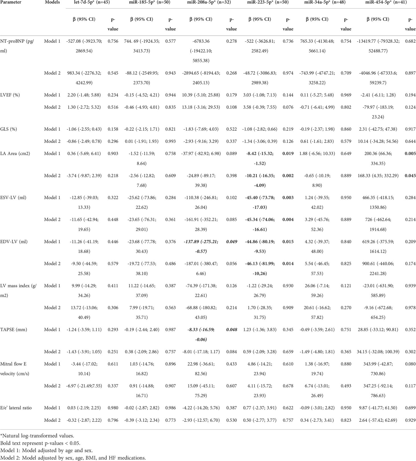
Table 1 The association between miRNAs levels and markers of myocardial injury in patients with Chronic Chagas Cardiomyopathy (n=74).
Considering the trends observed for miR-223-5p and the study’s small sample size, we compared the mean levels of this miRNA according to the NT-proBNP levels, the LVEF, and GLS divided into two groups (over the median value and below the median value). We observed that patients with an NT-proBNP value > 2150 pg/ul had a significantly lower hsa-miR-223-5p relative expression. A similar result was observed for the LVEF, highlighting that patients with an ejection fraction >29% had a significantly higher miR-223-5p relative expression. Finally, we did not observe a significant association for the GLS.
During the median follow-up of 40 months (Q1: 23; Q3: 52), 43% of participants reported an event of the CO, with a rate of 0.40 per 1000 person-years (95% CI 0.28-0.56). The Cox proportional hazard models suggested that none of the evaluated microRNAs was significantly associated with the composite outcome (Supplementary Table 4).
No significant interaction terms by sex were observed in any of the analyses. Similarly, we evaluated the possibility of non-linear associations between the microRNAs and the assessed variables, observing a significant quadratic term for miRNA-223-5p association with the left atrium area (Change in the R2: 0.12. p-value=0.044) (Supplementary Figure 4). No other significant associations regarding non-linear trends were observed.
We further performed a bioinformatic analysis on miR-223-5p, as it was the microRNA shown to be the most significantly associated with the evaluated outcomes. We observed a total of 123 potential target genes identified in the analysis, being eight present in the three queried databases, and the rest in two of them (Supplementary Table 5).
To elucidate the pathways potentially associated with disease severity in CCM related to miR-223-5p expression, the target genes were subjected to gene set enrichment analysis using miRWalk. The genes were observed to be involved in several biological processes, highlighting specific pathways related to signaling by receptor tyrosine kinases such as MAPK signaling pathway (hsa04010) and mTOR signaling pathway (hsa04150) (Tables 2, 3, and Figure 1).
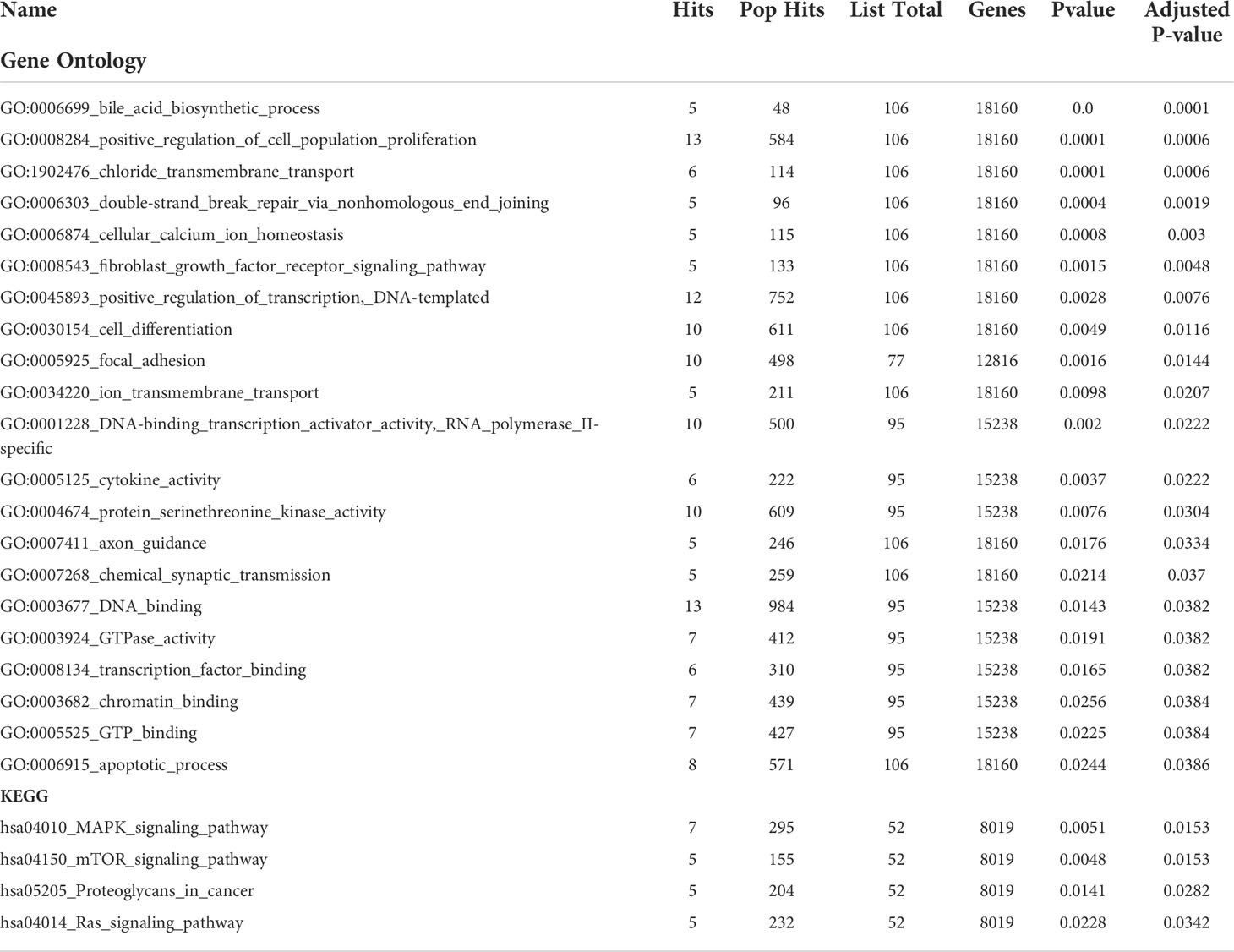
Table 2 Gene set enrichment analysis of target genes for hsa- miR-223-5p.
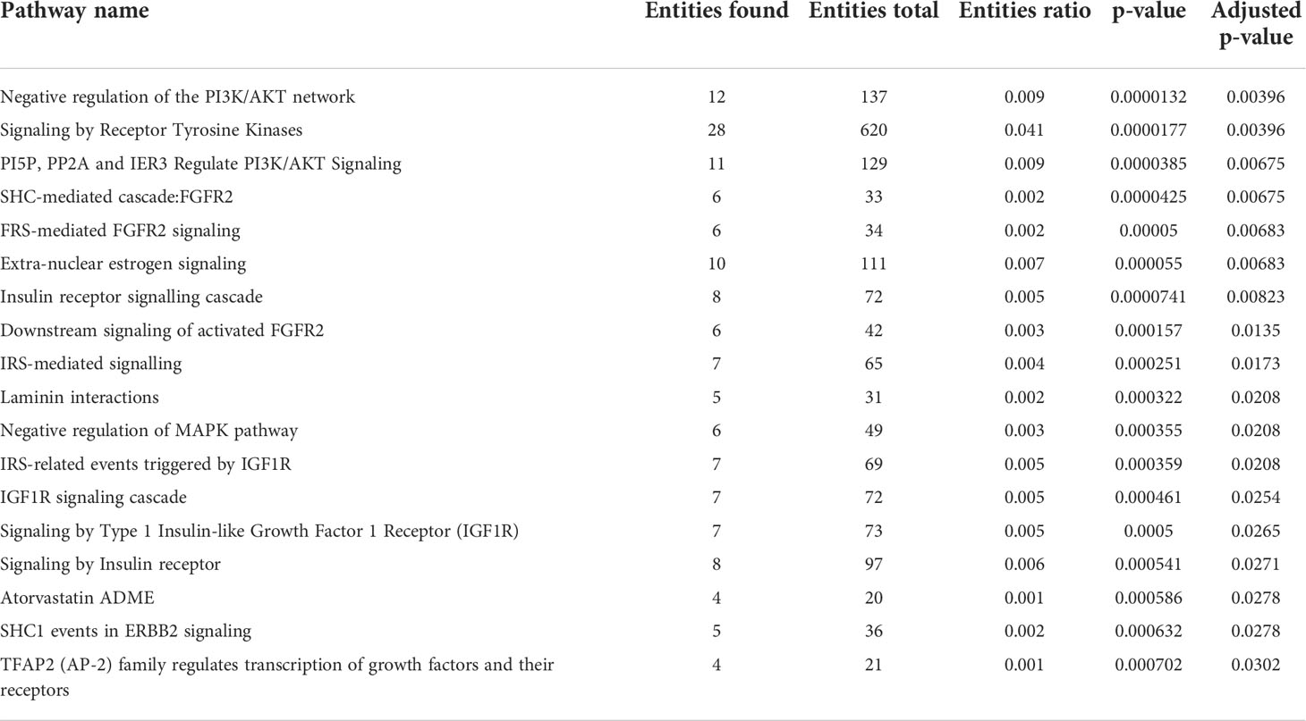
Table 3 Reactome pathway analysis of target genes for hsa- miR-223-5p.
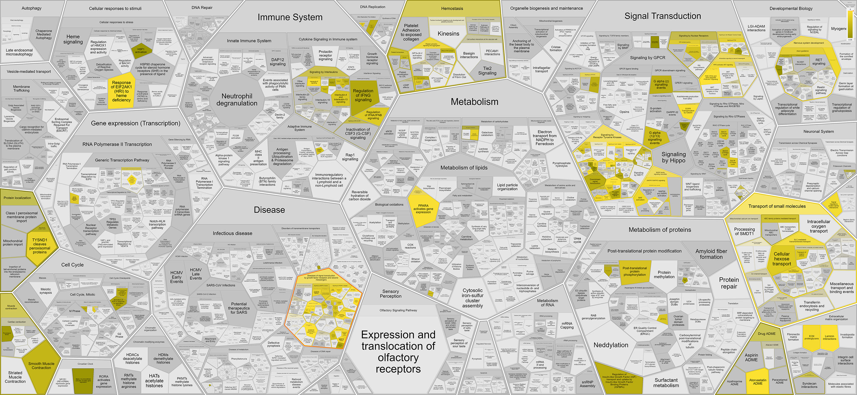
Figure 1 Graphical representation of the main Reactome pathways enriched in the target genes from hsa-miR-223-5p. The intensity of the color is related to the statistical significance of the miRNA involvement, while the cell size is related to the number of genes in each pathway.
Despite significant advances in the study of miRNAs as new biomarkers and therapeutic targets in the area of acute myocardial infarction and heart failure, lesser advances have been achieved in the setting of Chagas Disease and Chronic Chagas Cardiomyopathy (Wang and Cai, 2017; Peterlin et al., 2020). Our study results suggested that miR-223-5p expression is significantly associated with disease severity in CCM patients, independent of age and sex, with higher relative expression levels of this miRNA reflected in better myocardial function markers. Furthermore, our pathway enrichment analysis indicated that the role of this miRNA could be related to pathways involved in the adaptive immune system, specifically related to signaling by receptor tyrosine kinases. Our exploratory study provides new insights into the potential role of microRNAs in CD and CCM pathophysiology.
Several microRNAs have been reported in the literature to be involved in the pathophysiology and progression of CD. For example, the study of Jha BK et al. observed that miR-155 knockout (KO) mice had a significantly worse prognosis after T. cruzi infection compared with wild-type C57BL/6 mice. They found that, in the absence of this microRNA, a decreased release of tumor necrosis factor-alpha (TNF-α) and interferon-gamma (IFN-y) was observed. At the same time, there was an increased accumulation of neutrophils and inflammatory monocytes and a reduced amount of natural killer cells and CD8-positive T cells in the involved tissue. These results suggested a critical role of miR-155 in the immune regulation in response to T. cruzi infection (Jha et al., 2020). Similar to this, additional studies on murine models have observed a substantial role of several microRNA expression levels in multiple pathways related to the immune response and T. cruzi virulence mechanisms (Navarro et al., 2015; Linhares-Lacerda et al., 2015; Ferreira et al., 2017; Ballinas-Verdugo et al., 2021).
Similarly, studies performed in human myocardial tissue obtained from transplanted hearts have also reported relevant results, highlighting the study of Ferreira LRP et al. In this study, the authors analyzed the expression of nine microRNAs in myocardial tissue samples of three populations: CCM patients, dilated cardiomyopathy patients and healthy controls, observing that miR-1, miR-133a-2, miR-133b, miR-208a, and 208b were significantly down-regulated in CCM patients compared to controls. The pathway enrichment analysis suggested that the target genes for these microRNAs were involved in the network labeled as “Cardiovascular Disease, Connective Tissue Disorders, Dermatological Diseases” (Ferreira et al., 2014). Furthermore, the study of Laugier et al. suggested significantly different expression levels of miR-146a and miR-155 in CCM myocardium samples compared with controls. The pathway analyses of these tissue signatures suggested an enrichment in biological processes associated with the immune response, highlighting inflammation, Th1/IFN-γ-inducible genes, fibrosis, hypertrophy, and mitochondrial/oxidative stress/antioxidant response (Laugier et al., 2020). Nevertheless, up to date, only the study of Nonaka CKV et al. has evaluated the association between microRNA expression patterns and clinical variables in CCM patients, assessing six circulating microRNAs (miR-19a-3p, miR-21-5p, miR-29b-3p, miR-30a-5p, miR-199b-5p, and miR-208a-3p). In this study, the authors observed that miR-19a-3p, miR-21-5p, and miR-29b-3p were differentially expressed in CCM patients compared to those with the indeterminate form of the disease. Furthermore, a positive correlation between the relative expression of these microRNAs with functional class and fibrosis and a negative correlation with ejection fraction and left ventricular strain was observed. These results were validated by cardiac tissue analysis and in vitro studies, suggesting miR-19a-3p, miR-21-5p, and miR-29b-3p circulating levels as potential biomarkers for early detection of disease progression in CD (Nonaka et al., 2019).
While microRNAs have been previously suggested to play a role in Chagas disease, our study represents the first to investigate and observe the significant role of miR-223-5p in the pathophysiology of CD and CCM. MiR-223, a microRNA expressed in multiple cells such as platelets, macrophages, and cardiomyocytes, has been observed to be involved in several cardiovascular diseases. Nevertheless, most of the available evidence has evaluated the 3’-arm of precursor miR-223, as it was considered the only mature and functional component, while the complementary 5’-arm was thought to be degraded (Mitra et al., 2015; Córdova-Rivas et al., 2019). Recent evidence has suggested a co-expression of both arms of pre-miR-223 in relevant pathophysiological processes affecting the myocardium (Taïbi et al., 2014; Wang et al., 2014). In the study of Wang et al., the knockout (KO) of the miR-223 duplex (5p and 3p) promoted a most severe sepsis-induced cardiac dysfunction and higher mortality in KO mice compared with wild-type ones (Wang et al., 2014). A similar result was observed by Qin et al. in the setting of myocardial ischemia/reperfusion-induced necrosis in mice (Qin et al., 2016). In this study, miR-223-5p KO mice hearts exhibited worse recovery of contractile performance over the reperfusion period and a higher degree of necrosis than wild-type ones. The authors suggested the activation of the RIP1/RIP3/MLKL necroptotic pathway as responsible for the differences observed in both groups, as treatment of KO mice with necrostatin-1s, a RIP1 kinase activity suppressor and necroptosis inhibitor, decreased the infarction size and myocardial dysfunction after ischemia/reperfusion injury (Qin et al., 2016). Interestingly, regulated necrosis, or necroptosis, has been observed to be mediated by tumor necrosis factor-α (TNF-α)/TNFR1-induced protein complex RIP1-RIP3, also known as the necrosome (Silke et al., 2015). In this context, several studies have suggested that the TNF-α/TNFR-mediated pathway plays a critical role in the process of T. cruzi control in the acute stage and the development of CCM, being TNFR1 closely linked to CD8+ T cell dysregulation (Michailowsky et al., 2001; Tzelepis et al., 2007). Nonetheless, although necroptosis have been studied in heart failure as one of the potential drivers of myocardial remodeling, no study has assessed the role of this mechanism in CD or CCM (Li et al., 2014; Guo et al., 2017).
Finally, we also observed a trend concerning the expression of miR-208a-5p and markers of myocardial function such as the ESV and the TAPSE. Nevertheless, the associations had a borderline statistical significance, and no other trend was observed for the other biomarkers. Therefore, we did not include this microRNA in our bioinformatics analysis. In contrast to miR-223-5p, miR-208a-3p has been previously studied in CD and CCM, highlighting the study of Linhares-Lacerda L et al., which observed significantly higher circulating levels plasma miR-208a in patients with the indeterminate form of the disease than in controls (Linhares-Lacerda et al., 2018). On the other hand, the study of Ferreira LRP et al. observed that patients with CCM had a significantly lower relative expression of miR-208a, measured in human myocardial samples, than healthy controls (Ferreira et al., 2014). MiR-208a represents an essential regulator of several pathways involved in cardiac fibrosis and hypertrophy, being also associated with the development of conduction abnormalities and arrhythmias (Wang et al., 2014; Huang et al., 2021). Although the role of this microRNA in CD is not clear, published evidence suggests that TGF-β may activate miR-208a to regulate genes related to hypertrophy, as TGF-β neutralizing antibody therapy attenuated miR-208a induced expression in myocytes (Wang et al., 2013). In this context, the TGF-β signaling pathway has been shown to play a significant role in the development of CCM, as it may potentiate the parasitic burden by downregulating the intracellular immune response against T. cruzi (Ferreira et al., 2016; da et al., 2019). Moreover, the study of Araújo et al. reported that circulating levels of TGF-β were significantly associated with cardiac dysfunction and fibrosis in CD (Araújo-Jorge et al., 2002), while the study of Ferreira RR et al. observed a positive therapeutic effect in mice infected with T. cruzi and treated with a selective inhibitor of TβR1/ALK5 (Ferreira et al., 2019), highlighting the relevance of this cytokine in this setting and the potential of miR-208a as a critical mediator of this effect.
The present study has relevant strengths worthy of mention, highlighting the inclusion of a population of patients with advanced-stage heart failure and a relatively long follow-up. On the other hand, several limitations are present. First, our study has a small sample size and limited events for the composite outcomes and therefore may not have had enough power, especially for the CO. Second, the cross-sectional design of the study does not provide insights into the causality of the identified associations. Third, the measurement of the microRNAs also carried significant processing issues, highlighting the lack of pre-amplification of serum RNAs due to limited resources, the wide variability of results observed and the low concentrations of these microRNAs in serum. Furthermore, microRNA measurement was performed using a single serum sample, limiting the possibilities of assessing changes over time. Finally, it was not possible to perform an experimental validation of the potential target genes, which limited the possibility of confirming the findings of the prediction analysis performed.
Our exploratory study observed a significant association between the expression levels of circulating miR-223-5p and echocardiographic and laboratory biomarkers of myocardial injury in patients with CCM. Furthermore, the bioinformatics analysis suggested an important role of this microRNA in pathways related to signaling by receptor tyrosine kinases. Further large prospective cohort studies are required to potentially validate this and other relevant microRNAs as biomarkers or therapeutic targets in the setting of CD and CCM.
The raw data supporting the conclusions of this article will be made available by the authors upon request, without undue reservation.
The studies involving human participants were reviewed and approved by Comité de etica de la Fundación Cardiovascular de Colombia. The patients/participants provided their written informed consent to participate in this study.
SG-O, LR, LH, TM, and LE contributed to the conception and design of the study. PB-N did the microRNA extraction and measurements. SG-O, PB-N, and LR organized the database. SG-O, LR, and TM performed the statistical analysis. SG-O wrote the first draft of the manuscript. SG-O, LH, and TM wrote sections of the manuscript. All authors contributed to manuscript revision, read, and approved the submitted version.
LR and TM were supported by St. Gallen University through the Seed Money Grants Call of 2019.
The authors declare that the research was conducted in the absence of any commercial or financial relationships that could be construed as a potential conflict of interest.
All claims expressed in this article are solely those of the authors and do not necessarily represent those of their affiliated organizations, or those of the publisher, the editors and the reviewers. Any product that may be evaluated in this article, or claim that may be made by its manufacturer, is not guaranteed or endorsed by the publisher.
The Supplementary Material for this article can be found online at: https://www.frontiersin.org/articles/10.3389/fcimb.2022.922189/full#supplementary-material
Antinori, S., Galimberti, L., Bianco, R., Grande, R., Galli, M., Corbellino, M., et al. (2017). Chagas disease in Europe: A review for the internist in the globalized world. Eur. J. Intern. Med. 43, 6–15. doi: 10.1016/j.ejim.2017.05.001
Araújo-Jorge, T. C., Waghabi, M. C., Hasslocher-Moreno, A. M., Xavier, S. S., Higuchi de, M. L., Keramidas, M., et al. (2002). Implication of transforming growth factor-beta1 in chagas disease myocardiopathy. J. Infect. Dis. 186, 1823–1828. doi: 10.1086/345882
Ballinas-Verdugo, M. A., Jiménez-Ortega, R. F., Martínez-Martínez, E., Rivas, N, Contreras-López, EA, Carbó, R, et al. (2021). Circulating miR-146a as a possible candidate biomarker in the indeterminate phase of chagas disease. Biol. Res. 54, 21. doi: 10.1186/s40659-021-00345-3
Bernardo, B. C., Gao, X.-M., Tham, Y. K., Kiriazis, H, Winbanks, C. E., Ooi, J. Y. Y., et al. (2014). Silencing of miR-34a attenuates cardiac dysfunction in a setting of moderate, but not severe, hypertrophic cardiomyopathy. PloS One 9, e90337. doi: 10.1371/journal.pone.0090337
Bern, C., Messenger, L. A., Whitman, J. D., Maguire, J. H.. (2019). Chagas disease in the united states: a public health approach. Clin. Microbiol. Rev. 33, e00023–e00019. doi: 10.1128/CMR.00023-19
Bestetti, R. B. (2016). Chagas heart failure in patients from Latin America. Card. Fail Rev. 2, 90–94. doi: 10.15420/cfr.2016:14:2
Bonney, K. M., Engman, D. M. (2015). Autoimmune pathogenesis of chagas heart disease: looking back, looking ahead. Am. J. Pathol. 185, 1537–1547. doi: 10.1016/j.ajpath.2014.12.023
Bonney, K. M., Luthringer, D. J., Kim, S. A., Garg, N. J., Engman, D. M.. (2019). Pathology and pathogenesis of chagas heart disease. Annu. Rev. Pathol. 14, 421–447. doi: 10.1146/annurev-pathol-020117-043711
Conners, E. E., Vinetz, J. M., Weeks, J. R., Brouwer, K. C.. (2016). A global systematic review of chagas disease prevalence among migrants. Acta Trop. 156, 68–78. doi: 10.1016/j.actatropica.2016.01.002
Córdova-Rivas, S., Fraire-Soto, I., Mercado-Casas Torres, A., Servín-González, L. S., Granados-López, A. J., López-Hernández, Y., et al. (2019). 5p and 3p strands of miR-34 family members have differential effects in cell proliferation, migration, and invasion in cervical cancer cells. Int. J. Mol. Sci. 20, E545. doi: 10.3390/ijms20030545
da Costa, A. W. F., do Carmo Neto, J. R., Braga, Y. L. L., et al. (2019). Cardiac chagas disease: MMPs, TIMPs, galectins, and TGF-β as tissue remodelling players. Dis. Markers 2019, 3632906. doi: 10.1155/2019/3632906
Echeverria, L. E., Morillo, C. A. (2019). American Trypanosomiasis (Chagas disease). Infect. Dis. Clin. North Am. 33, 119–134. doi: 10.1016/j.idc.2018.10.015
Echeverría, L. E., Rojas, L. Z., Gómez-Ochoa, S. A. (2021). Coagulation disorders in chagas disease: A pathophysiological systematic review and meta-analysis. Thromb. Res. 201, 73–83. doi: 10.1016/j.thromres.2021.02.025
Ferreira, R. R., Abreu R da, S., Vilar-Pereira, G., et al. (2019). TGF-β inhibitor therapy decreases fibrosis and stimulates cardiac improvement in a pre-clinical study of chronic chagas’ heart disease. PloS Negl. Trop. Dis. 13, e0007602. doi: 10.1371/journal.pntd.0007602
Ferreira, R. R., de Souza, E. M., de Oliveira, F. L., Ferrão, P. M., Gomes, L. H. F., Mendonça-Lima L., , et al. (2016). Proteins involved on TGF-β pathway are up-regulated during the acute phase of experimental chagas disease. Immunobiology 221, 587–594. doi: 10.1016/j.imbio.2016.01.009
Ferreira, L. R. P., Ferreira, F. M., Laugier, L., Cabantous, S., Navarro, I. C., da Silva Cândido, D., et al. (2017). Integration of miRNA and gene expression profiles suggest a role for miRNAs in the pathobiological processes of acute trypanosoma cruzi infection. Sci. Rep. 7, 17990. doi: 10.1038/s41598-017-18080-9
Ferreira, L. R. P., Frade, A. F., Santos, R. H. B., Teixeira, P. C., Baron, M. A., Navarro, I. C., et al. (2014). MicroRNAs miR-1, miR-133a, miR-133b, miR-208a and miR-208b are dysregulated in chronic chagas disease cardiomyopathy. Int. J. Cardiol. 175, 409–417. doi: 10.1016/j.ijcard.2014.05.019
Guo, X., Yin, H., Li, L., Chen, Y., Li, J., Doan, J., et al. (2017). Cardioprotective role of tumor necrosis factor receptor-associated factor 2 by suppressing apoptosis and necroptosis. Circulation 136, 729–742. doi: 10.1161/CIRCULATIONAHA.116.026240
Gupta, H., Rubio, M., Sitoe, A., et al. (2021). Plasma MicroRNA profiling of plasmodium falciparum biomass and association with severity of malaria disease. Emerg. Infect. Dis. 27, 430–442. doi: 10.3201/eid2702.191795
Gupta, H., Sahu, P. K., Pattnaik, R., Varo, R., Cisteró, P., Madrid, L., et al. (2021). Plasma levels of hsa-miR-3158-3p microRNA on admission correlate with MRI findings and predict outcome in cerebral malaria. Clin. Transl. Med. 11, e396. doi: 10.1002/ctm2.396
Huang, X.-H., Li, J.-L., Li, X.-Y., Wang, S.-X., Jiao, Z.-H., Li, S.-Q., et al. (2021). miR-208a in cardiac hypertrophy and remodeling. Front. Cardiovasc. Med. 8, 773314. doi: 10.3389/fcvm.2021.773314
Jha, B. K., Varikuti, S., Seidler, G. R., Volpedo, G., Satoskar, A. R., McGwire, B. S.. (2020). MicroRNA-155 deficiency exacerbates trypanosoma cruzi infection. Infect. Immun. 88, e00948–e00919. doi: 10.1128/IAI.00948-19
Laugier, L., Ferreira, L. R. P., Ferreira, F. M., Cabantous, S., Frade, A. F., Nunes, J. P., et al. (2020). miRNAs may play a major role in the control of gene expression in key pathobiological processes in chagas disease cardiomyopathy. PloS Negl. Trop. Dis. 14, e0008889. doi: 10.1371/journal.pntd.0008889
Lee, B. Y., Bacon, K. M., Bottazzi, M. E., Hotez, PJ. (2013). Global economic burden of chagas disease: a computational simulation model. Lancet Infect. Dis. 13, 342–348. doi: 10.1016/S1473-3099(13)70002-1
Li, L., Chen, Y., Doan, J., Murray, J., Molkentin, J. D., Liu, Q., et al. (2014). Transforming growth factor β-activated kinase 1 signaling pathway critically regulates myocardial survival and remodeling. Circulation 130, 2162–2172. doi: 10.1161/CIRCULATIONAHA.114.011195
Linhares-Lacerda, L., Granato, A., Gomes-Neto, J. F., Conde, L., Freire-de-Lima, L., de Freitas, E. O., et al. (2018). Circulating plasma MicroRNA-208a as potential biomarker of chronic indeterminate phase of chagas disease. Front. Microbiol. 9, 269. doi: 10.3389/fmicb.2018.00269
Linhares-Lacerda, L., Palu, C. C., Ribeiro-Alves, M., Paredes, B. D., Morrot, A., Garcia-Silva, M. R., et al. (2015). Differential expression of microRNAs in thymic epithelial cells from trypanosoma cruzi acutely infected mice: Putative role in thymic atrophy. Front. Immunol. 6, 428. doi: 10.3389/fimmu.2015.00428
Lv, P., Zhou, M., He, J., Meng, W., Ma, X., Dong, S., et al. (2014). Circulating miR-208b and miR-34a are associated with left ventricular remodeling after acute myocardial infarction. Int. J. Mol. Sci. 15, 5774–5788. doi: 10.3390/ijms15045774
Michailowsky, V., Silva, N. M., Rocha, C. D., et al. (2001). Pivotal role of interleukin-12 and interferon-gamma axis in controlling tissue parasitism and inflammation in the heart and central nervous system during trypanosoma cruzi infection. Am. J. Pathol. 159, 1723–1733. doi: 10.1016/S0002-9440(10)63019-2
Mitra, R., Lin, C.-C., Eischen, C. M., Bandyopadhyay, S., Zhao, Z.. (2015). Concordant dysregulation of miR-5p and miR-3p arms of the same precursor microRNA may be a mechanism in inducing cell proliferation and tumorigenesis: a lung cancer study. RNA 21, 1055–1065. doi: 10.1261/rna.048132.114
Montgomery, R. L., Hullinger, T. G., Semus, H. M., Dickinson, B. A., Seto, A. G., Lynch, J. M., et al. (2011). Therapeutic inhibition of miR-208a improves cardiac function and survival during heart failure. Circulation 124, 1537–1547. doi: 10.1161/CIRCULATIONAHA.111.030932
Navarro, I. C., Ferreira, F. M., Nakaya, H. I., Baron, M. A., Vilar-Pereira, G., Pereira, I. R., et al. (2015). MicroRNA transcriptome profiling in heart of trypanosoma cruzi-infected mice: Parasitological and cardiological outcomes. PloS Negl. Trop. Dis. 9, e0003828. doi: 10.1371/journal.pntd.0003828
Nonaka, C. K. V., Macêdo, C. T., Cavalcante, B. R. R., Alcântara, A. C., de Silva, D. N., Bezerra da, M. R., et al. (2019). Circulating miRNAs as potential biomarkers associated with cardiac remodeling and fibrosis in chagas disease cardiomyopathy. Int. J. Mol. Sci. 20, E4064. doi: 10.3390/ijms20164064
Nunes, M. C. P., Beaton, A., Acquatella, H., Bern, C., Bolger, A. F., Echeverría, L. E., et al. (2018). Chagas cardiomyopathy: An update of current clinical knowledge and management: A scientific statement from the American heart association. Circulation 138, e169–e209. doi: 10.1161/CIR.0000000000000599
Peterlin, A., Počivavšek, K., Petrovič, D., Peterlin, B.. (2020). The role of microRNAs in heart failure: A systematic review. Front. Cardiovasc. Med. 7, 161. doi: 10.3389/fcvm.2020.00161
Qin, D., Wang, X., Li, Y., Yang, L., Wang, R., Peng, J., et al. (2016). MicroRNA-223-5p and -3p cooperatively suppress necroptosis in Ischemic/Reperfused hearts. J. Biol. Chem. 291, 20247–20259. doi: 10.1074/jbc.M116.732735
Savarese, G., Lund, L. H. (2017). Global public health burden of heart failure. Card. Fail Rev. 3, 7–11. doi: 10.15420/cfr.2016:25:2
Shen, L., Ramires, F., Martinez, F., Bodanese, L. C., Echeverría, L. E., Gómez, E. A., et al. (2017). Contemporary characteristics and outcomes in chagasic heart failure compared with other nonischemic and ischemic cardiomyopathy. Circ. Heart Fail 10, e004361. doi: 10.1161/CIRCHEARTFAILURE.117.004361
Silke, J., Rickard, J. A., Gerlic, M. (2015). The diverse role of RIP kinases in necroptosis and inflammation. Nat. Immunol. 16, 689–697. doi: 10.1038/ni.3206
Taïbi, F., Metzinger-Le Meuth, V., Massy, Z. A., Metzinger, L.. (2014). miR-223: An inflammatory oncomiR enters the cardiovascular field. Biochim. Biophys. Acta 1842, 1001–1009. doi: 10.1016/j.bbadis.2014.03.005
Tzelepis, F., Persechini, P. M., Rodrigues, M. M. (2007). Modulation of CD4(+) T cell-dependent specific cytotoxic CD8(+) T cells differentiation and proliferation by the timing of increase in the pathogen load. PloS One 2, e393. doi: 10.1371/journal.pone.0000393
Wang, H., Cai, J. (2017). The role of microRNAs in heart failure. Biochim. Biophys. Acta Mol. Basis Dis. 1863, 2019–2030. doi: 10.1016/j.bbadis.2016.11.034
Wang, X., Huang, W., Yang, Y., Wang, Y., Peng, T., Chang, J., et al. (2014). Loss of MiR-223 duplex (5p and 3p) aggravates myocardial depression and mortality in polymicrobial sepsis. Biochim. Biophys. Acta 1842, 701–711. doi: 10.1016/j.bbadis.2014.01.012
Wang, B.-W., Wu, G.-J., Cheng, W.-P., Shyu, K.-G.. (2013). Mechanical stretch via transforming growth factor-β1 activates microRNA-208a to regulate hypertrophy in cultured rat cardiac myocytes. J. Formos. Med. Assoc. 112, 635–643. doi: 10.1016/j.jfma.2013.01.002
Wang, B.-W., Wu, G.-J., Cheng, W.-P., Shyu, K.-G.. (2014). MicroRNA-208a increases myocardial fibrosis via endoglin in volume overloading heart. PloS One 9, e84188. doi: 10.1371/journal.pone.0084188
Yang, Y., Cheng, H.-W., Qiu, Y., et al. (2015). MicroRNA-34a plays a key role in cardiac repair and regeneration following myocardial infarction. Circ. Res. 117, 450–459. doi: 10.1161/CIRCRESAHA.117.305962
Keywords: chagas disease, chronic chagas cardiomyopathy, microRNAs, echocardiography, omics
Citation: Gómez-Ochoa SA, Bautista-Niño PK, Rojas LZ, Hunziker L, Muka T and Echeverría LE (2022) Circulating MicroRNAs and myocardial involvement severity in chronic Chagas cardiomyopathy. Front. Cell. Infect. Microbiol. 12:922189. doi: 10.3389/fcimb.2022.922189
Received: 17 April 2022; Accepted: 18 July 2022;
Published: 08 August 2022.
Edited by:
Bikash R. Giri, Tongji University, ChinaReviewed by:
Otacilio C. Moreira, Oswaldo Cruz Foundation (Fiocruz), BrazilCopyright © 2022 Gómez-Ochoa, Bautista-Niño, Rojas, Hunziker, Muka and Echeverría. This is an open-access article distributed under the terms of the Creative Commons Attribution License (CC BY). The use, distribution or reproduction in other forums is permitted, provided the original author(s) and the copyright owner(s) are credited and that the original publication in this journal is cited, in accordance with accepted academic practice. No use, distribution or reproduction is permitted which does not comply with these terms.
*Correspondence: Taulant Muka, dGF1bGFudC5tdWthQGlzcG0udW5pYmUuY2g=
Disclaimer: All claims expressed in this article are solely those of the authors and do not necessarily represent those of their affiliated organizations, or those of the publisher, the editors and the reviewers. Any product that may be evaluated in this article or claim that may be made by its manufacturer is not guaranteed or endorsed by the publisher.
Research integrity at Frontiers
Learn more about the work of our research integrity team to safeguard the quality of each article we publish.