
95% of researchers rate our articles as excellent or good
Learn more about the work of our research integrity team to safeguard the quality of each article we publish.
Find out more
ORIGINAL RESEARCH article
Front. Cell. Infect. Microbiol. , 06 September 2021
Sec. Clinical Microbiology
Volume 11 - 2021 | https://doi.org/10.3389/fcimb.2021.713873
This article is part of the Research Topic Advances in Diagnosis and Therapeutic Intervention for Foodborne Parasitic Diseases, Volume II View all 20 articles
 Si-Yuan Qin1,2†
 He-Ting Sun2†Chuang Lyu3,4†Jun-Hui Zhu1Zhen-Jun Wang1Tao Ma1
 Quan Zhao5*
 Yun-Gang Lan1*
 Wen-Qi He1*
Cryptosporidium is an enteric apicomplexan parasite, which can infect multiple mammals including livestock and wildlife. Tibetan Antelope (Pantholops hodgsonii) is one of the most famous wildlife species, that belongs to the first class protected wild animals in China. However, it has not been known whether Tibetan Antelope is infected with Cryptosporidium so far. The objective of the present study was to determine the prevalence and characterization of Cryptosporidium species infection in Tibetan Antelope and the corresponding species by using molecular biological method. In the current study, a total of 627 fecal samples were randomly collected from Tibetan Antelope in the Tibet Autonomous Region (2019–2020), and were examined by PCR amplification of the small subunit ribosomal RNA (SSU rRNA) gene. Among 627 samples, 19 (3.03%, 19/627) were examined as Cryptosporidium-positive, with 7 (2.33%, 7/300) in females and 12 (3.67%, 12/327) in males. The analysis of SSU rRNA gene sequence suggested that only two Cryptosporidium species, namely, C. xiaoi and C. ubiquitum, were identified in this study. This is the first evidence for an existence of Cryptosporidium in Tibetan Antelope. These findings extend the host range for Cryptosporidium spp. and also provide important data support for prevention and control of Cryptosporidium infection in Tibetan Antelope.
Cryptosporidium, the causative agent of cryptosporidiosis, causes an intestinal disease in a wide range of hosts worldwide, including wildlife, livestock, and humans. Human infection with Cryptosporidium is usually through a close contact with the infected animals or consuming contaminated water or food (Rossignol, 2010). At least 38 species and over 70 genotypes of Cryptosporidium can infect humans and animals (Deng et al., 2020). Among them, more than 20 have been considered as zoonotic potential risks, including C. hominis, C. parvum, C. meleagridis, C. felis, C. canis, C. cuniculus, C. ubiquitum, C. viatorum, C. muris, C. suis, C. fayeri, C. andersoni, C. bovis, C. scrofarum, C. xiaoi, C. tyzzeri, C. erinaceid, and C. horse, C. skunk, and C. chipmunk I genotype (Ren et al., 2012; Adamu et al., 2014; Koehler et al., 2014; Kváč et al., 2014; Ma et al., 2014; Qi et al., 2014; Qin et al., 2014; Yang et al., 2014; Galuppi et al., 2015; Lin et al., 2015; Yan et al., 2017; Firoozi et al., 2019; Takaki et al., 2020; Xu et al., 2020). C. hominis and C. parvum were most frequently found in human. C. xiaoi was generally considered as C. bovis-like genotype or C. bovis when Fayer and Santín identified it as a new species in 2009 based on morphology and molecular methods (Fayer and Santín, 2009).
Since C. xiaoi and C. ubiquitum were recognized firstly in sheep, many researches were focused on the prevalence of C. xiaoi and C. ubiquitum in humans and other animals which have closer relationship with the sheep like bovine and cervine. To date, C. xiaoi infection in sheep has been reported in many countries, including Ireland, Kuwait, Australia, Norway, Spain, France, Greece, Egypt, Tanzania, Jordan, Poland, Ghana, and Iran (Díaz et al., 2010; Robertson et al., 2010; Yang et al., 2011; Rieux et al., 2013; Mahfouz et al., 2014; Tzanidakis et al., 2014; Parsons et al., 2015; Hijjawi et al., 2016; Mirhashemi et al., 2016; Kaupke et al., 2017; Squire et al., 2017; Majeed et al., 2018; Firoozi et al., 2019). In addition, the pertinent literatures about C. ubiquitum infection in sheep were derived from Ireland, Kuwait, Australia, Spain, Greece, Poland, Ghana, Iran, and Algeria (Díaz et al., 2010; Yang et al., 2011; Tzanidakis et al., 2014; Mirhashemi et al., 2016; Kaupke et al., 2017; Squire et al., 2017; Baroudi et al., 2018; Majeed et al., 2018; Firoozi et al., 2019). In China, C. xiaoi and C. ubiquitum were also found in sheep in Anhui, Xinjiang, Jilin, Inner Mongolia, Ningxia, Shandong, Shanghai, Henan, Qinghai, and Beijing (Mi et al., 2018; Qi et al., 2019), Tibetan sheep in Qinghai (Li et al., 2016), and goat in Guangdong, Hubei, Shandong, Shanghai, Henan, Chongqing, Shaanxi (Mi et al., 2014; Wang et al., 2014; Peng et al., 2016). Interestingly, C. xiaoi and C. ubiquitum have also been occasionally found in yak (Ma et al., 2014). The infection of C. xiaoi and C. ubiquitum in hosts is usually asymptomatic. However, the infection occasionally causes diarrhea and weight loss (Santín, 2013). More importantly, C. xiaoi is also found in HIV/AIDS patients (Adamu et al., 2014), and C. ubiquitum, previously known as the cervine genotype, has been emerging as another major zoonotic species that infects persons (Li et al., 2014), thus posing a risk to public health. Therefore, C. xiaoi and C. ubiquitum are of public health concern because of its wide geographic distribution and broad host range.
China has abundant biodiversity resources. Tibetan Antelope (Pantholops hodgsonii) is one of the most important wild animal species, which is a very important part of the natural ecology in Qinghai-tibet plateau (Peng et al., 2018). In 1981, China had accessed to the convention on international trade about endangered species of wild fauna and flora, in which the Tibetan antelope was classified into appendix I species (http://www.iucnredlist.org/). Since 1988, the Tibetan Antelope was identified as a first-grade state protection of wildlife (http://www.forestry.gov.cn/main/3954/content-1063883.html). However, the information for this pathogen infection in Tibetan Antelope is limited. Importantly, there has been no available information concerning Cryptosporidium infection in Tibetan Antelope. Therefore, the objective of the present study was to molecularly determine the prevalence and characterization of Cryptosporidium species in Tibetan Antelope in Tibet Autonomous Region, China.
A total of 627 fecal samples of Tibetan Antelope were collected from Nyima County, Shuanghu County, Shenza County, and Baingoin County in Tibet Autonomous Region of China in 2019 and 2020 (Figure 1). A fresh fecal sample (approximately 5 g) for each Tibetan Antelope was collected from the ground using sterile gloves after defecation, and then was placed into ice boxes and sent to the laboratory. Tibetan Antelope with horns are males, otherwise, are females. The information regarding sampling time, region, and gender were recorded. This study was approved by the Ethics Committee of Jilin University.
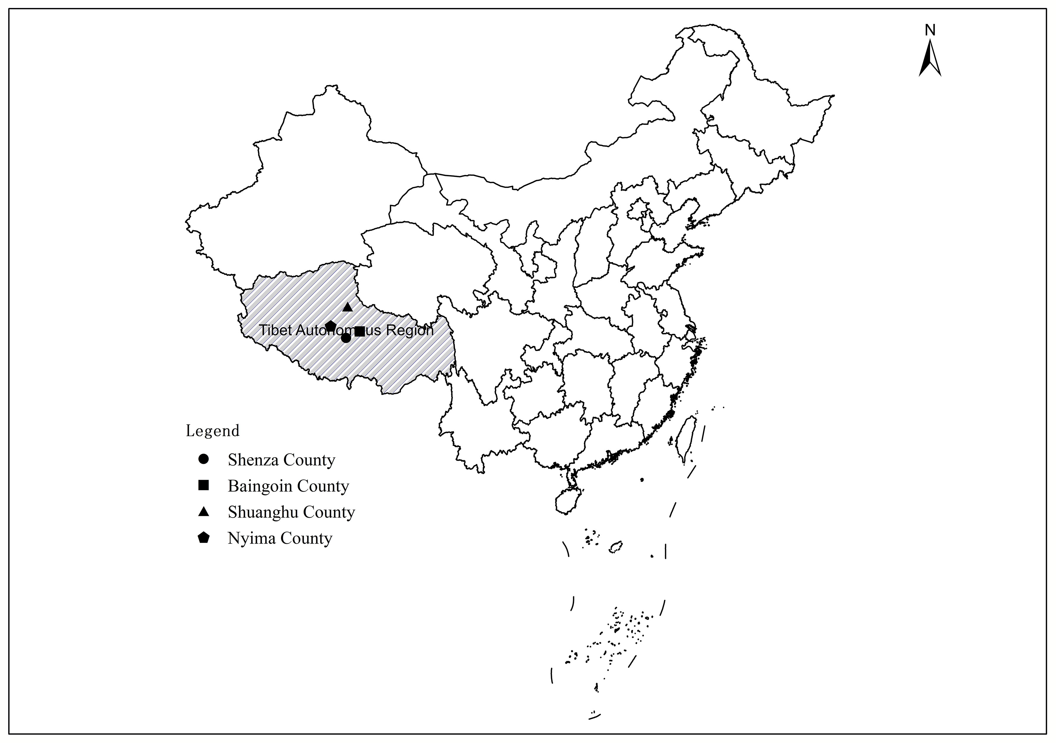
Figure 1 A map of Tibet Autonomous Region, China showing the geographical regions in Nyima County, Shuanghu County, Shenza County, and Baingoin County, in which Tibetan Antelope were sampled.
The fecal samples were diluted with 0.9% normal saline and filtered through 100-mesh stainless steel sieve. The filtrate was centrifuged at 4000 rpm/min for 5 min to enrich Cryptosporidium eggs. Genomic DNA was extracted from approximately 200 mg of each stool specimen using the E.Z.N.A.® Stool DNA Kit (Omega Biotek Inc., Norcross, GA, USA) according to the manufacturer’s instructions, and then were stored at -20°C prior to a PCR analysis. Cryptosporidium prevalence and their species/genotypes were identified by nested PCR amplification of the small subunit ribosomal RNA (SSU rRNA) gene, using the primers 18SiCF2 (5′-GACATATCATTCAAGTTTCTGACC-3′) and 18SiCR2 (5′-CTGAAGGAGTAAGGAACAACC-3′) that amplified a fragment of about 760 bp in length in the first round PCR and the primers 18SiCF1 (5′-CCTATCAGCTTTAGACGGTAGG-3′) and 18SiCR1 (5′-TCTAAGAATTTCACCTCTGACTG-3′) that amplified a fragment of about 590 bp in length in the second round PCR (Qin et al., 2014; Koehler et al., 2018). The positive and negative controls were included in each test. The second PCR products were observed using UV light after electrophoresis at a 1.5% (m/V) agarose gel containing ethidium bromide.
The positive PCR products were sent to Sangon Biotech Company (Shanghai, China) for sequencing. The PCR products were sequenced on both strands to guarantee the accuracy of the sequence. A new PCR product was subjected to sequencing when single nucleotide substitution, insertion, or deletion was found in the former sequencing. The alignment and analysis for the SSU rRNA nucleotide sequences and reference sequences were performed using the Clustal X 1.83 program and Basic Local Alignment Search Tool (BLAST) (https://blast.ncbi.nlm.nih.gov), in order to determine the species of Cryptosporidium. The phylogenetic trees were reconstructed by MEGA 5.0 software using a neighbor-joining (NJ) method with a Kimura 2-parameter model (1,000 replicates). The representative nucleotide sequences were disposed to GenBank with accession numbers MZ220364 and MZ220365.
To assess the possible risk factors (gender, region, and year) associated with an exposure to Cryptosporidium infection in Tibetan Antelope, a multivariable logistic regression analysis was carried out using the PASW Statistics 18.0 (SPSS, Inc., IBM Corporation, Somers, NY) (Zhao et al., 2013). When independent variables were contained in the multivariable logistic regression model, probability (P) value < 0.05 was considered as statistically significant between levels within factors and interactions, and their odd ratio (OR) and 95% confidence interval (CI) were calculated.
In the present study, 19 (3.0%, 95% CI 1.7–4.4) out of 627 Tibetan Antelope fecal samples from Tibet Autonomous Region were tested as Cryptosporidium-positive by PCR amplification of the SSU rRNA gene. The prevalence of Cryptosporidium infection in Tibetan Antelope was 2.2% (7/322, 95% CI 0.6–3.8) in 2019, and 3.9% (12/305, 95% CI 0.6–3.8) in 2020 (Table 1). Male Tibetan Antelope had a higher prevalence (3.7%, 95% CI 1.7–5.7, 12/327) as compared to that of females (2.3%, 95% CI 0.6–4.0, 7/300) (Table 1). The prevalence of Cryptosporidium in Tibetan Antelope in Nyima County, Shenza County, Shuanghu County, and Baingoin County was 3.8% (7/182, 95% CI 1.1–6.6), 4.8% (10/209, 95% CI 1.9–7.7), 0.9% (1/103, 95% CI 0.0–2.9), and 0.8% (1/133, 95% CI 0.0–2.2), respectively (Table 1).
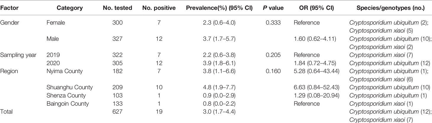
Table 1 Prevalence and subtypes of Cryptosporidium infection in Tibetan Antelope (Pantholops hodgsonii) among different related factors.
According to multivariable logistic regression, gender, sampling year, and region of Tibetan Antelope were not significant in the logistic regression analysis (P > 0.05) and left out of the final model (Hosmer and Lemeshow goodness of fit test P = 1.00). Therefore, gender, sampling year, and region of collecting samples were not considered as main risk factor to influence the seroprevalence significantly (Table 1).
In the present study, 19 samples were Cryptosporidium-positive tested based on the SSU rRNA gene (Figure 2). The analysis of SSU rRNA gene suggested that the samples were C. xiaoi (n = 7) and C. ubiquitum (n = 12) positive in investigated Tibetan Antelope (Table 1 and Figure 3). C. ubiquitum is the predominant Cryptosporidium species, which was responsible for 63.2%. C. xiaoi was only found in Nyima County (n = 6) and Baingoin County (n = 1) in 2019, and C. ubiquitum was only identified in three counties (n = 1 in Nyima County; n = 10 in Shuanghu County; n = 1 in Shenza County) in 2020 (Table 1). Moreover, C. xiaoi and C. ubiquitum were identified in both males (n = 12) and females (n = 7) in present study (Table 1). The representative sequences of C. xiaoi showed 100% similarity with sequences of C. xiaoi (MH049731, KF907825). The representative sequences of C. ubiquitum were identical to the sequences of C. ubiquitum (MT044147, MK573335).
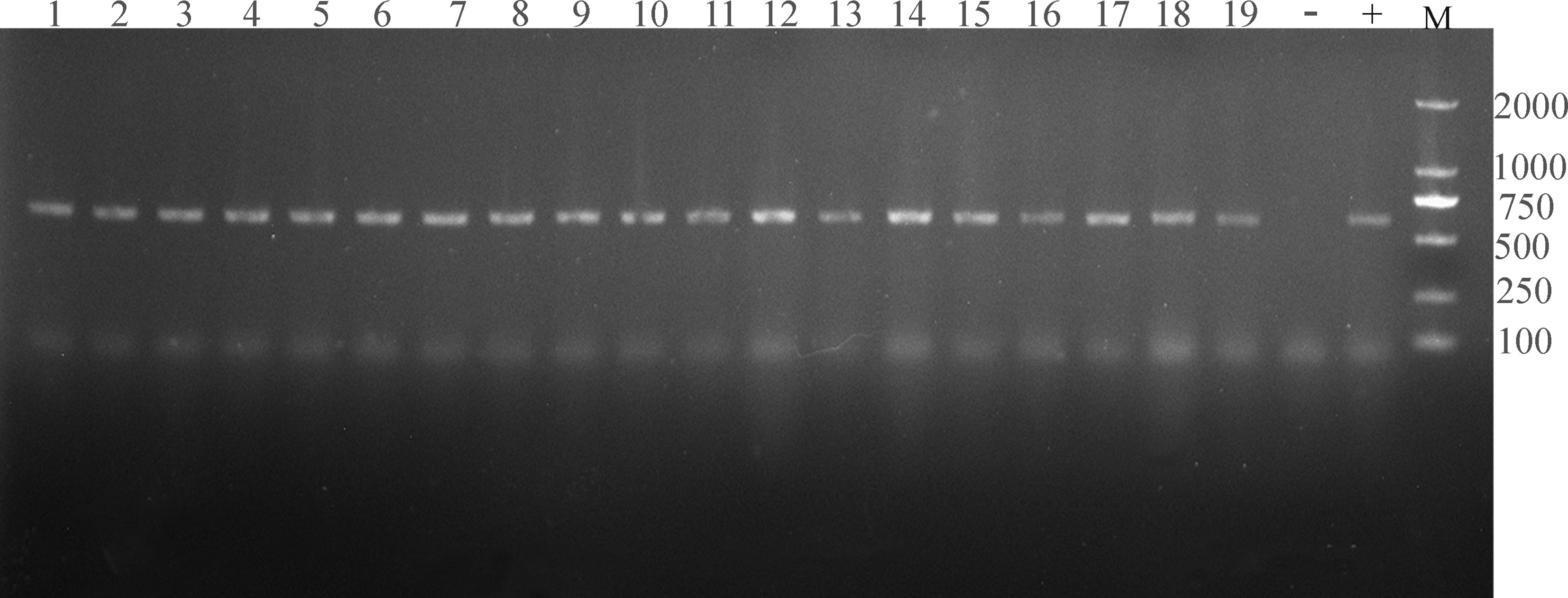
Figure 2 The electropherogram of PCR amplification of SSU rRNA gene of Cryptosporidium. Lanes 1–19 represent TA3, TA9, TA12, TA33, TA61, TA94, TA99, TA103, TA203, TA215, TA217, TA220, TA224, TA230, TA231, TA236, TA241, TA258, and TA301, respectively (19 Cryptosporidium-positive samples); “-” represents negative control; “+” represents positive control; “M” represents DL2000 DNA marker.
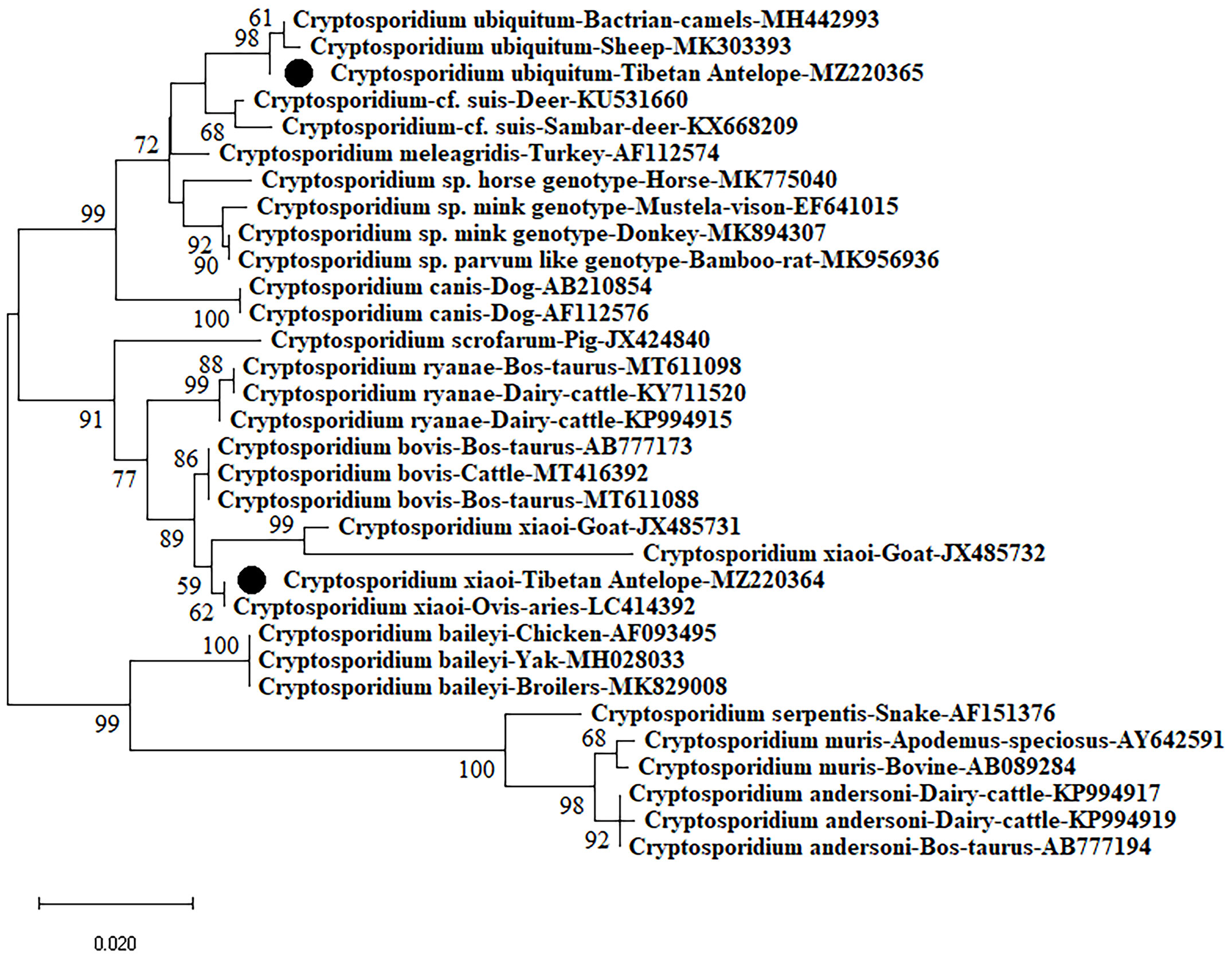
Figure 3 Phylogenetic analyses of Cryptosporidium using neighbor-joining (NJ) method (Kimura 2-parameter model). Bootstrap values below 50% are not shown (1,000 replicates). Cryptosporidium isolates identified in the present study are indicated by solid circles.
The overall Cryptosporidium prevalence was 3.03%, which was significantly lower than that in sheep and goats in Kuwait (9.71%, 54/556) (Majeed et al., 2018), Jordan (10.53%, 12/114) (Hijjawi et al., 2016), Poland (24.78%, 84/339) (Kaupke et al., 2017), Spain (5.9%, 33/58) (Díaz et al., 2018), sheep in Iran (9.1%) (Haghi et al., 2020), goat in Australia (27.2%) (Al-Habsi et al., 2017), and Norwegian sheep in Norway (15%) (Robertson et al., 2010). It is also lower than that in sheep and/or goats in many provinces of China, such as goats in Henan and Chongqing (3.48%, 44/1256) (Wang et al., 2014), Guangdong, Hubei, Shandong, and Shanghai (11.4%, 69/604) (Mi et al., 2014), Tibetan sheep in Qinghai (12.3%, 43/350) (Li et al., 2016), sheep in 10 provinces of China (28.5%, 295/1035) (Mi et al., 2018), but higher than that of sheep in Xinjiang (0.9%, 3/318) (Qi et al., 2019). In the present study, statistical analysis showed that there was no significant difference in Cryptosporidium prevalence with several risk factors (P > 0.05), suggesting that gender, sampling year, and region may not be crucial factors for Cryptosporidium infection in Tibetan Antelope. The difference in Cryptosporidium prevalence may be related to sampling position, sensitivity of the employed detection method, sample sizes, susceptibility in different animals, the pollution degree of environment caused by Cryptosporidium oocysts, as well as animal husbandry practices.
Cryptosporidium genus consists of more than 108 species/genotypes. To date, C. ryanae, C. bovis, C. xiaoi, C. parvum, C. andersoni, C. meleagridis, C. baileyi, C. hominis, C. ubiquitum, C. scrofarum, Cryptosporidium cervine genotype, sheep genotype I, and Cryptosporidium rat genotype II have been reported in various sheep worldwide (Wang et al., 2010; Sweeny et al., 2011; Silverlås et al., 2012; Rieux et al., 2013; Koinari et al., 2014; Yang et al., 2014; Koinari et al., 2014; Mirhashemi et al., 2016; Kaupke et al., 2017; Sqquire et al., 2017; Firoozi et al., 2019). However, only C. xiaoi and C. ubiquitum were identified in Tibetan Antelope in this study, thus suggesting the C. xiaoi and C. ubiquitum were epidemic in the investigated Tibetan Antelope in Tibet Autonomous Region. Moreover, the sequences of isolates from seven fecal samples carrying C. xiaoi shared 100% similarity with isolates from sheep in the Algeria (LC414392) and China (MH049731), goats in Poland (KY055403), and Tibetan sheep in China (KF907825), showing that the sequences of C. xiaoi from Tibetan Antelope have a certain correlation with sheep and goat in Algeria, Poland, and China. But the detailed transmission chain of Cryptosporidium in Tibetan Antelope should be conducted in-depth study in the future. Similarly, another sequence of the 12 isolates belonging to C. ubiquitum showed 100% similarity with an isolate from cattle in the India (MT044147), goats in Algeria (LC414387), and Tibetan sheep in China (MK573335), indicating that the sequences of C. ubiquitum from Tibetan Antelope have a connection with cattle, goat, and sheep in India, Algeria, and China. More importantly, C. ubiquitum and C. xiaoi were also found in other animals and even in HIV/AIDS patients (Adamu et al., 2014; Li et al., 2014). According to relevant literature reports, C. ubiquitum was identified as six subtype families (XIIa–XIIf) based on the 60-kDa glycoprotein (gp60) gene (Li et al., 2014). Among them, subtype XIIa of C. ubiquitum was found in ruminants worldwide, subtype families XIIb–XIId of C. ubiquitum were found in rodents in the United States, and XIIe and XIIf of C. ubiquitum were found in rodents in the Slovak Republic (Li et al., 2014). In addition, humans were found to be infected with subtypes XIIa and XIIb–XIId isolates of C. ubiquitum (Li et al., 2014). In the investigated regions, the population of Tibetan Antelope lived with other free-range animals on the same prairie, and shared with the same source of water, which showing the risk of Cryptosporidium transmission between domestic and wild animals. Contacting with sheep infected with C. ubiquitum and drinking water contaminated by wildlife infected could be sources of human infections (Li et al., 2014). These findings not only demonstrated that Cryptosporidium infection of Tibetan Antelope may result from nearby animals, local herdsmen, or polluted water source, but also suggested that the Tibetan Antelope might be one of the important resources transmitting Cryptosporidium to local people and other native animals, including goa, blue sheep, yak, takin, and wapiti.
In addition, the Tibetan Antelope freely lived in high altitude regions, and frequently moved in plenty of space. They can also contact with other animals. Moreover, the shedding of oocysts into environment by Tibetan Antelope becomes the most important resource for a transmission to other animals and humans. As is well-known, Cryptosporidium is widely regarded as the pathogen of livestock, poultry, companion animals, and wildlife, posing a threat to public health. Local Tibetan live a herding life for chronically, which result in contacting with wildlife and free-range livestock frequently. Local Tibetan occasionally drink water in the process of grazing. Drinking untreated water contaminated by wildlife might be a potential source of Cryptosporidium infecting local Tibetan in Tibet Autonomous Region. Thus, it is very important to take actions for protecting Tibetan Antelope, other free-range animals, and local Tibetan from infecting with Cryptosporidium and the infection status of pathogens (not only Cryptosporidium) in Tibetan Antelope should continue to be monitored in the future. Further studies will sample more Tibetan Antelope in different regions to determine the dynamics and full profiles of Cryptosporidium infection in Tibetan Antelope, to examine the infection status of the local Tibetans with Cryptosporidium, and to assess the zoonotic potential of Cryptosporidium from Tibetan Antelope.
This is the first report of C. xiaoi and C. ubiquitum infection in Tibetan Antelope worldwide. The overall prevalence of Cryptosporidium was 3.03%. The results also confirmed that C. xiaoi and C. ubiquitum were the most common Cryptosporidium species in Tibetan Antelope. Furthermore, C. xiaoi and C. ubiquitum, occasionally found in humans, were also identified in the Tibetan Antelope in this study. These results suggest the transmission of Cryptosporidium from Tibetan Antelope to other animals and/or humans should cause enough attention.
The datasets presented in this study can be found in online repositories. The names of the repository/repositories and accession number(s) can be found in the article/supplementary material.
This study was approved by the Ethics Committee of Jilin University.
QZ, Y-GL, and W-QH conceived and designed the study and critically revised the manuscript. S-YQ, H-TS, J-HZ, Z-JW, and TM collected the samples. S-YQ, H-TS, and CL performed the experiments, analyzed the data, and drafted the manuscript. All authors read and approved the final manuscript.
This work was supported by the “Independent research and development project from General Station of Forest and Grassland pest Management, National Forestry and Grassland Administration” (Grant No. LC-3-03), “Special Fund for Forestry Scientific Research in the Public Interest” (Grant No. 201504310), and “National Key Research and Development Program of China” (Grant no. 2017YFD0501706).
Author CL is employed by Shandong New Hope Liuhe Group Co., Ltd., and Qingdao Jiazhi Biotechnology Co., Ltd.
The remaining authors declare that the research was conducted in the absence of any commercial or financial relationships that could be construed as a potential conflict of interest.
All claims expressed in this article are solely those of the authors and do not necessarily represent those of their affiliated organizations, or those of the publisher, the editors and the reviewers. Any product that may be evaluated in this article, or claim that may be made by its manufacturer, is not guaranteed or endorsed by the publisher.
Adamu, H., Petros, B., Zhang, G., Kassa, H., Amer, S., Ye, J., et al. (2014). Distribution and Clinical Manifestations of Cryptosporidium Species and Subtypes in HIV/AIDS Patients in Ethiopia. PloS Negl. Trop. Dis. 8 (4), e2831. doi: 10.1371/journal.pntd.0002831
Al-Habsi, K., Yang, R., Williams, A., Miller, D., Ryan, U., Jacobson, C. (2017). Zoonotic Cryptosporidium and Giardia Shedding by Captured Rangeland Goats. Vet. Parasitol. Reg Stud. Rep. 7, 32–35. doi: 10.1016/j.vprsr.2016.11.006
Baroudi, D., Hakem, A., Adamu, H., Amer, S., Khelef, D., Adjou, K., et al. (2018). Zoonotic Cryptosporidium Species and Subtypes in Lambs and Goat Kids in Algeria. Parasitol. Vectors 11 (1), 582. doi: 10.1186/s13071-018-3172-2
Deng, L., Chai, Y. J., Luo, R., Yang, L. L., Yao, J. X., Zhong, Z. J., et al. (2020). Occurrence and Genetic Characteristics of Cryptosporidium Spp. And Enterocytozoon Bieneusi in Pet Red Squirrels (Sciurus Vulgaris) in China. Sci. Rep. 10, 1026. doi: 10.1038/s41598-020-57896-w
Díaz, P., Quílez, J., Robinson, G., Chalmers, R. M., Díez-Baños, P., Morrondo, P. (2010). Identification of Cryptosporidium Xiaoi in Diarrhoeic Goat Kids (Capra Hircus) in Spain. Vet. Parasitol. 172, 132–134. doi: 10.1016/j.vetpar.2010.04.029
Díaz, P., Navarro, E., Prieto, A., Pérez-Creo, A., Viña, M., Díaz-Cao, J. M., et al (2018). Cryptosporidium species in post-weaned and adult sheep and goats from N.W. Spain: Public and animal health significance. Vet. Parasitol. 254, 1–5. doi: 10.1016/j.vetpar.2018.02.040
Fayer, R., Santín, M. (2009). Cryptosporidium Xiaoi N. Sp. (Apicomplexa: Cryptosporidiidae) in Sheep (Ovis Aries). Vet. Parasitol. 164, 192–200. doi: 10.1016/j.vetpar.2009.05.011
Firoozi, Z., Sazmand, A., Zahedi, A., Astani, A., Fattahi-Bafghi, A., Kiani-Salmi, N., et al. (2019). Prevalence and Genotyping Identification of Cryptosporidium in Adult Ruminants in Central Iran. Parasitol. Vectors 12 (1), 510. doi: 10.1186/s13071-019-3759-2
Galuppi, R., Piva, S., Castagnetti, C., Iacono, E., Tanel, S., Pallaver, F., et al. (2015). Epidemiological Survey on Cryptosporidium in an Equine Perinatology Unit. Vet. Parasitol. 210, 10–18. doi: 10.1016/j.vetpar.2015.03.021
Haghi, M. M., Khorshidvand, Z., Khazaei, S., Foroughi-Parvar, F., Sarmadian, H., Barati, N., et al. (2020). Cryptosporidium Animal Species in Iran: A Systematic Review and Meta-Analysis. Trop. Med. Health 48, 97. doi: 10.1186/s41182-020-00278-9
Hijjawi, N., Mukbel, R., Yang, R., Ryan, U. (2016). Genetic Characterization of Cryptosporidium in Animal and Human Isolates From Jordan. Vet. Parasitol. 228, 116–120. doi: 10.1016/j.vetpar.2016.08.015
Kaupke, A., Michalski, M. M., Rzeżutka, A. (2017). Diversity of Cryptosporidium Species Occurring in Sheep and Goat Breeds Reared in Poland. Parasitol. Res. 116, 871–879. doi: 10.1007/s00436-016-5360-3
Koehler, A. V., Wang, T., Haydon, S. R., Gasser, R. B. (2018). Cryptosporidium Viatorum From the Native Australian Swamp Rat Rattus Lutreolus - An Emerging Zoonotic Pathogen? Int. J. Parasitol. Parasites Wildl 7, 18–26. doi: 10.1016/j.ijppaw.2018.01.004
Koehler, A. V., Whipp, M. J., Haydon, S. R., Gasser, R. B. (2014). Cryptosporidium Cuniculus–New Records in Human and Kangaroo in Australia. Parasitol. Vectors 7, 492. doi: 10.1186/s13071-014-0492-8
Koinari, M., Lymbery, A. J., Ryan, U. M. (2014). Cryptosporidium Species in Sheep and Goats From Papua New Guinea. Exp. Parasitol. 141, 134–137. doi: 10.1016/j.exppara.2014.03.021
Kváč, M., Hofmannová, L., Hlásková, L., Květoňová, D., Vítovec, J., McEvoy, J., et al. (2014). Cryptosporidium Erinacei N. Sp. (Apicomplexa: Cryptosporidiidae) in Hedgehogs. Vet. Parasitol. 201, 9–17. doi: 10.1016/j.vetpar.2014.01.014
Li, P., Cai, J., Cai, M., Wu, W., Li, C., Lei, M., et al. (2016). Distribution of Cryptosporidium Species in Tibetan Sheep and Yaks in Qinghai, China. Vet. Parasitol. 215, 58–62. doi: 10.1016/j.vetpar.2015.11.009
Lin, Q., Wang, X. Y., Chen, J. W., Ding, L., Zhao, G. H. (2015). Cryptosporidium Suis Infection in Post-Weaned and Adult Pigs in Shaanxi Province, Northwestern China. Korean J. Parasitol. 53, 113–117. doi: 10.3347/kjp.2015.53.1.113
Li, N., Xiao, L., Alderisio, K., Elwin, K., Cebelinski, E., Chalmers, R., et al. (2014). Subtyping Cryptosporidium Ubiquitum,a Zoonotic Pathogen Emerging in Humans. Emerg. Infect. Dis. 20, 217–224. doi: 10.3201/eid2002.121797
Ma, J., Cai, J., Ma, J., Feng, Y., Xiao, L. (2014). Occurrence and Molecular Characterization of Cryptosporidium Spp. In Yaks (Bos Grunniens) in China. Vet. Parasitol. 202, 113–118. doi: 10.1016/j.vetpar.2014.03.030
Mahfouz, M. E., Mira, N., Amer, S. (2014). Prevalence and Genotyping of Cryptosporidium Spp. In Farm Animals in Egypt. J. Vet. Med. Sci. 76, 1569–1575. doi: 10.1292/jvms.14-0272
Majeed, Q. A. H., El-Azazy, O. M. E., Abdou, N. M. I., Al-Aal, Z. A., El-Kabbany, A. I., Tahrani, L. M. A., et al. (2018). Epidemiological Observations on Cryptosporidiosis and Molecular Characterization of Cryptosporidium Spp. In Sheep and Goats in Kuwait. Parasitol. Res. 117, 1631–1636. doi: 10.1007/s00436-018-5847-1
Mirhashemi, M. E., Zintl, A., Grant, T., Lucy, F., Mulcahy, G., De Waal, T. (2016). Molecular Epidemiology of Cryptosporidium Species in Livestock in Ireland. Vet. Parasitol. 216, 18–22. doi: 10.1016/j.vetpar.2015.12.002
Mi, R., Wang, X., Huang, Y., Mu, G., Zhang, Y., Jia, H., et al. (2018). Sheep as a Potential Source of Zoonotic Cryptosporidiosis in China. Appl. Environ. Microbiol. 84, e00868–e00818. doi: 10.1128/AEM.00868-18
Mi, R., Wang, X., Huang, Y., Zhou, P., Liu, Y., Chen, Y., et al. (2014). Prevalence and Molecular Characterization of Cryptosporidium in Goats Across Four Provincial Level Areas in China. PLoS One 9, e111164. doi: 10.1371/journal.pone.0111164
Parsons, M. B., Travis, D., Lonsdorf, E. V., Lipende, I., Roellig, D. M., Collins, A., et al. (2015). Epidemiology and Molecular Characterization of Cryptosporidium Spp. In Humans, Wild Primates, and Domesticated Animals in the Greater Gombe Ecosystem, Tanzania. PloS Negl. Trop. Dis. 9, e0003529. doi: 10.1371/journal.pntd.0003529
Peng, P., Qin, S. Y., Sun, H. T., Xie, L. H., Chu, D., Geng, H. D., et al. (2018). Prevalence and Prevention and Control Strategy of Caprine Contagious Pleuropneumonia in Tibetan Antelope in Nagqu Region of Tibet, China. Chin. J. Wildlife 39, 972–977. doi: 10.19711/j.cnki.issn2310-1490.20180723.001 (in Chinese)
Peng, X. Q., Tian, G. R., Ren, G. J., Yu, Z. Q., Lok, J. B., Zhang, L. X., et al. (2016). Infection Rate of Giardia Duodenalis, Cryptosporidium Spp. And Enterocytozoon Bieneusi in Cashmere, Dairy and Meat Goats in China. Infect. Genet. Evol. 41, 26–31. doi: 10.1016/j.meegid.2016.03.021
Qi, M., Huang, L., Wang, R., Xiao, L., Xu, L., Li, J., et al. (2014). Natural Infection of Cryptosporidium Muris in Ostriches (Struthio Camelus). Vet. Parasitol. 205, 518–522. doi: 10.1016/j.vetpar.2014.06.035
Qin, S. Y., Zhang, X. X., Zhao, G. H., Zhou, D. H., Yin, M. Y., Zhao, Q., et al. (2014). First Report of Cryptosporidium Spp. In White Yaks in China. Parasitol. Vectors 7, 230. doi: 10.1186/1756-3305-7-230
Qi, M., Zhang, Z., Zhao, A., Jing, B., Guan, G., Luo, J., et al. (2019). Distribution and Molecular Characterization of Cryptosporidium Spp., Giardia Duodenalis, and Enterocytozoon Bieneusi Amongst Grazing Adult Sheep in Xinjiang, China. Parasitol. Int. 71, 80–86. doi: 10.1016/j.parint.2019.04.006
Ren, X., Zhao, J., Zhang, L., Ning, C., Jian, F., Wang, R., et al. (2012). Cryptosporidium Tyzzeri N. Sp. (Apicomplexa: Cryptosporidiidae) in Domestic Mice (Mus Musculus). Exp. Parasitol. 130, 274–281. doi: 10.1016/j.exppara.2011.07.012
Rieux, A., Paraud, C., Pors, I., Chartier, C. (2013). Molecular Characterization of Cryptosporidium Spp. In Pre-Weaned Kids in a Dairy Goat Farm in Western France. Vet. Parasitol. 192, 268–272. doi: 10.1016/j.vetpar.2012.11.008
Robertson, L. J., Gjerde, B. K., Furuseth Hansen, E. (2010). The Zoonotic Potential of Giardia and Cryptosporidium in Norwegian Sheep: A Longitudinal Investigation of 6 Flocks of Lambs. Vet. Parasitol. 171, 140–145. doi: 10.1016/j.vetpar.2010.03.014
Rossignol, J. F. (2010). Cryptosporidium and Giardia: Treatment Options and Prospects for New Drugs. Exp. Parasitol. 124, 45–53. doi: 10.1016/j.exppara.2009.07.005
Santín, M. (2013). Clinical and Subclinical Infections With Cryptosporidium in Animals. N. Z Vet. J. 61, 1–10. doi: 10.1080/00480169.2012.731681
Silverlås, C., Mattsson, J. G., Insulander, M., Lebbad, M. (2012). Zoonotic Transmission of Cryptosporidium Meleagridis on an Organic Swedish Farm. Int. J. Parasitol. 42, 963–967. doi: 10.1016/j.ijpara.2012.08.008
Squire, S. A., Yang, R., Robertson, I., Ayi, I., Ryan, U. (2017). Molecular Characterization of Cryptosporidium and Giardia in Farmers and Their Ruminant Livestock From the Coastal Savannah Zone of Ghana. Infect. Genet. Evol. 55, 236–243. doi: 10.1016/j.meegid.2017.09.025
Sweeny, J. P., Ryan, U. M., Robertson, I. D., Yang, R., Bell, K., Jacobson, C. (2011). Longitudinal Investigation of Protozoan Parasites in Meat Lamb Farms in Southern Western Australia. Prev. Vet. Med. 101, 192–203. doi: 10.1016/j.prevetmed.2011.05.016
Takaki, Y., Takami, Y., Watanabe, T., Nakaya, T., Murakoshi, F. (2020). Molecular Identification of Cryptosporidium Isolates From Ill Exotic Pet Animals in Japan Including a New Subtype in Cryptosporidium Fayeri. Vet. Parasitol. Reg Stud. Rep. 21, 100430. doi: 10.1016/j.vprsr.2020.100430
Tzanidakis, N., Sotiraki, S., Claerebout, E., Ehsan, A., Voutzourakis, N., Kostopoulou, D., et al. (2014). Occurrence and Molecular Characterization of Giardia Duodenalis and Cryptosporidium Spp. In Sheep and Goats Reared Under Dairy Husbandry Systems in Greece. Parasite 21, 45. doi: 10.1051/parasite/2014048
Wang, Y., Feng, Y., Cui, B., Jian, F., Ning, C., Wang, R., et al. (2010). Cervine Genotype is the Major Cryptosporidium Genotype in Sheep in China. Parasitol. Res. 106, 341–347. doi: 10.1007/s00436-009-1664-x
Wang, R., Li, G., Cui, B., Huang, J., Cui, Z., Zhang, S., et al. (2014). Prevalence, Molecular Characterization and Zoonotic Potential of Cryptosporidium Spp. In Goats in Henan and Chongqing, China. Exp. Parasitol. 142, 11–16. doi: 10.1016/j.exppara.2014.04.001
Xu, N., Liu, H., Jiang, Y. Y., Yin, J. H., Yuan, Z. Y., Shen, Y. J., et al. (2020). First Report of Cryptosporidium Viatorum and Cryptosporidium Occultus in Humans in China, and of the Unique Novel C. Viatorum Subtype XVaA3h. BMC Infect. Dis. 20, 16. doi: 10.1186/s12879-019-4693-9
Yan, W., Alderisio, K., Roellig, D. M., Elwin, K., Chalmers, R. M., Yang, F., et al. (2017). Subtype Analysis of Zoonotic Pathogen Cryptosporidium Skunk Genotype. Infect. Genet. Evol. 55, 20–25. doi: 10.1016/j.meegid.2017.08.023
Yang, R., Fenwick, S., Potter, A., Ng, J., Ryan, U. (2011). Identification of Novel Cryptosporidium Genotypes in Kangaroos From Western Australia. Vet. Parasitol. 179, 22–27. doi: 10.1016/j.vetpar.2011.02.011
Yang, R., Jacobson, C., Gardner, G., Carmichael, I., Campbell, A. J., Ng-Hublin, J., et al. (2014). Longitudinal Prevalence, Oocyst Shedding and Molecular Characterisation of Cryptosporidium Species in Sheep Across Four States in Australia. Vet. Parasitol. 200, 50–58. doi: 10.1016/j.vetpar.2013.11.014
Keywords: Cryptosporidium, Tibetan antelope (Pantholops hodgsonii), prevalence, characterization, PCR
Citation: Qin S-Y, Sun H-T, Lyu C, Zhu J-H, Wang Z-J, Ma T, Zhao Q, Lan Y-G and He W-Q (2021) Prevalence and Characterization of Cryptosporidium Species in Tibetan Antelope (Pantholops hodgsonii). Front. Cell. Infect. Microbiol. 11:713873. doi: 10.3389/fcimb.2021.713873
Received: 24 May 2021; Accepted: 09 August 2021;
Published: 06 September 2021.
Edited by:
Ehsan Ahmadpour, Tabriz University of Medical Sciences, IranReviewed by:
Hongxuan He, Institute of Zoology, Chinese Academy of Sciences (CAS), ChinaCopyright © 2021 Qin, Sun, Lyu, Zhu, Wang, Ma, Zhao, Lan and He. This is an open-access article distributed under the terms of the Creative Commons Attribution License (CC BY). The use, distribution or reproduction in other forums is permitted, provided the original author(s) and the copyright owner(s) are credited and that the original publication in this journal is cited, in accordance with accepted academic practice. No use, distribution or reproduction is permitted which does not comply with these terms.
*Correspondence: Quan Zhao, emhhb3F1YW4wODI1QDE2My5jb20=; Yun-Gang Lan, bGFueXVuZ2FuZ0BqbHUuZWR1LmNu; Wen-Qi He, aGV3cUBqbHUuZWR1LmNu
†These authors have contributed equally to this work
Disclaimer: All claims expressed in this article are solely those of the authors and do not necessarily represent those of their affiliated organizations, or those of the publisher, the editors and the reviewers. Any product that may be evaluated in this article or claim that may be made by its manufacturer is not guaranteed or endorsed by the publisher.
Research integrity at Frontiers
Learn more about the work of our research integrity team to safeguard the quality of each article we publish.