 Emídio Vale-Fernandes1,2,3,4
 Mafalda V. Moreira2,3,5
 Bárbara Rodrigues2,3,6Sofia S. Pereira2,3Carla Leal1,2,3Márcia Barreiro1,2,3,4António Tomé4
 Mariana P. Monteiro2,3*
- 1Centre for Medically Assisted Procreation/Public Gamete Bank, Gynaecology Department, Centro Materno-Infantil do Norte Dr. Albino Aroso (CMIN), Unidade Local de Saúde de Santo António (ULSSA), Porto, Portugal
- 2UMIB - Unit for Multidisciplinary Research in Biomedicine, ICBAS - School of Medicine and Biomedical Sciences, University of Porto, Porto, Portugal
- 3ITR - Laboratory for Integrative and Translational Research in Population Health, Porto, Portugal
- 4Gynaecology Department, Centro Materno-Infantil do Norte Dr. Albino Aroso (CMIN), Unidade Local de Saúde de Santo António (ULSSA), Porto, Portugal
- 5i3S - Instituto de Investigação e Inovação em Saúde, Universidade do Porto, Porto, Portugal
- 6Molecular Genetics Unit, Centro de Genética Médica Dr. Jacinto Magalhães (CGM), Unidade Local de Saúde de Santo António (ULSSA), Porto, Portugal
Polycystic ovary syndrome (PCOS) is the most common endocrinopathy in women at childbearing age. Anti-Müllerian hormone (AMH) is a widely accepted sensitive marker of ovarian reserve, which has been suggested that could also act as biomarker of ovarian morphology for PCOS diagnosis. Oxidative stress (OS) is known to be associated and have a negative impact factor in several reproductive conditions, including PCOS. However, the relationship between circulating AMH and OS within the follicular fluid (FF), and its potential impact on in vitro fertilization (IVF) outcomes of women with PCOS, remains largely unexplored. A total of 84 women, with PCOS (n = 30) or ovulatory controls (n = 54), were enrolled in this study. Women underwent individualized controlled ovarian stimulation for oocyte retrieval. Blood and FF obtained from mature follicles were collected at the time of oocyte retrieval, for measuring total testosterone, ∆4-androstenedione, progesterone, sex hormone binding globulin (SHBG) and AMH. OS in the FF was assessed by measuring total antioxidant capacity (TAC) through the ferric reducing antioxidant power (FRAP) and lipid peroxidation (LPO) by quantification of malondialdehyde (MDA) levels. Our results demonstrated that women with PCOS had significantly higher plasma levels of AMH, ∆4-androstenedione, total testosterone and a free androgen index (FAI) than observed in non-PCOS controls. In women with PCOS, total testosterone and AMH levels in the FF were also higher, while TAC was lower compared to non-PCOS. Furthermore, circulating AMH levels were positively correlated with ∆4-androstenedione, albeit negatively correlated with TAC. In this study we demonstrated that the susceptibility to OS, as assessed by the total antioxidant capacity in the FF, is higher in women with PCOS and inversely related to AMH levels. This study results lead us to forge the reasonable hypothesis that the greater susceptibility to OS within the follicle microenvironment is potentially at the end of a roadway that starts with elevated ∆4-androstenedione and AMH within the FF, which in turn are mirrored by circulating AMH and androgen levels. Thus, suggesting that circulating AMH levels could act as a surrogate biomarker of follicular fluid oxidative stress in women with PCOS.
1 Introduction
Polycystic ovary syndrome (PCOS) is the most common endocrinopathy in women at childbearing age with a prevalence ranging from five to 18% depending on the population (Rotterdam ESHRE/ASRM-Sponsored PCOS consensus workshop group, 2004). PCOS diagnosis relies on the Rotterdam criteria, which requires the presence of at least two out of three parameters: irregular menstrual cycles, clinical or biochemical hyperandrogenism and polycystic ovarian morphology (Christ and Cedars, 2023).
Anti-Müllerian hormone (AMH) is a polypeptide secreted by cells of the granulosa layer of primary/preantral ovarian follicles and small secondary/antral follicles (van Rooij et al., 2002). AMH is a widely accepted sensitive marker of ovarian reserve, given that the secretion tends to be proportional to the granulosa cell mass (La Marca et al., 2009; Tal et al., 2014; Vale-Fernandes et al., 2023b). Additionally, AMH has been pointed as a possible surrogate marker of ovarian morphology (Dumont et al., 2015), once it is typically elevated in women with PCOS, as compared to normoovulatory women (Cook et al., 2002). Despite controversial, it has been suggested that AMH should be integrated into the Rotterdam criteria for PCOS diagnosis (Teede et al., 2023). Moreover, there is also evidence that AMH levels may correlate with reproductive outcomes in Assisted Reproductive Technology (ART) procedures, including pregnancy and live birth rates, irrespective of age (Brodin et al., 2013).
PCOS accounts for a considerable proportion of the causes of infertility among couples seeking for ART. Despite most women with PCOS exhibiting positive responses to ovarian stimulation for in vitro fertilization (IVF), characterized by a higher-than-average number of growing follicles, their reproductive outcomes often prove less favorable compared to women experiencing infertility from other causes (Rajani et al., 2012; Piomboni et al., 2014; Yilmaz et al., 2016; Artimani et al., 2018). Factors appointed as potentially detrimental to reproductive outcomes in women with PCOS, include the competence of the endometrium (Piltonen, 2016) and the unfavorable follicular microenvironment compromising the physiological functions and quality of oocytes (Qiao and Feng, 2010; Palomba et al., 2017).
Oxidative stress (OS) is recognized as an impacting factor in the pathophysiology of several infertility-related problems, including PCOS. OS occurs when the production of reactive oxygen species (ROS) surpasses the antioxidant capacity. Despite the physiological role of ROS in the modulation of a spectrum of reproductive functions (Shkolnik et al., 2011; Wang et al., 2017; Lu et al., 2018), the disruption of the redox signaling can lead mitochondrial dysfunction, protein oxidation, lipid peroxidation and DNA damage, negatively impacting on reproductive function (Sasaki et al., 2019; Jamil et al., 2020). Previous studies have demonstrated higher levels of pro-oxidant markers and lower levels of antioxidants in the follicular fluid (FF) of women with PCOS when compared to normoovulatory women (Chattopadhayay et al., 2010; Artimani et al., 2018; Naigaonkar et al., 2021; Moreira et al., 2023). Given the complex nature of PCOS and potential deleterious impact of OS on the follicular microenvironment and follicle growth, it is important to deepen the knowledge on the putative triggers of OS in FF. In particular, the relationship between AMH and OS within the FF, and its potential impact on IVF outcomes, remains largely unexplored.
Therefore, to address this gap in knowledge, this study aimed to investigate the association between circulating and FF AMH and OS markers within the FF.
2 Materials and methods
2.1 Participants’ recruitment
A total of 84 women undergoing in vitro fertilization (IVF) were enrolled in this study. Women were allocated into two groups depending on the presence of PCOS (n = 30) or non-PCOS ovulatory controls (n = 54) The diagnosis of PCOS was based on the 2003 Rotterdam diagnostic criteria, which requires the presence of at least two of the following three manifestations: (1) oligo- and/or anovulation, (2) clinical and/or biochemical evidence of hyperandrogenism, and (3) polycystic ovaries on ultrasound examination (at least 10 follicles 2–9 mm in size or volume of the ovary greater than 10 mL). The control group included women who underwent ART treatments for oocyte donation or infertility treatment due to tubal and/or male factors. Exclusion criteria included women with diminished ovarian reserve, endometriosis, systemic diseases, abnormal prolactin levels, and/or thyroid dysfunction. Electronic medical records were used for data extraction, namely, age, body mass index (BMI), hormonal measurements, and IVF cycle characteristics. All patients were recruited to participate and enrolled in the study after providing written informed consent to participate in this study. The study protocol was authorized by the Ethics Committee of the Institution [2020.119 (097-DEFI/099-CE)].
2.2 Ovarian stimulation protocol
Women underwent a Gonadotropin-Releasing Hormone (GnRH) antagonist protocol with individualized controlled ovarian stimulation based on ovarian reserve testing and standard clinical practice. Two different types of ovarian stimulation protocols are used for assisted reproduction, the long protocol with a GnRH agonist and the short protocol with GnRH antagonist. However, for safety reasons given the risk of ovarian hyperstimulation syndrome, in women with PCOS only the short protocol with GnRH antagonist, avoiding the trigger with human chorionic gonadotropin (βHCG), is used.
2.3 Collection of blood plasma and follicular fluid
Follicular fluid (FF) of each woman was collected at the day of oocyte retrieval. The ovulatory trigger was performed in preparation for aspiration, whenever a minimum of two or three follicles reached the size of 17 mm in diameter. Nevertheless, all follicles were aspirated on the oocyte retrieval day, regardless of its size. Each follicle was aspirated separately and pooled into a single FF sample from each patient, obtained by combining equal aliquots from two or more FF collection tubes. Each fluid sample was centrifuged at 20000 g for 10 min at 4°C to eliminate cells and debris. The supernatant was collected and stored at −80°C until used.
Before oocyte puncture, a peripheral blood sample of each patient was collected by venepuncture of an antebrachial vein into EDTA-containing tubes. The blood was centrifuged at 500 g for 5 min at room temperature and the plasma was separated and stored at −80°C.
2.4 Hormone analysis of plasma and follicular fluid
Total testosterone, ∆4-androstenedione, progesterone and insulin were measured with an electrochemiluminescence immunoassay (ECLIA, Roche Diagnostics, Mannheim, Germany); sex hormone binding globulin (SHBG) was measured with a chemiluminescent immunometric assay (Immulite XPi instrument, Siemens Healthcare Diagnostics, UK); and Anti-Müllerian hormone (AMH) was measured with an enzyme-linked immunosorbent assay (Beckman Coulter Access AMH, Immunotech, France). All hormone analyses in plasma and FF were performed at the routine core laboratory that serves our centre, based at a public tertiary hospital.
2.5 Total antioxidant capacity
The total antioxidant capacity (TAC) was measured by Ferric Reducing Antioxidant Power (FRAP) assay in the FF. FRAP reagent was freshly prepared by mixing 300 mM acetate buffer (pH 3.6), 10 mM 2,4,6-tripyridyl-S-triazine (TPTZ) in 40 mM HCl and 20 mM FeCl3.6H2O (Sigma-Aldrich, St. Louis, Missouri, United States) in the ratio of 10:1:1. Briefly, in a 96-well plate, 6 μL of FF samples were added to 180 µL of FRAP reagent. Triplicates were made for each sample. Distilled water (dH2O) was used as negative control and ascorbic acid (1,000 µM) was used as an antioxidant standard. The absorbance was read on a Synergy™ H1 multi-mode microplate reader (BioTek, Winooski, VT, United States) at OD 593 nm, immediately after the addition of the FRAP reagent (0 min) and after 40 min. Between measurements, the plate was kept in the dark, at room temperature. The antioxidant potential of the samples was determined against the ascorbic acid (1,000 µM) standard and corrected using the absorbance value of dH2O absorbance. FRAP value (µmol/L), was calculated using the formula described in (Benzie and Strain, 1996).
2.6 Lipid peroxidation
The lipid peroxidation (LPO) was evaluated by quantifying malondialdehyde (MDA) levels in the FF. LPO was determined using the MDA Assay Kit (ab118970, Abcam Cambridge, MA, United States) according to the manufacturer’s instructions. The MDA that is present in plasma sample reacts with thiobarbituric acid (TBA) to generate an MDA-TBA adduct that is quantified colorimetrically. For the colorimetric assay, a 0.1 mol/L MDA standard was prepared, and serial dilutions were made for the standard curve. Each FF sample and standard was pipetted into a 96-well plate (clear bottom black plate) and colorimetric measurement was taken on a Synergy™ H1 multi-mode microplate reader (BioTek, Winooski, VT, United States) at OD 532 nm. The absorbance value of the blank (water) was used for background subtraction.
2.7 Statistical analysis
Results are presented as mean ± standard deviation (SD). The normality of the data was tested according to the Kolmogorov-Smirnov test. Continuous variables were assessed for normal distribution and homogeneity of variance, and the differences between the study groups were evaluated by Student’s t-test for parametric data. In cases where the data did not meet these assumptions (non-normal distribution or heterogeneity of variance), the Mann-Whitney test was employed. The Pearson test or Spearman rank test was used to assess the correlation between two different variables, depending on whether the distribution was normal or non-normal, respectively. The statistical analysis of this work was performed by GraphPad Prism eight software and IBM SPSS Statistics 29.0. Values of p < 0.05 were considered as statistically significant.
3 Results
3.1 Clinical characteristics of study patients
A total of 84 women undergoing IVF were divided into two groups. The anthropometric, clinical and biochemical data of women allocated to the PCOS group (n = 30) and non-PCOS group (n = 54) are shown in Table 1. The study groups did not vary in terms of age, body mass index (BMI), prolactin, thyroid stimulating hormone (TSH), glucose and insulin levels. Circulating levels of follicle-stimulating hormone (FSH) were significantly lower in women with PCOS. Circulating levels of luteinizing hormone (LH) and AMH and LH:FSH ratio were significantly higher in women with PCOS. Hormone levels were also measured in plasma and FF was collected at oocyte retrieval day. Women in the PCOS group had significantly higher plasma levels of ∆4-androstenedione, total testosterone and a higher free androgen index (FAI) than observers in non-PCOS controls. In women with PCOS, FF total testosterone levels were also higher, while there were no significant differences in the other parameters analysed, which were comparable between the study groups.
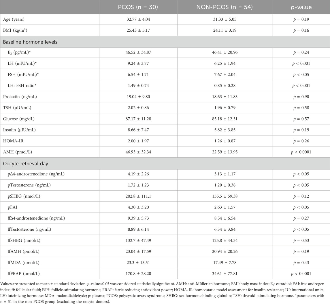
Table 1. Demographic and clinical characteristics of women according to study group.
3.2 IVF cycle parameters
The IVF cycle characteristics of PCOS groups (n = 30) and non-PCOS (n = 31, excluding the oocyte donors) are present in Table 2. Among IVF cycle characteristics, the number of cumulus-oocyte complexes (COCs) and the number of two pronucleated oocytes were significantly higher in women with PCOS. There were no significant differences in the other parameters analyzed between groups.
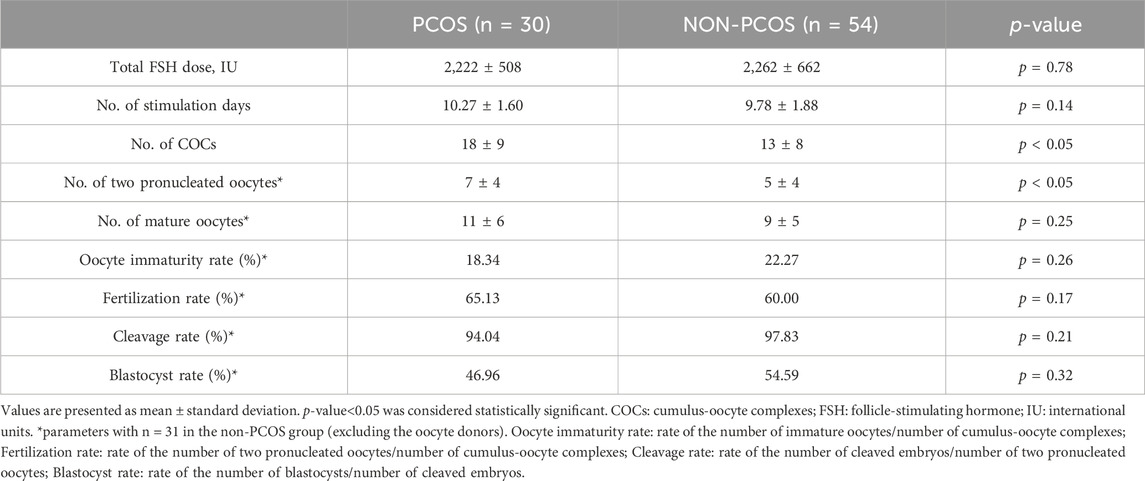
Table 2. In vitro fertilization (IVF) cycle characteristics.
3.3 Oxidative stress parameters in the follicular fluid
We evaluated the FRAP and the MDA levels in FF of women with PCOS and compared it to those of non-PCOS (Figure 1). We observed significantly lower FRAP levels and a trend for higher MDA levels in the FF of women with PCOS (Table 1).
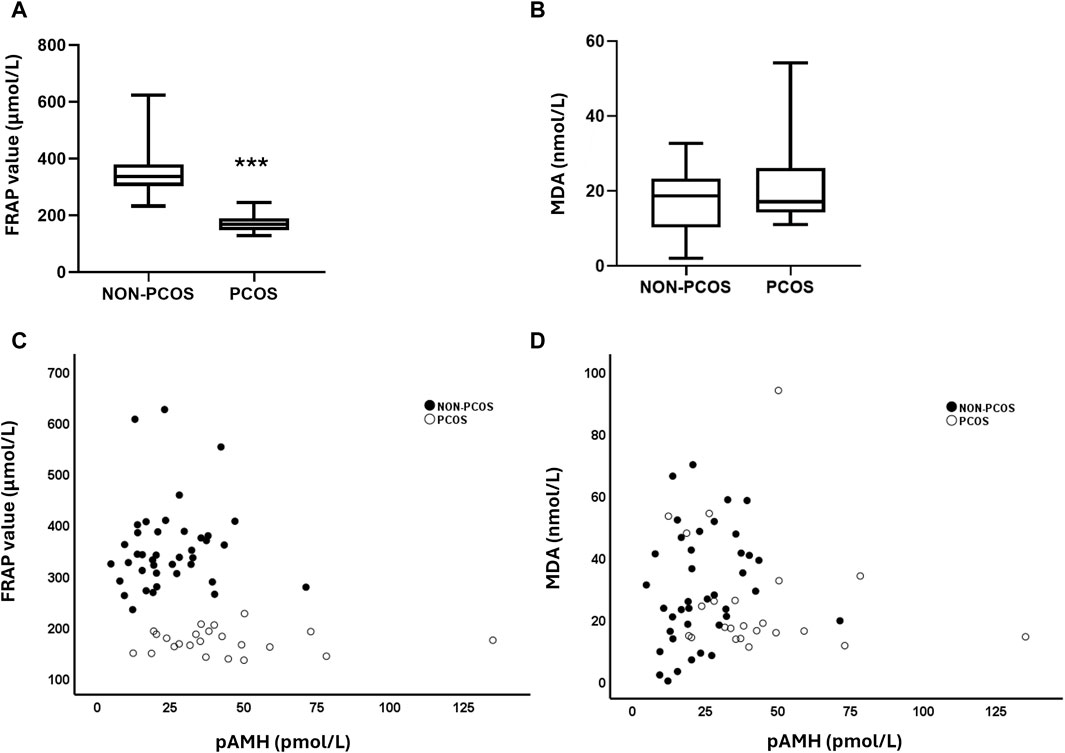
Figure 1. (A) Total antioxidant capacity (TAC) levels among non-PCOS vs. PCOS groups. (B) Lipid peroxidation (LPO) levels among non-PCOS vs. PCOS groups. (C) Correlation between FRAP value and plasma AMH among non-PCOS vs. PCOS groups. (D) Correlation between MDA levels and plasma AMH among non-PCOS vs. PCOS groups. FRAP: ferric reducing antioxidant power; MDA: malondialdehyde. Statistical analysis was performed using the Mann-Whitney U test, ***p < 0.0001.
3.4 Relationship between AMH and hormone levels and oxidative stress parameters
To investigate whether there was an association between circulating hormone levels and oxidative stress parameters on the FF, we performed a correlation analysis across our entire population (women with PCOS and without PCOS) (Table 3). A positive correlation was found between plasma AMH and FF AMH, as well as between plasma AMH and plasma ∆4-androstenedione. Additionally, plasma AMH levels were negatively correlated to those of FF progesterone. Moreover, plasma AMH was negatively correlated with FF FRAP levels, while there was no statistically significant correlation between plasma AMH levels and FF LPO (Figures 1C, D; Table 3).
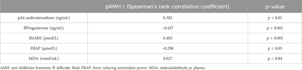
Table 3. Correlation between serum anti-Müllerian hormone (AMH) and clinical parameters and oxidative stress markers.
4 Discussion
The finding that AMH in the circulation of women with PCOS is elevated, not only raised the question of whether this could be a biomarker with potential diagnostic applications (Teede et al., 2023), but also prompted significant inquiries into which could be its role in the pathophysiological mechanisms underlying this condition.
PCOS is characterized by gonadotrophic axis dysfunction besides other hormone imbalances. Elevated circulating levels of LH, testosterone and AMH are the hallmarks of this gonadotrophic axis dysregulation. In women with PCOS, the typical gonadotrophin secretion pattern consists of increased LH secretion with normal follicle-stimulating hormone secretion, which results in an abnormal LH/FSH ratio that is considered a valuable marker for evaluating ovarian function while assisting PCOS diagnosis (Dewailly et al., 2020). In this study, as it would have been expected and further supporting the diagnosis, women with PCOS had higher levels of AMH and LH, lower levels of FSH and a higher LH:FSH ratio, when compared to ovulatory controls. Additionally, women with PCOS had significantly higher androgen levels, namely, total testosterone, ∆4-androstenedione, and FAI than women in the control group. Likewise, the total testosterone levels in the FF of women with PCOS were also significantly higher than observed in FF of control women. Remarkably, the presence of elevated androgens is one of the parameters included in the Rotterdam criteria for PCOS diagnosis (Rotterdam ESHRE/ASRM-Sponsored PCOS consensus workshop group, 2004).
Furthermore, we found a positive correlation between circulating levels of AMH and ∆4-androstenedione. While both hormones can be similarly affected in PCOS and the correlation between AMH and ∆4-androstenedione levels has been previously described, the significance of this direct correlation is not well-established (Leonhardt et al., 2014; Piltonen et al., 2023; Barbagallo et al., 2024). Additionally, relationship between AMH and ∆4-androstenedione in different physiological and pathological contexts remains to be elucidated in order to understand whether and how they may influence each other (Piltonen et al., 2019). Notwithstanding, this finding further supports the potential relevance of considering the use of AMH for diagnosing and grading PCOS severity, in line with the growing trend of international groups working towards the refinement of the 2003 Rotterdam criteria (Teede et al., 2023).
In our study, circulating and FF AMH levels were found to be positively correlated, allowing us to reasonably infer that circulating AMH levels reflect the follicular microenvironment in the ovary, lined by the granulosa cell layer responsible for AMH secretion (van Rooij et al., 2002).
The implications of FF AMH levels for follicle physiology are not well-established, nor is the predictive value of FF AMH on reproductive outcomes, particularly in women with PCOS. However, it is recognized that the balance between FSH and AMH within granulosa cells is pivotal in the shift from androgen-to oestrogen-driven follicles (Dewailly et al., 2016) Besides, high levels of AMH in the FF may have harmful consequences for the oocyte quality and final maturation (Desforges-Bullet et al., 2010), as well as fertilization and cleavage rate (Mashiach et al., 2010), although the mechanisms are unknown.
Several studies have explored the relationship between baseline serum AMH levels and pregnancy outcomes following IVF treatment (Liu et al., 2022; Hou et al., 2023; Li et al., 2023; Yuwen et al., 2023). In women with PCOS results have been inconsistent with some authors reporting higher AMH levels to be associated with better reproductive outcomes (Xi et al., 2012; Tal et al., 2015), while others found the opposite (Xi et al., 2012; Tal et al., 2020; Guo et al., 2021; Sun et al., 2021; Vale-Fernandes et al., 2023a). Our results showed that the number of cumulus-oocyte complexes (COCs) and the number of two pronucleated oocytes were significantly higher in women with PCOS. These disparities could be attributed to intrinsic differences between study populations and study population sizes. A previous study conducted by our group uphold the hypothesis that in women with PCOS, high circulating levels of AMH, instead of reflecting the ovarian reserve, act as a biomarker of disease severity and worse reproductive prognosis (Vale-Fernandes et al., 2023a). Overall, the available evidence highlights the unmet need of further understanding the role of AMH in the processes that drive folliculogenesis in the normal and PCOS, which will be key for the treatment of anovulation.
Besides, a negative correlation was found between circulating AMH levels and progesterone levels in the FF. The relationship between AMH and progesterone levels in the FF has not been previously described. While both hormones are involved in reproductive processes, these are secreted by different ovarian cell types and regulated by distinct mechanisms. AMH is primarily involved in ovarian follicle development and is mainly produced by small follicles in the ovaries (van Rooij et al., 2002). Progesterone, on the other hand, is a hormone produced mainly by the corpus luteum after ovulation and plays a crucial role in the menstrual cycle and pregnancy (Chemerinski et al., 2024). In PCOS, AMH levels are often elevated due to an increased number of small ovarian follicles, while progesterone levels may be reduced or irregular due to anovulation or luteal phase defects (Teede et al., 2023). Considering that progesterone is the hormone that highlights ovulation and women with PCOS are predominantly anovulatory, this is an interesting finding not previously described in the literature, that requires greater contextualization and pathophysiological clarification, given that the progesterone measurement was carried out in the context of controlled ovarian hyperstimulation. Additionally, it should be recalled that high progesterone levels at the end of the follicular phase in women undergoing controlled ovarian hyperstimulation for IVF is a reason for suspending fresh embryo transfer and undergoing embryos cryopreservation with subsequent endometrial preparation for transfer of cryopreserved embryos (Bosch et al., 2024).
There is growing evidence suggesting that OS may play a role in the reproductive outcomes of women with PCOS, namely, fertilization and pregnancy rates (Yilmaz et al., 2016; Naigaonkar et al., 2021; Moreira et al., 2023). Studies have indicated that women with PCOS often exhibit higher levels of oxidative stress markers, such as increased ROS production and LPO, and decreased TAC, at the FF compared to women without PCOS (Chattopadhayay et al., 2010; Naigaonkar et al., 2021). In our herein study, we found that the TAC of the FF, as evaluated by FRAP, was significantly lower in women with PCOS as compared to controls. Regarding LPO, although we found a tendency for MDA levels to be higher in the FF of women with PCOS, we did not observe statistically significant differences between women with PCOS and controls. LPO is a consequence of oxidative stress on cell membrane structure and function. This process is characterized by the molecular modification of lipids containing carbon-carbon double bond(s), especially polyunsaturated fatty acids, when exposed to free radicals (Ayala et al., 2014). During LPO different aldehydes can be formed as secondary products, among which MDA and 4-hydroxynonenal (4-HNE) have been widely used as OS markers. However, MDA measures a specific consequence of OS, which could not be particularly affected in this condition and thus explain why no significant differences were observed in our study.
Most importantly, we were able to demonstrate that the higher the circulating AMH and the FF AMH levels, the lower is the TAC. Given that women with PCOS present significantly higher levels of circulating androgens and AMH, in parallel with those observed in the FF, and that these are inversely correlated with the TAC, it seems reasonable enough to raise the hypothesis that AMH could be involved in the processes leading to OS within the FF of women with PCOS, which could then be reflected in poorer reproductive outcomes, as compared to normal ovulatory women particularly during IVF treatments (Vale-Fernandes et al., 2023a).
PCOS is associated with sub-optimal oocyte quality and poor outcomes of ART treatments (Palomba et al., 2017). The evaluation of IVF treatments efficacy still relies mainly on morphological parameters of egg and embryo quality. The redox dynamics in the oocyte microenvironment that encompasses the FF is not assessed nor used in clinical practice in the decision algorithm in ART treatments. However, more and more studies raise the possibility that the study of FF biomarkers could be a fundamental tool to identify the factors that influence the quality of the oocyte/embryo (Naigaonkar et al., 2021; Vale-Fernandes et al., 2023a). By examining the relationship between circulating and FF AMH and TAC, this study provides insights into the potential role of AMH in modulating oxidative stress and its implications for IVF success. Moreover, grounded on AMH levels, it now allows to identify a subset of women with PCOS potentially amenable for intervention to reduce OS aimed at improving the reproductive outcomes.
It is noteworthy to acknowledge that despite the relevant contributions that this study provided, there are several limitations to mention. First of all, this was a single center study and therefore the recruitment capacity was limited by that fact. Second, given the small number of women per group that were enrolled, further stratification of women, with regards to BMI and insulin resistance as assessed by HOMA-IR, were rendered impossible. These would be of particular interest given the well-established association between obesity, insulin resistance and oxidative stress. Notwithstanding the fact that despite the BMI and HOMA-IR tended to be higher in women of the PCOS group compared to the control group these were well balanced between groups and the average did not fall in the obesity nor insulin resistant range. Third, since the circulating levels of AMH in women undergoing IVF treatments are routinely measured between the third to fifth day of the menstrual cycle, up to 6 months before starting the ovarian stimulation, as a surrogate measure of the ovarian reserve, and reassessment of AMH levels at the day of follicular puncture was not conducted, the correlation with FF AMH has to be interpreted taking this limitation into account. And last but not least, since our control group encompassed predominantly healthy women oocyte donors, who did not seek fertility, precluded the possibility of evaluating end stage reproductive parameters, such as pregnancy rates and live births.
5 Conclusion
This study results lead us to the reasonable hypothesis of greater susceptibility of the follicle microenvironment to OS being at the end of a roadway that starts with elevated ∆4-androstenedione and AMH within the FF, which in turn are mirrored by circulating AMH and androgen levels that are inversely related to the total anti-oxidant capacity. This end of the spectrum of the hormonal profile is what distinguishes women with and without PCOS, highlighting the central role of androgens and AMH for the phenotype characterization and predicting reproductive outcomes in women affected. The hypothesis that circulating levels of AMH could act as a surrogate biomarker of follicular fluid oxidative stress in PCOS, along with its importance in predicting disease severity and reproductive prognosis in women with PCOS undergoing IVF ttreatments, make the proposal to include AMH in the review of PCOS diagnostic and classification criteria quite pertinent and useful.
Data availability statement
The raw data supporting the conclusions of this article will be made available by the authors, without undue reservation.
Ethics statement
The studies involving humans were approved by Ethics Committee of the Unidade Local de Saúde de Santo António [2020.119(097-DEFI/099-CE)]. The studies were conducted in accordance with the local legislation and institutional requirements. The participants provided their written informed consent to participate in this study.
Author contributions
EV-F: Conceptualization, Data curation, Funding acquisition, Investigation, Methodology, Writing–original draft. MVM: Conceptualization, Data curation, Formal Analysis, Investigation, Methodology, Writing–original draft. BR: Investigation, Methodology, Writing–review and editing. SP: Formal Analysis, Writing–review and editing. CL: Validation, Writing–review and editing. MB: Validation, Writing–review and editing. AT: Supervision, Validation, Writing–review and editing. MPM: Conceptualization, Funding acquisition, Supervision, Writing–review and editing.
Funding
The author(s) declare that financial support was received for the research, authorship, and/or publication of this article. This work was funded by the Foundation for Science and Technology (FCT) through the following funds: Mafalda V. Moreira (2022.12784.BD); UMIB (UIDB/00215/2020 and UIDP/00215/2020); ITR-Laboratory for Integrative and Translational Research in Population Health (LA/P/0064/2020). This work was funded by MERCK project “Impact of clinical and metabolic profile, FMR1 gene and other genetic modifiers on female fertility”.
Acknowledgments
The authors would like to thank all the staff of the Centre for Medically Assisted Procreation/Public Gamete Bank, Gynaecology Department, Centro Materno-Infantil do Norte Dr Albino Aroso (CMIN), Unidade Local de Saúde de Santo António (ULSSA), for collaborating, particularly the Clinical Embryologists Dr Raquel Brandão and Dr Daniela Sousa and the Nurses Manuela Perpétuo and Irene Rodrigues who were responsible for collecting the biological samples.
Conflict of interest
The authors declare that the research was conducted in the absence of any commercial or financial relationships that could be construed as a potential conflict of interest.
Publisher’s note
All claims expressed in this article are solely those of the authors and do not necessarily represent those of their affiliated organizations, or those of the publisher, the editors and the reviewers. Any product that may be evaluated in this article, or claim that may be made by its manufacturer, is not guaranteed or endorsed by the publisher.
References
Artimani, T., Karimi, J., Mehdizadeh, M., Yavangi, M., Khanlarzadeh, E., Ghorbani, M., et al. (2018). Evaluation of pro-oxidant-antioxidant balance (PAB) and its association with inflammatory cytokines in polycystic ovary syndrome (PCOS). Gynecol. Endocrinol. 34 (2), 148–152. doi:10.1080/09513590.2017.1371691
Ayala, A., Muñoz, M. F., and Argüelles, S. (2014). Lipid peroxidation: production, metabolism, and signaling mechanisms of malondialdehyde and 4-hydroxy-2-nonenal. Oxidative Med. Cell. Longev. 2014, 360438. doi:10.1155/2014/360438
Barbagallo, F., van der Ham, K., Willemsen, S. P., Louwers, Y. V., and Laven, J. S. (2024). Age-related curves of AMH using the Gen II, the picoAMH and the Elecsys assays in women with polycystic ovary syndrome. J. Clin. Endocrinol. Metab., dgae153. doi:10.1210/clinem/dgae153
Benzie, I. F., and Strain, J. J. (1996). The ferric reducing ability of plasma (FRAP) as a measure of “antioxidant power”: the FRAP assay. Anal. Biochem. 239 (1), 70–76. doi:10.1006/abio.1996.0292
Bosch, E., Alama, P., Romero, J. L., Mari, M., Labarta, E., and Pellicer, A. (2024). Serum progesterone is lower in ovarian stimulation with highly purified HMG compared to recombinant FSH owing to a different regulation of follicular steroidogenesis: a randomized controlled trial. Hum. Reprod. 39 (2), 393–402. doi:10.1093/humrep/dead251
Brodin, T., Hadziosmanovic, N., Berglund, L., Olovsson, M., and Holte, J. (2013). Antimüllerian hormone levels are strongly associated with live-birth rates after assisted reproduction. J. Clin. Endocrinol. Metab. 98 (3), 1107–1114. doi:10.1210/jc.2012-3676
Chattopadhayay, R., Ganesh, A., Samanta, J., Jana, S. K., Chakravarty, B. N., and Chaudhury, K. (2010). Effect of follicular fluid oxidative stress on meiotic spindle formation in infertile women with polycystic ovarian syndrome. Gynecol. Obstet. Invest. 69 (3), 197–202. doi:10.1159/000270900
Chemerinski, A., Shen, M., Valero-Pacheco, N., Zhao, Q., Murphy, T., George, L., et al. (2024). The impact of ovarian stimulation on the human endometrial microenvironment. Hum. Reprod. 39, 1023–1041. doi:10.1093/humrep/deae048
Christ, J. P., and Cedars, M. I. (2023). Current guidelines for diagnosing PCOS. Diagn. (Basel) 13 (6), 1113. doi:10.3390/diagnostics13061113
Cook, C. L., Siow, Y., Brenner, A. G., and Fallat, M. E. (2002). Relationship between serum müllerian-inhibiting substance and other reproductive hormones in untreated women with polycystic ovary syndrome and normal women. Fertil. Steril. 77 (1), 141–146. doi:10.1016/S0015-0282(01)02944-2
Desforges-Bullet, V., Gallo, C., Lefebvre, C., Pigny, P., Dewailly, D., and Catteau-Jonard, S. (2010). Increased anti-Mullerian hormone and decreased FSH levels in follicular fluid obtained in women with polycystic ovaries at the time of follicle puncture for in vitro fertilization. Fertil. Steril. 94 (1), 198–204. doi:10.1016/j.fertnstert.2009.03.004
Dewailly, D., Barbotin, A.-L., Dumont, A., Catteau-Jonard, S., and Robin, G. (2020). Role of anti-müllerian hormone in the pathogenesis of polycystic ovary syndrome. Front. Endocrinol. 11, 641. doi:10.3389/fendo.2020.00641
Dewailly, D., Robin, G., Peigne, M., Decanter, C., Pigny, P., and Catteau-Jonard, S. (2016). Interactions between androgens, FSH, anti-Müllerian hormone and estradiol during folliculogenesis in the human normal and polycystic ovary. Hum. Reprod. Update 22 (6), 709–724. doi:10.1093/humupd/dmw027
Dumont, A., Robin, G., Catteau-Jonard, S., and Dewailly, D. (2015). Role of anti-müllerian hormone in pathophysiology, diagnosis and treatment of polycystic ovary syndrome: a review. Reprod. Biol. Endocrinol. 13, 137. doi:10.1186/s12958-015-0134-9
Guo, Y., Liu, S., Hu, S., Li, F., and Jin, L. J. F. i.e. (2021). High serum anti-müllerian hormone concentrations are associated with poor pregnancy outcome in fresh IVF/ICSI cycle but not cumulative live birth rate in PCOS patients. Front. Endocrinol. 12, 673284. doi:10.3389/fendo.2021.673284
Hou, Y., Wang, L., Li, Y., Ai, J., and Tian, L. (2023). Serum levels of anti-Müllerian hormone influence pregnancy outcomes associated with gonadotropin-releasing hormone antagonist treatment: a retrospective cohort study. Sci. Rep. 13 (1), 2127. doi:10.1038/s41598-023-28724-8
Jamil, M., Debbarh, H., Aboulmaouahib, S., Aniq Filali, O., Mounaji, K., Zarqaoui, M., et al. (2020). Reactive oxygen species in reproduction: harmful, essential or both? Zygote 28 (4), 255–269. doi:10.1017/S0967199420000179
La Marca, A., Sighinolfi, G., Radi, D., Argento, C., Baraldi, E., Artenisio, A. C., et al. (2009). Anti-Müllerian hormone (AMH) as a predictive marker in assisted reproductive technology (ART). Hum. Reprod. Update 16 (2), 113–130. doi:10.1093/humupd/dmp036
Leonhardt, H., Hellström, M., Gull, B., Lind, A.-K., Nilsson, L., Janson, P. O., et al. (2014). Ovarian morphology assessed by magnetic resonance imaging in women with and without polycystic ovary syndrome and associations with antimüllerian hormone, free testosterone, and glucose disposal rate. Fertil. Steril. 101 (6), 1747–1756. doi:10.1016/j.fertnstert.2014.02.022
Li, N.-j., Yao, Q.-y., Yuan, X.-q., Huang, Y., and Li, Y.-F. (2023). Anti–müllerian hormone as a predictor for live birth among women undergoing IVF/ICSI in different age groups: an update of systematic review and meta-analysis. Archives Gynecol. Obstetrics 308 (1), 43–61. doi:10.1007/s00404-022-06683-1
Liu, L., Sun, X.-Y., Yang, H., Feng, X.-J., and Lan, Y.-Z. (2022). Predictive value of anti-Mullerian hormone for pregnancy outcomes following assisted reproductive techniques (ART) in Southwest China. Reprod. Health 19 (1), 224. doi:10.1186/s12978-022-01524-5
Lu, J., Wang, Z., Cao, J., Chen, Y., and Dong, Y. (2018). A novel and compact review on the role of oxidative stress in female reproduction. Reproductive Biol. Endocrinol. 16 (1), 80. doi:10.1186/s12958-018-0391-5
Mashiach, R., Amit, A., Hasson, J., Amzalzg, S., Almog, B., Ben-Yosef, D., et al. (2010). Follicular fluid levels of anti-Mullerian hormone as a predictor of oocyte maturation, fertilization rate, and embryonic development in patients with polycystic ovary syndrome. Fertil. Steril. 93 (7), 2299–2302. doi:10.1016/j.fertnstert.2009.01.125
Moreira, M. V., Vale-Fernandes, E., Albergaria, I. C., Alves, M. G., and Monteiro, M. P. (2023). Follicular fluid composition and reproductive outcomes of women with polycystic ovary syndrome undergoing in vitro fertilization: a systematic review. Rev. Endocr. Metab. Disord. 24 (6), 1045–1073. doi:10.1007/s11154-023-09819-z
Naigaonkar, A., Dadachanji, R., Hinduja, I., and Mukherjee, S. (2021). Altered redox status may contribute to aberrant folliculogenesis and poor reproductive outcomes in women with polycystic ovary syndrome. J. Assist. Reprod. Genet. 38 (10), 2609–2623. doi:10.1007/s10815-021-02241-x
Palomba, S., Daolio, J., and La Sala, G. B. (2017). Oocyte competence in women with polycystic ovary syndrome. Trends Endocrinol. Metabolism 28 (3), 186–198. doi:10.1016/j.tem.2016.11.008
Piltonen, T. T. (2016). Polycystic ovary syndrome: endometrial markers. Best Pract. Res. Clin. Obstetrics Gynaecol. 37, 66–79. doi:10.1016/j.bpobgyn.2016.03.008
Piltonen, T. T., Giacobini, P., Edvinsson, Å., Hustad, S., Lager, S., Morin-Papunen, L., et al. (2019). Circulating antimüllerian hormone and steroid hormone levels remain high in pregnant women with polycystic ovary syndrome at term. Fertil. Steril. 111 (3), 588–596. doi:10.1016/j.fertnstert.2018.11.028
Piltonen, T. T., Komsi, E., Morin-Papunen, L. C., Korhonen, E., Franks, S., Jarvelin, M. R., et al. (2023). AMH as part of the diagnostic PCOS workup in large epidemiological studies. Eur. J. Endocrinol. 188 (6), 547–554. doi:10.1093/ejendo/lvad065
Piomboni, P., Focarelli, R., Capaldo, A., Stendardi, A., Cappelli, V., Cianci, A., et al. (2014). Protein modification as oxidative stress marker in follicular fluid from women with polycystic ovary syndrome: the effect of inositol and metformin. J. Assisted Reproduction Genet. 31 (10), 1269–1276. doi:10.1007/s10815-014-0307-z
Qiao, J., and Feng, H. L. (2010). Extra- and intra-ovarian factors in polycystic ovary syndrome: impact on oocyte maturation and embryo developmental competence. Hum. Reprod. Update 17 (1), 17–33. doi:10.1093/humupd/dmq032
Rajani, S., Chattopadhyay, R., Goswami, S. K., Ghosh, S., Sharma, S., and Chakravarty, B. (2012). Assessment of oocyte quality in polycystic ovarian syndrome and endometriosis by spindle imaging and reactive oxygen species levels in follicular fluid and its relationship with IVF-ET outcome. J. Hum. Reprod. Sci. 5 (2), 187–193. doi:10.4103/0974-1208.101020
Rotterdam ESHRE/ASRM-Sponsored PCOS consensus workshop group (2004). Revised 2003 consensus on diagnostic criteria and long-term health risks related to polycystic ovary syndrome (PCOS). Hum. Reprod. 19 (1), 41–47. doi:10.1093/humrep/deh098
Sasaki, H., Hamatani, T., Kamijo, S., Iwai, M., Kobanawa, M., Ogawa, S., et al. (2019). Impact of oxidative stress on age-associated decline in oocyte developmental competence. Front. Endocrinol. 10, 811. doi:10.3389/fendo.2019.00811
Shkolnik, K., Tadmor, A., Ben-Dor, S., Nevo, N., Galiani, D., and Dekel, N. (2011). Reactive oxygen species are indispensable in ovulation. Proc. Natl. Acad. Sci. U. S. A. 108 (4), 1462–1467. doi:10.1073/pnas.1017213108
Sun, T. C., Zhou, S. J., Song, L. L., Li, J. H., Chen, X., and Tian, L. (2021). High anti-Müllerian hormone levels might not reflect the likelihood of clinical pregnancy rate in IVF/ICSI treatment. JBRA Assist. Reprod. 25 (2), 266–271. doi:10.5935/1518-0557.20200094
Tal, R., Seifer, C. M., Khanimov, M., Seifer, D. B., and Tal, O. (2020). High serum Antimullerian hormone levels are associated with lower live birth rates in women with polycystic ovarian syndrome undergoing assisted reproductive technology. Reproductive Biol. Endocrinol. 18 (1), 20. doi:10.1186/s12958-020-00581-4
Tal, R., Seifer, D. B., Khanimov, M., Malter, H. E., Grazi, R. V., and Leader, B. (2014). Characterization of women with elevated antimüllerian hormone levels (AMH): correlation of AMH with polycystic ovarian syndrome phenotypes and assisted reproductive technology outcomes. Am. J. Obstetrics Gynecol. 211 (1), 59.e51–e8. doi:10.1016/j.ajog.2014.02.026
Tal, R., Tal, O., Seifer, B. J., and Seifer, D. B. (2015). Antimüllerian hormone as predictor of implantation and clinical pregnancy after assisted conception: a systematic review and meta-analysis. Fertil. Steril. 103 (1), 119–130. doi:10.1016/j.fertnstert.2014.09.041
Teede, H. J., Tay, C. T., Laven, J., Dokras, A., Moran, L. J., Piltonen, T. T., et al. (2023). Recommendations from the 2023 international evidence-based guideline for the assessment and management of polycystic ovary syndrome†. Hum. Reprod. 38 (9), 1655–1679. doi:10.1093/humrep/dead156
Vale-Fernandes, E., Barreiro, M., Leal, C., Macedo, R. Z., Tomé, A., and Monteiro, M. P. (2023a). Elevated anti-müllerian hormone as a prognostic factor for poor outcomes of in vitro fertilization in women with polycystic ovary syndrome. Biomedicines 11 (12), 3150. doi:10.3390/biomedicines11123150
Vale-Fernandes, E., Barreiro, M., and Monteiro, M. P. (2023b). Candidates selection for oocyte donation in a public gamete bank-Predictive value of the anti-Mullerian hormone. Porto Biomed. J. 8 (1), e199. doi:10.1097/j.pbj.0000000000000199
van Rooij, I. A., Broekmans, F. J., te Velde, E. R., Fauser, B. C., Bancsi, L. F., de Jong, F. H., et al. (2002). Serum anti-Müllerian hormone levels: a novel measure of ovarian reserve. Hum. Reprod. 17 (12), 3065–3071. doi:10.1093/humrep/17.12.3065
Wang, S., He, G., Chen, M., Zuo, T., Xu, W., and Liu, X. (2017). The role of antioxidant enzymes in the ovaries. Oxidative Med. Cell. Longev. 2017, 4371714. doi:10.1155/2017/4371714
Xi, W., Gong, F., and Lu, G. (2012). Correlation of serum Anti-Müllerian hormone concentrations on day 3 of the in vitro fertilization stimulation cycle with assisted reproduction outcome in polycystic ovary syndrome patients. J. Assisted Reproduction Genet. 29 (5), 397–402. doi:10.1007/s10815-012-9726-x
Yilmaz, N., Inal, H. A., Gorkem, U., Sargin Oruc, A., Yilmaz, S., and Turkkani, A. (2016). Follicular fluid total antioxidant capacity levels in PCOS. J. Obstet. Gynaecol. 36 (5), 654–657. doi:10.3109/01443615.2016.1148683
Keywords: Anti-Müllerian hormone, oxidative stress, follicular fluid, in vitro fertilization, polycystic ovary syndrome
Citation: Vale-Fernandes E, Moreira MV, Rodrigues B, Pereira SS, Leal C, Barreiro M, Tomé A and Monteiro MP (2024) Anti-Müllerian hormone a surrogate of follicular fluid oxidative stress in polycystic ovary syndrome?. Front. Cell Dev. Biol. 12:1408879. doi: 10.3389/fcell.2024.1408879
Received: 28 March 2024; Accepted: 11 June 2024;
Published: 01 July 2024.
Edited by:
Michał Kunicki, Medical University of Warsaw, PolandReviewed by:
Marta Tesone, University of Buenos Aires, ArgentinaYigit Cakiroglu, Acıbadem University, Türkiye
Copyright © 2024 Vale-Fernandes, Moreira, Rodrigues, Pereira, Leal, Barreiro, Tomé and Monteiro. This is an open-access article distributed under the terms of the Creative Commons Attribution License (CC BY). The use, distribution or reproduction in other forums is permitted, provided the original author(s) and the copyright owner(s) are credited and that the original publication in this journal is cited, in accordance with accepted academic practice. No use, distribution or reproduction is permitted which does not comply with these terms.
*Correspondence: Mariana P. Monteiro, bXBtb250ZWlyb0BpY2Jhcy51cC5wdA==