94% of researchers rate our articles as excellent or good
Learn more about the work of our research integrity team to safeguard the quality of each article we publish.
Find out more
CORRECTION article
Front. Cell Dev. Biol. , 22 March 2023
Sec. Cellular Biochemistry
Volume 11 - 2023 | https://doi.org/10.3389/fcell.2023.1177893
This article is a correction to:
Spontaneous Browning of White Adipose Tissue Improves Angiogenesis and Reduces Macrophage Infiltration After Fat Grafting in Mice
A Corrigendum on
Spontaneous browning of white adipose tissue improves angiogenesis and reduces macrophage infiltration after fat grafting in mice
by Lin J, Zhu S, Liao Y, Liang Z, Quan Y, He Y, Cai J and Lu F (2022). Front. Cell Dev. Biol. 10:845158. doi: 10.3389/fcell.2022.845158
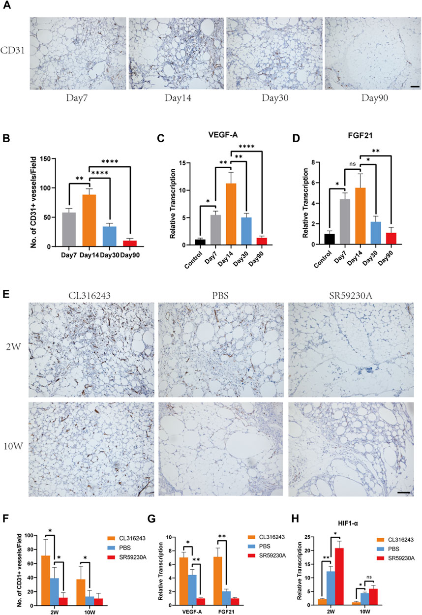
In the published article, there was an error in Figures 4A, E as published. We accidently applied mismatched images to Figures 4A, E. The corrected Figure 4 appears below.

FIGURE 4. Beige adipocyte formation was associated with early angiogenesis and the production of VEGF-A and FGF21. (A) Angiogenesis of fat grafts was superior at day 14 than day 7, 30, and 90. (B) Quantification of CD31-positive cells at different time points. (C, D) Expression levels of angiogenic genes, VEGF-A and FGF21. (E) Angiogenesis in the fat grafts 2 weeks and 10 weeks after transplantation, identified using immunohistochemical staining for CD31. (F) Number of CD31+ vessels at week 2 and week 10. (G) Expression of the Vegfa and Fgf21 genes in the fat grafts at week 2, measured using quantitative RT-PCR. (H) Expression of HIF1-α associated with hypoxia at week 2 and week 10. (*p < 0.05; **p < 0.01; ****p < 0.0001). Scale bar = 50 μm.
The authors apologize for this error and state that this does not change the scientific conclusions of the article in any way. The original article has been updated.
All claims expressed in this article are solely those of the authors and do not necessarily represent those of their affiliated organizations, or those of the publisher, the editors and the reviewers. Any product that may be evaluated in this article, or claim that may be made by its manufacturer, is not guaranteed or endorsed by the publisher.
Keywords: fat grafting, browning, beige adipocytes, angiogenesis, inflammation
Citation: Lin J, Zhu S, Liao Y, Liang Z, Quan Y, He Y, Cai J and Lu F (2023) Corrigendum: Spontaneous browning of white adipose tissue improves angiogenesis and reduces macrophage infiltration after fat grafting in mice. Front. Cell Dev. Biol. 11:1177893. doi: 10.3389/fcell.2023.1177893
Received: 02 March 2023; Accepted: 10 March 2023;
Published: 22 March 2023.
Edited and reviewed by:
Nadia Akawi, United Arab Emirates University, United Arab EmiratesCopyright © 2023 Lin, Zhu, Liao, Liang, Quan, He, Cai and Lu. This is an open-access article distributed under the terms of the Creative Commons Attribution License (CC BY). The use, distribution or reproduction in other forums is permitted, provided the original author(s) and the copyright owner(s) are credited and that the original publication in this journal is cited, in accordance with accepted academic practice. No use, distribution or reproduction is permitted which does not comply with these terms.
*Correspondence: Junrong Cai, ZHJqdW5yb25nY2FpQG91dGxvb2suY29t; Feng Lu, ZG9jdG9ybHVmZW5nQGhvdG1haWwuY29t
†These authors have contributed equally to this work and share first authorship
‡These authors have contributed equally to this work and share last authorship
Disclaimer: All claims expressed in this article are solely those of the authors and do not necessarily represent those of their affiliated organizations, or those of the publisher, the editors and the reviewers. Any product that may be evaluated in this article or claim that may be made by its manufacturer is not guaranteed or endorsed by the publisher.
Research integrity at Frontiers

Learn more about the work of our research integrity team to safeguard the quality of each article we publish.