- 1Integrated Hospital of Traditional Chinese Medicine, Southern Medical University, Guangzhou, China
- 2Cancer Center, TCM-Integrated Hospital of Southern Medical University, Guangzhou, China
- 3Department of Hepatology, TCM-Integrated Hospital of Southern Medical University, Guangzhou, China
REST corepressors (RCORs) are the core component of the LSD1/CoREST/HDACs transcriptional repressor complex, which have been revealed differently expressed in various cancers, but the therapeutic and prognostic mechanisms in cancer are still poorly understood. In this study, we analyzed expression, prognostic value, molecular subtypes, genetic alteration, immunotherapy response and drug sensitivity of RCORs in pan-cancer. Clinical correlation, stemness index, immune infiltration and regulatory networks of RCORs in hepatocellular carcinoma (HCC) were detected through TCGA and GSCA database. In-vitro experiments were conducted to explore the role of RCOR1 in HCC cells. The expression of RCORs varied among different cancers, and have prognostic values in several cancers. Cancer subtypes were categorized according to the expression of RCORs with clinical information. RCORs were significantly correlated with immunotherapy response, MSI, drug sensitivity and genetic alteration in pan-cancer. In HCC, RCORs were considered as potential predictor of stemness and also had association with immune infiltration. The ceRNA-TF-kinase regulatory networks of RCORs were constructed. Besides, RCOR1 acts as an oncogene in HCC and promotes the proliferation of HCC cells by inhibiting cell cycle arrest and cell apoptosis. Taken together, our study revealed the potential molecular mechanisms of RCORs in pan-cancer, offering a benchmark for disease-related research.
1 Introduction
Despite the emerging role of immunotherapy in cancer treatment, cancer is still a major factor which has caused high mortality all over the world, therefore, explorations for more effective anti-tumor therapeutic targets and methods are urgently needed (Sung et al., 2021). In recent years, with the continuous developments of public databases such as The Cancer Genome Atlas (TCGA), more and more oncogenes which might be potential novel molecular targets for cancer therapy have been studied by analyzing their expression and correlation with clinical prognosis and relevant signaling pathways (Blum et al., 2018).
CoREST (REST corepressor), a 66 KD protein with two highly structural conservative SANT domains. It binds to the REST (RE1 silencing transcription factor) to repress the transcription of some neuron-specific genes in non-neuronal cells (Andrés et al., 1999). Recently, CoREST was discovered to be a complex consists of three sub-members: CoREST1 (encoded by RCOR1), CoREST2 (encoded by RCOR2) and CoREST3 (encoded by RCOR3), which functionally related to the programming of cell fate (Saleque et al., 2007; Upadhyay et al., 2014). Each member of this family exhibits unique properties in many physiological processes. RCOR1 is a co-inhibitor of the transcription factor REST/NRSF, and it interacts with REST to silence the neuron-related gene expression in neural stem cells or non-neural cells (Monaghan et al., 2017) and also regulates hematopoietic differentiation (Yao et al., 2014). RCOR2 plays a role in maintaining the pluripotency and cortical development of embryonic stem cells (ESCs) by inhibiting the proliferation of ESCs and formation of induced pluripotent stem (iPS) cells (Yang et al., 2011). Both RCOR1 and RCOR2 positively influence LSD1 demethylating activity, leading to more active erythro-megakaryocytic differentiation. However, RCOR3 serves as a competitor for LSD1 in hematopoietic cells (Upadhyay et al., 2014). In the last few years, researchers have paid extensive attention to the roles that RCORs played in cancer. Compared with the control normal groups, RCOR1 displays different expression level among oral squamous cell carcinoma, prostate cancer, breast cancer, ovarian cancer, lymphoma, glioma patients. RCOR2 is identified downregulated in emodin-treated lung adenocarcinomas cells (Maimaiti et al., 2022). Except for differential expression, accumulating studies indicated that RCORs probably have some functional effects on initiation and progression of tumors. For example, RCOR1 directly interacts with MED28 and supresses cancer stem cell (CSC)-like properties in oral cavity squamous cell carcinoma (OCSCC) cells (Xiang et al., 2020). Knock-down of RCOR1 reflects a gene signature which is associated with novel molecular subtype and predictable for unfavorable PFS of R-CHOP-treated DLBCL patents (Chan et al., 2015). Upregulation of RCOR1 may maintain the tumor stem-like phenotype in diffuse astrocytoma (DA), anaplastic oligodendroglioma (AO), and glioblastoma multiforme (GBM) (Yucebas et al., 2016). RCOR1 activates the secretion of angiogenic and inflammatory factors, strengthening tumor-induced angiogenesis and inflammatory responses in breast cancer (Mazumdar et al., 2015). Xue et al. (2011) revealed that serum RCOR3 level can reflect liver injury degree and it is a biomarker of intrahepatic cholangiocarcinoma (Lv et al., 2017). In addition, RCOR3 shows lower expression in colorectal cancer patients, with hypermethylation on promoters (Liu et al., 2019). However, the underlying therapeutic and prognostic mechanisms of RCORs in cancer remain unclear.
In this study, we detected the expression and prognostic values of RCORs in pan-cancer, as well as the association between RCORs expression and cancer subtypes, responsiveness to immunotherapy, signaling pathway, drug sensitivity and genetic alteration. Besides, correlation analysis of RCORs expression with clinicopathological parameters, stemness signature, immune infiltrations and regulatory networks were completed in HCC. Additionally, we identified RCOR1 is upregulated in HCC, and promotes cell proliferation by inhibiting cell cycle arrest and cell apoptosis. Generally, our study revealed the potential molecular mechanisms of RCORs in pan-cancer, offering a benchmark for cancer-related research.
2 Materials and methods
2.1 TCGA data acquisition and processing
The data of 11,069 tumor patients across 33 different tumor types was downloaded from TCGA database (https://portal.gdc.cancer.gov/), which contains transcriptome RNAseq data, survival information and clinicopathological characteristics including age, sex, and tumor stage. Abbreviations of the 33 tumor types were provided in Supplementary Table S1. All expression data were coded as fragments per kilobase per million (FPKM) and normalized by log2 transformation. R software and its packages were used to process all these data.
2.2 Expression of RCORs in pan-cancer
Expression profile of RCORs in tumor tissues, adjacent non-tumor tissues and tumor cell lines were extracted from TCGA database. RCORs expression in normal tissues was acquired from the Human Protein Altas database (https://www.proteinatlas.org/), where RNA-seq tissue data was generated from Genotype-Tissue Expression (GTEx). Differential expression studies were performed between tumor and adjacent non-tumor tissues by Wilcox test.
2.3 Prognostic value analysis of RCORs in pan-cancer
Univariate cox proportional hazards model and Kaplan-Meier method were applied to assess the relationship between RCORs expression and clinical outcomes, including overall survival (OS), progression-free survival (PFS), disease-free survival (DFS) and disease-specific survival (DSS). Hazard ratios (HRs) were calculated by Cox model, with 95% confidence intervals (95% CI). Schoenfeld’s residuals test was used to perform proportional hazards (PH) hypothesis on risk models. Log-rank test was used for Kaplan-Meier survival analysis.
2.4 Molecular subtype models in pan-cancer
Non-negative matrix factorization (NMF) clustering algorithm was used to categorize patients into different subtypes (Yuan et al., 2020). Differences in clinicopathological characteristics including sex, age, tumor stage and etc., of each subtype among cancers were compared, and the clinical data were processed and analyzed by the “CreateTableOne” function within R package “TableOne” (https://github.com/agapiospanos/TableOne). Details for the parameter setting in statistical analysis were displayed in Supplementary Table S2.
2.5 Pathway activity and drug sensitivity analysis in pan-cancer
GSCALite (http://bioinfo.life.hust.edu.cn/web/GSCALite/) is an online platform for genomic cancer research which contains 33 cancers’ data, and GSCA (http://bioinfo.life.hust.edu.cn/GSCA/#/) is the updated version of GSCALite (Liu et al., 2023). Correlation of RCORs expression with 10 common cancer-related signaling pathways (TSC/mTOR, RTK, RAS/MAPK, PI3K/AKT, Hormone ER, Hormone AR, EMT, DNA Damage Response, Cell Cycle, Apoptosis) were conducted through GSCALite. Student’s t test was performed in this module. Anti-tumor drug sensitivity was analyzed using the “Drug” module of GSCA by Pearson test. p-value was adjusted by FDR.
2.6 Responsiveness to immunotherapy and genetic alterations analysis of RCORs in pan-cancer
Spearman test was used to analyze the correlation between RCORs expression and Tumor Mutation Burden (TMB) and Microsatellite instability (MSI) respectively. Genetic alteration of RCORs and its correlation with prognosis (OS, PFS, DFS, and DSS) in pan-cancer were investigated using the cBioPortal (http://www.cbioportal.org/).
2.7 RCORs in HCC
Correlation between RCORs expression and clinical features, immune checkpoint genes in HCC was utilized based on TCGA data. Stemness models were established, multivariate Cox proportional hazards regression was used to evaluate the association of clinical features, stemness indices with OS and Kruaskal-Wallis analysis was used to examine the of stemness indices among different HCC subtypes. GSCA was employed to analyze the correlation between RCORs expression and immune infiltration in HCC. Upstream miRNAs of RCORs were searched from mirecords (https://mirecords.biolead.org/), mirtarbase (https://mirtarbase.cuhk.edu.cn/) and tarbase (https://dianalab.e-ce.uth.gr/). MiRNA-affiliated lncRNAs were retrieved by R package “multiMiR” (Ru et al., 2014). Competing endogenous RNA (ceRNA) networks were constructed, eliminating mRNAs which were unrelated to RCORs. Transcription factors (TFs) of HCC were downloaded from AnimalTFDB3.0 (http://bioinfo.life.hust.edu.cn/AnimalTFDB#!/). Pearson correlation coefficients of RCORs-affiliated TFs were calculated by setting threshold |cor| ≥ 0.7 and p ≤ 0.05. The upstream kinases of RCORs were predicted by using X2Kgui. All networks were mapped by Cytoscape.
2.8 Tissue microarrays and immunohistochemistry
Tissue Microarray (TMA) were obtained from Shanghai Outdo Biotech Co., Ltd. (Cat No: HLivH180Su18; Shanghai, China) with 90 cases of HCC tissues and adjacent non-tumor tissues. After deparaffinization and rehydration, using microwave treatment to perform heat-mediated antigen retrieval with a pH 9.0 Tris/EDTA buffer. Incubating tissues in 3% H2O2 at room temperature for 15 min to block endogenous peroxidase activity and 30 min’ incubation of goat serum (Zhongshan Golden Bridge Biotechnology Co., Ltd., Beijing, China) at 37°C to block non-specific staining. A rabbit anti-human RCOR1 mAb (1:50, ab183711; Abcam, United States) with overnight incubation at 4°C, followed by incubation with a secondary anti-rabbit/mouse HRP-conjugated antibody (Zhongshan Golden Bridge Biotechnology Co., Ltd., Beijing, China) at room temperature for 1 h. Subsequently, TMA was washed by phosphate-buffered saline (PBS) for 3 times, and colorized with DAB-positive substrate. The slides were counterstained with hematoxylin. Staining intensity was classified into 4 grades: 0 (negative), 1 (weak), 2 (moderate), and 3 (strong). The percentage of staining-positive cells was scored as 0 (0%), 1 (1%–10%), 2 (11%–50%), 3 (51%–80%), and 4 (81%–100%). The overall score was calculated using the following formula: overall score = intensity score × percentage score. The total scores of 0–4, 5–8, and 9–12 were defined as weak positive, moderate positive, and strong positive, respectively (Scharl et al., 1990).
2.9 Cell culture and transfection
Four HCC cell lines: SK-Hep1, Hep3B, Huh7, and HCCLM3 were obtained from TCM-Integrated Hospital of Southern Medical University (Guangzhou, China). Cells were routinely maintained in high-glucose DMEM (Gibco Co., Ltd., Grand Island, NY, United States) and supplemented with 10% fetal bovine serum (Gibco) at 37°C with 5% CO2. RCOR1 siRNAs and negative control (NC) siRNA were purchased from RIBOBIO (Guangzhou, China). The RCOR1 overexpression and negative control plasmids were purchased from OBIO (Shanghai, China). Hep3B was used for loss-of-function studies and Huh7 was used for gain-of-function studies. Lipofectamine 3,000 and P3000 (Invitrogen Life Technologies, Carlsbad, CA, United States) were used to transfect cells with siRNAs and plasmids. Three siRNA-targeted sequences (provided by RIBOBIO) as follows were used to silence RCOR1: si-1: 5′-CCAGATAAATCTATAGCAA-3′; si-2: 5′-GCATGGGTACAACATGGAA-3′; si-3: 5′-GGCAGAACATGGTAAAGAA-3′. Cells were seeded in 6-well plates and transfected for 48 h, then harvested for further assays.
2.10 Real-time quantitative PCR
Total RNA was isolated with Trizol reagent (#YY101, EpiZyme Biotech Co., Ltd., Shanghai, China). 1 μg total RNA was reversely transcribed into cDNA using PrimeScript RT Master Mix (#RR036A, Takara Biotechnology (Dalian) Co., Ltd., China) according to the manufacturers’ protocols. TB Green™ Premix Ex Taq™ II (#RR820A, Takara Biotechnology (Dalian) Co., Ltd., China) was used as the fluorescent dye. Real-time (RT) PCR was performed by LightCycler480 system (Roche, Switzerland) with the RCOR1-specific primers as follows: forward, 5′-CGAGGACTAAAACTAGTGTGATGG-3′, reverse, 5′-TGCCTCTTCCAGTTCATCCT-3′; β-actin: forward, 5′-GGGAAATCGTGCGTGACATTAAGG-3′, reverse, 5′-CAGGAAGGAAGGCTGGAAGAGTG-3′. The β-actin gene was used as the housekeeping control gene. Relative gene expression was calculated using the 2^−ΔΔct method. All the oligonucleotide primers were synthesized from Invitrogen.
2.11 Western blotting
RIPA lysis buffer (#PC101, EpiZyme Biotech; Shanghai, China) was used to extract total protein. It was supplemented with protease inhibitors (#GRF101, EpiZyme Biotech) and phosphatase inhibitors (#GRF102, EpiZyme Biotech), separated by SDS-PAGE, and transferred to a PVDF membrane (Millipore, Billerica, MA). The membranes were blocked with 5% non-fat milk at room temperature for 1 h. The membranes were incubated with primary antibody at 4°C overnight. Tris-HCl solution with Tween-20 (TBST) was used to wash the membranes three times for 10 min. Subsequently, they were incubated with horseradish peroxidase-conjugated secondary antibody (goat-anti-rabbit/mouse IgG, LI-COR, United States) for 1 h at room temperature. Protein bands were visualized by using the enhanced chemiluminescence (ECL) Plus kit (Millipore). The membranes were detected by the Odyssey infrared imaging system (LICOR, United States). The primary antibodies are as follows: anti-CoREST (RCOR1) rabbit-mAb (1:1,000, #14567T; CST, United States), anti-β-actin mouse mAb (1:5,000, A5316; Sigma, United States).
2.12 Cell viability analysis
SiRNAs and overexpression plasmids which were used to transfect HCC cells were seeded into 96-well plates at a density of 1,500 cells per well in triplicate and incubated at 37°C in 5% CO2. After 6 h, 24 h, 48 h, 72 h, and 96 h of cultivation, 10 µL CCK-8 (Dojindo, Kumamoto, Japan) reagent was added to each well, followed by incubating for 2.5 h at 37°C in 5% CO2. The absorbance at 450 nm was measured using a microplate reader (BioRad, Berkeley, CA, United States).
2.13 5-Ethynyl-2′-deoxyuridine (EDU) assay
Cells were seeded in serum-free media for 6 h prior to treatment to allow for cell cycle synchronization. After 48 h transfection of siRNAs or overexpression plasmids, cells were pulsed with Cell-Light EdU buffer (Apollo567 In Vitro Kit; #C10310-1; RIBOBIO, Guangzhou, China) and incubated for 2 h before fixation in 4% paraformaldehyde (PFA) for 15 min. The results were imaged under an inverted fluorescence microscope. Quantification of S phase cells was operated using ImageJ. The fraction of S phase cells for each field of view was captured and analyzed.
2.14 Cell cycle analysis
5 × 10^4 cells were seeded into each well of six-well plates. After transfection with siRNAs or plasmids, cells were cultured for 48 h, then collected and fixed with 75% ethanol overnight at −20°C. The fixed cells were washed with cold PBS and suspended in 500 μL PBS containing RNAase and stained in the dark with propidium iodide at room temperature for 30 min. Cell suspension was subjected to FACS (BD Co., San Jose, CA, United States) to analyze the percentage of cells at G1, S and G2/M phases of the cell cycle.
2.15 Apoptosis analysis
Cells were stained with Annexin V-FITC and propidium iodide kit (AO 2001-02P-G, Tianjin Sungene Biotech Co., Ltd., Tianjin, China) to evaluate apoptosis by flow cytometry. 5 × 10^4 cells were washed twice with PBS and stained with 5 µL Annexin V-FITC and 5 µL propidium iodide solution in 1 × binding buffer for 15 min at room temperature in the dark. Additional 400 µL of 1 × binding buffer was added to the cell suspension. Apoptosis rates were determined by flow cytometry (BD).
2.16 Statistical analysis
Statistical methods which were used in bioinformatic validations are shown above. For in-vitro validation data, values are presented as the mean ± SD for three independent experiments. All statistical analysis were performed and imaged with GraphPad Prism 8.0 software (GraphPad, La Jolla, CA, United States). Differences between groups were analyzed by Student’s t-test for two groups. The median expression level of RCOR1 was used as the cut-off for the high and low RCOR1 group in IHC scoring assessment. Test level was set at both sides α = 0. 05, p < 0.05 was considered statistically significant. Significant differences in figures and tables are marked as follows: *p < 0.05, **p < 0.01, and ***p < 0.001.
3 Results
3.1 Pan-caner expression lanscape of RCORs
Gene expression analyses of RCORs in pan-cancer were employed. As shown in Figures 1A–C, the expression of RCORs in various cancer tissues were analyzed and the expression levels of all RCOR family members in LIHC were relatively low compared to other cancer types. Besides, both RCOR1 and RCOR3 displayed highest expression in LAML, whereas RCOR2 was obviously upregulated in UCS and LGG, indicating that RCORs expression may varied among different cancer types. Consistently, expression of RCORs also varied among multiple normal samples and tumor cell lines (Supplementary Figures S1A–C, S2A–C). Further differential expression analyses of RCORs expression between tumor tissues and adjacent non-tumor tissues were also performed, we found that RCOR1 and RCOR2 are significantly higher in tumor than in adjacent non-tumor tissues in BRCA, CHOL, COAD, ESCA, KIRP, LIHC, LUAD, PRAD, STAD, THCA, and UCEC (p < 0.05, Figures 1D, E) and RCOR3 was highly expressed in most cancer tissues except KICH (Figure 1F). These results revealed that the expression of RCORs varies among different cancers and normal tissues and differentially expressed between tumor tissues and adjacent non-tumor tissues, indicating that RCORs may be potential cancer biomarkers.
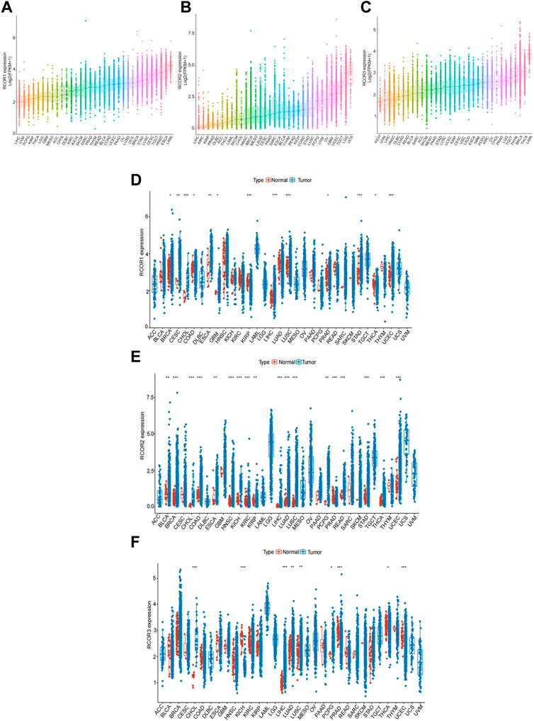
FIGURE 1. Pan-caner expression lanscape of RCORs. (A) RCOR1, (B) RCOR2 and (C) RCOR3 expression levels among different cancer tissues. (D) RCOR1, (E) RCOR2 and (F) RCOR3 expression in different cancer tissues (blue) and adjacent non-tumor tissues (red). *p < 0.05, **p < 0.01, and ***p < 0.001.
3.2 Prognostic value of RCORs in pan-cancer
Univariate Cox models were utilized to estimate the association between the RCORs expression and the prognosis of patients in pan-cancer, Schoenfeld’s residuals test was used to perform proportional hazards (PH) hypothesis on risk models, and the negative results for PH test were showed in Supplementary Figures S3–S6. The survival metrics included OS, PFS, DFS, and DSS. As shown in Figure 2A, univariate Cox regression analysis of the results from 33 types of cancer revealed that RCOR1 expression was unfavorably associated with OS of ACC (HR = 2.31, 95% CI 1.30–4.10), BLCA (HR = 1.38, 95% CI 1.10–1.74), LIHC (HR = 1.41, 95% CI 1.02–1.94), LAUD (HR = 1.40, 95% CI 1.05–1.86) and PCPG (HR = 2.64, 95% CI 1.03–6.76) (p < 0.05). Upregulation of RCOR2 asscociated to poor OS of KIRC (HR = 1.60, 95% CI 1.26–2.02), LIHC (HR = 1.76, 95% CI 1.37–2.25) and UVM (HR = 2.25, 95% CI 1.36–3.71) (p < 0.05). High RCOR3 expression level was correlated with poor OS of ACC (HR = 3.83, 95% CI 1.68–8.72) (p < 0.05).
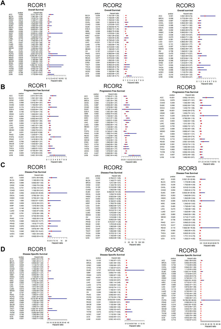
FIGURE 2. Univariate cox proportional hazards model of RCORs in pan-cancer. Forest maps for OS (A), PFS (B), DFS (C) and DSS (D) with hazard ratios (log10) and 95% confidence intervals from TCGA; HR < 1 represents low risk and HR > 1 represents high risk.
As shown in Figure 2B, increased RCOR1 expression was associated with better PFS in KIRC (HR = 0.65, 95% CI 0.48–0.87) and OV (HR = 0.81, 95% CI 0.67–0.98) (p < 0.05). High RCOR2 expression was correlated with better PFS in GBM (HR = 0.80, 95% CI 0.70–0.92). Conversely, upregulated RCOR3 expression was correlated to poor PFS of ACC (HR = 4.40, 95% CI 2.18–8.91) and READ (HR = 2.50, 95% CI 1.03–6.05) (p < 0.05). In Figure 2C, RCOR1 expression has association with DFS in ACC, OV and UCEC (p < 0.05). RCOR2 could serve as a risk factor for BRCA (HR = 1.27, 95% CI 1.06–1.51) (p < 0.05), and DFS-related hazard ratios for RCOR3 expression were significant in PCPG and UCEC (p < 0.05). In Figure 2D, RCOR1 expression was significantly related to DSS in ACC, BLCA, LAUD, PCPG and THYM (p < 0.05). RCOR2 expression was associated with DSS in BRCA, KIRC, LIHC, and UVM (p < 0.05). RCOR3 expression was significantly correlated with DSS in ACC, BRCA, and GBM (p < 0.05).
On the other hand, Kaplan-Meier analyses were used to further assess the association between RCORs expression and cancer prognosis. RCOR1 expression positively associated with OS of KIRC and LAML. On the contrary, it negatively related to OS of ACC and PRAD (p < 0.05, Figures 3A–D). Higher expression of RCOR2 indicated poor OS in MESO, UCEC, and UVM, whereas it suggested longer OS in GBM and LGG (p < 0.05, Figures 3E–I). RCOR3 expression was correlated to prognosis of ACC (p < 0.05, Figure 3J). PFS, DFS and DSS in 33 tumors were also analyzed, and the results with significance can be seen in Supplementary Figures S7–S8. All the above data revealed RCORs family was related to prognosis and may serve as cancer biomarkers.
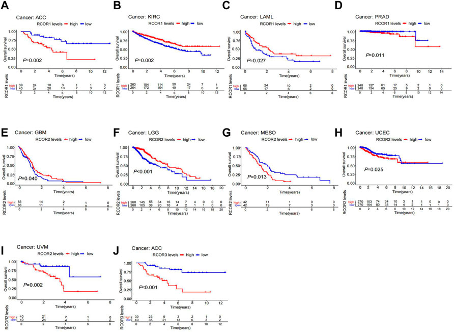
FIGURE 3. Kaplan-Meier survival analysis of RCORs in different cancers. RCOR1 as a potential prognosis factor in ACC (A), KIRC (B), LAML (C) and PRAD (D); RCOR2 as a potential prognosis factor in LGG (E), MESO (F), UCEC (G), GBM (H) and UVM (I); RCOR3 as a potential prognosis factor in ACC (J).
3.3 Analysis between RCORs and cancer molecular subtypes
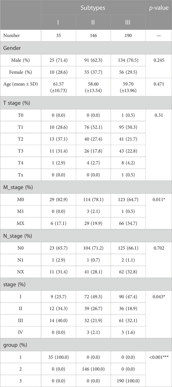
Based on the expression of RCOR members across multiple cancers, we divided cancer samples into different molecular subtypes through non-negative matrix factorization (NMF) clustering algorithm and R package “NMF” was used for classification. Setting a rank = 2:7 as the basis of classification, the criteria was to recognize the point which locate at the ahead of the largest decline of the cophenetic curve. As the cophenetic curve represented, HCC patients were divided into 3 subtypes (Figures 4A, D). BRCA patients were divided into 4 subtypes (Figures 4B, E) and BLCA patients were divided into 3 subtypes (Figures 4C, F) using the same approach. Interestingly, correlations between molecular subtypes and clinicopathological characteristics were discussed, significant difference was found in M-stage and TNM stage among different subtypes in HCC (Table 1). In addition, different age distributions were also found in different subtypes in BRCA and BLCA (Supplementary Tables S3–S4). Taken together, different cancer molecular subtypes based on RCORs expression exhibit different clinicopathological features.
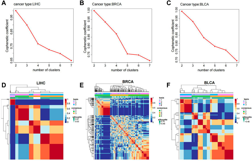
FIGURE 4. Molecular subtype analysis in various cancers. Cophenetic coefficient in RCORs in HCC (A), BRCA (B) and BLCA (C); heatmaps of molecular subtypes of RCORs in HCC (D), BRCA (E) and BLCA (F).
3.4 Correlation of RCOR family with responsiveness to immunotherapy, drug sensitivity and signaling pathways
TMB (Tumor Mutation Burden) and MSI (Microsatellite Instability) are two novel biomarkers that relevant to responsiveness to immunotherapy. Spearman analysis was used to analyze the correlation of RCORs expression with TMB and MSI. The results were displayed in Figures 5A, B respectively. According to our results, RCOR1 expression was positively associated with TMB in ACC, COAD, HNSC, LAML, LGG, PAAD, PCPG, PRAD, SARC, STAD, whereas it negatively associated with TMB in KIRC and THYM. RCOR2 expression positively connected with TMB in BRCA, COAD, HNSC, KICH, LAML, LAUD, LUSC, MESO, PAAD, STAD, and TGCT, but adverse correlations were obtained from ACC, KIRC, LIHC, PRAD, and THYM. Expression of RCOR3 was positively correlated with TMB in ACC, LAML, LGG, and SKCM, it was inversely linked with TMB in BLCA, BRCA, OV, SARC. For MSI, positive correlations with RCOR1 expression were illustrated in COAD, KICH, LAML, LUSC, SARC, STAD, and UCEC, opposite effects were found in BRCA, DLBC, HNSC, and PRAD. Only positive correlations were found with RCOR2 in ACC, BLCA, BRCA, COAD, ESCA, KICH, HNSC, LUSC, OV, STAD, and UVM. Positive connections between RCOR3 and MSI were displayed in LGG, LAUD, LUSC, READ, and UCEC, whereas it was negative in DLBC.
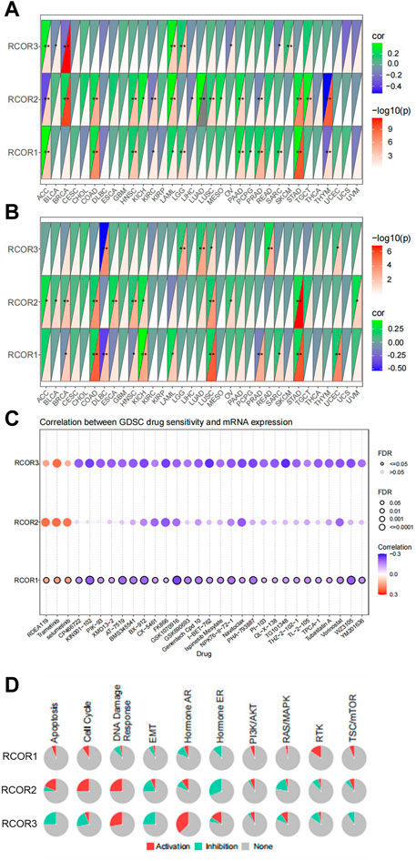
FIGURE 5. Pan-cancer analysis of RCORs in responsiveness to immunotherapy, drug sensitivity and signaling pathway. Correlation of RCORs expression with TMB (A), MSI (B). Drug sensitivity analysis (C) and proportion of activation or inhibition of signaling pathway (D) involved in GSCALite. *p < 0.05, **p < 0.01, and ***p < 0.001.
Furthermore, we conducted anti-tumor drug sensitivity analysis through the online database GSCA. Correlation analysis between gene expression and IC50 of drugs was performed by Pearson test. We selected the top 30 ranked anti-tumor drugs to show their correlations with RCORs expression, as shown in Figure 5C, It was concluded that RDEA119, Trametinib and Selumetinib positively associated with all RCOR members. However, no statistical significance appeared with RCOR2 among CP466722, KIN001-102, PIK93, and XMD13-2. Complete information for drugs study can be seen in Supplementary Table S5.
Finally, GSCALite website was used for speculating the potential roles of RCORs in 10 classic signaling pathways (TSC/mTOR, RTK, RAS/MAPK, PI3K/AKT, Hormone ER, Hormone AR, EMT, DNA Damage Response, Cell Cycle, Apoptosis), which are considered extensively involved in the process of tumorgenesis. RCOR1 showed obvious activation of RTK and Cell Cycle, whereas it significantly correlated with inhibition of Hormone ER. Significant correlations were found between RCOR2 and activation of Cell Cycle and Apoptosis. RCOR2 had similar inhibition effect of Hormone ER as RCOR1. RCOR3 remarkably correlated with activation of Hormone AR and DNA Damage Response and inhibition of EMT (Figure 5D).
3.5 Analysis of genetic alteration of RCORs in pan-cancer
cBioPortal (TCGA PanCancer Atlas) was employed to analyze genetic alterations in RCOR family. Genetic alterations of RCOR1 were detected in 28 cancer types, among them UCEC and KICH had higher alteration frequency (>4%). “Mutation” was the dominant type of alteration of UCEC, and “amplification” was the primary alteration form of KICH (Figure 6A). The highest gene alteration frequency of RCOR2 (>12%) occurred in UCSC, revealing that “amplification” is the primary type of alteration (Figure 6B). For RCOR3, patients with CHOL, BRCA and LIHC had relatively obvious alteration frequency (>4%), “amplification” was also the most common type alteration (Figure 6C). In addition, effects of RCORs alteration status on cancer prognosis were also covered. For RCOR1 and RCOR3, altered groups correlated with better OS (p < 0.05) (Figures 6D–F). Figures 6G–I displayed that altered RCOR1 positively linked with PFS (p < 0.05), whereas no significance appeared in altered RCOR2 and RCOR3. Altered RCORs did not show significant effects on DFS compared to unaltered groups (Figures 6J–L). Figures 6M–O indicated that RCOR1 and RCOR3 alteration had positive associations with DSS (p < 0.05).
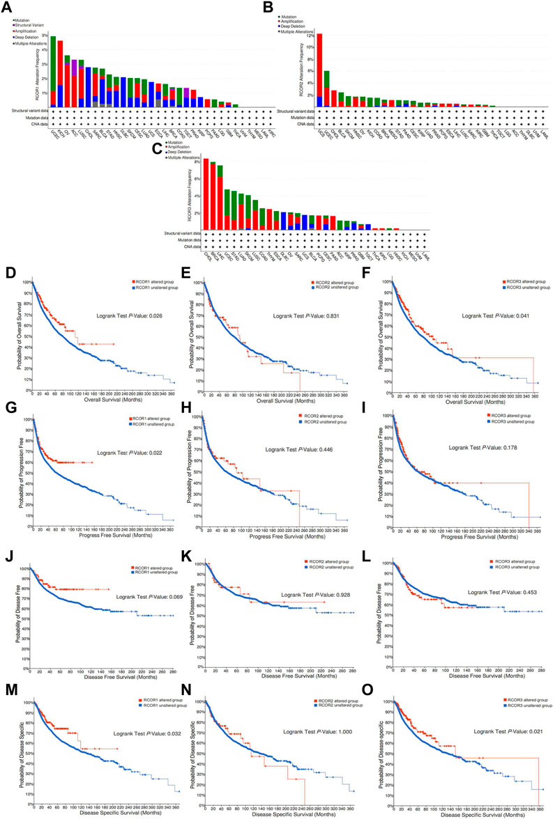
FIGURE 6. Genetic alteration of RCORs in pan-cancer. The alteration frequency of RCOR1 (A), RCOR2 (B) and RCOR3 (C) in different cancers. Effect of RCOR1 (D), RCOR2 (E) and RCOR3 (F) alteration status on OS. Effect of RCOR1 (G), RCOR2 (H) and RCOR3 (I) alteration status on PFS. Effect of RCOR1 (J), RCOR2 (K) and RCOR3 (L) alteration status on DFS. Effect of RCOR1 (M), RCOR2 (N) and RCOR3 (O) alteration status on DSS.
3.6 RCORs were significantly correlated with age, sex and tumor stage of HCC
According to RCORs expression in HCC, correlation analysis with clinical features such as age, sex and tumor stage was conducted. Box plots demonstrated that RCOR2 expression in HCC patients with age <65 was significantly higher than in age ≥65 group. RCOR1 and RCOR3 expression also seemed higer in age <65 groups without statistical significance (Figures 7A–C). RCOR1 and RCOR3 were significantly upregulated in HCC female samples (Figures 7D–F). RCOR1 and RCOR2 expression differed significantly among tumor stages, whereas no significance exists between RCOR3 expression and tumor stages, we still noticed that RCOR3 expression elevated progressively among stage II, III, and IV (Figures 7G–I). Taken together, these results revealed that RCORs expression was significantly correlated with cliniclpathological parameters in HCC.
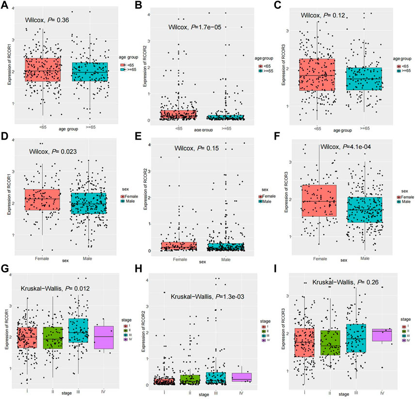
FIGURE 7. Clinical features analysis in HCC. Expression of RCOR1 (A), RCOR2 (B) and RCOR3 (C) in different age groups. Expression of RCOR1 (D), RCOR2 (E) and RCOR3 (F) in different sex. Expression of RCOR1 (G), RCOR2 (H) and RCOR3 (I) at different tumor stages.
3.7 Correlation between RCORs expression and immune infiltration, immune checkpoints and stemness of HCC
To estimate the correlation of RCORs expression with 24 immune cell types infiltration, “Immune” module of GSCA online tool was used. In Figure 8A, significant positive correlations were presented between RCOR1 expression and infiltration of central memory T cell and iTreg (p < 0.05). RCOR2 was negatively correlated with Neutrophil and Th17 cells infiltration, but positively with NKT cell (p < 0.05). RCOR3 was negatively correlated with infiltration score, macrophage and NK cell (p < 0.05). To determine whether there is association between RCOR members and some immune checkpoint genes, we categorized the HCC samples into high and low groups in terms of median expression value of RCORs, then compared the expression of immune checkpoint molecules (CD274, CTLA-4, LAG-3, LGALS9, HAVCR2, PDCD1, PDCD1LG2) between different groups of RCORs. Figure 8B showed that high RCOR1 group appeared to upregulate CD274 (p < 0.05), whereas no significance was seen on other immune checkpoint molecules. Conversely, RCOR2 favorably correlated with all immune checkpoint molecules except CD274 (Figure 8C). However, none of these immune checkpoints demonstrated significant difference between high and low groups of RCOR3 (Figure 8D).
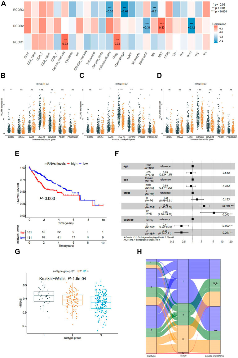
FIGURE 8. Association of RCORs with HCC. (A) Correlation between RCORs and immune infiltration from GSCA database; Expression of immune checkpoint moleculars between differentially expressed groups of RCOR1(B), RCOR2 (C) and RCOR3 (D) in HCC; (E) Survival analysis between high and low HCC stemness groups; (F) Multivariate cox regression analysis; (G) Stemness indices among different molecular subtypes; (H) Relationship in tumor stage, subtype and stemness status. *p < 0.05, **p < 0.01, and ***p < 0.001.
According to RCORs expression, HCC stemness indices were also calculated. Results can be seen in Supplementary Table S6. Samples were divided into two groups based on stemness indices. As a result, higher stemness indices group of RCORs correlated with poor OS (p < 0.05) (Figure 8E). In addition, multivariate Cox regression analysis was conducted, where the mRNAsi group was removed through PH test (Supplementary Figure S9). As displayed in Figure 8F, significance can be found in tumor stage (Ⅲ, Ⅳ), tumor subtype (2, 3) in this model. Meanwhile, stemness indices among cancer molecular subtypes were compared by Kruaskal-wallis test (Figure 8G). Sankey diagram was used to display the relationship of stages, subtypes and stemness (Figure 8H).
3.8 Construction of regulatory networks of RCORs in HCC
Upstream miRNAs of RCORs were extracted from Mirecords, Mirtarbase and Tarbase databases and further retrieval was achieved by R package “multiMiR”. As a result, 177 RCORs-miRNA pairs were obtained. Then, we retrieved 462 miRNA-lncRNA pairs also by “multiMiR”, a ceRNA network was established through cytoscope, as shown in Supplementary Figure S10A. Subsequently, we downloaded 1,665 human transcription factor (TF) data from “AnimalTFDB” database, and 1,614 TFs were extracted from the TCGA-LIHC expression profile. By calculating the Pearson correlation coefficient between TFs and RCORs expression, 304 RCORs-TFs pairs were captured. The RCORs-TFs regulatory networks such as RCOR1-SP3, RCOR3-YY1 were shown in Supplementary Figure S10B. X2Kgui was used to predict the upstream kinases of RCORs, a sum of 138 RCORs-kinase pairs were acquired. The RCORs-kinase regulatory networks were demonstrated in Supplementary Figure S10C, including RCOR1-ABL1, RCOR2-NLK, RCOR3-CDK2, etc. Totally, a comprehensive kinase-TF-ceRNA networks were constructed, comprising FRK-SP1-RCOR1-hsa-miR-520f-3p-TMEM105, PRKCZ-MYNN-RCOR3-hsa-miR-26b-5p-SNHG17 and so on (Supplementary Figure S10D).
3.9 RCOR1 is elevated in HCC and promotes HCC cell proliferation by inhibiting cell cycle arrest and cell apoptosis
Since RCOR1 is the only member of the RCORs family which showed significant difference both in sex and TNM stage, we chose RCOR1 for further experiment. To detect the expression of RCOR1 in HCC tissue, we performed IHC staining of HCC tissue microarrays, indicating that RCOR1 protein was located in both nucleus and cytoplasm, and RCOR1 protein level was higher in tumor specimens than in matched adjacent non-tumor tissues (Figures 9A, B). RCOR1 mRNA level in 4 HCC cell lines (SK-Hep1, Hep3B, Huh7, and HCCLM3) was also investigated. Compared with SK-Hep3 cells, RCOR1 was significantly upregulated in Hep3B and HCCLM4, whereas it appeared downregulation in Huh7 (Figure 9C). The results were confirmed by detections in protein (Figure 9D). Therefore, Hep3B was selected for siRNA studies, and Huh7 was for overexpression researches. Their efficiency were verified by qPCR and western blotting respectively (Figures 9E, F). Si-3 were chosen for subsequent loss-of-functions analysis, and vector-RCOR1 was for gain-of-function analysis.
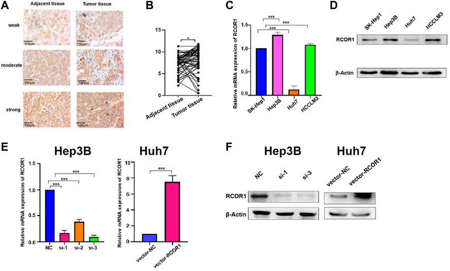
FIGURE 9. RCOR1 expression in HCC. (A) IHC staining of RCOR1 levels using HCC tissue microarrays; (B) IHC scoring of RCOR1 in tumor and adjacent non-tumor tissues; Expression of RCOR1 in HCC cell lines at mRNA (C) and protein (D) levels. Knock-down and overexpression efficiency in Hep3B and Huh7 cells at mRNA (E) and protein (F) levels. *p < 0.05, **p < 0.01, and ***p < 0.001.
CCK-8 and EDU assays were used to characterize RCOR1 effects on HCC cell proliferation, knockdown of RCOR1 can significantly inhibit cell viability compared with siRNA-negative control (NC) group (Figures 10A, C), whereas upregulation of RCOR1 raised cell viability (Figures 10B, D). Flow cytometry was used to analyze cell cycle and apoptosis. In RCOR1-knockdown Hep3B cells, the percentage of cells in the G1 phase increased while the percentage of cells in the S phase decreased significantly (Figure 10E). Compared with si-NC cells, annexin V positive cells in si-RCOR1 Hep3B cells increased significantly (Figure 10G). The percentage of cells in the G1 phase decreased and the S phase cell percentage increased significantly in vector-RCOR1 Huh7 cells compared to vector-NC cells (Figure 10F) and vector-RCOR1 groups showed remarkably lower proportion of Annexin V positivity than that in vector-NC groups (Figure 10H).
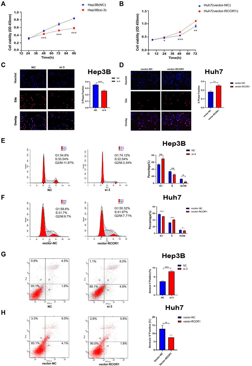
FIGURE 10. RCOR1 promotes cell growth in HCC cells. CCK8 assay in Hep3B (A) and Huh7 (B) cell line. EDU assays in Hep3B (C) and Huh7 cells (D). Effect of RCOR1 on cell cycle in Hep3B (E) and Huh7 (F) cells, Apoptosis analysis of RCOR1 in Hep3B (G) and Huh7 (H) cells. *p < 0.05, **p < 0.01, and ***p < 0.001.
4 Discussion
RCORs are generally considered as components of silencing transcription factor REST. They have crucial function in regulating neuro-development, and mediating neuron biological processes in a REST-independent way (Maksour et al., 2020). Moreover, a few evidences showed that RCORs produce marked effects on various disease. For example, RCORs were considered to be involved in the terminal differentiation of OA chondrocytes (Xiao et al., 2010). In recent years, an increasing number of studies revealed that RCORs might play a role in the process of tumorigenesis. So far, there is no systematic analysis of RCORs in cancers. Our study elaborated the tumor-related features of RCOR family genes in multiple dimensions.
GF60-mutant RCOR1 was isolated in drosophila melanogaster follicular epithelium and activated Notch signaling pathway (Domanitskaya and Schüpbach, 2012). In our study, genetic alteration of RCORs in pan-cancer was detected, revealing that the alteration status of RCORs has a significant impact on the survival rate of cancer patients. The relatively highly potential anti-neoplastic drugs that associated to RCORs were screened in this study, including RDEA119, Trametinib, Selumatinib. They are all belong to selective MEK1/MEK2 inhibitor, which can activate cancer cell autophagy, inhibiting cell proliferation, migration and inducing apoptosis. This indicated that RCORs probably regulate tumorgenesis through MAPK signaling pathway. Thus, drug sensitivity analysis of RCORs can offer more clues for further signaling pathway researches in cancers. GSCALite analysis showed that each member of RCORs exhibited different activation or inhibition in common signaling pathways, indicating that RCOR1, RCOR2, RCOR3 probably work independently or competitively in tumor progression so that leading to opposite effect on the prognosis of cancer patients. Visible inhibition of Hormone ER couid be seen significantly associated with RCOR1 and RCOR2, whereas activation of this signaling pathway strongly associated with RCOR3. Recently, Martinez et al. has come up with that RCORs (CoREST) drive the tumorgenesis of ER + breast cancer and induce resistantance to endocrine therapy by switching the recruiting site of the complex (Garcia-Martinez et al., 2022). Consistent of our study, there is an imagination that RCORs are able to function as potential targets of advanced breast cancer treatment.
Several studies clarified that RCORs may be effective in tumor immunity. Xiong et al. (2020) proved that the conditional deletion of RCOR1 in Foxp3+ Tregs had waken the function of Tregs, while the proportion of IL-2 and IFN-γraised up in peripheral lymphoid tissues and promoted antitumor immunity. In our study, expression of RCORs correlated to the infiltration of congenital and specific immune cells in HCC. Given that, it’s worthwhile to detect the tumorous immune characteristics of RCOR genes.
Emerging of competitive endogenous RNA (ceRNA) has started a new form of gene expression regulation, which is composed of mRNA, pseudogenes encoding genes, long chain non coding RNA (lncRNA) and miRNA (Salmena et al., 2011). Compared with miRNA regulation network, it is more sophisticated and complex, which provides a more detectable perspective for researchers to conduct transcriptome research, and is conducive to a more in-depth and comprehensive explanation of biological phenomena in cancer. Here, we constructed ceRNA regulatory networks by directly retrieving the miRNAs that were verified related to RCORs and the lncRNAs which were related to miRNAs. A large account of researches has illustrated that RCORs were targeted by microRNA such as miR-22 (Volvert et al., 2014), miR-124 (Baudet et al., 2011), miR9* (Packer et al., 2008), miR-432 (Das and Bhattacharyya, 2014) etc., and played crucial role in many biological processes. According to our study, in HCC, we extracted RCORs-miRNA-lncRNA pairs, such as RCOR1-hsa-miR-9-3p-CT62, RCOR2-hsa-miR-1343-3p-LINC02693, RCOR3-hsa-miR-128-3p, etc., offering potential targets for cancer therapy. Besides, based on the silencing effect on transcription of RCORs, we also filtered transcription factors (TF), which might be the potential targets in occurrence and development of HCC. Our study captured 304 TF-mRNA pairs in RCOR1 and RCOR3. There is no effective discovery in RCOR2, lack of detailed researches. More evidences are needed to reveal how RCORs affect TF in cancer progression. Zhang et al. (2015) declared that PLK1 kinase reduced levels of RCORs through degradation of ZNF198 in HBV-replicating cells, which might induce the process of liver carcinogenesis. In our study, we predicted the upstream kinases of RCORs, obtained 138 RCORs-kinase pairs, and constructed RCORs-kinase regulatory networks. Totally, we drew the Kinase-TF-ceRNA regulatory networks of HCC, which systematically demonstrated the potential regulation landscape.
In signaling pathway analysis, Apoptosis and Cell cycle pathways seemed visibly activated by RCOR1 and RCOR2, whereas RCOR3 presented evident inhibition in these two pathways. Based on this observation, we consider RCORs probably regulate tumor growth through mediating cell cycle and/or apoptosis. Our study verified RCOR1 enhance HCC cell growth by affecting cell cycle and cell apoptosis. Deregulation of cell cycle is regarded as one of the common causalities of the early steps of hepatocarcigenesis (Greenbaum, 2004). Our results demonstrated that RCOR1 play a role in regulating G1/S cell cycle phase in HCC cells according to cell cycle flow-cytometry analysis. Lack of apoptosis is related to the development and progression of liver tumors (Fabregat et al., 2007). Here we also proved overexpressed RCOR1 induce cell apoptosis in HCC cells. Above all, our current study suggested that RCOR1 regulates cell cycle and apoptosis, thereby promoting the growth of HCC cells. However, further studies especially detailed mechanism researches and in-vivo studies are needed in the near future to further explore and validate the role of RCORs family in HCC.
In conclusion, our findings systematically illustrated the potential molecular mechanisms of RCORs in pan-cancer, offering a benchmark for cancer-related research. Moreover, RCOR1 acts as an oncogene in HCC and promotes the proliferation of HCC cells by inhibiting cell cycle arrest and cell apoptosis.
Data availability statement
The datasets presented in this study can be found in online repositories. The names of the repository/repositories and accession number(s) can be found in the article/Supplementary Material.
Ethics statement
The studies involving human participants were reviewed and approved by Ethics Committee of Human Research at TCM-Integrated Hospital of Southern Medical University. The patients/participants provided their written informed consent to participate in this study. Written informed consent was obtained from the individual(s) for the publication of any potentially identifiable images or data included in this article.
Author contributions
YL, DZ, and AL conceived and designed this study. RZ and YP did the bioinformatic analysis, experiments, data curation, statistical analysis. RZ and YP mainly wrote the manuscript. XL, FL, and YL revised the manuscript and provided comments. All authors contributed to the article and approved the submitted version.
Funding
This work was supported by National Natural Science Foundation of China (grant nos 81802733 and 82160506); Natural Science Foundation of Guangdong Province (grant no. 2022A1515012620); Science and Technology Program of Guangzhou (grant no. 2023A04J0422); Department of Education of Guangdong Province (grant no. 2022KTSCX023); President’s fund of Integrated hospital of traditional Chinese Medicine (grant no. 1202103008).
Conflict of interest
The authors declare that the research was conducted in the absence of any commercial or financial relationships that could be construed as a potential conflict of interest.
Publisher’s note
All claims expressed in this article are solely those of the authors and do not necessarily represent those of their affiliated organizations, or those of the publisher, the editors and the reviewers. Any product that may be evaluated in this article, or claim that may be made by its manufacturer, is not guaranteed or endorsed by the publisher.
Supplementary material
The Supplementary Material for this article can be found online at: https://www.frontiersin.org/articles/10.3389/fcell.2023.1162344/full#supplementary-material
References
Andrés, M. E., Burger, C., Peral-Rubio, M. J., Battaglioli, E., Anderson, M. E., Grimes, J., et al. (1999). CoREST: A functional corepressor required for regulation of neural-specific gene expression. Proc. Natl. Acad. Sci. U. S. A. 96, 9873–9878. doi:10.1073/pnas.96.17.9873
Baudet, M. L., Zivraj, K. H., Abreu-Goodger, C., Muldal, A., Armisen, J., Blenkiron, C., et al. (2011). miR-124 acts through CoREST to control onset of Sema3A sensitivity in navigating retinal growth cones. Nat. Neurosci. 15, 29–38. doi:10.1038/nn.2979
Blum, A., Wang, P., and Zenklusen, J. C. (2018). SnapShot: TCGA-analyzed tumors. Cell. 173, 530. doi:10.1016/j.cell.2018.03.059
Chan, F. C., Telenius, A., Healy, S., Ben-Neriah, S., Mottok, A., Lim, R., et al. (2015). An RCOR1 loss-associated gene expression signature identifies a prognostically significant DLBCL subgroup. Blood 125, 959–966. doi:10.1182/blood-2013-06-507152
Das, E., and Bhattacharyya, N. P. (2014). MicroRNA-432 contributes to dopamine cocktail and retinoic acid induced differentiation of human neuroblastoma cells by targeting NESTIN and RCOR1 genes. FEBS Lett. 588, 1706–1714. doi:10.1016/j.febslet.2014.03.015
Domanitskaya, E., and Schüpbach, T. (2012). CoREST acts as a positive regulator of Notch signaling in the follicle cells of Drosophila melanogaster. J. Cell. Sci. 125, 399–410. doi:10.1242/jcs.089797
Fabregat, I., Roncero, C., and Fernández, M. (2007). Survival and apoptosis: A dysregulated balance in liver cancer. Liver Int. 27, 155–162. doi:10.1111/j.1478-3231.2006.01409.x
Garcia-Martinez, L., Adams, A. M., Chan, H. L., Nakata, Y., Weich, N., Stransky, S., et al. (2022). Endocrine resistance and breast cancer plasticity are controlled by CoREST. Nat. Struct. Mol. Biol. 29, 1122–1135. doi:10.1038/s41594-022-00856-x
Greenbaum, L. E. (2004). Cell cycle regulation and hepatocarcinogenesis. Cancer Biol. Ther. 3, 1200–1207. doi:10.4161/cbt.3.12.1392
Liu, C., Fennell, L. J., Bettington, M. L., Walker, N. I., Dwine, J., Leggett, B. A., et al. (2019). DNA methylation changes that precede onset of dysplasia in advanced sessile serrated adenomas. Clin. Epigenetics 11, 90. doi:10.1186/s13148-019-0691-4
Liu, C. J., Hu, F. F., Xie, G. Y., Miao, Y. R., Li, X. W., Zeng, Y., et al. (2023). Gsca: An integrated platform for gene set cancer analysis at genomic, pharmacogenomic and immunogenomic levels. Brief. Bioinform 24, bbac558. doi:10.1093/bib/bbac558
Lv, L., Wei, M., Lin, P., Chen, Z., Gong, P., Quan, Z., et al. (2017). Integrated mRNA and lncRNA expression profiling for exploring metastatic biomarkers of human intrahepatic cholangiocarcinoma. Am. J. Cancer Res. 7, 688–699.
Maimaiti, A., Xingliang, L., and Shi, L. (2022). In vitro and in vivo anti-lung cancer activity of emodin: A RNA-seq transcriptome analysis. Curr. Med. Chem. [Epub ahead of print] doi:10.2174/0929867329666220921120314
Maksour, S., Ooi, L., and Dottori, M. (2020). More than a corepressor: The role of CoREST proteins in neurodevelopment. eNeuro 7. doi:10.1523/eneuro.0337-19.2020
Mazumdar, S., Arendt, L. M., Phillips, S., Sedic, M., Kuperwasser, C., and Gill, G. (2015). CoREST1 promotes tumor formation and tumor stroma interactions in a mouse model of breast cancer. PLoS One 10, e0121281. doi:10.1371/journal.pone.0121281
Monaghan, C. E., Nechiporuk, T., Jeng, S., Mcweeney, S. K., Wang, J., Rosenfeld, M. G., et al. (2017). REST corepressors RCOR1 and RCOR2 and the repressor INSM1 regulate the proliferation-differentiation balance in the developing brain. Proc. Natl. Acad. Sci. U. S. A. 114, E406–e415. doi:10.1073/pnas.1620230114
Packer, A. N., Xing, Y., Harper, S. Q., Jones, L., and Davidson, B. L. (2008). The bifunctional microRNA miR-9/miR-9* regulates REST and CoREST and is downregulated in Huntington's disease. J. Neurosci. 28, 14341–14346. doi:10.1523/jneurosci.2390-08.2008
Ru, Y., Kechris, K. J., Tabakoff, B., Hoffman, P., Radcliffe, R. A., Bowler, R., et al. (2014). The multiMiR R package and database: Integration of microRNA-target interactions along with their disease and drug associations. Nucleic Acids Res. 42, e133. doi:10.1093/nar/gku631
Saleque, S., Kim, J., Rooke, H. M., and Orkin, S. H. (2007). Epigenetic regulation of hematopoietic differentiation by Gfi-1 and Gfi-1b is mediated by the cofactors CoREST and LSD1. Mol. Cell. 27, 562–572. doi:10.1016/j.molcel.2007.06.039
Salmena, L., Poliseno, L., Tay, Y., Kats, L., and Pandolfi, P. P. (2011). A ceRNA hypothesis: The rosetta stone of a hidden RNA language? Cell. 146, 353–358. doi:10.1016/j.cell.2011.07.014
Scharl, A., Vierbuchen, M., Conradt, B., Moll, W., Würz, H., and Bolte, A. (1990). Immunohistochemical detection of progesterone receptor in formalin-fixed and paraffin-embedded breast cancer tissue using a monoclonal antibody. Arch. Gynecol. Obste.t 247, 63–71. doi:10.1007/bf02390663
Sung, H., Ferlay, J., Siegel, R. L., Laversanne, M., Soerjomataram, I., Jemal, A., et al. (2021). Global cancer statistics 2020: GLOBOCAN estimates of incidence and mortality worldwide for 36 cancers in 185 countries. CA Cancer J. Clin. 71, 209–249. doi:10.3322/caac.21660
Upadhyay, G., Chowdhury, A. H., Vaidyanathan, B., Kim, D., and Saleque, S. (2014). Antagonistic actions of Rcor proteins regulate LSD1 activity and cellular differentiation. Proc. Natl. Acad. Sci. U. S. A. 111, 8071–8076. doi:10.1073/pnas.1404292111
Volvert, M. L., Prévot, P. P., Close, P., Laguesse, S., Pirotte, S., Hemphill, J., et al. (2014). MicroRNA targeting of CoREST controls polarization of migrating cortical neurons. Cell. Rep. 7, 1168–1183. doi:10.1016/j.celrep.2014.03.075
Xiang, Z., Zhou, S., Liang, S., Zhang, G., and Tan, Y. (2020). RCOR1 directly binds to MED28 and weakens its inducing effect on cancer stem cell-like activity of oral cavity squamous cell carcinoma cells. J. Oral Pathol. Med. 49, 741–750. doi:10.1111/jop.13022
Xiao, J., Li, T., Wu, Z., Shi, Z., Chen, J., Lam, S. K., et al. (2010). REST corepressor (CoREST) repression induces phenotypic gene regulation in advanced osteoarthritic chondrocytes. J. Orthop. Res. 28, 1569–1575. doi:10.1002/jor.21151
Xiong, Y., Wang, L., Di Giorgio, E., Akimova, T., Beier, U. H., Han, R., et al. (2020). Inhibiting the coregulator CoREST impairs Foxp3+ Treg function and promotes antitumor immunity. J. Clin. Invest. 130, 1830–1842. doi:10.1172/jci131375
Xue, J. H., Zheng, M., Xu, X. W., Wu, S. S., Chen, Z., and Chen, F. (2011). Involvement of REST corepressor 3 in prognosis of human Hepatitis B. Acta Pharmacol. Sin. 32, 1019–1024. doi:10.1038/aps.2011.49
Yang, P., Wang, Y., Chen, J., Li, H., Kang, L., Zhang, Y., et al. (2011). RCOR2 is a subunit of the LSD1 complex that regulates ESC property and substitutes for SOX2 in reprogramming somatic cells to pluripotency. Stem Cells 29, 791–801. doi:10.1002/stem.634
Yao, H., Goldman, D. C., Nechiporuk, T., Kawane, S., Mcweeney, S. K., Tyner, J. W., et al. (2014). Corepressor Rcor1 is essential for murine erythropoiesis. Blood 123, 3175–3184. doi:10.1182/blood-2013-11-538678
Yuan, Y., Chen, J., Wang, J., Xu, M., Zhang, Y., Sun, P., et al. (2020). Development and clinical validation of a novel 4-gene prognostic signature predicting survival in colorectal cancer. Front. Oncol. 10, 595. doi:10.3389/fonc.2020.00595
Yucebas, M., Yilmaz Susluer, S., Onur Caglar, H., Balci, T., Dogan Sigva, Z. O., Akalin, T., et al. (2016). Expression profiling of RE1-silencing transcription factor (REST), REST corepressor 1 (RCOR1), and Synapsin 1 (SYN1) genes in human gliomas. J. Buon. 21, 964–972.
Keywords: RCORs, pan-cancer, prognosis, biomarker, HCC, cell growth
Citation: Zheng R, Pan Y, Liu X, Liu F, Li A, Zheng D and Luo Y (2023) Comprehensive analysis of REST corepressors (RCORs) in pan-cancer. Front. Cell Dev. Biol. 11:1162344. doi: 10.3389/fcell.2023.1162344
Received: 09 February 2023; Accepted: 24 May 2023;
Published: 05 June 2023.
Edited by:
Alexander Brodsky, Lifespan, United StatesReviewed by:
Dongbo Xu, University at Buffalo, United StatesPei Li, University at Buffalo, United States
Zinian Wang, University at Buffalo, United States
Copyright © 2023 Zheng, Pan, Liu, Liu, Li, Zheng and Luo. This is an open-access article distributed under the terms of the Creative Commons Attribution License (CC BY). The use, distribution or reproduction in other forums is permitted, provided the original author(s) and the copyright owner(s) are credited and that the original publication in this journal is cited, in accordance with accepted academic practice. No use, distribution or reproduction is permitted which does not comply with these terms.
*Correspondence: Aimin Li, bGlhaW1pbjIwMDVAMTYzLmNvbQ==; Dayong Zheng, emhlbmdkeUBzbXUuZWR1LmNu; Yue Luo, bHVveXVlOTIxMkAxNjMuY29t
†These authors have contributed equally to this work and share first authorship
 Rong Zheng
 Yingying Pan
 Xinhui Liu
 Feiye Liu
 Aimin Li
 Dayong Zheng
 Yue Luo