 Mabruka Alfaidi
 Paul C. Evans2*
 J. Geoffrey Pickering
95% of researchers rate our articles as excellent or good
Learn more about the work of our research integrity team to safeguard the quality of each article we publish.
Find out more
EDITORIAL article
Front. Cardiovasc. Med. , 12 October 2023
Sec. Heart Valve Disease
Volume 10 - 2023 | https://doi.org/10.3389/fcvm.2023.1290050
This article is part of the Research Topic EndMT in Cardiovascular Diseases View all 6 articles
Editorial on the Research Topic
Endothelial-to-mesenchymal transition in cardiovascular disease
Endothelial-to-mesenchymal transition (EndMT) is a process through which endothelial cells (ECs) transition into mesenchymal cells and gain invasive and migratory properties. During this process, ECs can delaminate from their cell layer and invade the underlying tissue. In the classical form of EndMT, this is accompanied by downregulation of EC markers such as CD31 and VE-cadherin with concomitant upregulation of mesenchymal markers such as α-SMA (alpha-smooth muscle actin) and PDGFRα (platelet-derived growth factor receptor alpha), vimentin (VIM), and N-cadherin (CDH2) (1). However, it is now known that EndMT can be partial (2) and in some cases transient (3). EndMT is a fundamental process during early development (4) and has also been identified in a multitude of cardiovascular disease processes, including atherosclerosis (5–8), valvular heart disease, peripheral artery disease (9), and myocardial infarction (10). Growing evidence for EndMT in human pathologies point to the clinical relevance of EndMT in cardiovascular diseases (5, 11, 12).
The collection of the research articles presented herein provides new insights into the molecular mechanisms and importance of EndMT in cardiovascular physiology and disease. The research highlights the complexities of the EndMT process, including distinctions among different EndMT-inducing stimuli and between heart valves and the vessel wall (Table 1). The relevant actions of TGFβ1, TGFβ2, TNFα, and flow, among other mediators, are explored.
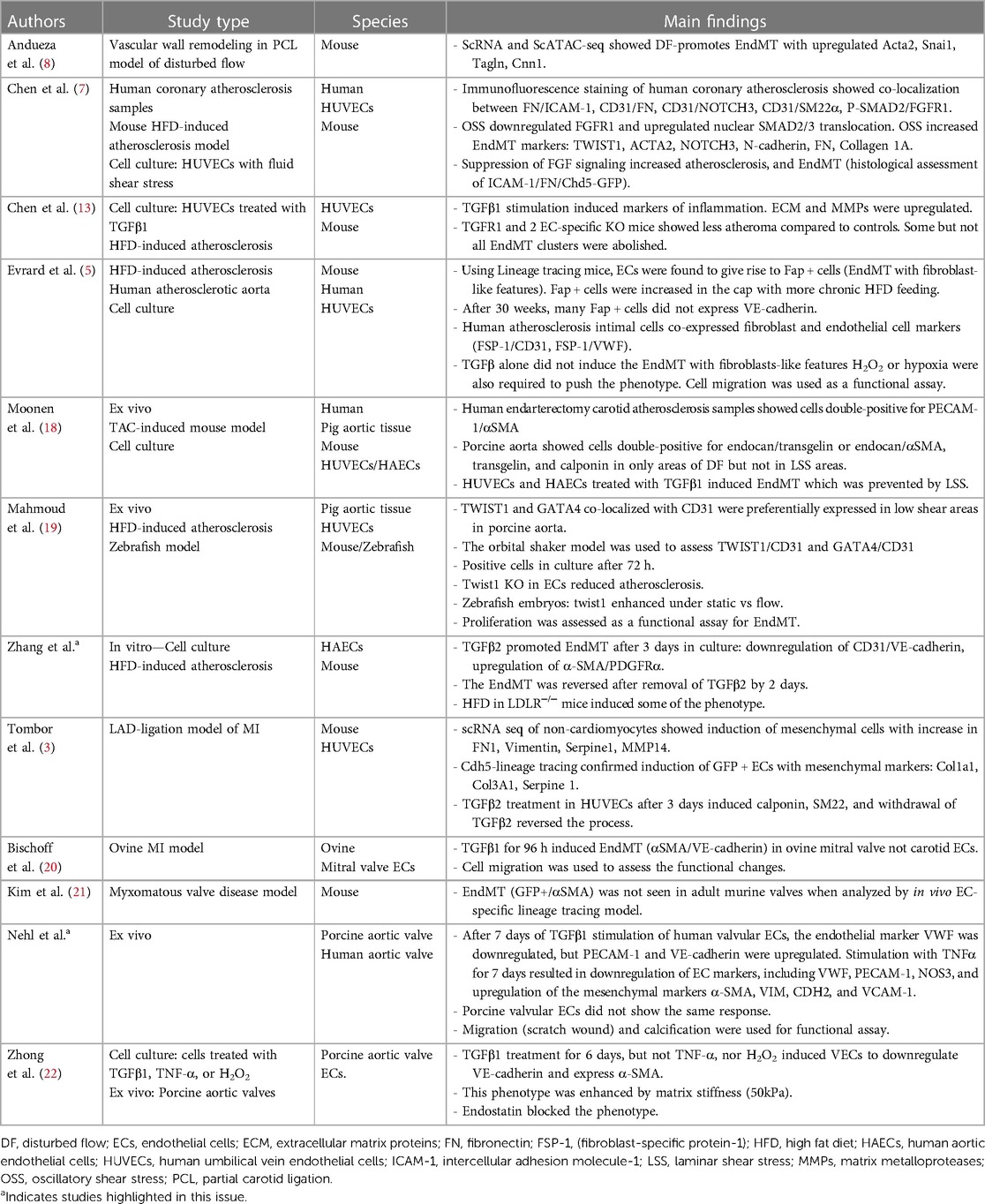
Table 1. Summary to the endMT in cardiac valves vs. in vasculature and differences among species.
Zhang et al. present a novel mechanistic link between TGFβ2 and Wnt signaling pathway in human aortic endothelial cells and mouse atherosclerotic plaques. Exposure of cultured endothelial cells to TGFβ2 for 3 days upregulated α-SMA and PDGFRα and downregulated CD31 and VE-cadherin. After removal of TGFβ2 from the media, endothelial cell adhesion marker genes re-expressed, highlighting the plasticity of the response. Interestingly, deletion of Wnt2 significantly abolished the TGFβ2- driven EndMT. Wnt2 also colocalized with α-SMA in aortic atherosclerosis in LDLR−/− mice fed Western diet for 12 weeks, but not in the chow diet-fed mice, indicating that Wnt2 expression is associated with atherosclerosis. A recent study by Chen et al. (13) reported that TGFβ induced populations of EndMT with proinflammatory features, and that Wnt signaling was altered upon EC-specific knock-out of TGFβ receptors 1 and 2 in atherogenic mice. The interplay among TGFβ2-mediated EndMT formation and Wnt2 in regulating the atherosclerosis burden is thus a key topic for further study.
Another disease context for EndMT is calcified aortic valve disease (CAVD) (14). Valvular endothelial cells can undergo EndMT and transdifferentiate into myofibroblast-like cells, with subsequent immune cell infiltration and calcification (15, 16). There is no medical treatment currently for CAVD and delineating molecular and cellular mechanisms of EndMT in this disease thus merits attention (17). Nehl et al. isolated ECs from porcine and human valvular tissue and exposed the cells with TGFβ1 or TNFα. Interestingly, the phenotype responses differed considerably between stimuli. After 7 days of TGFβ1 stimulation of human valvular ECs, the endothelial marker VWF (Von Willebrand factor) was downregulated, but PECAM-1 and VE-cadherin were upregulated. In contrast, stimulation with TNFα for 7 days resulted in consistent downregulation of EC markers, including VWF, PECAM-1, NOS3, and upregulation of the mesenchymal markers α-SMA, VIM, CDH2, and VCAM-1. The porcine valvular ECs on the other hand did not substantially change their phenotype markers, nor was their migratory response like that of the human valvular ECs. This research highlights the diversity of EndMT profiles depending on the stimulus and potential for species-specific responses.
Also in this issue, Chen et al. review the emerging concept of EndMT as “an extreme spectrum of endothelial activation”. The authors discuss TGFβ as a major inducer of EndMT. Disturbed flow was also sufficient to induce EndMT and under disturbed flow, FGF (Fibroblast growth factor) was downregulated, which exerts a positive effect on TGFR1. FGF and TGFβ have reciprocal actions in this regard. Furthermore, gene expression data analyses of ECs vs. cells having undergone EndMT vs. fibroblasts suggest that the set of EndMT genes differs from both ECs and fibroblasts.
In another review, Huang et al. provide a discussion of the mechanisms of EndMT in atherosclerosis and the different stimuli used by researchers to induce EndMT in culture, including TGFβ, interleukin-1 (IL-1β), oxidized low-density lipoprotein (oxLDL), Hydrogen peroxide (H2O2), and shear stress. The TGFβ signaling pathway, bone morphogenic protein (BMP) signaling pathway and NOTCH signaling pathway in EndMT induction are reviewed. Preventing EndMT to treat atherosclerosis is considered Huang et al. Finally, Jiang et al. review the role of EndMT in vascular calcification, also with consideration to therapeutic strategies.
Collectively, these research articles and reviews add to our understanding of EndMT in cardiovascular disease. The diversity of phenotypes and the differences among drivers of EndMT highlight the complexity of this remarkable re-wiring of endothelial cells. Ultimately, proving disease-altering roles for EndMT requires further attention, with the exciting possibility of disease-mitigating strategies.
MA: Writing – original draft, Writing – review & editing. PCE: Writing – original draft, Writing – review & editing. JGP: Writing – original draft, Writing – review & editing.
This work was supported by the American Heart Association Career Development Award [21CDA853487] to MA.
The authors declare that the research was conducted in the absence of any commercial or financial relationships that could be construed as a potential conflict of interest.
All claims expressed in this article are solely those of the authors and do not necessarily represent those of their affiliated organizations, or those of the publisher, the editors and the reviewers. Any product that may be evaluated in this article, or claim that may be made by its manufacturer, is not guaranteed or endorsed by the publisher.
1. Welch-Reardon KM, Wu N, Hughes CC. A role for partial endothelial-mesenchymal transitions in angiogenesis? Arterioscler Thromb Vasc Biol. (2015) 35:303–8. doi: 10.1161/ATVBAHA.114.303220
2. Li X, Souilhol C, Canham L, Jia X, Diagbouga M, Ayllon BT, et al. DLL4 promotes partial endothelial-to-mesenchymal transition at atherosclerosis-prone regions of arteries. Vascul Pharmacol. (2023) 150:107178. doi: 10.1016/j.vph.2023.107178
3. Tombor LS, John D, Glaser SF, Luxan G, Forte E, Furtado M, et al. Single cell sequencing reveals endothelial plasticity with transient mesenchymal activation after myocardial infarction. Nat Commun. (2021) 12:681. doi: 10.1038/s41467-021-20905-1
4. Vicovac L, Aplin JD. Epithelial-mesenchymal transition during trophoblast differentiation. Acta Anat (Basel). (1996) 156:202–16. doi: 10.1159/000147847
5. Evrard SM, Lecce L, Michelis KC, Nomura-Kitabayashi A, Pandey G, Purushothaman KR, et al. Endothelial to mesenchymal transition is common in atherosclerotic lesions and is associated with plaque instability. Nat Commun. (2016) 7:11853. doi: 10.1038/ncomms11853
6. Kidder E, Pea M, Cheng S, Koppada SP, Visvanathan S, Henderson Q, et al. The interleukin-1 receptor type-1 in disturbed flow-induced endothelial mesenchymal activation. Front Cardiovasc Med. (2023) 10:1190460. doi: 10.3389/fcvm.2023.1190460
7. Chen PY, Qin L, Baeyens N, Li G, Afolabi T, Budatha M, et al. Endothelial-to-mesenchymal transition drives atherosclerosis progression. J Clin Invest. (2015) 125:4514–28. doi: 10.1172/JCI82719
8. Andueza A, Kumar S, Kim J, Kang DW, Mumme HL, Perez JI, et al. Endothelial reprogramming by disturbed flow revealed by single-cell RNA and chromatin accessibility study. Cell Rep. (2020) 33:108491. doi: 10.1016/j.celrep.2020.108491
9. Chevalier J, Yin H, Arpino JM, O'Neil C, Nong Z, Gilmore KJ, et al. Obstruction of small arterioles in patients with critical limb ischemia due to partial endothelial-to-mesenchymal transition. iScience. (2020) 23:101251. doi: 10.1016/j.isci.2020.101251
10. Souilhol C, Harmsen MC, Evans PC, Krenning G. Endothelial-mesenchymal transition in atherosclerosis. Cardiovasc Res. (2018) 114:565–77. doi: 10.1093/cvr/cvx253
11. Pan H, Xue C, Auerbach BJ, Fan J, Bashore AC, Cui J, et al. Single-cell genomics reveals a novel cell state during smooth muscle cell phenotypic switching and potential therapeutic targets for atherosclerosis in mouse and human. Circulation. (2020) 142:2060–75. doi: 10.1161/CIRCULATIONAHA.120.048378
12. Alsaigh T, Evans D, Frankel D, Torkamani A. Decoding the transcriptome of calcified atherosclerotic plaque at single-cell resolution. Commun Biol. (2022) 5:1084. doi: 10.1038/s42003-022-04056-7
13. Chen PY, Qin L, Li G, Wang Z, Dahlman JE, Malagon-Lopez J, et al. Endothelial TGF-beta signalling drives vascular inflammation and atherosclerosis. Nat Metab. (2019) 1:912–26. doi: 10.1038/s42255-019-0102-3
14. Kovacic JC, Mercader N, Torres M, Boehm M, Fuster V. Epithelial-to-mesenchymal and endothelial-to-mesenchymal transition: from cardiovascular development to disease. Circulation. (2012) 125:1795–808. doi: 10.1161/CIRCULATIONAHA.111.040352
15. Wirrig EE, Yutzey KE. Conserved transcriptional regulatory mechanisms in aortic valve development and disease. Arterioscler Thromb Vasc Biol. (2014) 34:737–41. doi: 10.1161/ATVBAHA.113.302071
16. Aikawa E, Nahrendorf M, Figueiredo JL, Swirski FK, Shtatland T, Kohler RH, et al. Osteogenesis associates with inflammation in early-stage atherosclerosis evaluated by molecular imaging in vivo. Circulation. (2007) 116:2841–50. doi: 10.1161/CIRCULATIONAHA.107.732867
17. Makkar RR, Fontana GP, Jilaihawi H, Kapadia S, Pichard AD, Douglas PS, et al. Transcatheter aortic-valve replacement for inoperable severe aortic stenosis. N Engl J Med. (2012) 366:1696–704. doi: 10.1056/NEJMoa1202277
18. Moonen JR, Lee ES, Schmidt M, Maleszewska M, Koerts JA, Brouwer LA, et al. Endothelial-to-mesenchymal transition contributes to fibro-proliferative vascular disease and is modulated by fluid shear stress. Cardiovasc Res. (2015) 108:377–86. doi: 10.1093/cvr/cvv175
19. Mahmoud MM, Kim HR, Xing R, Hsiao S, Mammoto A, Chen J, et al. TWIST1 integrates endothelial responses to flow in vascular dysfunction and atherosclerosis. Circ Res. (2016) 119:450–62. doi: 10.1161/CIRCRESAHA.116.308870
20. Bischoff J, Casanovas G, Wylie-Sears J, Kim DH, Bartko PE, Guerrero JL, et al. CD45 expression in mitral valve endothelial cells after myocardial infarction. Circ Res. (2016) 119:1215–25. doi: 10.1161/CIRCRESAHA.116.309598
21. Kim AJ, Alfieri CM, Yutzey KE. Endothelial cell lineage analysis does not provide evidence for EMT in adult valve homeostasis and disease. Anat Rec (Hoboken). (2019) 302:125–35. doi: 10.1002/ar.23916
Keywords: EndMT, cardiovascular diseases, pathogenesis, molecular pathways, endothelial-to-mesenchymal transition
Citation: Alfaidi M, Evans PC and Pickering JG (2023) Editorial: Endothelial-to-mesenchymal transition in cardiovascular disease. Front. Cardiovasc. Med. 10:1290050. doi: 10.3389/fcvm.2023.1290050
Received: 6 September 2023; Accepted: 22 September 2023;
Published: 12 October 2023.
Edited and Reviewed by: Elena Aikawa, Harvard Medical School, United States
© 2023 Alfaidi, Evans and Pickering. This is an open-access article distributed under the terms of the Creative Commons Attribution License (CC BY). The use, distribution or reproduction in other forums is permitted, provided the original author(s) and the copyright owner(s) are credited and that the original publication in this journal is cited, in accordance with accepted academic practice. No use, distribution or reproduction is permitted which does not comply with these terms.
*Correspondence: Mabruka Alfaidi bWFicnVrYS5hbGZhaWRpQGxzdWhzLmVkdQ== Paul C. Evans cGF1bC5ldmFuc0BxbXVsLmFjLnVr J. Geoffrey Pickering Z3BpY2tlcmluZ0Byb2JhcnRzLmNh
Disclaimer: All claims expressed in this article are solely those of the authors and do not necessarily represent those of their affiliated organizations, or those of the publisher, the editors and the reviewers. Any product that may be evaluated in this article or claim that may be made by its manufacturer is not guaranteed or endorsed by the publisher.
Research integrity at Frontiers
Learn more about the work of our research integrity team to safeguard the quality of each article we publish.