- 1Department of Vascular Surgery, Beijing Hospital, National Center of Gerontology, Institute of Geriatric Medicine, Chinese Academy of Medical Sciences, Beijing, China
- 2Graduate School of Peking Union Medical College, Beijing, China
Background: Varicose veins are found to be associated with increased risk of venous thromboembolism (VTE) in many observational studies, but whether varicose veins are causally associated with VTE remains unclear. Therefore, we used a series of Mendelian randomization (MR) methods to investigate that association.
Methods: 23 independent single-nucleotide polymorphisms (SNPs) for varicose veins were obtained from the Pan UK Biobank analysis. The outcomes datasets for deep vein thrombosis (DVT), pulmonary embolism (PE) and venous thromboembolism (VTE) were obtained from the FinnGen study. Before analysis, body mass index (BMI) and height were included as confounders in our MR model. Basic MR [inverse-variance weighted (IVW), weight-median, penalized weighted-median and MR-Egger methods] and MR-PRESSO were performed against each outcome using the whole SNPs and SNPs after excluding those associated with confounders. If causal associations were suggested for any outcome, a basic MR validation analysis, a multivariable MR analysis with BMI and height, a Causal Analysis Using Summary Effect estimates (CAUSE), and a two-step MR analysis with BMI and height, would follow.
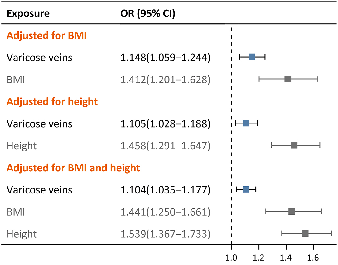
Results: Using 21 qualified SNPs, the IVW method (OR: 1.173, 95% CI: 1.070–1.286, p < 0.001, FDR = 0.002), the weighted median method (OR: 1.255, 95% CI: 1.106–1.423, p < 0.001, FDR = 0.001), the penalized weighted median method (OR: 1.299, 95% CI: 1.128–1.495, p < 0.001, FDR = 0.001) and the MR-PRESSO (OR: 1.165, 95% CI: 1.067–1.273, p = 0.003, FDR = 0.009) suggested potential causal effect of varicose veins on DVT, but no cause effect was found for PE and VTE. Excluding SNPs associated with confounders yielded similar results. The causal association with DVT was validated using a self-reported DVT cohort (IVW, OR: 1.107, 95% CI: 1.041–1.178, p = 0.001). The causal association maintained after adjustment for height (OR = 1.105, 95% CI: 1.028–1.188, p = 0.007), BMI (OR = 1.148, 95% CI: 1.059–1.244, p < 0.001) and them both (OR = 1.104, 95% CI: 1.035–1.177, p = 0.003). The causal association also survived the strict CAUSE (p = 0.018). Finally, in two-step MR, height and BMI were found to have causal effects on both varicose veins and DVT.
Conclusion: Genetically predicted varicose veins may have a causal effect on DVT and may be one of the mediators of obesity and taller height that predispose to DVT.
Introduction
Varicose veins are an important manifestation of chronic venous disease (CVD), and they can be present thought nearly the whole course of CVD from small varicosities to venous ulcer (1). Varicose veins are generally recognized as a weak risk factor of venous thromboembolic diseases, i.e., deep vein thrombosis (DVT), pulmonary embolism (PE) and venous thrombosis (VTE) (2, 3). For patients receiving surgical procedures, the presence of lower extremity varicose veins adds one point to the total Caprini score that may result in VTE prophylaxis upgrade.
Several observational studies have examined the association between varicose veins and the risk of VTE. A cross-sectional study reported 8-fold increased odds of DVT in 2,357 patients documented with varicose veins in German population (4). A recent population-based longitudinal study revealed 4-fold increased risk of DVT in patients with varicose veins during a median follow-up of 7.5 years (5). Besides, varicose veins have also been found to confer additional DVT risk to patients who already had high VTE risk factors, e.g., cancer and orthopedic surgery (6, 7). However, to our best knowledge, no study has tested whether the risk of VTE can be reduced if varicose veins are surgically removed or ablated. Therefore, it is of considerable clinical interest to investigate whether varicose veins are causally associated with VTE (3).
Previous epidemiological studies on varicose veins and VTE were susceptible to confounding factors, making causal inference difficult. By contrast, Mendelian randomization (MR) uses genetic variations as instrumental variables to mimic a randomized trial, and is capable of uncovering the causal relationship between exposures and outcomes (8, 9). MR has been wildly carried out to test causalities in the field of cardiovascular research (10). For example, MR analysis revealed that genetically determined alcohol consumption, at all dose, increases the risk of coronary heart disease and hypertension (11, 12). In the present study, we utilized a series of two-sample-based MR methods to explore whether there are causal associations between varicose veins and venous thromboembolic diseases.
Methods
Exposure and Outcome Datasets
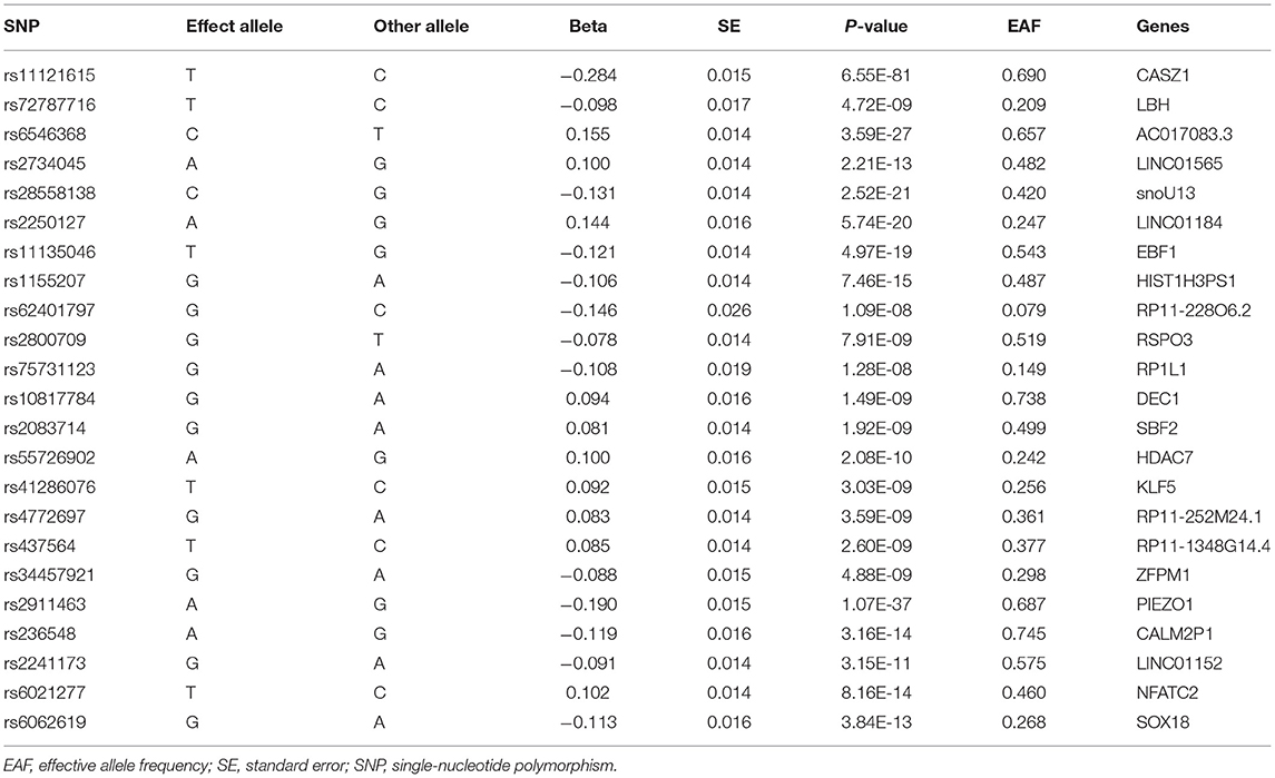
Genetic variants, i.e., single-nucleotide polymorphisms (SNP), were chosen as instrumental variables (IV). Genome-wide association study (GWAS) summary statistics for varicose veins of European ancestry were obtained from the Pan-ancestry Genetic Analysis of the UK Biobank adjusted for age, sex, age*sex, age2, age2*sex and the first 10 principal components (https://pan.ukbb.broadinstitute.org/). A total of 1,567 SNPs reached the genome-wide significance threshold (P < 5E-8), of which 23 appeared to be independent after clumping (based on the 1,000 genomes reference panel for Europeans, r2 = 0.001, kb = 10,000) for linkage disequilibrium. The SNPs were searched in the PhenoScanner database (http://www.phenoscanner.medschl.cam.ac.uk/) for associated genes and phenotypes other than varicose veins. The details of the 23 SNPs were listed in Table 1 and Supplementary Table 1. The strength of each of the IVs were evaluated by R2 statistic (the proportion of variance explained) and F statistic. R2 was calculated with formula 2 × MAF × (1–MAF) × (β estimate in SD units)2, whereas F statistic was calculated from R2 as F = (N – K – 1)/K × R2/(1 – R2) (13). MAF refers to minor allele frequency, K is the number of selected IVs and N is the samples size. A F statistic > 10 indicates a strong IV.
The outcome summary statistics for venous thromboembolic diseases were obtained from the 5th release of the FinnGen study (https://r5.finngen.fi/, Supplementary Table 2). The respective datasets for lower extremity DVT, PE, and VTE were “I9_PHLETHROMBDVTLOW” (4,576 cases and 190,028 controls), “I9_PULMEMB” (4,185 cases and 214,228 controls), and “I9_VTE” (9,176 cases and 209,616 controls). The cases were identified through either hospital discharge or cause of death ICD codes attached to the disease. Since all analyses were based on publicly available summary statistics derived from biobanks which had already been approved by their local ethical committees, no further ethical approval was required.
MR Model and Study Design
There are three key assumptions for genetic variants to be valid. First, the genetic variants should be associated with exposure. Second, the genetic variant should not be associated with confounders of the exposure-outcome relationship. Third, the genetic variants exert effects on the outcome only via the exposure (8). However, the second and third assumption are often violated since most genetic variants are actually pleiotropic. Therefore, beyond conventional MR analyses, we conducted a series of robust MR analyses, including Mendelian randomization pleiotropy residual sum and outlier (MR-PRESSO) (14), multivariable MR (15), Causal Analysis Using Summary Effect estimates (CAUSE) (16) to account for pleiotropic effects.
As shown in the Supplementary Table 1, these SNPs also displayed genome-wide associations with other traits that mainly enriched in anthropometric and blood cell parameters, causing potential IV assumption violations. Taller height (17–19) and elevated BMI (20–23) are well-established risk factors for venous thromboembolic diseases according to both clinical observational and genetic association studies. In addition, obesity is a tradition risk factor (24) and taller height is a newly discovered risk factor (25, 26) of varicose veins. Given these clinical and genetic clues, we included these two traits as potential confounders in the varicose veins and VTE relationship in our MR model, and the following MR methods were applied to test whether there was a true causality between varicose veins and VTE. The illustrations of our MR model and its MR solutions were shown in Figures 1, 2. It is worth noting that only causality suggested by basic MR analysis would undergo further tests.

Figure 1. Mendelian randomization model in this study. BMI, body mass index; DVT, deep vein thrombosis; IV, instrumental variables; PE, pulmonary embolism; SNP, single-nucleotide polymorphism; VTE, venous thromboembolism. Assumption 1: the genetic variants should be associated with exposure. Assumption 2: the genetic variant should not be associated with confounders of the exposure-outcome relationship. Assumption 3: the genetic variants exert effects on the outcome only via the exposure.
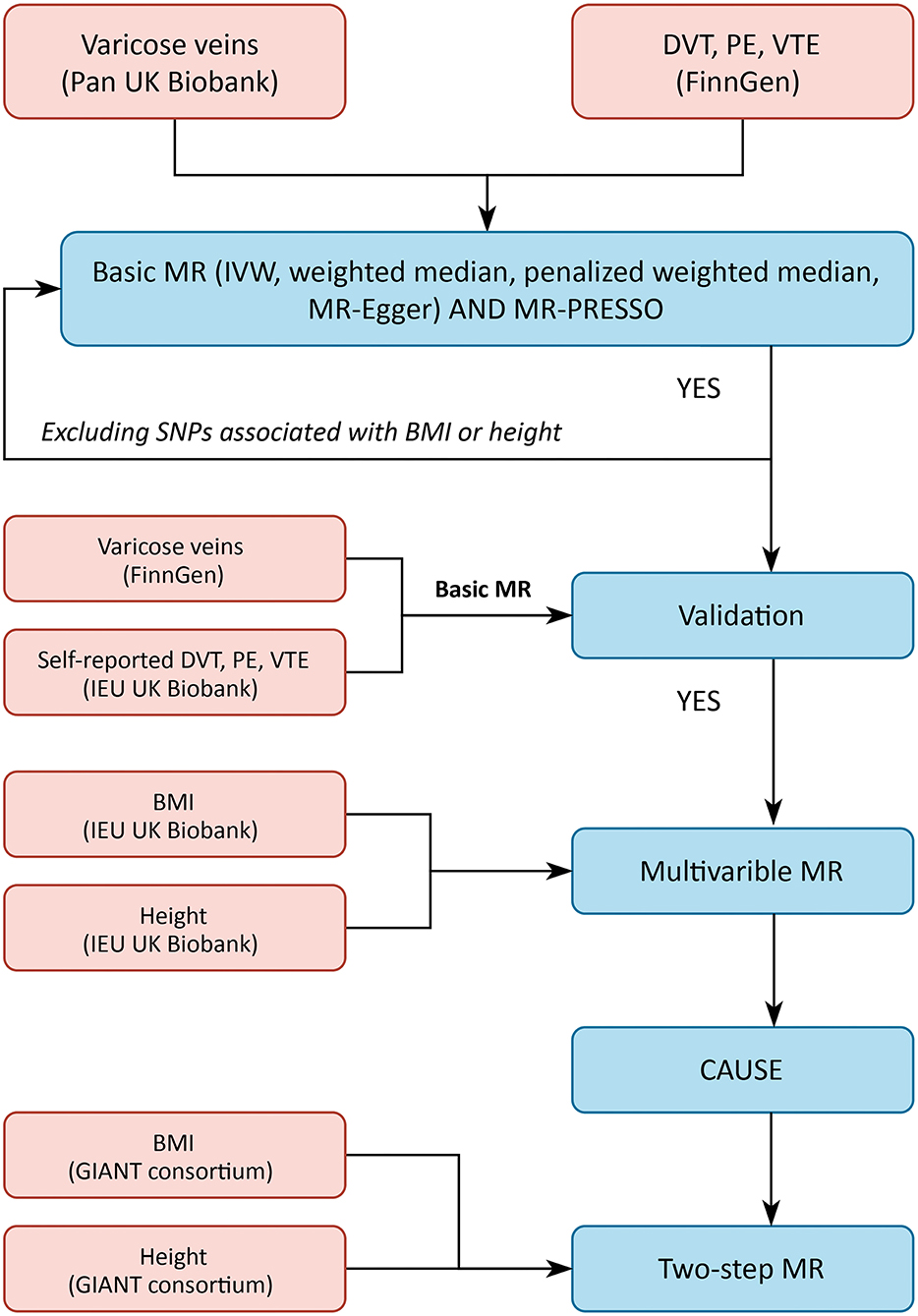
Figure 2. Flow of the study. CAUSE, Causal Analysis Using Summary Effect estimates; IVW, inverse-variance weighted; MR, Mendelian randomization; MR-PRESSO, Mendelian randomization pleiotropy residual sum and outlier.
Basic MR and MR-PRESSO
For basic MR analysis, we chose the inverse-variance-weighted (IVW) method as the main method, and the weighted-median (WM) method, the penalized-weighted-median (PWM) methods and the MR-Egger regression method as supplementary analyses (27). The IVW method gives unbiased estimates if all the instrumental variables are valid or directional pleiotropic is absent (28). The WM method provides a consistent estimate if at least half of the instrumental variables are valid, but may be less efficient (27). The PWM method down-weights the outlying variants most while affects the other variants at minimum (27). The MR-Egger method allows one or more genetic variants to have pleiotropic effects, as long as the size of these pleiotropic effects is independent of the size of the genetic variants' effects on the exposure (29). In addition, we applied MR-PRESSO test to identify horizontal pleiotropic outliers (14). After excluding SNPs concomitantly associated with height or BMI, we re-performed basic MR analysis and MR-PRESSO.
MR Using Validation Cohorts
The FinnGen study also contains GWAS summary statistics of varicose veins (https://r5.finngen.fi/, I9_VARICVE), which may serve as another source of IVs. Besides, self-reported venous outcomes are available in the UK Biobank cohort, too (https://gwas.mrcieu.ac.uk/, ukb-b-12040). By interchanging the two cohort, basic MR analysis as mentioned above was used to validate any causal associations suggested by the primary datasets (details available in Supplementary Table 2).
Multivariable MR
Multivariable MR is an extension to conventional MR that uses genetic variants associated with multiple, potentially related exposures to estimate the effect of each exposure on a single outcome (15). This approach gives unbiased causal estimates of direct effect if confounders are adjusted for (15, 30). Summary statistics for height and BMI were obtained from the IEU Open GWAS Project (https://gwas.mrcieu.ac.uk/, Supplementary Table 2). We conducted three rounds of multivariable MR analyses: varicose veins against the outcomes adjusted for (1) height alone, (2) BMI along, and (3) height and BMI combined. Because the primary exposure is not included in the IEU Open GWAS Project and automatic data formatting is not possible, we manually formatted the data for multivariable MR analyses. In brief, independent SNPs for each exposure were collected and combined, and then the essential information (i.e., betas, standard errors, p-values and effect allele frequencies) for the combined SNPs were extracted from each exposure's GWAS summary statistics.
MR With CAUSE
There are two kinds of horizontal pleiotropy, one is uncorrelated pleiotropy and the other is correlated pleiotropy. Uncorrelated pleiotropy occurs when a genetic variant affects the outcome and the exposure of interest through separate mechanisms (violation of the third IV assumption), whereas correlated pleiotropy occurs when a genetic variant affects the outcome and the tested exposure through a shared heritable factor (violation of the second IV assumption) (16). In this case, correlated pleiotropy may occur because of height and BMI, leading to false positive. Therefore, CAUSE developed by He et al. (16) were utilized to account for both correlated and uncorrelated pleiotropic effects. In brief, complete GWAS summary statistics of the exposure and the outcomes were merged, and nuisance parameters were calculated using one million unique variants for the merged data. Finally, nuisance parameters and top variants after LD pruning were used to fit CASUSE. We performed CAUSE following the online instruction with default parameters for LD pruning.
Two-Step MR
Once varicose veins were proved to be causally associated with one or more venous thromboembolic diseases, it was of interest to investigate whether varicose veins play as mediators between height and/or BMI and VTE. Therefore, two-step MR was utilized to assess potential mediation effects (31). Genetic instrumental variables for height and BMI were obtained from the Genetic Investigation of Anthropometric Traits (GIANT) Consortium via the IEU Open GWAS Project (https://gwas.mrcieu.ac.uk/, Supplementary Table 2). In the first step, the genetic variants of height and BMI were used to perform MR analysis (IVW method) against varicose veins. And in the second step the genetic variants were used to perform MR analysis against venous thromboembolic diseases. Mediation effects were suggested if evidence of causalities appeared in both steps.
Statistical Analysis
All analyses were conducted using R version 4.1.1 (The R Foundation for Statistical Computing, Vienna, Austria) under Windows environment. The R packages for MR analyses were “TwoSampleMR” (https://mrcieu.github.io/TwoSampleMR/index.html), “MR-PRESSO” (https://github.com/rondolab/MR-PRESSO) and “CAUSE” (https://jean997.github.io/cause/). A p two-sided p-value lower than 0.05 indicated statistical significance and supported a causal relationship. In addition, false discovery rate (FDR) adjusted p-values proposed by Benjamini and Hochberg were used to address multiple hypotheses testing (32).
Results
Strength of Selected Genetic Variables
The total variance explained by these SNPs were 15.0% (Supplementary Table 3), which is similar to a variance-explained of 13.4% based on 12 SNPs in a previous GWAS (26) of the same cohort. The mean and total F statistics were 120.92 and 2,871.55, respectively, indicating strong IVs.
Basic MR and MR-PRESSO
Among 23 selected SNPs derived from the exposure dataset, 19 were initially matched with the outcome datasets. 3 LD proxied SNPs were qualified (r2 > 0.9) for the missing SNPs and were used for harmonization (Supplementary Table 4). rs34457921 were excluded from harmonization for no qualified proxy and rs28558138 was exclude for being palindromic, leaving 21 SNPs for basic MR analysis and MR-PRESSO.
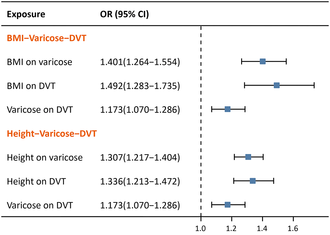
For DVT, a causal association was suggested using the IVW method (OR: 1.173, 95% CI: 1.070–1.286, p < 0.001, FDR = 0.002), the weighted median method (OR: 1.255, 95% CI: 1.106–1.423, p < 0.001, FDR = 0.001) and the penalized weighted median method (OR: 1.299, 95% CI: 1.128–1.495, p < 0.001, FDR = 0.001), the MR-PRESSO (OR: 1.165, 95% CI: 1.067–1.273, p = 0.003, FDR = 0.009), with the exception of the MR-Egger method (OR: 1.026, 95% CI: 0.811–1.298, p = 0.830). The effect sizes and their corresponding CIs of basic MR analyses were illustrated in Figure 3 (left). No significant directional pleiotropic effect was detected by the Egger-intercept test (intercept = 0.018, p = 0.241). Besides, no pleiotropic outlier was detected by MR-PRESSO. Details of other supporting statistics were listed in Supplementary Table 5. Since rs41286076 and rs1155207 were associated with height, we excluded them and re-performed basic MR analysis, and similar results were obtained (Figure 3, right). The scatter plots, forest plots, and leave-one-out plots for DVT were available in Supplementary Figure 1.
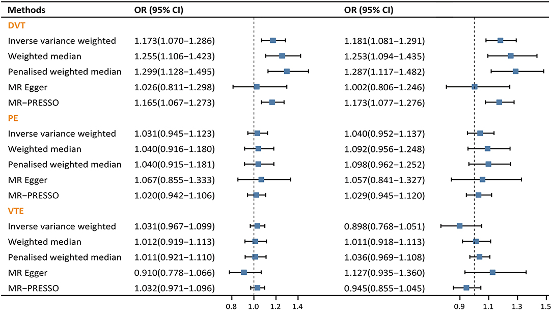
Figure 3. Forest plots of causal effect of varicose vein on venous thromboembolic diseases. (Left) MR analysis using all 21 qualified SNPs. (Right) MR analysis using 19 SNPs that have no genome-wide association with confounders. CI, confidence interval; OR, odds ratio.
However, as for PE and VTE, no sign of causal association was observed in either basic MR analysis or MR-PRESSO. Non-significant effect estimates remained even after excluding those SNPs associated with height (Figure 3). As a result, PE and VTE were excluded from further MR analyses.
Validation
Using the validation cohort where the DVT outcome was self-reported, the IVW (OR: 1.107, 95% CI: 1.041–1.178, p = 0.001), the WM method (OR: 1.126, 95% CI: 1.033–1.226, p = 0.007) and the PWM method (OR: 1.127, 95% CI: 1.036–1.227, p = 0.005) consistently showed that varicose veins were causally associated with DVT, whereas the MR-Egger method suggested no association (OR: 1.157, 95% CI: 0.956–1.400, p = 0.144). Scatter plot is available in Supplementary Figure 2.
Multivariable MR
In multivariable MR (Figure 4), varicose veins consistently showed a causality with DVT after adjustment for height (OR = 1.105, 95% CI: 1.028–1.188, p = 0.007), BMI (OR = 1.148, 95% CI: 1.059–1.244, p < 0.001) and both of them (OR = 1.104, 95% CI: 1.035–1.177, p = 0.003). The effect sizes of causal association slightly attenuated in multivariable MR as compared with univariable MR.
CAUSE
In the strict CAUSE, the causal model was shown to be a better fit than the sharing model (p = 0.018), indicating a causal association between varicose veins and DVT. More supporting statistics were listed in Supplementary Table 6.
Two-Step MR
As expected, in two-step MR (Figure 5), BMI was shown to be casually associated with both varicose veins (OR = 1.401, 95% CI: 1.264–1.554, p < 0.001) and lower extremity DVT (OR = 1.492, 95% CI: 1.283–1.735, p = p < 0.001). Similarly, taller height was also found to have casual associations with both varicose veins (OR = 1.307, 95% CI: 1.217–1.404, p < 0.001), and lower extremity DVT (OR = 1.336, 95% CI: 1.213–1.472, p < 0.001).
Discussion
In this comprehensive MR study, our results highlighted that genetically predicted varicose veins may have a causal association with DVT and may be one of the mediators of traditional DVT risk factors that predispose to DVT, e.g., obesity and taller height. Our findings concur with previous observational studies that varicosity is an independent risk factor of DVT (4–6, 33), but are less susceptible to unmeasured confounders.
The causal associations were, however, non-significant for PE and VTE. Since DVT is seen as the most common cause of PE, a causal risk factor of DVT is also considered a causal risk factor for PE. Several reasons may explain this paradox in our studies. At first, the effect size and of varicose veins on PE would be smaller than that of DVT because PE is the downstream of DVT, and the CI would be wider as well. Furthermore, the percentage of DVT and PE overlap was low in the FinnGen study (9.1%), which means that the causal effect of varicose veins on PE via DVT may have been too small to be detected using MR methods. In fact, the difference between DVT and PE is more remarkable than previous thought (34). For example, patients carrying the factor V Leiden mutation are reported to have a substantially increased risk to develop DVT but only a mildly increased risk to acquire PE (35). In contrast, several risk factors like pneumonia, COPD, atrial fibrillation, and sickle cell disease lead to a higher risk of PE and seem to have a much smaller effect on DVT (36, 37). Similar to the FinnGen study, many studies have reported high rates of isolated PE in the absence of DVT (20–79.3%), and patients with isolated PE are more likely to have cancer, atrial fibrillation and heart failure and be exposed to hormone therapy (37–41). One recent meta-analysis concluded that PE is not associated with lower extremity DVT in adult trauma patients (42). Furthermore, one study used comprehensive magnetic resonance imaging to detect origins of pulmonary emboli, but could only find a origin in less than half of the patients, suggesting PE may arise de novo in the lungs (in-situ thrombosis) (40). These evidences indicate that DVT and PE have differences in risk factors, etiology and pathophysiology and these differences may have genetic backgrounds. Hence, it was possible that no causal association were found for the remaining two outcomes. Future releases of biobank-level GWAS with larger sample sizes are need to clarify these issues.
In an early population-based case-control study, varicose veins were found to be an independent risk factor of VTE in an age-dependent manner, with people aged 45 suffering the highest risk (OR: 4.19) (33). In another observation confined to DVT, the presence of varicose veins was associated with 8 times the odds of DVT in German population (4). In one case-control study enrolling patients aged over 70 years, the adjusted odds ratio for VTE patients to have varicose veins was 1.6 (95% CI: 1.2–2.3) (43). And more recently, Chang et al. (5) found the hazard ratios for developing DVT and PE in varicose vein cases were 5.3 and 1.73, respectively, as compares with non-varicose vein cases in a 7.5-year long follow-up. Despite the findings form epidemiological investigations, studies aiming to elucidate the mechanisms underlying the risk difference of DVT were scarce. One possible explanation could be that the turbulent flow and venous stasis cause by reflux in primary venous insufficiency predisposes to a prothrombotic state of lower extremity (44). Another hypothesis is about chronic inflammation. Some inflammatory and prothrombotic markers (e.g., IL-6, TNF-α, vWF, and PAI-1) has been reported to be significantly elevated in varicose veins (45). And that it is well-recognized that inflammation is an important trigger of thrombosis (46). However, no direct biological evidence has been found that varicose veins are causes of DVT, therefore, the exact role of varicose veins in the occurrence of DVT is under-explored. An external mechanism easy to think of is that varicose vein surgeries lead to the increased risk of DVT, but the Chang et al.'s investigation found that the magnitudes only slightly attenuated after excluding those who received varicose vein surgeries (47), indicating potential mechanisms from within.
In contrast, the mechanism by which a DVT lead to varicose veins is clearer. Venous obstruction and valvular reflux may appear after a chronic DVT event, causing persistent venous hypertension and finally leading to post-thrombotic syndrome (PTS), of which varicose veins is an important manifestation (48). Our bi-directional MR analysis also supported that DVT has a causal effect on varicose veins (IVW, OR: 1.111, 95% CI: 1.040–1.187, p = 0.002), which corroborated previous study (25).
The strength of our studies is obvious, that the MR methods were robust and comprehensive, and the MR model was designed from a clinical perspective. However, several limitations of the studies should be mentioned as well. First, although primary varicose veins and secondary varicose veins (e.g., PTS) differ in genetics and pathophysiology, they might be coded under the same diagnostic code in real world settings (44). Due to a lack of individual-level data, we cannot exclude PTS cases in the exposure dataset, and thus the results may not completely represent the VTE risks associated with primary varicose veins. Nonetheless, one smaller GWAS of varicose veins (26) using the UK Biobank cohort had adjusted for DVT and BMI and yielded 13 qualified IVs, still the MR result supported a causal association between varicose veins and DVT (IVW, OR: 1.184, 95% CI: 1.083–1.293). Second, the outcomes were also based on ICD codes without further verification, and coding errors may lead to misclassification of diseases as previous observational studies (4, 5). To remedy this, we chose self-reported DVT cohort as supplement, and the primary finding was reproductive using the basic MR methods. Third, varicose veins affect much more women than men, but current datasets were not sufficient to conduct a gender-specific MR analysis. Fourth, all the study population were of European ancestry in our study but the disease patterns may vary across different ancestries, therefore, generalizing the finding to non-European ancestries may not be suitable. Given these limitations, the causal association between varicose vein and DVT suggested by our MR analyses should be interpreted with caution. More studies are warranted to better clarify the causal association between varicose veins and VTE.
Conclusion
In conclusion, using a series of robust MR methods, we found that genetically determined varicose veins may have causal effect on DVT. In addition, we revealed that varicose veins may serve as mediators of obesity and taller height on the increased risk of DVT. Our study brings some new insight into the relationship between varicose veins and DVT, and future basic experiments or well-designed clinical studies are warranted to corroborate these findings.
Data Availability Statement
The datasets presented in this study can be found in online repositories. The names of the repository/repositories and accession number(s) can be found in the article/Supplementary Material.
Ethics Statement
The studies involving human participants were reviewed and approved by Local Ethics Committees of the FinnGen Project and the UK Biobank Project, no additional ethical approval are required. The patients/participants provided their written informed consent to participate in these study.
Author Contributions
RL: conception, design, data analysis and interpreting, and writing. ZC and LG: conception, data analysis, and revision. ZW and YM: data analysis and interpreting. QG: data interpreting. YD: critical revision. YL: conception and critical revision. Final approval was obtained from all authors.
Funding
This work was support by a grant from Wu Jieping Medical Foundation (320.6750.19089-36).
Conflict of Interest
The authors declare that the research was conducted in the absence of any commercial or financial relationships that could be construed as a potential conflict of interest.
Publisher's Note
All claims expressed in this article are solely those of the authors and do not necessarily represent those of their affiliated organizations, or those of the publisher, the editors and the reviewers. Any product that may be evaluated in this article, or claim that may be made by its manufacturer, is not guaranteed or endorsed by the publisher.
Acknowledgments
The authors thank all participants in the UK biobank, the FinnGen project, the GIANT project for their contribution, and all workers of these consortia for making GWAS data freely accessible. We also thank all the developers and researchers behind the webs and software tools.
Supplementary Material
The Supplementary Material for this article can be found online at: https://www.frontiersin.org/articles/10.3389/fcvm.2022.849027/full#supplementary-material
References
2. Anderson FA, Jr., Spencer FA. Risk factors for venous thromboembolism. Circulation. (2003) 107 (23 Suppl. 1):I9–16. doi: 10.1161/01.CIR.0000078469.07362.E6
3. Kemp MT, Obi AT, Henke PK, Wakefield TW. A narrative review on the epidemiology, prevention, and treatment of venous thromboembolic events in the context of chronic venous disease. J Vasc Surg Venous Lymphat Disord. (2021) 9:1557–67. doi: 10.1016/j.jvsv.2021.03.018
4. Müller-Bühl U, Leutgeb R, Engeser P, Achankeng EN, Szecsenyi J, Laux G. Varicose veins are a risk factor for deep venous thrombosis in general practice patients. Vasa. (2012) 41:360–5. doi: 10.1024/0301-1526/a000222
5. Chang SL, Huang YL, Lee MC, Hu S, Hsiao YC, Chang SW, et al. Association of varicose veins with incident venous thromboembolism and peripheral artery disease. JAMA. (2018) 319:807–17. doi: 10.1001/jama.2018.0246
6. Königsbrügge O, Lötsch F, Reitter EM, Brodowicz T, Zielinski C, Pabinger I, et al. Presence of varicose veins in cancer patients increases the risk for occurrence of venous thromboembolism. J Thromb Haemost. (2013) 11:1993–2000. doi: 10.1111/jth.12408
7. Prandoni P, Bruchi O, Sabbion P, Tanduo C, Scudeller A, Sardella C, et al. Prolonged thromboprophylaxis with oral anticoagulants after total hip arthroplasty: a prospective controlled randomized study. Arch Intern Med. (2002) 162:1966–71. doi: 10.1001/archinte.162.17.1966
8. Davies NM, Holmes MV, Davey Smith G. Reading Mendelian randomisation studies: a guide, glossary, and checklist for clinicians. BMJ. (2018) 362:k601. doi: 10.1136/bmj.k601
9. Emdin CA, Khera AV, Kathiresan S. Mendelian randomization. JAMA. (2017) 318:1925–6. doi: 10.1001/jama.2017.17219
10. Holmes MV, Ala-Korpela M, Smith GD. Mendelian randomization in cardiometabolic disease: challenges in evaluating causality. Nat Rev Cardiol. (2017) 14:577–90. doi: 10.1038/nrcardio.2017.78
11. Chen L, Smith GD, Harbord RM, Lewis SJ. Alcohol intake and blood pressure: a systematic review implementing a Mendelian randomization approach. PLoS Med. (2008) 5:e52. doi: 10.1371/journal.pmed.0050052
12. Holmes MV, Dale CE, Zuccolo L, Silverwood RJ, Guo Y, Ye Z, et al. Association between alcohol and cardiovascular disease: Mendelian randomisation analysis based on individual participant data. BMJ. (2014) 349:g4164. doi: 10.1136/bmj.g4164
13. Burgess S, Dudbridge F, Thompson SG. Combining information on multiple instrumental variables in Mendelian randomization: comparison of allele score and summarized data methods. Stat Med. (2016) 35:1880–906. doi: 10.1002/sim.6835
14. Verbanck M, Chen CY, Neale B, Do R. Detection of widespread horizontal pleiotropy in causal relationships inferred from Mendelian randomization between complex traits and diseases. Nat Genet. (2018) 50:693–8. doi: 10.1038/s41588-018-0099-7
15. Sanderson E. Multivariable Mendelian randomization and mediation. Cold Spring Harb Perspect Med. (2021) 11:a038984. doi: 10.1101/cshperspect.a038984
16. Morrison J, Knoblauch N, Marcus JH, Stephens M, He X. Mendelian randomization accounting for correlated and uncorrelated pleiotropic effects using genome-wide summary statistics. Nat Genet. (2020) 52:740–7. doi: 10.1038/s41588-020-0631-4
17. Lai FY, Nath M, Hamby SE, Thompson JR, Nelson CP, Samani NJ. Adult height and risk of 50 diseases: a combined epidemiological and genetic analysis. BMC Med. (2018) 16:187. doi: 10.1186/s12916-018-1175-7
18. Flinterman LE, van Hylckama Vlieg A, Rosendaal FR, Cannegieter SC. Body height, mobility, and risk of first and recurrent venous thrombosis. J Thromb Haemost. (2015) 13:548–54. doi: 10.1111/jth.12860
19. Roetker NS, Armasu SM, Pankow JS, Lutsey PL, Tang W, Rosenberg MA, et al. Taller height as a risk factor for venous thromboembolism: a Mendelian randomization meta-analysis. J Thromb Haemost. (2017) 15:1334–43. doi: 10.1111/jth.13719
20. Gregson J, Kaptoge S, Bolton T, Pennells L, Willeit P, Burgess S, et al. Cardiovascular risk factors associated with venous thromboembolism. JAMA Cardiol. (2019) 4:163–73. doi: 10.1001/jamacardio.2018.4537
21. Larsson SC, Bäck M, Rees JMB, Mason AM, Burgess S. Body mass index and body composition in relation to 14 cardiovascular conditions in UK Biobank: a Mendelian randomization study. Eur Heart J. (2020) 41:221–6. doi: 10.1093/eurheartj/ehz388
22. Tan JS, Liu NN, Guo TT, Hu S, Hua L. Genetically predicted obesity and risk of deep vein thrombosis. Thromb Res. (2021) 207:16–24. doi: 10.1016/j.thromres.2021.08.026
23. Ogren M, Eriksson H, Bergqvist D, Sternby NH. Subcutaneous fat accumulation and BMI associated with risk for pulmonary embolism in patients with proximal deep vein thrombosis: a population study based on 23 796 consecutive autopsies. J Intern Med. (2005) 258:166–71. doi: 10.1111/j.1365-2796.2005.01517.x
24. Lee AJ, Evans CJ, Hau CM, Fowkes FG. Fiber intake, constipation, and risk of varicose veins in the general population: Edinburgh Vein Study. J Clin Epidemiol. (2001) 54:423–9. doi: 10.1016/S0895-4356(00)00300-0
25. Fukaya E, Flores AM, Lindholm D, Gustafsson S, Zanetti D, Ingelsson E, et al. Clinical and genetic determinants of varicose veins. Circulation. (2018) 138:2869–80. doi: 10.1161/CIRCULATIONAHA.118.035584
26. Shadrina AS, Sharapov SZ, Shashkova TI, Tsepilov YA. Varicose veins of lower extremities: insights from the first large-scale genetic study. PLoS Genet. (2019) 15:e1008110. doi: 10.1371/journal.pgen.1008110
27. Bowden J, Davey Smith G, Haycock PC, Burgess S. Consistent estimation in Mendelian randomization with some invalid instruments using a weighted median estimator. Genet Epidemiol. (2016) 40:304–14. doi: 10.1002/gepi.21965
28. Burgess S, Butterworth A, Thompson SG. Mendelian randomization analysis with multiple genetic variants using summarized data. Genet Epidemiol. (2013) 37:658–65. doi: 10.1002/gepi.21758
29. Bowden J, Davey Smith G, Burgess S. Mendelian randomization with invalid instruments: effect estimation and bias detection through Egger regression. Int J Epidemiol. (2015) 44:512–25. doi: 10.1093/ije/dyv080
30. Burgess S, Thompson SG. Multivariable Mendelian randomization: the use of pleiotropic genetic variants to estimate causal effects. Am J Epidemiol. (2015) 181:251–60. doi: 10.1093/aje/kwu283
31. Relton CL, Davey Smith G. Two-step epigenetic Mendelian randomization: a strategy for establishing the causal role of epigenetic processes in pathways to disease. Int J Epidemiol. (2012) 41:161–76. doi: 10.1093/ije/dyr233
32. Benjamini Y, Hochberg Y. Controlling the false discovery rate - a practical and powerful approach to multiple testinG. J R Stat Soc Series B Stat Methodol. (1995) 57:289–300. doi: 10.1111/j.2517-6161.1995.tb02031.x
33. Heit JA, Silverstein MD, Mohr DN, Petterson TM, O'Fallon WM, Melton LJ 3rd. Risk factors for deep vein thrombosis and pulmonary embolism: a population-based case-control study. Arch Intern Med. (2000) 160:809–15. doi: 10.1001/archinte.160.6.809
34. Wenger N, Sebastian T, Engelberger RP, Kucher N, Spirk D. Pulmonary embolism and deep vein thrombosis: similar but different. Thromb Res. (2021) 206:88–98. doi: 10.1016/j.thromres.2021.08.015
35. van Stralen KJ, Doggen CJ, Bezemer ID, Pomp ER, Lisman T, Rosendaal FR. Mechanisms of the factor V Leiden paradox. Arterioscler Thromb Vasc Biol. (2008) 28:1872–7. doi: 10.1161/ATVBAHA.108.169524
36. Huisman MV, Barco S, Cannegieter SC, Le Gal G, Konstantinides SV, Reitsma PH, et al. Pulmonary embolism. Nat Rev Dis Primers. (2018) 4:18028. doi: 10.1038/nrdp.2018.28
37. Morella P, Sacco M, Carafa M, Ferro G, Curcio F, Gargiulo G, et al. Permanent atrial fibrillation and pulmonary embolism in elderly patients without deep vein thrombosis: is there a relationship? Aging Clin Exp Res. (2019) 31:1121–8. doi: 10.1007/s40520-018-1060-4
38. Palareti G, Antonucci E, Dentali F, Mastroiacovo D, Mumoli N, Pengo V, et al. Patients with isolated pulmonary embolism in comparison to those with deep venous thrombosis. Differences in characteristics and clinical evolution. Eur J Intern Med. (2019) 69:64–70. doi: 10.1016/j.ejim.2019.08.023
39. Schwartz T, Hingorani A, Ascher E, Marks N, Shiferson A, Jung D, et al. Pulmonary embolism without deep venous thrombosis. Ann Vasc Surg. (2012) 26:973–6. doi: 10.1016/j.avsg.2012.01.014
40. van Langevelde K, Srámek A, Vincken PW, van Rooden JK, Rosendaal FR, Cannegieter SC. Finding the origin of pulmonary emboli with a total-body magnetic resonance direct thrombus imaging technique. Haematologica. (2013) 98:309–15. doi: 10.3324/haematol.2012.069195
41. Van Gent JM, Zander AL, Olson EJ, Shackford SR, Dunne CE, Sise CB, et al. Pulmonary embolism without deep venous thrombosis: de novo or missed deep venous thrombosis? J Trauma Acute Care Surg. (2014) 76:1270–4. doi: 10.1097/TA.0000000000000233
42. Aziz HA, Hileman BM, Chance EA. No correlation between lower extremity deep vein thrombosis and pulmonary embolism proportions in trauma: a systematic literature review. Eur J Trauma Emerg Surg. (2018) 44:843–50. doi: 10.1007/s00068-018-1043-3
43. Engbers MJ, Karasu A, Blom JW, Cushman M, Rosendaal FR, van Hylckama Vlieg A. Clinical features of venous insufficiency and the risk of venous thrombosis in older people. Br J Haematol. (2015) 171:417–23. doi: 10.1111/bjh.13579
44. Baylis RA, Smith NL, Klarin D, Fukaya E. Epidemiology and genetics of venous thromboembolism and chronic venous disease. Circ Res. (2021) 128:1988–2002. doi: 10.1161/CIRCRESAHA.121.318322
45. Castro-Ferreira R, Cardoso R, Leite-Moreira A, Mansilha A. The role of endothelial dysfunction and inflammation in chronic venous disease. Ann Vasc Surg. (2018) 46:380–93. doi: 10.1016/j.avsg.2017.06.131
46. Stark K, Massberg S. Interplay between inflammation and thrombosis in cardiovascular pathology. Nat Rev Cardiol. (2021) 18:666–82. doi: 10.1038/s41569-021-00552-1
47. Chang SL, Chen PC. Varicose veins and deep venous thrombosis-reply. JAMA. (2018) 320:510. doi: 10.1001/jama.2018.7331
48. Kahn SR, Comerota AJ, Cushman M, Evans NS, Ginsberg JS, Goldenberg NA, et al. The postthrombotic syndrome: evidence-based prevention, diagnosis, and treatment strategies: a scientific statement from the American Heart Association. Circulation. (2014) 130:1636–61. doi: 10.1161/CIR.0000000000000130
Keywords: Mendelian randomization, varicose veins, deep vein thrombosis, pulmonary embolism, venous thromboembolism
Citation: Li R, Chen Z, Gui L, Wu Z, Miao Y, Gao Q, Diao Y and Li Y (2022) Varicose Veins and Risk of Venous Thromboembolic Diseases: A Two-Sample-Based Mendelian Randomization Study. Front. Cardiovasc. Med. 9:849027. doi: 10.3389/fcvm.2022.849027
Received: 05 January 2022; Accepted: 22 March 2022;
Published: 14 April 2022.
Edited by:
Marina Panova-Noeva, Johannes Gutenberg University Mainz, GermanyReviewed by:
Oliver Königsbrügge, Medical University of Vienna, AustriaNaeimeh Atabaki Pasdar, Lund University, Sweden
Copyright © 2022 Li, Chen, Gui, Wu, Miao, Gao, Diao and Li. This is an open-access article distributed under the terms of the Creative Commons Attribution License (CC BY). The use, distribution or reproduction in other forums is permitted, provided the original author(s) and the copyright owner(s) are credited and that the original publication in this journal is cited, in accordance with accepted academic practice. No use, distribution or reproduction is permitted which does not comply with these terms.
*Correspondence: Yongjun Li, bGl5b25nanVuNDY3OSYjeDAwMDQwO2JqaG1vaC5jbg==
 Ruihao Li
 Zuoguan Chen1
 Zhiyuan Wu
 Qing Gao