
95% of researchers rate our articles as excellent or good
Learn more about the work of our research integrity team to safeguard the quality of each article we publish.
Find out more
ORIGINAL RESEARCH article
Front. Bioinform. , 26 July 2024
Sec. Integrative Bioinformatics
Volume 4 - 2024 | https://doi.org/10.3389/fbinf.2024.1411935
This article is part of the Research Topic Omics Technologies and Bioinformatic Tools in Probiotic Research View all 7 articles
 Abdullahi Tunde Aborode1
 Neeraj Kumar2
 Christopher Busayo Olowosoke3,4
 Tope Abraham Ibisanmi5
 Islamiyyah Ayoade6,7Haruna Isiyaku Umar6,7
 Abdullahi Temitope Jamiu8Basit Bolarinwa9Zainab Olapade10Abidemi Ruth Idowu11
 Ibrahim O. Adelakun12
 Isreal Ayobami Onifade13Benjamin Akangbe14Modesta Abacheng15Odion O. Ikhimiukor16
 Aeshah A. Awaji17
 Ridwan Olamilekan Adesola18*
Introduction: This work utilizes predictive modeling in drug discovery to unravel potential candidate genes from Escherichia coli that are implicated in antimicrobial resistance; we subsequently target the gidB, MacB, and KatG genes with some compounds from plants with reported antibacterial potentials.
Method: The resistance genes and plasmids were identified from 10 whole-genome sequence datasets of E. coli; forty two plant compounds were selected, and their 3D structures were retrieved and optimized for docking. The 3D crystal structures of KatG, MacB, and gidB were retrieved and prepared for molecular docking, molecular dynamics simulations, and ADMET profiling.
Result: Hesperidin showed the least binding energy (kcal/mol) against KatG (−9.3), MacB (−10.7), and gidB (−6.7); additionally, good pharmacokinetic profiles and structure–dynamics integrity with their respective protein complexes were observed.
Conclusion: Although these findings suggest hesperidin as a potential inhibitor against MacB, gidB, and KatG in E. coli, further validations through in vitro and in vivo experiments are needed. This research is expected to provide an alternative avenue for addressing existing antimicrobial resistances associated with E. coli’s MacB, gidB, and KatG.
The growing problem of antibiotic resistance presents substantial risks to global public health and requires novel approaches to address the emergence of resistant bacterial strains (Lázár et al., 2013). Escherichia coli demonstrates the capacity of a bacterial pathogen and resists classical antimicrobial treatments by accumulating resistance mechanisms; its ongoing evolution of resistance mechanisms presents an opportunity for therapeutic intervention through identifying and targeting candidate genes related to resistance (Lázár et al., 2013). In this context, the utilization of the extensive genomic data on E. coli, along with prediction identification and development of inhibitors targeting resistance-induced candidate genes, presents a highly promising strategy for mitigating antibiotic resistance.
E. coli is a Gram-negative bacterium that is widely distributed in the environment and commonly found in the gastrointestinal tracts of both humans and warm-blooded animals (Carattoli et al., 2014). Although this is true for the majority of strains, some pathogenic variations have the potential to induce various diseases from relatively simple urinary tract infections to serious bloodstream infections (Carattoli, 2013). The urgent need for novel therapeutic compounds is highlighted by the growth of multidrug-resistant (MDR) bacteria, which are resistant to various classes of antibiotics. A comprehensive study of the genetic basis of antibiotic resistance in E. coli is therefore crucial for facilitating the advancement of antimicrobial medicines.
The use of whole-genome sequencing (WGS) has significantly transformed our capacity to unravel the genetic structures of bacterial pathogens, facilitating thorough examinations of their genomes with unparalleled precision (Didelot et al., 2012). By clarifying the genetic factors that contribute to antibiotic resistance, WGS enables identification of possible genes that may be targeted for suppression. Furthermore, comparative genomics enables investigation of evolutionary connections between the strains that are resistant and those that are susceptible, providing insights into the mechanisms driving the acquisition and spread of resistance (Didelot et al., 2012; Roemer et al., 2017).
The objective of this work is to utilize predictive modeling and computational methodologies to uncover potential genes that are involved in the resistance mechanisms within the E. coli genome. Our objective is to prioritize the candidate genes with the greatest potential for therapeutic interventions by integrating genomic, structural, and functional data. Moreover, in silico screening methodologies such as molecular docking, molecular dynamics (MD) simulations, and ADMET profiling are used in our objective to develop small molecule inhibitors that specifically target these potential genes. The ultimate aim of this work is to reinstate the ability to respond to antibiotic therapy.
This goal relies on the collaborative interactions among bioinformatics, computational biology, and medicinal chemistry, highlighting the interconnectedness of these fields in the pursuit of developing drugs to combat antibiotic resistance. By employing methodical and logical approaches, our objective is to accelerate the process of converting genetic knowledge into therapeutic interventions that are applicable in clinical settings. This aims to effectively tackle the increasing challenges presented by antibiotic-resistant strains of E. coli.
A total of 10 WGS datasets (FASTQ) of E. coli with identification numbers SRX19510069, SRX19510068, SRX19510067, SRX19510066, SRX19510065, SRX19510064, SRX19510063, SRX19510062, SRX19510061, and SRX19510060 were retrieved from the Sequence Read Archive (SRA; www.ncbi.nlm.nih.gov) maintained by the National Center for Biotechnology Information (NCBI). All data were filtered and clipped, and quality control procedures were implemented to remove low-quality reads and adapter sequences to improve the accuracy and reliability of downstream analyses. Using ResFinder (Bortolaia et al., 2020), the threshold for the percentage of identification of a resistance gene was set (≥90% identity over ≥60% of the length of the target gene). E. coli was selected as the chromosomal point mutation reference database, and antimicrobial configurations were used to select the desired antimicrobial resistance (AMR) genes.
The outputs were analyzed to define the AMR genotypes, i.e., patterns of resistance determinants observed for each antimicrobial substance in each dataset. The analyses and results visualization were carried out using R packages. PlasmidFinder 2.1 Database version (18-01-2023) was used to determine the plasmid replicon types of the assembled genomes of E. coli; the threshold for minimum percentage identity was 95%, and the minimum coverage of the contigs was set at 60%. pMLST 2.0 was used to determine the in silico plasmid MLST typing of the replicons and assembled genomes of E. coli, and the MLST configuration was set to IncF RST. Comprehensive genome analysis was carried out on the assembled data using the method described by Brettin et al. (2015), which was then annotated using the RAST tool kit (RASTtk) (Figure 1).
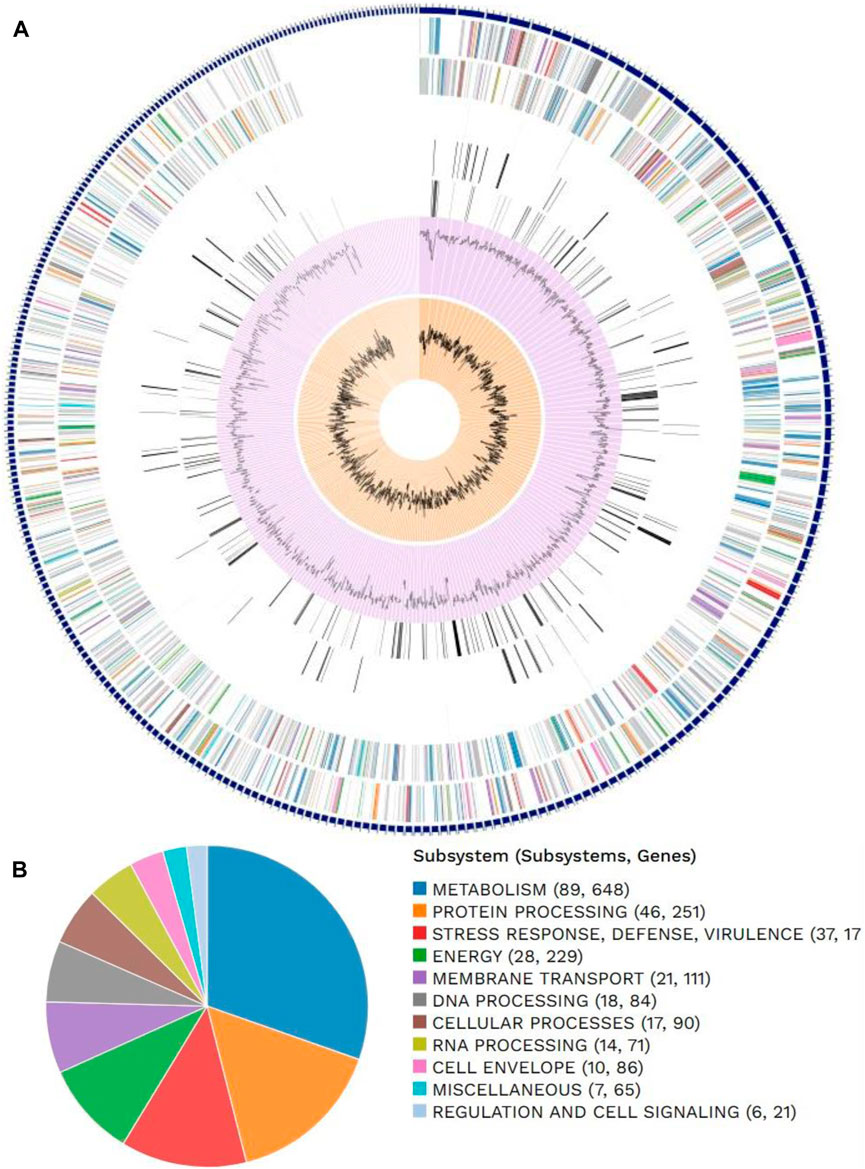
Figure 1. Distributions of (A) genome annotations and (B) subsystems in Escherichia coli.
A total of 42 compounds from different plants (Felipe et al., 2014; Thumann and Moissl-eichinger 2019; Dimitrijevi et al., 2021; Chen et al., 2022; Santhiravel et al., 2022) that have shown antibacterial properties against E. coli were chosen for the in silico evaluations. The control drugs for this project include isoniazid (PubChem ID: 3767) for KatG (PDB ID: 1U2J), chlorpromazine (PubChem ID: 2726) for gidB (PDB ID: 5LJ8), and sinefungin (PubChem ID: 65482) for MacB.
The three-dimensional (3D) structures of the proteins for the KatG, MacB, and gidB of E. coli (Zhang et al., 2019) were modeled using the Swiss model server (https://swissmodel.expasy.org/). The proteins were made nascent by freeing them from heteroatoms, such as water molecules, ions, and ligands, followed by energy minimization using the protein preparation and minimization tools in Chimera© software (version 1.13.1; https://www.cgl.ucsf.edu/chimera/) (Pettersen et al., 2004).
The 3D conformers in the structure data files (SDFs) of the control drugs were obtained and downloaded from the PubChem chemical repository linked to the NCBI, which is one of the largest collections of freely accessible chemical information in the world. Further, the structures of the obtained compounds were converted to their best energetic and stable conformations through the Merck molecular force field (MMFF94) (Halgren, 1996) using the Open Babel tool and Python Prescription (version 0.8) (Umar et al., 2021).
The prediction of KatG of E. coli using the protein-plus server (http://proteinsplus.zbh.uni-hamburg.de) (Volkamer et al., 2012) showed 11 possible binding pockets. The best binding pocket had a maximum drug score and a simple score, which were in agreement with the information supporting the server. After calculating the prediction, the P_0 pocket showed the best score for druggability and a simple score of 0.87 and 0.37, respectively. The same protein-plus server was used to predict the MacB of E. coli, for which 7 possible binding pockets were found. After running the prediction, the P_0 pocket was found to have the maximum drug score and a simple score of 0.82 and 0.63, respectively. These prediction outcomes are depicted in Figure 2 as well as Tables 4 and 5.
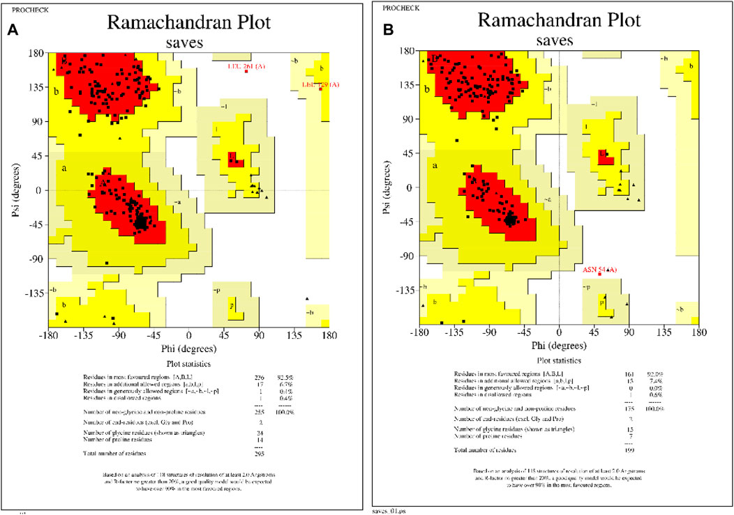
Figure 2. Homology modeled protein structural assessments of KatG and MacB genes of E. coli. (A) Ramachandran plot of KatG of E. coli, showing that 92.5% of the amino acid residues occupy the favored regions. (B) Ramachandran plot of MacB of E. coli, showing that 92% of the amino acid residues occupy the favored regions.
Molecular docking was performed using AutoDock Vina and open-source Python Prescription 0.8 (Trott and Olson, 2010; Dallakyan and Olson, 2015) to acquire the possible orientations and binding energies (BEs) of the compounds at the binding sites of the proteins. A target region for KatG equivalent to the binding regions of the other monomers of the KatG gene was attuned with the aid of a grid box with dimensions 26.3979 × 11.2720 × 21.5337 Å, and the center was adjusted based on the sites of the monomer bindings in KatG comprising Leu48, Ser56, Arg58, Thr59, Thr62, Leu63, Ala66, Gly103, Ile104, Ala105, Arg107, Ala124, Val125, Val126, Ala128, Leu129, His134, Leu136, Gln137, Ala140, Asn164, Asn165, Val166, Asp220, Val221, Thr223, His259, Cys260, Leu261, Pro262, Val281, Gln284, Ala285, Arg288, Thr291, Ala292, Val295, Leu296, Leu299, and Leu300, which were obtained from the protein binding server Zentrum fur bioinformatik (https://proteins.plus/). A target region for MacB corresponding to the binding regions of the other monomers of the MacB gene was adjusted with the aid of a grid box with dimensions 22.1946 × 22.5720 × 26.3415 Å, and the center was adjusted based on the sites of the monomer bindings in MacB comprising Asp6, Tyr8, Pro9, Gly10, Lys11, Asp12, Phe13, Gly14, Asp15, Asp16, Ala44, Val45, Ser46, Gln47, Ala62, Asn63, Gly64, Val65, Tyr69, Tyr73, Met75, Glu132, Ser135, Met136, Phe137, Gly138, Ser139, Ser140, Lys141, Arg144, Trp146, Tyr149, Met152, Ser153, Trp161, Asn163, Ser164, Phe196, Trp198, and Met200, which were also obtained from the Zentrum fur bioinformatik server. A target region for gidB corresponding to the binding regions of the other monomers of the gidB gene was attuned with the aid of a grid box with dimensions 26.3979 × 11.2720 × 26.3415 Å, and the center was adjusted based on the sites of the monomer bindings in gidB including Asn35, Asp71, Gly73, Gly77, Leu78, Asp96, Ser97, Leu98, Arg101, and Arg123 (Zhang et al., 2019). After the docking runs, the three compounds with the best docking scores (binding energies) below those of the control drugs were subjected to molecular visualization to analyze their molecular interaction fingerprints using PyMOL© Molecular Graphics (version 2.4, 2016, Schrödinger LLC) (Seeliger and Groot, 2010) and Maestro (version 12.5, 2016, Schrödinger LLC). The molecular interaction fingerprints analyzed for each protein–ligand complex included hydrogen bonds, hydrophobic interactions, and electrostatic linkages between the amino acids of the protein and ligand atoms.
The absorption, distribution, metabolism, excretion, and toxicity (ADMET) are important at the early stages of drug discovery and design pipeline to analyze the pharmacokinetics of the proposed compounds that could serve as drugs. The ADMETSar server was used to forecast the ADMET properties of the compounds with the best hits after molecular docking analysis (Cheng et al., 2012; Yang et al., 2018). The server was fed with the SMILE strings of the compounds from PubChem (https://pubchem.ncbi.nlm.nih.gov/compound/) through the search bar to predict the ADMET properties.
The three targets complexed with hesperidin served as the initial structures for the MD simulations in the Desmond package (Schrödinger Release 2019-3) based on the method described by Tutumlu et al. (2020). The simulations were performed for 200 ns durations, and various parameters were carefully examined to assess the structural stabilities of the tested compounds. To ascertain seamless amalgamation with the Schrodinger interface, the starting structures underwent meticulous preparation exploiting the Protein Preparation Wizard. Multiple crucial tasks were carried out to achieve the research objectives, including adding hydrogen atoms, assigning bond orders, and addressing any absent amino acid side chains and loops by optimizing the hydrogen bond assignments. Additionally, water orientation sampling at pH 7.0 was conducted. Through the System Builder option, the simulation periodic box was created, and solvation was accomplished by engaging the all-atom force field optimized potentials for liquid simulations (OPLSs) in combination with the single point charge (SPC) water model. An exhaustive system minimization was conducted, involving 1,000 iterations of the steepest descent technique and equilibration under the constant number of atoms, pressure, and temperature (NPT) ensemble conditions. The equilibration was implemented for 200 ns at a temperature of 300 K and pressure of 1.01325 bar. To regulate the temperature during the simulations, the Nosé–Hoover thermostat with a relaxation time of 1 ps was used, while the isotropic Martyna–Tobias–Klein barostat with a relaxation time of 2 ps was used to maintain constant pressure. Short-range interactions were considered with a cutoff of 9 Å, while the smooth particle mesh Ewald (PME) method was used in combination with the reversible reference system propagator algorithm (RESPA) integrator to calculate the long-range Coulombic interactions. To capture the dynamics of the system successfully, conformational snapshots were exported at regular intervals of 5 ps during the course of the simulations. At the end of the simulations, systemic stability was appraised using root mean-squared deviations (RMSDs), root mean-squared fluctuations (RMSFs), and assessments of the protein–ligand contacts. These analyses provided valuable insights into the behaviors and interactions of the protein–ligand complexes studied.
PCA was conducted through the ProDy library (version 1.5.1) within Python (www.python.org). This mainly involves aligning the structures based on the Cα atoms, which was accomplished using ProDy’s iterative superposition approach. The covariance matrix was calculated from the ensemble, and its diagonalization yielded the principal components, which were saved in ProDy’s NMD file format (Bakan et al., 2011). Notably, the analysis solely considered the Cα atoms, while the gaps were assigned weights of 0.0.
The DCC maps were constructed for the Cα atoms from the concatenated trajectories with respect to the reference structures using the “Bio3D” option in the R- and ProDy-based analysis tool (Grant et al., 2021).
For each of the selected models, we produced free energy landscapes (FELs) with respect to the radius of gyration (Rg) and RMSD trajectories using the Geo-Measures plugin in the PyMOL package (Kagami et al., 2020).
The assembly details outlined in Table 1 offer a comprehensive view of the genomic characteristics under study. With a total of 478 contigs, the genome appears to be somewhat fragmented, necessitating further investigations into potential scaffoldings or misassemblies. The GC content of 50.68% falls within the expected range for many organisms and serves as a baseline indicator of the nucleotide composition. Notably, the absence of plasmids in the assembly suggests a lack of extrachromosomal genetic elements in the sampled organism.
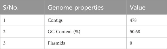
Table 1. Details of the genome assembly.
Table 2 presents the annotated genome features of the whole-genome analysis of E. coli, which contains 3,456 coding sequences (CDSs) demonstrating the vast genetic information stored within its DNA as well as 20 transfer RNA (tRNA) that are required for protein syntheses and cellular processes. The abundance of CDSs within E. coli’s genomic makeup is particularly important in drug discovery because it represents a rich source of potential drug targets. However, the current work is primarily in the interest of those implicated in antibiotic resistance.
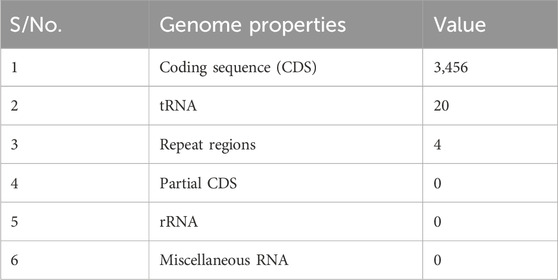
Table 2. Features from the genome annotation.
Table 3 presents the various AMR genes discovered from the whole-genome analysis of E. coli; these genes highlight the diverse strategies employed by E. coli to resist the effects of antibiotics. Notably, the presence of genes encoding the antibiotic activation enzyme KatG suggests a sophisticated ability to modify or neutralize antibiotics. The presence of antibiotic resistance gene clusters, such as MarA, MarB, and MarR, further underscores the complexity of the genetic basis for AMR in this pathogen; these genes that are associated with antibiotic targets in susceptible species and those conferring protection to these targets (e.g., BcrC) suggest a nuanced approach to evading the effects of antibiotics at the molecular level. Moreover, the presence of genes related to efflux pump systems (e.g., AcrAB-TolC, AcrZ, EmrAB-TolC, MacA, MacB, MdfA/Cmr, MdtEF-TolC, MdtL, and TolC/OpmH) indicates an active role in expelling antibiotics from the bacterial cell, contributing to a broader spectrum of resistance mechanisms. The presence of gidB, which contributes to resistance via absence, adds another layer of intricacy to the genetic landscape of antibiotic resistance in E. coli. Additionally, the involvement of the protein altering cell wall charge (PgsA) and regulators modulating the expressions of antibiotic resistance genes (e.g., AcrAB-TolC, EmrAB-TolC, GadE, H-NS, and OxyR) suggests a coordinated and adaptive response to antibiotic exposure. The diverse array of AMR genes identified in E. coli shows the urgent need for a drug with a multifaceted approach or mechanism of action in addressing antibiotic resistance in this pathogen.
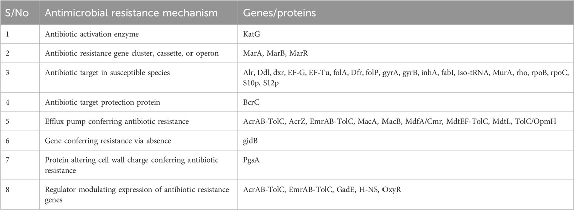
Table 3. Potential drug targets in Escherichia coli that confer antibiotic resistance.
The 3D protein structures of E. coli’s KatG and MacB have been crystallized and deposited in the popular RCSB protein database but have numerous missing residues and loops. Therefore, the homology modeling approach was employed to build suitable protein structures for this study. After building the structures, the models were assessed and showed that the KatG and MacB modeling for E. coli had 92.5% identity and 92.0% of the amino acid residues occupying the favored region, respectively (Figure 2). These indicate that the modeled structures are suitable for molecular docking and MD simulations.
The protein-plus server was used to identify 11 and 7 possible binding pockets for KatG and MacB, respectively (Tables 4, 5). The best binding pocket is the one with the maximum score for druggability and simple score; this is in accordance with the information supporting the server (Volkamer et al., 2012). After implementing the predictions, the P_0 pockets for both proteins were found to be the best choices as they had the maximum drug scores and simple scores each. The density map representations of the predicted pockets of both proteins are presented in Figure 3.
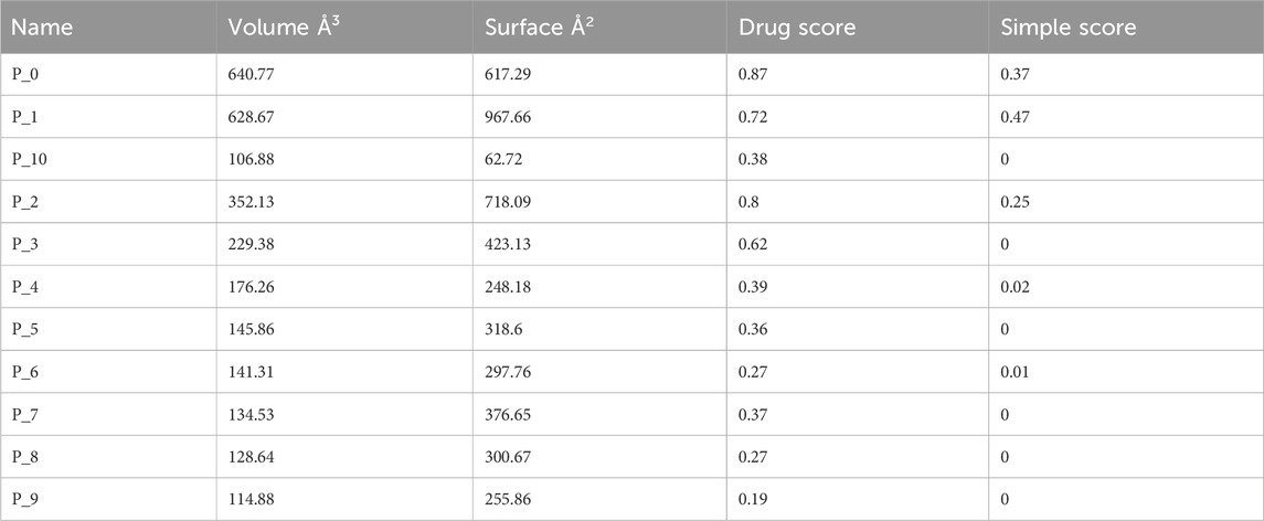
Table 4. Binding sites predicted from KatG of E. coli using the protein-plus server (http://proteinsplus.zbh.uni-hamburg.de).
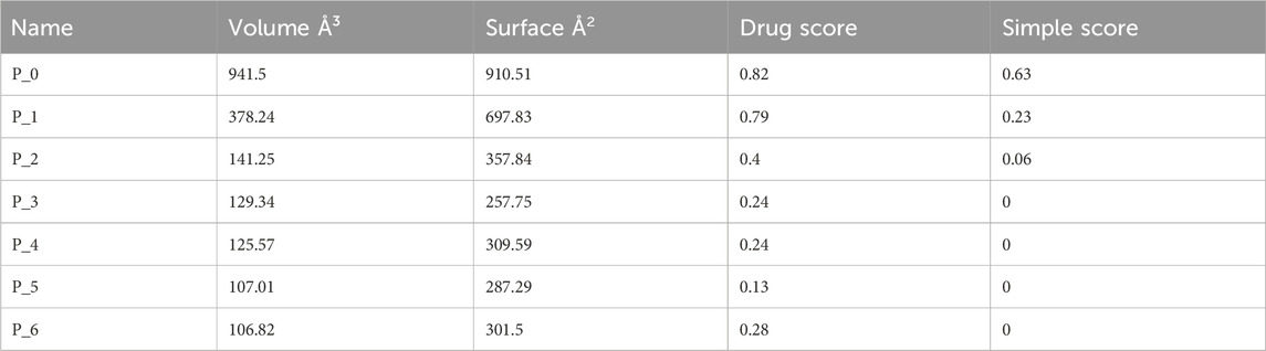
Table 5. Binding sites predicted from MacB of E. coli using the protein-plus server (http://proteinsplus.zbh.uni-hamburg.de).
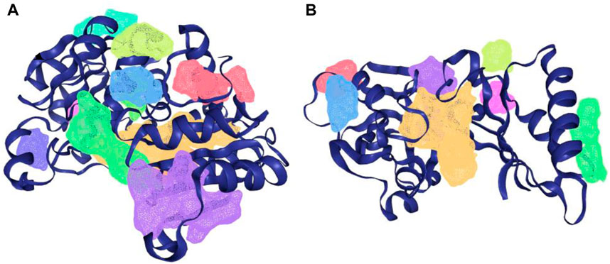
Figure 3. Density map presentation of the predicted binding sites of KatG and MacB of E. coli using the DoGsiteScorer module of protein-plus server (http://proteinsplus.zbh.uni-hamburg.de). (A) Density map representation of the predicted binding sites of KatG of E. coli, with the best binding site shown in purple. (B) Density map representation of the predicted binding sites of MacB of E. coli, with the best binding site shown in yellow.
Three out of the 42 compounds docked against the protein targets displayed better binding potentials based on their BEs (between −6.5 and −6.7 kcal/mol for gidB than −5.2 kcal/mol for sinefungin; between −8.5 and −9.4 kcal/mol for KatG than −5.4 kcal/mol for isoniazid, and −10.7 kcal/mol for MacB than −7.9 kcal/mol for chlorpromazine). The details for the 42 compounds and three targets are provided in Table 6. Hesperidin (−9.3 kcal/mol), rutin (−9.1 kcal/mol), and naringin (−8.8 kcal/mol) returned the least BEs against KatG. Similarly, biacalein (−6.7 kcal/mol), chrysin (−6.5 kcal/mol), and hesperidin (−6.7 kcal/mol) had the least BEs against gidB. Finally, epicatechin gallate (−10.7 kcal/mol), hesperidin (−10.7 kcal/mol), and epigallocatechin gallate (−10.7 kcal/mol) had the least BEs against MacB. Interestingly, hesperidin was able to bind all three targets in E. coli that are implicated in antibiotic resistance through different pathways (Table 6).
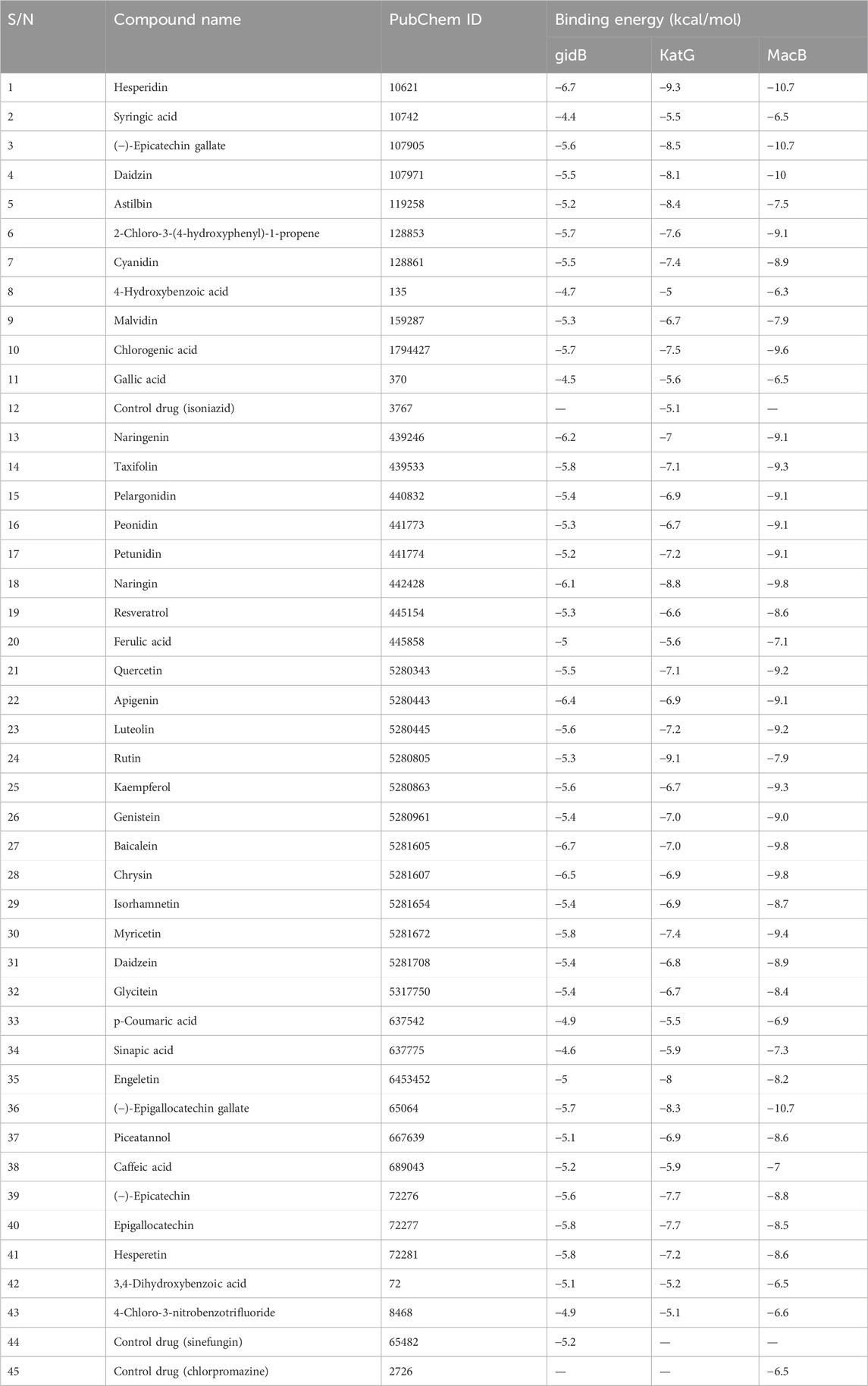
Table 6. Binding energies (kcal/mol) of compounds docked against gidB, KatG, and MacB of E. coli using AutoDock Vina.
Given the BEs observed in Table 6, there is a need to further probe the mechanisms involved in the binding of these compounds with the target proteins. These indicate the relevant relationships between the protein–compound complexes of these compounds. From a recent work, hydrogen bonds and hydrophobic interactions were majorly observed to occur between the atoms of the compounds and side chains of the amino acid residues occupying the binding sites in KatG, MacB, and gidB of E. coli. These details are provided in Figures 4–6.
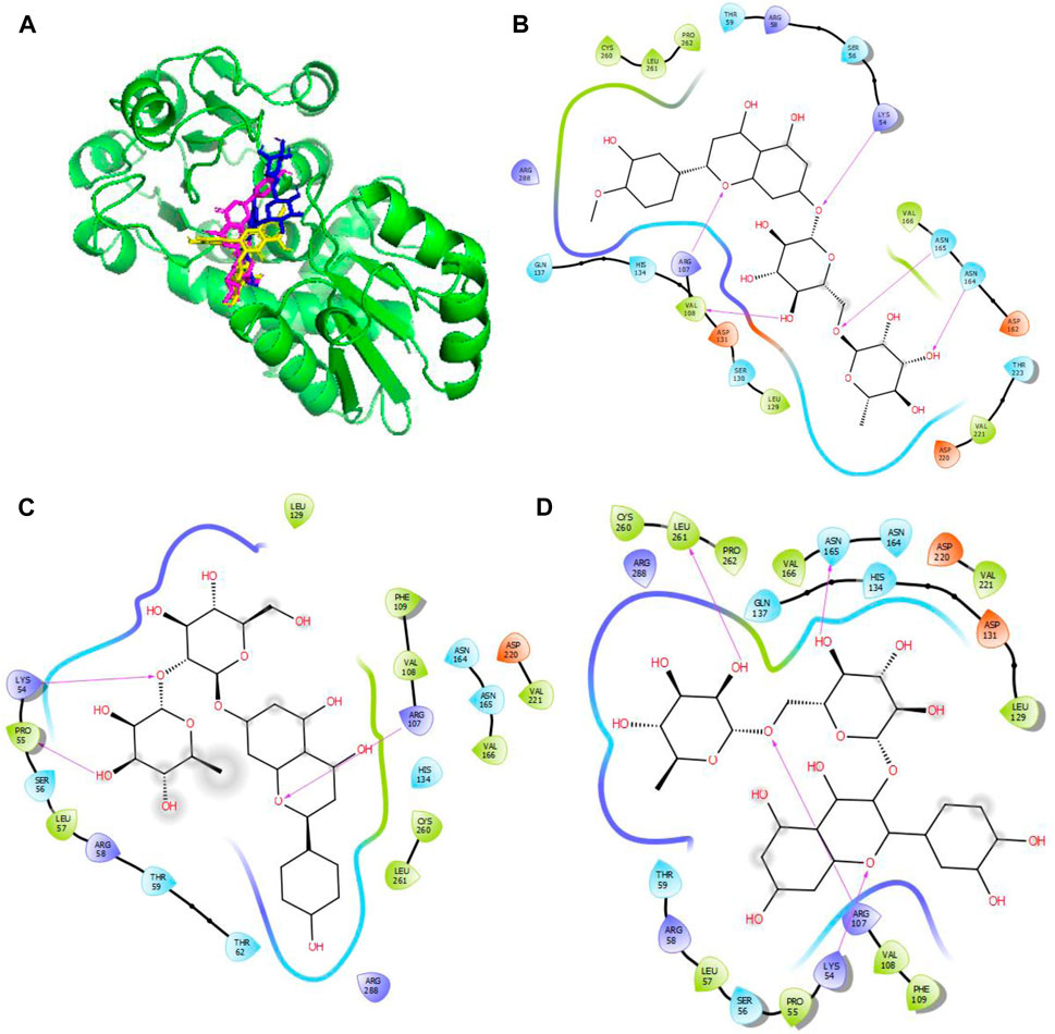
Figure 4. Molecular docking of the bioactive compounds in KatG of E. coli. (A) 3D binding positions of KatG (green), hesperidin (blue), naringin (purple), and rutin (yellow) in the binding pockets of KatG of E. coli. 2D molecular interaction analyses of (B) hesperidin, (C) naringin, and (D) rutin with the amino acid residues in the binding pockets of KatG of E. coli.
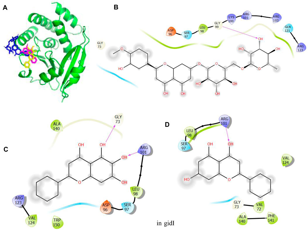
Figure 5. Molecular docking of the bioactive compounds in gidB of E. coli. (A) 3D binding positions of gidB (green), hesperidin (blue), chrysin (purple), and baicalein (yellow) in the binding pockets of gidB of E. coli. 2D molecular interaction analyses of (B) hesperidin, (C) baicalein, and (D) chrysin with the amino acid residues in the binding pockets of gidB of E. coli.
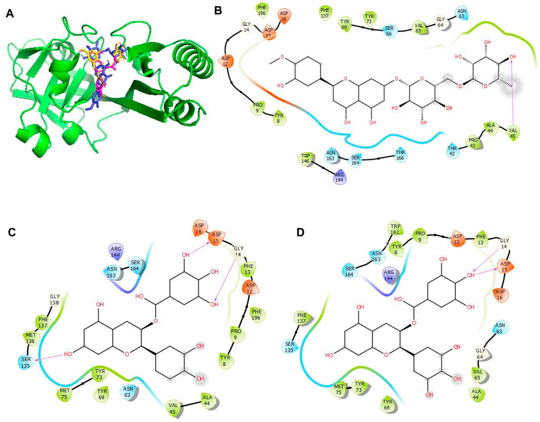
Figure 6. Molecular docking of the bioactive compounds in MacB of E. coli. (A) 3D binding positions of MacB (green), hesperidin (blue), epigallocatechin gallate (purple), and epicatechin gallate (yellow) in the binding pockets of MacB of E. coli. 2D molecular interaction analyses of (B) hesperidin, (C) epicatechin gallate, and (D) epigallocatechin gallate with the amino acid residues in the binding pockets of MacB of E. coli.
In Figure 4, the binding positions show that the three compounds occupy the same areas at the binding sites of KatG. It was observed that hydrogen bonds were established between the three compounds as well as Lys54 and Arg107. Aside from these two amino acid residues, Val108, Asn165, and Asn164 were found to link with hesperidin via hydrogen bonding, while Leu261 and Asn165 were involved with rutin via hydrogen bonding. Finally, Pro55 was the only extra amino acid to interact with naringin via hydrogen bonds. It was also observed that all three compounds interacted with Cys260, Thr59, Arg58, Ser56, Arg288, His134, Leu129, Asp220, Val221, and Val166 via hydrophobic interactions. Interestingly, four out of the six amino acids (Arg107, Asn164, Leu261, and Ans165) that interacted with the chosen compounds via hydrogen bonds were earlier predicted to be members of the binding site residues. Moreover, nine out of ten amino acids that interacted with the compounds were among the predicted amino acids of the binding sites.
In Figure 5, the binding positions show that the three compounds occupy the same areas at the binding sites of gidB. It was observed that hydrogen bonds were established between the atoms of biacalein as well as Gly73 and Arg101, between chrysin and Arg101, and between hesperidin and gly99. In addition, all three compounds interacted with Ala140, Leu98, Ser97, Asp96, Trp150, Val124, Arg123, Val72, Gly73, Phe141, Gln121, and Arg103 via hydrophobic interactions. Interestingly, Gly73 and Arg101 that interacted with the chosen compounds via hydrogen bonds were earlier predicted to be members of the binding site residues, while Asp96, Gly73, Arg123, Leu98, and Ser97 that interacted with the compounds via hydrophobic interaction were among the predicted amino acids of the binding sites.
In Figure 6, the binding positions show that the three compounds occupy the same areas at the binding sites of MacB. It was observed that hydrogen bonds were established between the atoms of epicatechin gallate as well as Ser135, Gly14, and Asp15; between epigallocatechin gallate as well as Gly14 and Asp15; and finally between hesperidin and Val45. In addition, all three compounds interacted with Asp12, Arg144, Asn163, Ser164, Tyr8, Pro9, Phe137, Tyr73, Tyr69, Asn63, and Ala44 via hydrophobic interactions. Interestingly, Asp15, Gly14, Ser135, and Val45 that interacted with the chosen compounds via hydrogen bonds were earlier predicted to be among the binding site residues, while Asp12, Ser164, Tyr8, Pro9, Phe137, Ala44, Tyr69, Asn63, and Ala44 that interacted with the compounds via hydrophobic interactions were among the predicted amino acids of the binding sites.
The ADMET predictions were carried out to ascertain how the three chosen compounds for each of the proteins were absorbed, distributed, metabolized, and eliminated as well as how toxic they can be. The outcomes of the evaluations are presented in Table 7. Hesperidin, epicatechin gallate, chrysin, and naringin showed good ADMET profiles and do not tend to cause mutations against Salmonella typhenurium (AMES). Epigallocatechin gallate and rutin tend to cause mutations against S. typhenurium (AMES). Rutin was observed to be a non-inhibitor of the human ether-a-go-go (herG) gene, while the other compounds were inhibitors to the human herG gene. Correspondingly, all these compounds did not display the ability to cross the blood–brain barrier (BBB) and may not be absorbed through caco-2. They tend to have good intestinal absorptions except for hesperidin and naringin. None of the compounds were predicted to serve as substrates to P-glycoprotein, and all the compounds were found to be substrates to cytochrome P450 isoform 3A4 except biacalein and chrysin.
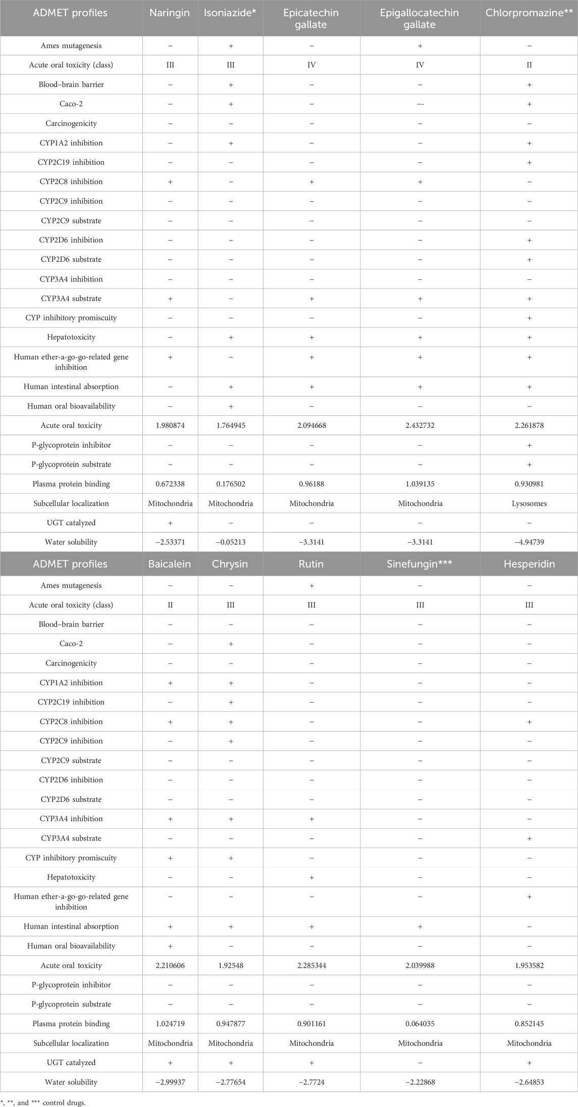
Table 7. ADMET properties of the three best compounds selected from compounds docked against the three proteins implicated in antibiotic resistance in E. coli using ADMETSar server.
The toxicities of the chosen compounds ranged between classes II to IV. In addition, they were predicted to be localized in the mitochondria. Biacalein, chrysin, hesperidin, and naringin were predicted to undergo phase-two drug metabolisms through glucorunidation. However, studying their structures keenly for the purpose of generating chemosimilars with improved safety profiles may be of great relevance and should be considered as a subject of future investigation.
Hesperidin (hesperetin-7-O-rutinoside), which shows great affinity across the three protein targets, is a flavone made up of the rhamnose, glucose, and hesperetin aglycone moieties. It is abundantly available in citrus plants and is majorly produced in the citrus industry (Rudrapal et al., 2023). According to recent preclinical and clinical studies on its biological usage as an active element, it possesses antioxidant, anti-inflammatory, lipid-lowering, and insulin sensitivity properties as well as potency in neurological disorders, psychiatric disorders, and cardiovascular diseases because of its effects on the blood pressure (Atoki et al., 2023; Hosawi, 2023; Ileriturk et al., 2023).
Hesperidin was found to have good binding abilities against the three selected protein targets in this work. Therefore, a unique study engaging MD simulation was undertaken for the three complex systems to delve into the structural and dynamic behaviors of gidB, KatG, and MacB in the presence of hesperidin. The study focused on investigating the intrinsic dynamic stability of each complex by analyzing their backbone RMSD (Figure 7) and RMSF (Figure 8) in 200 ns simulations.
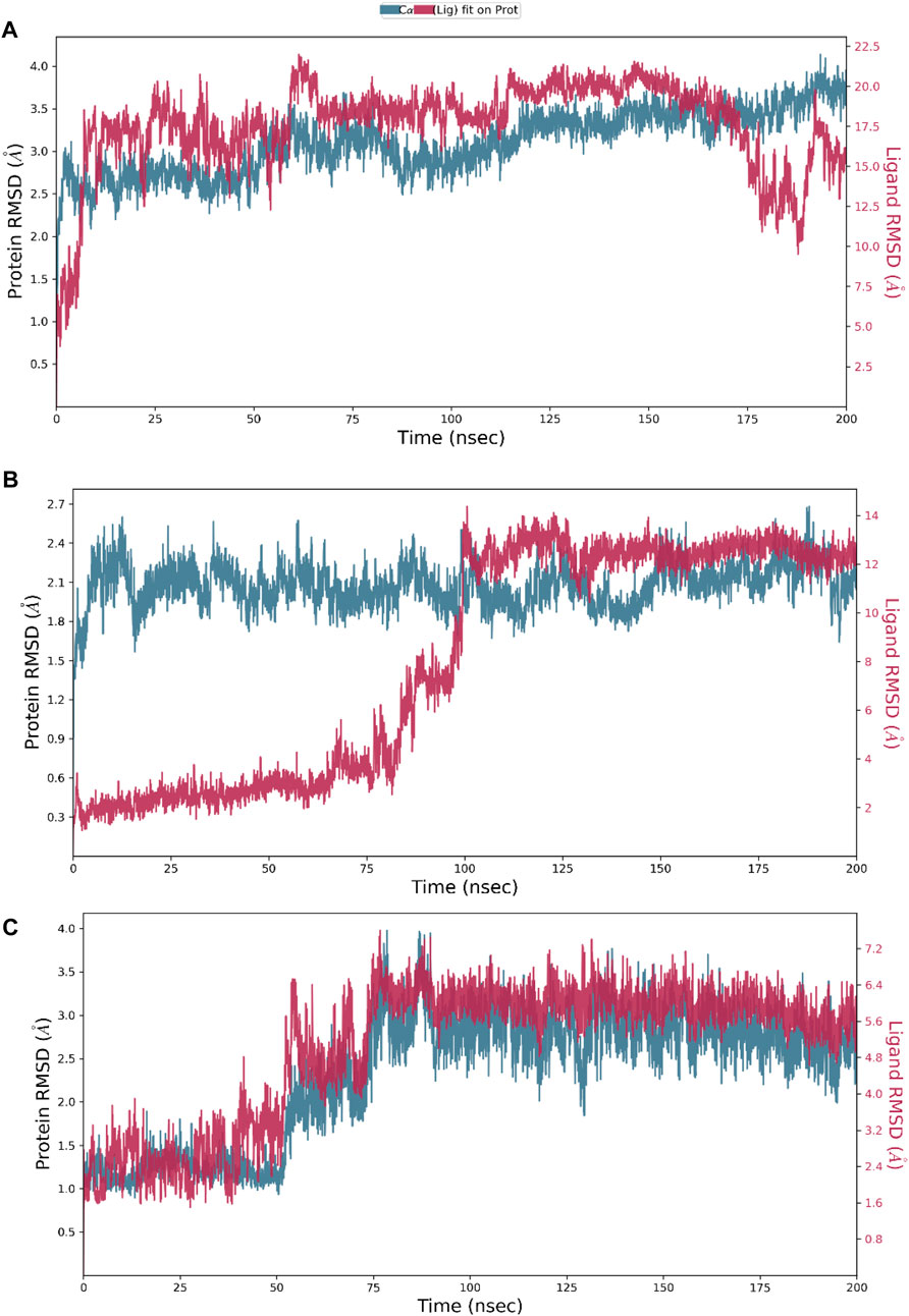
Figure 7. Dynamic stability metrics of hesperidin-bound protein complexes over 200 ns of molecular dynamics (MD) simulations. Time-dependent RMSD profiles of the backbone atoms of (A) hesperidin–gidB, (B) hesperidin–KatG, and (C) hesperidin–MacB complexes.
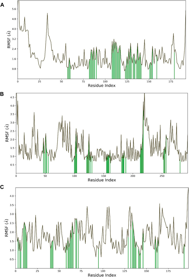
Figure 8. Dynamic stability metrics of hesperidin-bound protein complexes over 200 ns of MD simulations. Cα-RMSF profiles of the (A) hesperidin–gidB, (B) hesperidin–KatG, and (C) hesperidin–MacB complexes. The green lines indicate the points of contact of hesperidin with the amino acids during the MD runs.
The RMSD serves as a measure of variance between a protein backbone from its starting to final conformations and offers beneficial insights into the stability of the protein–ligand complex (Cavasotto et al., 2019; Solo-Aben et al., 2021; Danazumi and Umar, 2023). A plot of divergences observed during the simulations was used to measure the relative stabilities, whereby more negligible deviations indicate more stable protein structures (Hollingsworth and Dror, 2018; Tran et al., 2022). The RMSD values for the backbones were calculated via 200 ns simulations to establish the stabilities of the three systems. At the early stage of the simulation, the hesperidin–gidB complex (Figure 7A) exhibited an average RMSD (indicated in red) of 3.5 Å from 0 ns to 60 ns. From 100 ns to 175 ns, equilibrium was reached, as shown in the trajectory plot, followed by a gradual decline, reaching an average RMSD peak of 2.0 Å at 180 ns. Subsequently, the curve rises to 3.5 Å at 190 ns and is stabilized for the next 10 ns before declining to 2.4 Å and remaining stable until the end of the simulation period. A keen inspection of the gidB complex system revealed a moderately stable trend in RMSD, showcasing an average deviation of approximately <3.5 Å (Singharoy et al., 2016; Zuo et al., 2017). Such profiles demonstrate the inherent stability of the system investigated herein.
However, exploring the RMSD of the hesperidin–KatG complex (Figure 7B) yielded intriguing findings, where the ligand had notable changes in RMSD throughout the simulation, thereby highlighting a significant orientation within the binding pocket of KatG. The hesperidin-bound complex (indicated in red) exhibited an initial increment in RMSD up to ∼0.6 Å during the early stage of the simulation and lasting until around 75 ns, with an unusual rise at around 60 ns. Thereafter, it gradually increased to 2.65 Å at 110 ns, which dipped to 2.1 Å at 120 ns before rising above 2.4 Å at 125 ns. A dip was then observed at around 130 ns to 1.8 Å, after which the curve remained stable till the end of the simulation. However, a notable observation in the hesperidin–MacB complex was the occurrence of fluctuations in the RMSD between 0 ns and 80 ns, characterized by few crests and troughs, indicating substantial conformational changes within the hesperidin-bound complex during this timeframe.
These fluctuations suggest the transient destabilization and restabilization of the complex, possibly due to dynamic interactions between hesperidin and the surrounding amino acid residues. Following this period of fluctuations, the RMSD stabilized at around 3.0 Å and remained relatively stable until the end of the 200 ns simulation. This remarkable stability highlights the robust and consistent natures of the complexes.
To investigate the flexibility and local changes in the secondary structural elements involved in hesperidin binding, the residual fluctuations of the three complexes were examined carefully by mainly focusing on the Cα motions (Figure 8). The Cα RMSFs of the three complexes displayed noteworthy fluctuations in the residues situated at the N-terminal and C-terminal regions, corroborating the findings obtained from the RMSD analysis (Figure 7). Interestingly, the ligand-bound structures exhibited lower fluctuations (Figure 8) at the points of contact (green lines) with the amino acid residues of the proteins, with an average RMSF of 2.4 Å for hesperidin–gidB and 2.0 Å for hesperidin–KatG even when the fluctuations around the residues between 210 ns and 220 ns had an RMSF value of above 4.0 Å.
In the hesperidin–MacB complex (Figure 8C), the fluctuations averaged between 2.5 Å and 3.0 Å around the points of contact of hesperidin and the amino acid residues of MacB. The observed changes in the Cα-RMSF values are probably a consequence of the stabilizing effects prompted by hesperidin binding to the protein’s flexible regions, emphasizing the inherent adaptability of the proteins (Dokainish et al., 2022; Ali et al., 2023). All three systems displayed steady RMSF trends with marginal distinctions, confirming cohesive behaviors under dissimilar conditions.
The model trajectories were used to measure hesperidin’s binding affinity for identifying the stabilities of the three protein targets against it. The resulting structural chemistries witnessed during these modeling runs are depicted graphically in Figures 9, 10. These figures exemplify several interactions, such as hydrogen bonds in green, hydrophobic interactions in gray, ionic interactions in magenta, and water bridges in blue. The stacked bars (Figure 5) artfully represent the proportion of simulation time dedicated to maintaining each definite interaction.
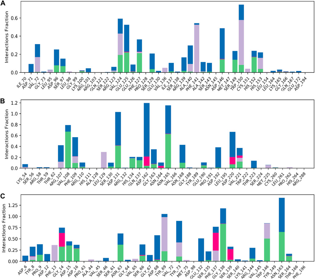
Figure 9. Normalized stacked bar charts representing the contact mappings of hesperidin with (A) gidB, (B) KatG, and (C) MacB. The green, gray, blue, and pink colors represent hydrogen bonds, hydrophobic interactions, water bridges, and ionic interactions, respectively.
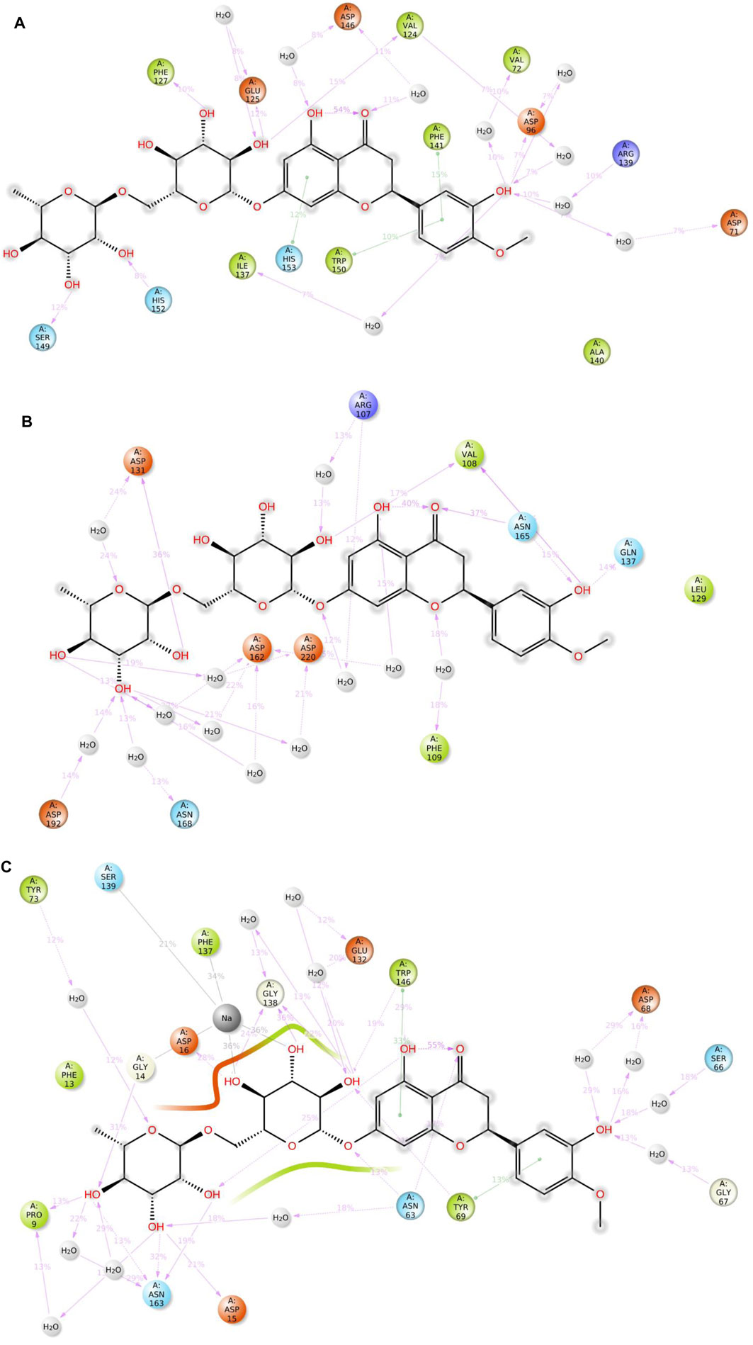
Figure 10. 2D summary diagrams showcasing the preserved contacts from 200 ns of MD simulations between hesperidin and the E. coli proteins (A) gidB, (B) KatG, and (C) MacB.
For example, a value of 0.6 would indicate that 60% of the simulation time involved maintaining a particular interaction. In some cases, values exceeding 1.0 were acceptable, signifying the manifestation of multiple contacts between a ligand and the same protein subclass due to conformational changes in the protein structure. Furthermore, 2D interaction charts for the hesperidin and three target protein complexes are shown in Figure 10, furnishing insights into the preservation of contacts over the entire simulation trajectory.
It is noted that both the Vina-based docking (Figures 4–6) and MD simulations (Figures 9, 10) were able to uncover significant H-bond interactions between hesperidin and the active site residues of the three protein targets, which may likely indicate the correctness of compound docking within the binding pockets of the target proteins. This is an interesting outcome that hesperidin may likely be a good inhibitor of these proteins and consequently prevent the spread of AMR in E. coli. Post-simulation in hesperidin–gidB, most of the OH groups of hesperidin were found to form five water-mediated hydrogen bond contacts with six residues: Asp96, Asp146, Glu125, Val124, Val72, and Arg139. Interestingly, these interactions occurred in 7%–31% of the frames throughout the simulation trajectory. Furthermore, three residues were observed to contact hesperidin via hydrogen bonds without mediation by water: His152, Phe127, and Ser149. Finally, His153 and Trp150 showed pi-pi interactions with rings A and C of hesperidin (Figure 10).
As in gidB, the hesperidin-bound KatG had many H-bond formations during the docking event despite being ranked second in terms of affinity among the chosen natural compounds (Figure 4). However, the H-bond interactions were mostly maintained between 13% and 40% during the MD simulations (Figure 10B), which may be due to conformational changes in the protein and ligand [47–48]. Such molecular changes were attributed to the moderate fitness of hesperidin in the binding pockets. However, the complex exhibited six water-mediated interactions, all involving the beta-oriented OH: four with the positively charged Asp192, Asp131, Asp162, and Asp220 residues as well as two with Phe109 and Arg107. Some additional residues had interactions with the OH groups of hesperidin without water mediation, namely Gln137, Asn165, and Val108. These contacts were maintained between 13% and 40% of the MD timescale (Figure 10B), signifying their modest stabilities.
Finally, the hesperidin-bound MacB had few H-bond formations during the docking event despite being ranked best in terms of affinity among the chosen natural compounds (Figure 6). However, the H-bond interactions were mostly maintained between 12% and 36% during the MD simulations (Figure 10C), which may be attributed to positional variations in MacB and the ligand (Girdhar et al., 2019; Redij et al., 2019). Such molecular changes were ascribed to the reasonable fitness of hesperidin in the binding pockets. However, the complex exhibited ten water-mediated interactions, all involving the beta-oriented OH: three with the positively charged Asp16, Glu132, and Asp68 residues, with the remaining residues being Ser66, Gly67, Asn63, Asn163, Tyr73, Pro9, and Phe137. Some additional residues had interactions with the OH groups of hesperidin without water mediation, namely Asp15, Gly14, and Trp146. In addition, Trp146 and Tyr69 were observed to interact with rings A and C of hesperidin during the MD simulations (Figure 10C), signifying their modest stabilities as a result of the H bonds and pi-pi interactions.
PCA was implemented to meticulously measure the routes of the protein–ligand complexes during MD simulations. The goals of this inquiry were to attempt to make sense of the seemingly random, global atomic displacements observed within the amino acid residues (Hayward, 2023; Vuillemot et al., 2023). The protein’s layout includes stochasticity and globally uncorrelated motions, contributing to the diversity and adaptability of the trajectories (Alghamdi et al., 2023; Debnath et al., 2023). The covariance matrix records the movements of the internal coordinates over 200 ns, as projected onto a 3D space. With this information, orthogonal systems or eigenvectors can be used to better comprehend the predictable motions inside each trajectory. The Cα atoms of the hesperidin-bound gidB (Figure 11A) demonstrated a disordered configuration in the MD simulation trajectories associated with the PC1 and PC2 modes in the early and last frames.
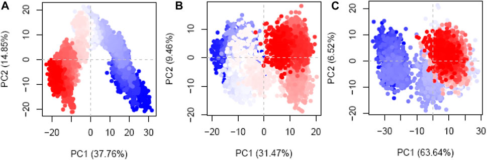
Figure 11. Principal component analysis (PCA) plots of the Cα atoms disclosing the first two eigenvectors in the conformational spaces of three different systems: (A) hesperidin–gidB, (B) hesperidin–KatG, and (C) hesperidin–MacB complexes.
In contrast, a more systematic arrangement emerged between the frames (highlighted in light to white), suggesting stable global correlated motions. In the case of KatG–hesperidin (Figure 11B), initial anisotropic motion was observed, which is attributed to the high mobility of the protein’s terminal regions in various directions; however, these motions became more ordered with time. Meanwhile, the Cα atoms of MacB linked to hesperidin (Figure 11C) exhibited more disordered configurations with moderate orientations in the PC1 and PC2 modes. This clustering of frames indicates cyclical motion, as shown in the MD trajectories, which results from the robust global conformational movements.
To clarify the complex, symbiotic conformational dynamics within the three ligand-bound protein complexes expressed across spatially divergent protein domains in different systems, a meticulous and sophisticated DCC analysis was employed (Zhang et al., 2022; Zhu et al., 2022). It involved averaging over three replicates of precisely curated MD trajectories for each system. The cross-correlation (Cij) coefficients, which are a display of mathematical sophistication, gracefully traversed the entire range from −1 (purple) to 1 (blue), reflecting the exquisitely collinear correlation between the two Cα atoms (i and j). Here, positive coefficients (Cij > 0) indicate harmonious and synchronized movements of the two residues, whereas negative coefficients (Cij < 0) suggest that the two residues move in opposite directions. As the coefficients approach zero (Cij = 0), the correlated motions between the two residues reach a standstill, indicating no noticeable correlation. Throughout the analysis, the absolute values of Cij highlight the intensity of the correlated motions within the complexes, providing insights into the magnitudes of the molecular movements (Zhang et al., 2022; Zhu et al., 2022).
The Cij matrices, represented as two-by-two plots of the Cα Cij coefficients, revealed near-similar diagonal patterns of correlated and anti-correlated motions within the ligand-bound protein systems as well as interdomain motions between these systems (Figure 12A–C). Across all three systems, the intradomain movements of the three proteins showed enhanced correlated motions (visually depicted as diagonal blue lines), indicating concerted motions within these regions.
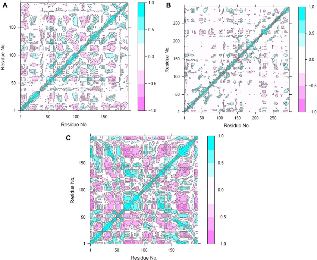
Figure 12. Dynamic cross-correlation maps (DCCMs) of the (A) hesperidin–gidB, (B) hesperidin–KatG, and (C) hesperidin–MacB complexes.
After a meticulous appraisal of the configurational space explored via simulations, the FEL representation was constructed in contours. The FEL is a valuable model that involves mapping the free energy values to the configurational space, enabling identification of energetically favorable regions and conversion blockades to the system during its conformational changes or evolutions between different states (Fang et al., 2022). In this investigation, the FEL was calculated via RMSD and Rg values as the reaction coordinates for evaluating relevant clusters of the three systems of hesperidin (Figure 13). A noteworthy finding was the significant divergence in the free-energy profiles of the ligand-bound complexes (Figures 13A–C). The hesperidin-bound gidB showed one main energy basin with similar free-energy values of 0 kJ/mol and an extended spread along the Rg value from 1.64 nm to 1.74 nm (Figure 13A), whereas the hesperidin-bound KatG displayed only a single and sharp global energy minimum (Figure 13B).
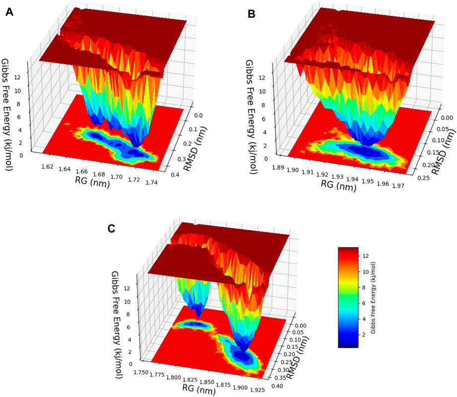
Figure 13. 3D free-energy landscape (FEL) contour plots of the (A) hesperidin–gidB, (B) hesperidin–KatG, and (C) hesperidin–MacB clusters with respect to the RSMD and Rg.
The hesperidin-bound MacB showed two main energy basins (Figure 13C). Such deviations suggest that the binding of hesperidin induces noteworthy changes in the energetic landscapes of the three protein molecules, stabilizing them in preferred conformations during the interactions. The sizes and shapes of the minimal energy areas, represented by dark blue regions on the free-energy contour plots, provide crucial insights into the enhanced stabilities of the complexes. The presence of smaller, more centralized blue area(s) resembling funnel-like bottoms indicate more excellent stabilities of the corresponding complexes. This funnel-like shape suggests that the ligand-bound complex adopts a preferred conformation with minimal energy, making it highly stable and less susceptible to significant conformational changes (Sang et al., 2020; Baidya et al., 2022).
In principle, the binding with hesperidin stretches the protein and results in a change of the overall motions of the protein. Convincingly, applying PCA in the current work sheds light on the dynamic properties of the gidB, KatG, and MacB proteins as well as how they are influenced by ligand binding. The results suggest that hesperidin interactions are vital in shaping the conformational landscapes and firmness of the proteins, possibly impacting their functional behaviors in cellular processes.
Hesperidin behaviors noted in this work are not accidental as they have been reported to have significant bioactivities that can combat bacterial and other microorganisms to enhance immune functions (Alam et al., 2023; Rudrapal et al., 2023). Furthermore, hesperidin can disrupt the cell walls of bacteria and cause leakage of biological macromolecules, such as proteins and DNA, by generating reactive oxygen species (Bahador and Vaezi, 2023). Regarding antimicrobial activity, hesperidin has been known to exhibit robust effects against countless microorganisms, including bacteria, viruses, and fungi; its mechanisms of action involve the disruption of microbial cell membranes and inhibition of microbial enzymes (Akbari et al., 2023; Bahador and Vaezi, 2023; Hosawi, 2023). These findings suggest that hesperidin could be a potential natural antimicrobial agent for the prevention and treatment of microbial infections.
This study aimed to contribute to the ongoing quest for better therapeutics that can circumvent the AMR of E. coli by focusing on its MacB, gidB, and KatG proteins by identifying potential inhibitors from compounds attributed to plants with established antibacterial activities and utilizing a comprehensive approach involving resistance gene identification, molecular docking, MD simulations, and ADMET analysis. Remarkably, our findings reveal that hesperidin exhibits good binding affinities with MacB, KatG, and gidB, with BEs of −10.7 kcal/mol, −9.3 kcal/mol, and −6.7 kcal/mol, respectively, compared to their respective control drugs. Notably, hesperidin demonstrates plausible binding positions, good structural stability, and favorable pharmacokinetic profile. These findings suggest that hesperidin holds promise as a potential therapeutic agent for AMR associated with the MacB, gidB, and KatG genes of E. coli.
However, it is vital to consider the preliminary nature of these in silico findings, so further experimental studies are imperative for validating the efficacy of hesperidin as a viable treatment option for AMR associated with the MacB, gidB, and KatG genes of E. coli. These future investigations are necessary for valuable insights into bridging the gap between computational predictions and practical therapeutic applications, thereby paving the path for more effective and targeted treatments.
The datasets presented in this study can be found in online repositories. The names of the repository/repositories and accession number(s) can be found in the article/Supplementary Material.
ATA: Conceptualization, Data curation, Formal analysis, Investigation, Supervision, Validation, Writing–original draft, Writing–review and editing. NK: Data curation, Formal analysis, Methodology, Project administration, Resources, Writing–original draft, Writing–review and editing. CO: Data curation, Investigation, Methodology, Resources, Software, Writing–original draft, Writing–review and editing. TI: Data curation, Investigation, Resources, Validation, Visualization, Writing–original draft, Writing–review and editing. IsA: Investigation, Methodology, Resources, Software, Validation, Writing–original draft, Writing–review and editing. HU: Investigation, Methodology, Resources, Writing–original draft, Writing–review and editing. AJ: Investigation, Resources, Validation, Writing–original draft, Writing–review and editing. BB: Data curation, Investigation, Resources, Software, Writing–original draft, Writing–review and editing. ZO: Resources, Software, Supervision, Validation, Writing–review and editing. AI: Resources, Writing–review and editing, Formal analysis, Investigation. IbA: Formal analysis, Methodology, Resources, Software, Writing–review and editing. IO: Validation, Visualization, Writing–review and editing. BA: Methodology, Resources, Supervision, Writing–review and editing. MA: Supervision, Validation, Writing–review and editing. OI: Resources, Validation, Visualization, Writing–review and editing. AAA: Funding acquisition, Supervision, Validation, Writing–review and editing. RA: Project administration, Supervision, Validation, Visualization, Writing–review and editing.
The author(s) declare that no financial support was received for the research, authorship, and/or publication of this article.
The authors declare that the research was conducted in the absence of any commercial or financial relationships that could be construed as a potential conflict of interest.
All claims expressed in this article are solely those of the authors and do not necessarily represent those of their affiliated organizations or those of the publisher, editors, and reviewers. Any product that may be evaluated in this article or claim that may be made by its manufacturer is not guaranteed or endorsed by the publisher.
Akbari, S., Assaran Darban, R., Javid, H., Esparham, A., and Hashemy, S. I. (2023). The anti-tumoral role of Hesperidin and Aprepitant on prostate cancer cells through redox modifications. Naunyn-Schmiedeberg’s Archives Pharmacol. 396 (12), 3559–3567. doi:10.1007/s00210-023-02551-0
Alam, P., Imran, M., Jahan, S., Akhtar, A., and Hasan, Z. (2023). Formulation and characterization of hesperidin-loaded transethosomal gel for dermal delivery to enhance antibacterial activity: comprehension of in vitro, ex vivo, and dermatokinetic analysis. Gels 9 (10), 791. doi:10.3390/gels9100791
Alghamdi, A., Abouzied, A. S., Alamri, A., Anwar, S., Ansari, M., Khadra, I., et al. (2023). Synthesis, molecular docking, and dynamic simulation targeting main protease (Mpro) of new, thiazole clubbed pyridine scaffolds as potential COVID-19 inhibitors. Curr. Issues Mol. Biol. 45 (2), 1422–1442. doi:10.3390/cimb45020093
Ali, S., Ali, U., Qamar, A., Zafar, I., Yaqoob, M., Ain, Q., et al. (2023). Predicting the effects of rare genetic variants on oncogenic signaling pathways: a computational analysis of HRAS protein function. Front. Chem. 11, 1173624. doi:10.3389/fchem.2023.1173624
Atoki, A. V., Aja, P. M., Shinkafi, T. S., Ondari, E. N., and Awuchi, C. G. (2023). Hesperidin plays beneficial roles in disorders associated with the central nervous system: a review. Int. J. Food Prop. 26 (1), 1867–1884. doi:10.1080/10942912.2023.2236327
Bahador, A., and Vaezi, M. (2023). Novel formulations of hesperidin for antitumor, antimicrobial, and neuroprotective effects: a review. Micro Nano Bio Asp. 2 (2), 13–19. doi:10.22034/mnba.2023.394932.1030
Baidya, A. T. K., Kumar, A., Kumar, R., and Darreh-Shori, T. (2022). Allosteric binding sites of Aβ peptides on the acetylcholine synthesizing enzyme ChAT as deduced by in silico molecular modeling. Int. J. Mol. Sci. 23 (11), 6073. doi:10.3390/ijms23116073
Bakan, A., Meireles, L. M., and Bahar, I. (2011). ProDy: protein dynamics inferred from theory and experiments. Bioinformatics 27 (11), 1575–1577. doi:10.1093/bioinformatics/btr168
Bortolaia, V., Kaas, R. S., Ruppe, E., Roberts, M. C., Schwarz, S., Cattoir, V., et al. (2020). ResFinder 4.0 for predictions of phenotypes from genotypes. J. Antimicrob. Chemother. 75 (12), 3491–3500. doi:10.1093/jac/dkaa345
Brettin, T., Davis, J. J., Disz, T., Edwards, R. A., Gerdes, S., Olsen, G. J., et al. (2015). RASTtk: a modular and extensible implementation of the RAST algorithm for building custom annotation pipelines and annotating batches of genomes. Sci. Rep. 5 (1), 8365. doi:10.1038/srep08365
Carattoli, A. (2013). Plasmids and the spread of resistance. Int. J. Med. Microbiol. 303 (6-7), 298–304. doi:10.1016/j.ijmm.2013.02.001
Carattoli, A., Zankari, E., García-Fernández, A., Voldby Larsen, M., Lund, O., Villa, L., et al. (2014). In silico detection and typing of plasmids using PlasmidFinder and plasmid multilocus sequence typing. Antimicrob. Agents Chemother. 58 (7), 3895–3903. doi:10.1128/AAC.02412-14
Cavasotto, C. N., Aucar, M. G., and Adler, N. S. (2019). Computational chemistry in drug lead discovery and design. Int. J. Quantum Chem. 119 (2). doi:10.1002/qua.25678
Chen, X., Pan, S., Li, F., Xu, X., and Xing, H. (2022). Plant-derived bioactive compounds and potential health benefits: involvement of the gut microbiota and its metabolic activity. Biomolecules 12, 1871–1915. doi:10.3390/biom12121871
Cheng, F., Li, W., Zhou, Y., Shen, J., Wu, Z., Liu, G., et al. (2012). admetSAR: a comprehensive source and free tool for assessment of chemical ADMET properties. J. Chem. Inf. Model 52, 3099–3105. doi:10.1021/ci300367a
Dallakyan, S., and Olson, A. J. (2015). Small-molecule library screening by docking with PyRx. Methods Mol. Biol. 1263, 243–250. doi:10.1007/978-1-4939-2269-7_19
Danazumi, A. U., and Umar, H. I. (2023). You must be flexible enough to be trained, Mr. Dynamics simulator. Mol. Divers., 5–7. doi:10.1007/s11030-023-10689-5
Debnath, S., Kant, A., Bhowmick, P., Malakar, A., Purkaystha, S., Jena, B. K., et al. (2023). The enhanced affinity of WRKY reinforces drought tolerance in Solanum lycopersicum L.: an innovative bioinformatics study. Plants 12 (4), 762. doi:10.3390/plants12040762
Didelot, X., Méric, G., Falush, D., and Darling, A. E. (2012). Impact of homologous and non-homologous recombination in the genomic evolution of Escherichia coli. BMC Genomics 13, 256. doi:10.1186/1471-2164-13-256
Dimitrijevi, S., Dimitrijević-Branković, S., and Rajilić-Stojanović, M. (2021). Plant extracts rich in polyphenols as potent modulators in the growth of probiotic and pathogenic intestinal microorganisms. Front. Nutr. 8, 1–11. doi:10.3389/fnut.2021.688843
Dokainish, H. M., Re, S., Mori, T., Kobayashi, C., Jung, J., and Sugita, Y. (2022). The inherent flexibility of receptor binding domains in SARS-CoV-2 spike protein. ELife 11, e75720. doi:10.7554/eLife.75720
Fang, R., Hon, J., Zhou, M., and Lu, Y. (2022). An empirical energy landscape reveals mechanism of proteasome in polypeptide translocation. ELife 11, e71911. doi:10.7554/eLife.71911
Felipe, F. L., Rivas, B., and Muñoz, R. (2014). Bioactive compounds produced by gut microbial tannase: implications for colorectal cancer development. Front. Microbiol. 5, 1–4. doi:10.3389/fmicb.2014.00684
Girdhar, K., Dehury, B., Kumar Singh, M., Daniel, V. P., Choubey, A., Dogra, S., et al. (2019). Novel insights into the dynamics behavior of glucagon-like peptide-1 receptor with its small molecule agonists. J. Biomol. Struct. Dyn. 37 (15), 3976–3986. doi:10.1080/07391102.2018.1532818
Grant, B. J., Skjærven, L., and Yao, X. Q. (2021). The Bio3D packages for structural bioinformatics. Protein Sci. 30 (1), 20–30. doi:10.1002/pro.3923
Halgren, T. A. (1996). Merck molecular force field. I. Basis, form, scope, parameterization, and performance of MMFF94. J. Comput. Chem. 17, 490–519. doi:10.1002/(sici)1096-987x(199604)17:5/6<490::aid-jcc1>3.0.co;2-p
Hayward, S. (2023). A Retrospective on the development of methods for the analysis of protein conformational ensembles. Protein J. 42 (3), 181–191. doi:10.1007/s10930-023-10113-9
Hollingsworth, S. A., and Dror, R. O. (2018). Molecular dynamics simulation for all. Neuron 99 (6), 1129–1143. doi:10.1016/j.neuron.2018.08.011
Hosawi, S. (2023). Current update on role of hesperidin in inflammatory lung diseases: chemistry, pharmacology, and drug delivery approaches. Life 13 (4), 937. doi:10.3390/life13040937
Ileriturk, M., Kandemir, O., Akaras, N., Simsek, H., Genc, A., and Kandemir, F. M. (2023). Hesperidin has a protective effect on paclitaxel-induced testicular toxicity through regulating oxidative stress, apoptosis, inflammation and endoplasmic reticulum stress. Reprod. Toxicol. 118, 108369. doi:10.1016/j.reprotox.2023.108369
Kagami, L. P., das Neves, G. M., Timmers, LFSM, Caceres, R. A., and Eifler-Lima, V. L. (2020). Geo-Measures: a PyMOL plugin for protein structure ensembles analysis. Comput. Biol. Chem. 87, 107322–107420. doi:10.1016/j.compbiolchem.2020.107322
Lázár, V., Pal Singh, G., Spohn, R., Nagy, I., Horváth, B., Hrtyan, M., et al. (2013). Bacterial evolution of antibiotic hypersensitivity. Mol. Syst. Biol. 9, 700. doi:10.1038/msb.2013.57
Pettersen, E. F., Goddard, T. D., Huang, C. C., Couch, G. S., Greenblatt, D. M., Meng, E. C., et al. (2004). UCSF chimera-A visualization system for exploratory research and analysis. J. Comput. Chem. 25, 1605–1612. doi:10.1002/jcc.20084
Redij, T., Chaudhari, R., Li, Z., Hua, X., and Li, Z. (2019). Structural modeling and in silico screening of potential small-molecule allosteric agonists of a glucagon-like peptide 1 receptor. ACS Omega 4 (1), 961–970. doi:10.1021/acsomega.8b03052
Roemer, T., et al. (2017). Systems chemical biology and the antibiotic resistome. J. Antibiot. (Tokyo) 70 (4), 401–407. doi:10.1038/ja.2017.17
Rudrapal, M., Sarkar, B., Deb, P., Bendale, A. R., and Nagar, A. (2023). “Addressing antimicrobial resistance by repurposing polyphenolic phytochemicals with novel antibacterial potential,” in Polyphenols. Editor M. Rudrapal 1st ed. (Wiley), 260–289. doi:10.1002/9781394188864.ch13
Sang, P., Liu, S. Q., and Yang, L. Q. (2020). New insight into mechanisms of protein adaptation to high temperatures: a comparative molecular dynamics simulation study of thermophilic and mesophilic subtilisin-like serine proteases. Int. J. Mol. Sci. 21 (9), 3128. doi:10.3390/ijms21093128
Santhiravel, S., Bekhit, A. E. A., Mendis, E., Jacobs, J. L., Dunshea, F. R., Rajapakse, N., et al. (2022). The impact of plant phytochemicals on the gut microbiota of humans for a balanced life. Int. J. Moleular Sci. 23, 8124. doi:10.3390/ijms23158124
Seeliger, D., and Groot, B. L. (2010). Ligand docking and binding site analysis with PyMOL and Autodock/Vina. J. Comput. Aided Mol. Des. 24, 417–422. doi:10.1007/s10822-010-9352-6
Singharoy, A., Teo, I., McGreevy, R., Stone, J. E., Zhao, J., and Schulten, K. (2016). Molecular dynamics-based refinement and validation for sub-5 Å cryo-electron microscopy maps. ELife 5, e16105. doi:10.7554/eLife.16105
Solo-Aben, O. M. H., Alanko, I., Bhadane, R., Bonvin, AMJJ, Honorato, R. V., Hussain, S., et al. (2021). Molecular dynamics simulations in drug discovery and pharmaceutical development. Processes 9 (71). doi:10.1007/978-3-030-36260-7_10
Thumann, T. A., and Moissl-eichinger, C. (2019). The role of gut microbiota for the activity of medicinal plants traditionally used in the European Union for gastrointestinal disorders. J. Ethnopharmacol. 112153. doi:10.1016/j.jep.2019.112153
Tran, Q. H., Nguyen, Q. T., Vo, N. Q. H., Mai, T. T., Tran, T. T. N., Tran, T. D., et al. (2022). Structure-based 3D-pharmacophore modeling to discover novel interleukin 6 inhibitors: an in silico screening, molecular dynamics simulations and binding free energy calculations. PLoS ONE 17 (4), e0266632. doi:10.1371/journal.pone.0266632
Trott, O., and Olson, A. J. (2010). AutoDock Vina: improving the speed and accuracy of docking with a new scoring function, efficient optimization and multithreading. J. Comput. Chem. 31, 455–461. doi:10.1002/jcc.21334
Tutumlu, G., Dogan, B., Avsar, T., Orhan, M. D., Calis, S., and Durdagi, S. (2020). Integrating ligand and target-driven based virtual screening approaches with in vitro human cell line models and time-resolved fluorescence resonance energy transfer assay to identify novel hit compounds against BCL-2. Front. Chem. 8, 167. doi:10.3389/fchem.2020.00167
Umar, H. I., Ajayi, A., Bello, R. O., Alabere, H. O., Sanusi, A. A., Awolaja, O. O., et al. (2021). Novel molecules derived from 3-O-(6-galloylglucoside) inhibit main protease of SARS-CoV 2 in silico. Chem. Pap. 76, 785–796. doi:10.1007/s11696-021-01899-y
Volkamer, A., Kuhn, D., Grombacher, T., Rippmann, F., and Rarey, M. (2012). Combining global and local measures for structure-based druggability predictions. J. Chem. Inf. Model 52, 360–372. doi:10.1021/ci200454v
Vuillemot, R., Rouiller, I., and Jonić, S. (2023). MDTOMO method for continuous conformational variability analysis in cryo electron subtomograms based on molecular dynamics simulations. Sci. Rep. 13, 10596. doi:10.1038/s41598-023-37037-9
Yang, H., Lou, C., Sun, L., et al. (2018). admetSAR 2. 0: web-service for prediction and optimization of chemical ADMET properties. Bioinformatics, 2016–2017. doi:10.1093/bioinformatics/bty707/5085368
Zhang, H., Zhu, M., Li, M., Ni, D., Wang, Y., Deng, L., et al. (2022). Mechanistic insights into Co-administration of allosteric and orthosteric drugs to overcome drug-resistance in T315I BCR-ABL1. Front. Pharmacol. 13, 862504. doi:10.3389/fphar.2022.862504
Zhu, Y. P., Gao, X. Y., Xu, G. H., Qin, Z. F., Ju, H. X., Li, D. C., et al. (2022). Computational dissection of the role of Trp305 in the regulation of the death-associated protein kinase–calmodulin interaction. Biomolecules 12 (10), 1395. doi:10.3390/biom12101395
Keywords: E. coli, resistance, antimicrobial, inhibitors, genes, whole-genome sequences
Citation: Aborode AT, Kumar N, Olowosoke CB, Ibisanmi TA, Ayoade I, Umar HI, Jamiu AT, Bolarinwa B, Olapade Z, Idowu AR, Adelakun IO, Onifade IA, Akangbe B, Abacheng M, Ikhimiukor OO, Awaji AA and Adesola RO (2024) Predictive identification and design of potent inhibitors targeting resistance-inducing candidate genes from E. coli whole-genome sequences. Front. Bioinform. 4:1411935. doi: 10.3389/fbinf.2024.1411935
Received: 03 April 2024; Accepted: 12 June 2024;
Published: 26 July 2024.
Edited by:
Gerard M. Moloney, University College Cork, IrelandReviewed by:
Apoorv Tiwari, National Agri-Food Biotechnology Institute, IndiaCopyright © 2024 Aborode, Kumar, Olowosoke, Ibisanmi, Ayoade, Umar, Jamiu, Bolarinwa, Olapade, Idowu, Adelakun, Onifade, Akangbe, Abacheng, Ikhimiukor, Awaji and Adesola. This is an open-access article distributed under the terms of the Creative Commons Attribution License (CC BY). The use, distribution or reproduction in other forums is permitted, provided the original author(s) and the copyright owner(s) are credited and that the original publication in this journal is cited, in accordance with accepted academic practice. No use, distribution or reproduction is permitted which does not comply with these terms.
*Correspondence: Ridwan Olamilekan Adesola, cmFkZXNvbGE3NThAc3R1LnVpLmVkdS5uZw==
Disclaimer: All claims expressed in this article are solely those of the authors and do not necessarily represent those of their affiliated organizations, or those of the publisher, the editors and the reviewers. Any product that may be evaluated in this article or claim that may be made by its manufacturer is not guaranteed or endorsed by the publisher.
Research integrity at Frontiers
Learn more about the work of our research integrity team to safeguard the quality of each article we publish.