 Qi Liu
 Haibo Mei3†
 Jieyu Liang
 Yi Zhang
94% of researchers rate our articles as excellent or good
Learn more about the work of our research integrity team to safeguard the quality of each article we publish.
Find out more
ORIGINAL RESEARCH article
Front. Bioeng. Biotechnol. , 12 July 2022
Sec. Biomaterials
Volume 10 - 2022 | https://doi.org/10.3389/fbioe.2022.929699
This article is part of the Research Topic Bioactive Materials and Musculoskeletal Disease View all 10 articles
Background: Distraction osteogenesis (DO) is an approach for bone lengthening and reconstruction. The pixel value ratio (PVR), an indicator calculated from X-ray images, is reported to assess the final timing for the external fixator removal. However, the early PVR and its potential influencing factors and the relationship between the early PVR and clinical outcomes are rarely discussed. Therefore, this study was employed to address these issues.
Methods: A total of 125 patients with bone lengthening were investigated retrospectively. The early PVR of regenerated bone was monitored in the first 3 months after osteotomy. The potential effect of sex, chronological age, BMI, lengthening site, and involvement of internal fixation during the consolidation period was analyzed. Moreover, the associations of the healing index (HI) and lengthening index (LI) with early PVR were also investigated.
Results: The early PVRs were 0.78 ± 0.10, 0.87 ± 0.06, and 0.93 ± 0.06 in the first 3 months after osteotomy, respectively. Moreover, the PVR in juvenile was significantly higher than that in adults in the first 3 months after osteotomy (0.80 ± 0.09 vs. 0.74 ± 0.10; p = 0.008), (0.89 ± 0.06 vs. 0.83 ± 0.06; p = 0.018), and (0.94 ± 0.05 vs. 0.87 ± 0.05; p = 0.003). In addition, the PVR in males was significantly higher than that in females in the first month after osteotomy (0.80 ± 0.09 vs. 0.76 ± 0.10; p = 0.015), and the PVR in femur site was significantly higher than that in the tibia site in the second and third months after osteotomy (0.88 ± 0.07 vs. 0.87 ± 0.06; p = 0.015) and (0.93 ± 0.06 vs. 0.92 ± 0.06, p = 0.037). However, the BMI and involvement of the internal fixator during the consolidation period seem to not influence the early PVR of regenerated callus during DO. Interestingly, the early PVR seems to be moderately inversely associated with HI (mean = 44.98 ± 49.44, r = -0.211, and p = 0.029) and LI (mean = 0.78 ± 0.77, r = -0.210, and p = 0.029), respectively.
Conclusion: The early PVR is gradually increasing in the first 3 months after osteotomy, which may be significantly influenced by chronological age, sex, and the lengthening site. Moreover, the early PVR of callus may reflect the potential clinical outcome for DO. Our results may be beneficial to the clinical management of the subjects with bone lengthening.
Distraction osteogenesis (DO) is an approach for bone lengthening and reconstruction. Generally speaking, the regeneration system in living tissue is activated under a physiological continuous, stable, and slow distraction force: the bone and its attached muscles, fascia, blood vessels, and nerve tissue grow synchronously. This technique is utilized to treat severely damaged limb tissues and complicated orthopedic disorders (Malkova and Borzunov, 2021). Basically, DO is an effective treatment for significant bone defects, limb shortening, bone non-union, limb deformities, and neurovascular skin injuries (Shchudlo et al., 2017).
Currently, a variety of evaluation methods have been employed to monitor bone lengthening during DO, including standard radiography (X-ray), dual-energy X-ray absorptiometry (DXA), quantitative computed tomography (QCT), ultrasound, biomechanical evaluation, and biochemical markers (Tesiorowski et al., 2005; Babatunde et al., 2010). In general, the cost and radiation of QCT and DXA are high, and the scope of their application is also limited (Babatunde et al., 2010; Engelke et al., 2013; Anna et al., 2021). Moreover, ultrasound cannot penetrate the cortex of mature bone, and the limb line of force is not intuitive enough to be presented either (Eyres et al., 1993). Biomechanical testing is usually considered for laboratory fundamental research (Floerkemeier et al., 2005; Ishimoto et al., 2011). Nevertheless, X-ray is the most common choice for being inexpensive and convenient. However, this evaluation is relatively subjective (relied on the experience of clinicians) (Starr et al., 2004; Anand et al., 2006). Therefore, an objective quantitative assessment based on X-ray is quite needed.
The pixel value (PV) is an assessment of the bone mineral density in pixels. On this basis, the pixel value ratio (PVR) is calculated by comparing the PV of regenerated bone with that of the adjacent bone (Bafor et al., 2020). Hazra et al. (2008) found that there was a good correlation between the BMD (bone mineral density) ratio and PVR, which suggested decent reliability of the PVR. The density of the regenerated bone increases with healing and leads to a higher PVR (approach to 1). Furthermore, the PVR can be calculated in the clinical setting without any additional expense or radiation for the patient, which makes it a potentially attractive method to objectively measure the status of regenerated bone healing. Therefore, PVR is a quantifiable, convenient and reliable evaluative indicator to monitor the formation of newborn callus.
To the best of our knowledge, PVR is mainly utilized to assess the maturity of the late callus and to confirm the timing to remove the external fixator (Hazra et al., 2008; Zhao et al., 2009; Song et al., 2012; Vulcano et al., 2018; Bafor et al., 2020). However, the early PVR of the callus during DO is rarely discussed. Importantly, the early PVR can assess the progress of callus maturation, which may be important for deciding the lengthening speed during the early DO stage (slow down or speed up the lengthening). Moreover, it has been reported that age, sex, BMI, lengthening site, and the involvement of internal fixation during the consolidation period may significantly affect osteogenesis (Mehta et al., 2011; Sun et al., 2011; Ko et al., 2019; Zak et al., 2021), which may further exert impact on PVR as a consequence. On the other hand, the lengthening index (LI) is the number of months required to achieve 1 cm lengthening, whereas the healing index (HI) is calculated as the duration of complete consolidation in days divided by the length gained in centimeters. Both LI and HI are reliable indicators of the bone healing potential (Koczewski and Shadi, 2013; Wright et al., 2020). However, the associations of LI and HI with early PVR have never been explored yet. Therefore, this study was employed to investigate: 1) the early PVR of the callus in bone lengthening; 2) the potential influencing factors for the early PVR; and 3) the associations of LI and HI with the early PVR of the callus.
This study was approved by the ethics committee of the Xiangya Hospital of Central South University. We retrospectively reviewed the clinical and imaging data of patients who completed bone lengthening in the Xiangya Hospital of Central South University and Hunan Children’s Hospital from January 2010 to December 2021. The inclusion criteria were as follows: 1) lower limb lengthening by using the Ilizarov technique; 2) primary surgery; and 3) patients with successful bone lengthening. The exclusion criteria were as follows: 1) patients with the bone non-union or delayed union; 2) patients with a skeletal disorder affecting the healing index, for example, congenital pseudarthrosis of the tibia; and 3) patients with missing follow-up data. Finally, a total of 125 subjects were recruited for our study. A chart for the study design has been shown in Figure 1.
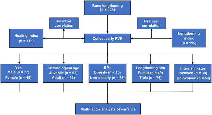
FIGURE 1. Flowchart for the study design of this study.
All patients were operated on by experienced surgeons, and the Ilizarov technique was used for bone lengthening in the femur and tibia. The patients were subjected with or without the involvement of an internal fixator during the consolidation period (after reaching the final length) randomly. As a consequence, 72 patients were kept with an external fixator until the callus was completely healed, whereas 53 patients were replaced with an internal fixator during the consolidation period. The distraction was initiated 1 week after the osteotomy (at the speed of 0.75–1 mm per day). Then, the patients were examined by X-ray monthly. The conditions to end the bone lengthening and remove the external fixator are listed as follows (patients kept with external fixator): 1) bridging callus is shown on three of the four cortices based on the anteroposterior and lateral X-ray photos of the extension segment; 2) the fixation time is generally in line with the average extension index (the total fixation time of the external fixator is the average time of soft callus consolidation calculated from the date of lengthening. Each 1 cm is fixed for 1 month, called the average extension index); and 3) no abnormal feeling of weight-bearing after loosening the nut (Iobst et al., 2017).
The X-ray image measurement tool of the picture archiving and communication system (PACS) was employed to depict the regenerated bone area and its distal and proximal normal bone areas (Figure 2). In order to improve the accuracy of the PVR, the part of the metal bar was rigorously avoided in the targeted area. Then, the ratio of the regenerated bone PV to the average value of the distal and proximal normal bone PV was calculated. The higher PVR (approach to 1) indicates that the regenerated callus is closer to the adjacent normal bone, whereas the lower PVR reflects a lower immaturity (Zhao et al., 2009). The formula for calculating PVR is as follows:
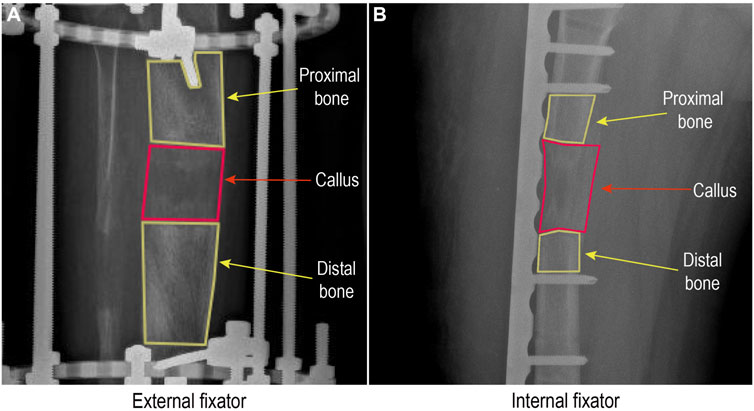
FIGURE 2. Pixel value assessment from a radiograph in a picture archiving and communication system.
The overall PVR is analyzed first. Then, the subjects were divided into several subgroups according to sex, chronological age, BMI, lengthening site, and the involvement of the internal fixator during the consolidation period, respectively.
The LI is the number of months required to achieve 1 cm lengthening, whereas the HI is calculated as the duration of complete consolidation (three cortices in the distraction callus) in days divided by the length gained in centimeters (Koczewski and Shadi, 2013; Wright et al., 2020). The mean of the HI and LI in these patients and the associations of HI and LI with the early PVR of the callus have been analyzed.
All the analyses were performed by using the SPSS 26.0 version. The mean value and standard deviation (SD) of the PVR for the first 3 months were calculated. The PVR differences according to sex (male vs. female), chronological age (juvenile: under 18 years old vs. adult: 18 years or older), BMI (non-obesity vs. obesity), lengthening site (femur vs. tibia), and involvement of the internal fixator were assessed by a multi-factor analysis of variance. The associations of the HI and LI with the early PVR of callus were assessed by Pearson’s correlation coefficient. p < 0.05 was regarded as statistically significant.
A total of 125 patients were recruited for this analysis. The PVRs in subjects with bone lengthening were 0.78 ± 0.10, 0.87 ± 0.06, and 0.93 ± 0.06 in the first 3 months, respectively (Table 1). Obviously, the PVR increased gradually during the first 3 months after osteotomy (Figure 3).

TABLE 1. Early PVR value of the regenerated callus during distraction osteogenesis.
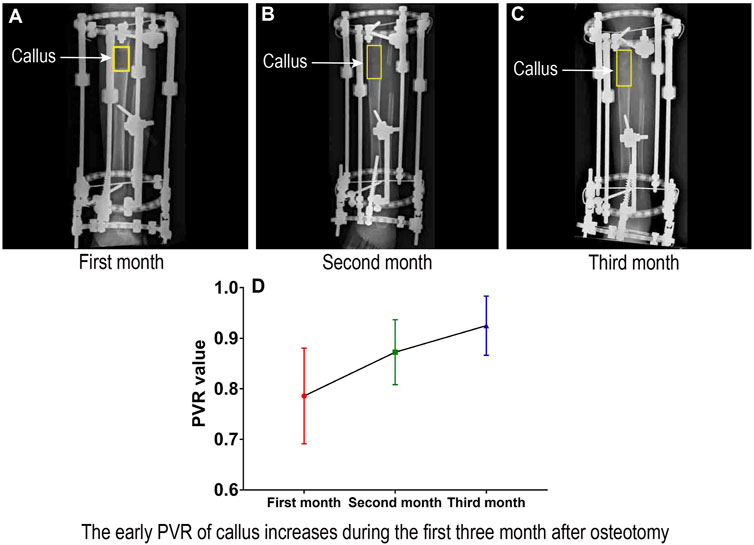
FIGURE 3. Early PVR of the regenerated callus during distraction osteogenesis.
A total of 77 male and 48 female subjects were recruited for this analysis. The PVR in males with bone lengthening was significantly higher than that in females in the first month after osteotomy (0.80 ± 0.09 vs. 0.76 ± 0.10; p = 0.015). However, there was no significant difference in the second and third month (0.87 ± 0.07 vs. 0.87 ± 0.06; p = 0.690), (0.93 ± 0.06 vs. 0.92 ± 0.06; p = 0.504) (Table 2).
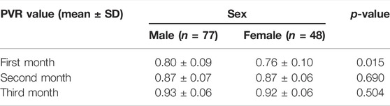
TABLE 2. Subgroup analysis for the early PVR based on sex.
A total of 92 juvenile and 33 adult subjects were recruited in this analysis. The PVR in juveniles with bone lengthening was significantly higher than that in adults in the first 3 months after osteotomy (0.80 ± 0.09 vs. 0.74 ± 0.10; p = 0.008), (0.89 ± 0.06 vs. 0.83 ± 0.06; p = 0.018), and (0.94 ± 0.05 vs. 0.87 ± 0.05; p = 0.003) (Table 3).
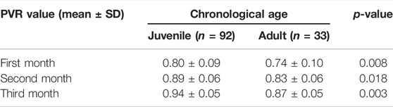
TABLE 3. Subgroup analysis for the early PVR based on chronological age.
A total of 15 obese and 73 non-obese subjects were recruited for this analysis. There was no significant difference in the PVR between the obese and non-obese subjects with bone lengthening in the first 3 months after osteotomy (0.76 ± 0.11 vs. 0.79 ± 0.10; p = 0.854), (0.87 ± 0.07 vs. 0.88 ± 0.06; p = 0.116), and (0.90 ± 0.06 vs. 0.94 ± 0.05; p = 0.154) (Table 4).
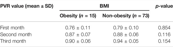
TABLE 4. Subgroup analysis for the early PVR based on BMI.
A total of 49 femoral and 76 tibial site subjects were recruited for this analysis. The PVR in the femur site was significantly higher than that in the tibia site in the second and third months after osteotomy (0.88 ± 0.07 vs. 0.87 ± 0.06; p = 0.015) and (0.93 ± 0.06 vs. 0.92 ± 0.06; p = 0.037). However, there was no difference in the first month (0.80 ± 0.10 vs. 0.76 ± 0.09; p = 0.349) (Table 5).
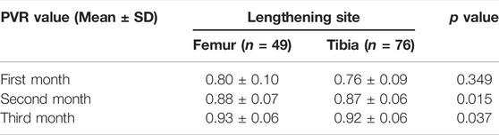
TABLE 5. Subgroup analysis for the early PVR based on the lengthening site.
A total of 38 and 50 subjects with or without the involvement of an internal fixator during the consolidation period were recruited for this analysis. There was no significant difference in the PVR growth value between the subjects with and without the involvement of the internal fixator (0.04 ± 0.04 vs. 0.04 ± 0.04; p = 0.422) (Table 6).

TABLE 6. Subgroup analysis for the PVR growth based on the involvement of the internal fixator during the consolidation period.
A total of 113 patients were recruited for HI analysis, whereas 115 patients were employed for LI analysis. The average HI was 44.98 ± 49.44 days per centimeter, and the LI was 0.78 ± 0.77 months per centimeter, respectively. The results showed that the early PVR of the regenerated callus in the first month after osteotomy was moderately inversely associated with the HI (r = −0.211; p = 0.029) and LI (r = −0.210; p = 0.029) (Table 7).
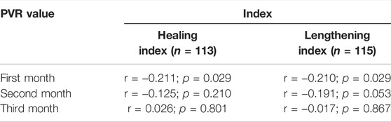
TABLE 7. Associations of the healing index and lengthening index with the early PVR value.
Our results showed that the early PVR is gradually increasing in the first 3 months after osteotomy, and the early PVRs in juvenile, male, and femur sites are significantly higher than those in adult, female, and tibial site subjects. Moreover, the early PVR is moderately inversely associated with the HI and LI, respectively (Figure 4).
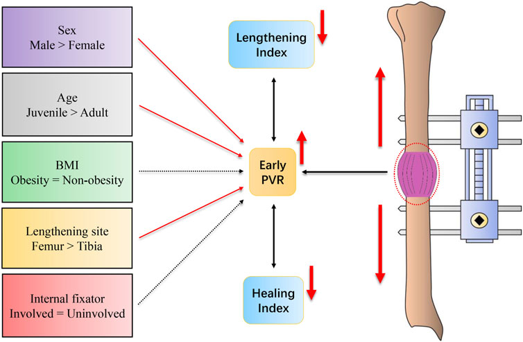
FIGURE 4. Schematic diagram for the results of this study.
Generally speaking, the PVR is mainly utilized to assess the maturity of the late bone callus in order to identify the time to remove the external fixator. Zhao et al. (2009) demonstrated that the PVR could be served as an objective parameter for callus measurement, which provided guidance for the timing of external fixator removal. Bafor et al. found that there were no adverse effects when subjects commenced full weight-bearing when three out of the four cortices of the anteroposterior and lateral radiographs had a PVR of 0.93. Moreover, both Song et al. (2012) and Vulcano et al. (2018) indicated that the PVR could be utilized as a criterion for callus maturation and full weight-bearing. On this basis, our study further analyzed the early PVR of the callus during DO and its potential influencing factors and association with HI and LI.
Interestingly, Koczewski and Shadi (2013) found that the LI was increased with aging, and the LI for the femur was significantly lower than that for the tibia. Indeed, the bone metabolism system with an equilibrium of bone resorption and formation changes with aging (Kloss and Gassner, 2006), and the arterial blood supply of the femur is also richer than that of the tibia (Kizilkanat et al., 2007) (the nutrient artery is the main blood supply to the long bone). Moreover, Mehta et al. (2011) found that being female was an independent risk factor for bone healing in vivo, which may be attributed to a reduction in the mesenchymal stem cell quantity in the bone marrow (Strube et al., 2009). Consistently, we did find that the early PVRs in male, juvenile, and femur sites were significantly higher than those in female, adult, and tibial site subjects, respectively. On the other hand, obesity may induce ectopic adipocyte accumulation in bone marrow cavities, which is considered to impair osteogenic regeneration (Ambrosi et al., 2017). Furthermore, Sun et al. (2011) found more favorable progress in callus regeneration during bone lengthening with the involvement of an internal fixator. Nevertheless, no significant difference in the PVR with regard to BMI and the involvement of the internal fixator was identified in our study. It was speculated that the PVR might not be sensitive enough to reflect the issues. More importantly, the number of obese subjects is rather small, which may inevitably influence our results. Basically, a series of clinical issues caused by an external fixator (for example, inconvenient activities, psychological impact, and uncomfortableness) (Castelein and Docquier, 2016; Nguyen Van and Le Van, 2021) may be avoided by the involvement of an internal fixator. However, our results showed that the early PVR value might not reflect the clinical benefit of an internal fixator (additional medical resources are also consumed). Therefore, the involvement of the internal fixator in bone lengthening still needs to be discussed further. In addition, it is well known that both the LI and HI indicate the bone healing potential (Koczewski and Shadi, 2013; Wright et al., 2020). Our study suggests that the early PVR is moderately inversely associated with LI and HI, which may partly reflect the potential clinical outcome of bone lengthening. However, our results are restricted to the nature of the retrospective design. Taken together, further large well-designed prospective studies are still needed.
The advantages of this study are as follows: first, this is the first study to assess the early PVR value and its potential influencing factors (sex, chronological age, and lengthening site) in bone lengthening. Second, the associations of the HI and LI with the early PVR were also discussed first. Third, this is the largest sample-sized study for PVR analysis until now (others only involved tens of subjects). Fourth, our results may provide the potential clinical value of the early PVR in subjects with bone lengthening. The limitations to the present study should also be acknowledged. First, several issues cannot be addressed due to the nature of the retrospective study design. Second, due to the limited available evidence, bone nonunion cannot be considered in the present study. Third, the disturbance by metal fixtures during the PVR measurement may still slightly influence the PVR assessment. Fourth, some data for BMI and PVR growth were lost in our study, which leads to a different sample size between overall and subgroup analysis. Fifth, the disuse osteopenia of the adjacent bone caused by DO has been ignored in our study. Last but not the least, the number of obese subjects is relatively small in our study.
Our results showed that the early PVR is gradually increasing in the first 3 months after osteotomy, which may be significantly influenced by chronological age, sex, and lengthening site. Moreover, the early PVR of the callus may reflect the potential clinical outcome for DO. Our results may be beneficial to the clinical management of the subjects with bone lengthening.
The original contributions presented in the study are included in the article/supplementary material; further inquiries can be directed to the corresponding authors.
JL and YZ decided and conceptualized this manuscript and revised the draft. QL and HM wrote the manuscript. GZ and ZL collected and analyzed the data. HG and MW prepared the figures and tables. JL and YZ was the guarantor of the overall content. All authors approved the final version of the manuscript and agreed to be accountable for all specs of the work.
This study was supported by the National Natural Science Foundation of China (82102581), the National Postdoctoral Science Foundation of China (2021M693562), the Provincial Natural Science Foundation of Hunan (2019JJ40517), the Provincial Outstanding Postdoctoral Innovative Talents Program of Hunan (2021RC2020), the Young Investigator Grant of Xiangya Hospital, Central South University (2020Q14), the FuQing Postdoc Program of Xiangya Hospital, Central South University (176), and the Fund of Reform and Practice of Ideological and Political in Xiangya Hospital, Central South University (36, 40).
The authors declare that the research was conducted in the absence of any commercial or financial relationships that could be construed as a potential conflict of interest.
All claims expressed in this article are solely those of the authors and do not necessarily represent those of their affiliated organizations, or those of the publisher, the editors, and the reviewers. Any product that may be evaluated in this article, or claim that may be made by its manufacturer, is not guaranteed or endorsed by the publisher.
Ambrosi, T. H., Scialdone, A., Graja, A., Gohlke, S., Jank, A.-M., Bocian, C., et al. (2017). Adipocyte Accumulation in the Bone Marrow during Obesity and Aging Impairs Stem Cell-Based Hematopoietic and Bone Regeneration. Cell Stem Cell 20, 771–784. doi:10.1016/j.stem.2017.02.009
Anand, A., Feldman, D. S., Patel, R. J., Lehman, W. B., van Bosse, H. J. P., Badra, M. I., et al. (2006). Interobserver and Intraobserver Reliability of Radiographic Evidence of Bone Healing at Osteotomy Sites. J. Pediatr. Orthop. B 15, 271–272. doi:10.1097/01202412-200607000-00007
Anna, U.-M., Maria, S., and Kerstin, B. (2021). Comparison of Quantitative Ultrasound of Calcaneus and Dual Energy X-Ray Absorptiometry in Measuring Bone Density and Predicting Fractures in Patients with Diabetic Polyneuropathy: A Prospective Cohort Study. Diabetes Res. Clin. Pract. 180, 109064. doi:10.1016/j.diabres.2021.109064
Babatunde, O. M., Fragomen, A. T., and Rozbruch, S. R. (2010). Noninvasive Quantitative Assessment of Bone Healing after Distraction Osteogenesis. HSS Jrnl 6, 71–78. doi:10.1007/s11420-009-9130-y
Bafor, A., Iobst, C., and Duncan, M. E. (2020). Evaluating the Utility of the Pixel Value Ratio in the Determination of Time to Full Weight-Bearing in Patients Undergoing Intramedullary Limb Lengthening. Strateg. Trauma Limb Reconstr. 15, 74–78. doi:10.5005/jp-journals-10080-1461
Castelein, S., and Docquier, P. L. (2016). Complications Associated with Bone Lengthening of the Lower Limb by Callotasis. Acta Orthop. Belg 82, 806–813.
Engelke, K., Libanati, C., Fuerst, T., Zysset, P., and Genant, H. K. (2013). Advanced CT Based In Vivo Methods for the Assessment of Bone Density, Structure, and Strength. Curr. Osteoporos. Rep. 11, 246–255. doi:10.1007/s11914-013-0147-2
Eyres, K., Bell, M., and Kanis, J. (1993). Methods of Assessing New Bone Formation during Limb Lengthening. Ultrasonography, Dual Energy X-Ray Absorptiometry and Radiography Compared. J. Bone Jt. Surg. Br. volume 75-B, 358–364. doi:10.1302/0301-620X.75B3.8496200
Floerkemeier, T., Hurschler, C., Witte, F., Wellmann, M., Thorey, F., Vogt, U., et al. (2005). Comparison of Various Types of Stiffness as Predictors of the Load-Bearing Capacity of Callus Tissue. J. Bone Jt. Surg. Br. volume 87-B, 1694–1699. doi:10.1302/0301-620X.87B12.16247
Hazra, S., Song, H.-R., Biswal, S., Lee, S.-H., Lee, S. H., Jang, K.-M., et al. (2008). Quantitative Assessment of Mineralization in Distraction Osteogenesis. Skelet. Radiol. 37, 843–847. doi:10.1007/s00256-008-0495-7
Iobst, C. A., Mohammed, W., and Colley, R. (2017). Determining when it Is Safe to Remove the External Fixator: Results from a Survey of the Limb Lengthening and Reconstruction Society. Orthopedics 40, e876–e879. doi:10.3928/01477447-20170810-06
Ishimoto, T., Nakano, T., Yamamoto, M., and Tabata, Y. (2011). Biomechanical Evaluation of Regenerating Long Bone by Nanoindentation. J. Mater Sci. Mater Med. 22, 969–976. doi:10.1007/s10856-011-4266-y
Kizilkanat, E., Boyan, N., Ozsahin, E. T., Soames, R., and Oguz, O. (2007). Location, Number and Clinical Significance of Nutrient Foramina in Human Long Bones. Ann. Anat. - Anatomischer Anzeiger 189, 87–95. doi:10.1016/j.aanat.2006.07.004
Kloss, F. R., and Gassner, R. (2006). Bone and Aging: Effects on the Maxillofacial Skeleton. Exp. Gerontol. 41, 123–129. doi:10.1016/j.exger.2005.11.005
Ko, K. R., Shim, J. S., Chung, C. H., and Kim, J. H. (2019). Surgical Results of Limb Lengthening at the Femur, Tibia, and Humerus in Patients with Achondroplasia. Clin. Orthop. Surg. 11, 226–232. doi:10.4055/cios.2019.11.2.226
Koczewski, P., and Shadi, M. (2013). Factors Influencing Bone Regenerate Healing in Distraction Osteogenesis. Ortop. Traumatol. Rehabil. 15, 591–599. doi:10.5604/15093492.1091515
Malkova, T. A., and Borzunov, D. Y. (2021). International Recognition of the Ilizarov Bone Reconstruction Techniques: Current Practice and Research (Dedicated to 100th Birthday of G. A. Ilizarov). Wjo 12, 515–533. doi:10.5312/wjo.v12.i8.515
Mehta, M., Duda, G. N., Perka, C., and Strube, P. (2011). Influence of Gender and Fixation Stability on Bone Defect Healing in Middle-Aged Rats: a Pilot Study. Clin. Orthop. Relat. Res. 469, 3102–3110. doi:10.1007/s11999-011-1914-y
Nguyen Van, L., and Le Van, D. (2021). Complications and Functional, Psychological Outcomes of Bilateral Tibial Lengthening over Intramedullary Nail: Evidence from Vietnam. Int. Orthop. (SICOT) 45, 2007–2015. doi:10.1007/s00264-021-05059-5
Shchudlo, N., Varsegova, T., Stupina, T., Shchudlo, M., Saifutdinov, M., and Yemanov, A. (2017). Benefits of Ilizarov Automated Bone Distraction for Nerves and Articular Cartilage in Experimental Leg Lengthening. Wjo 8, 688–696. doi:10.5312/wjo.v8.i9.688
Song, S.-H., Agashe, M., Kim, T.-Y., Sinha, S., Park, Y.-E., Kim, S.-J., et al. (2012). Serial Bone Mineral Density Ratio Measurement for Fixator Removal in Tibia Distraction Osteogenesis and Need of a Supportive Method Using the Pixel Value Ratio. J. Pediatr. Orthop. B 21, 137–145. doi:10.1097/BPB.0b013e32834f04f3
Starr, K. A., Fillman, R., and Raney, E. M. (2004). Reliability of Radiographic Assessment of Distraction Osteogenesis Site. J. Pediatr. Orthop. 24, 26–29. doi:10.1097/00004694-200401000-0000610.1097/01241398-200401000-00006
Strube, P., Mehta, M., Baerenwaldt, A., Trippens, J., Wilson, C. J., Ode, A., et al. (2009). Sex-specific Compromised Bone Healing in Female Rats Might Be Associated with a Decrease in Mesenchymal Stem Cell Quantity. Bone 45, 1065–1072. doi:10.1016/j.bone.2009.08.005
Sun, X.-T., Easwar, T. R., Stephen, M., Song, S.-H., Kim, S.-J., and Song, H.-R. (2011). Comparative Study of Callus Progression in Limb Lengthening with or without Intramedullary Nail with Reference to the Pixel Value Ratio and the Ru Li's Classification. Arch. Orthop. Trauma Surg. 131, 1333–1340. doi:10.1007/s00402-011-1302-9
Tesiorowski, M., Kacki, W., Jasiewicz, B., Rymarczyk, A., and Sebastianowicz, P. (2005). Methods for the Evaluation of Bone Regeneration during Distraction Osteogenesis. Chir. Narzadow Ruchu Ortop. Pol. 70, 127–130.
Vulcano, E., Markowitz, J. S., Ali, S., Nguyen, J., Fragomen, A. T., and Rozbruch, S. R. (2018). Assessment of Bone Healing during Antegrade Intramedullary Rod Femur Lengthening Using Radiographic Pixel Density. J. Am. Acad. Orthop. Surg. 26, e388–e394. doi:10.5435/JAAOS-D-16-00949
Wright, S. E., Goodier, W. D., and Calder, P. (2020). Regenerate Deformity with the Precice Tibial Nail. Strateg. Trauma Limb Reconstr. 15, 98–105. doi:10.5005/jp-journals-10080-1457
Zak, L., Arnhold, R., Tiefenboeck, T. M., and Wozasek, G. E. (2021). The Influence of Advanced Age in Bone Healing after Intramedullary Limb Lengthening. Orthop. Traumatology Surg. Res. 107, 103055. doi:10.1016/j.otsr.2021.103055
Keywords: distraction osteogenesis, bone lengthening, pixel value ratio, X-ray, external fixator
Citation: Liu Q, Mei H, Zhu G, Liu Z, Guo H, Wang M, Liang J and Zhang Y (2022) Early Pixel Value Ratios to Assess Bone Healing During Distraction Osteogenesis. Front. Bioeng. Biotechnol. 10:929699. doi: 10.3389/fbioe.2022.929699
Received: 27 April 2022; Accepted: 13 June 2022;
Published: 12 July 2022.
Edited by:
Jun Lin, First Affiliated Hospital of Soochow University, ChinaReviewed by:
Sien Lin, The Chinese University of Hong Kong, ChinaCopyright © 2022 Liu, Mei, Zhu, Liu, Guo, Wang, Liang and Zhang. This is an open-access article distributed under the terms of the Creative Commons Attribution License (CC BY). The use, distribution or reproduction in other forums is permitted, provided the original author(s) and the copyright owner(s) are credited and that the original publication in this journal is cited, in accordance with accepted academic practice. No use, distribution or reproduction is permitted which does not comply with these terms.
*Correspondence: Yi Zhang, emhhbmd5aTAyMDVAY3N1LmVkdS5jbg==; Jieyu Liang, amFtZXNsaWFuZzhAYWxpeXVuLmNvbQ==
†These authors have contributed equally to this work and share the first authorship
Disclaimer: All claims expressed in this article are solely those of the authors and do not necessarily represent those of their affiliated organizations, or those of the publisher, the editors and the reviewers. Any product that may be evaluated in this article or claim that may be made by its manufacturer is not guaranteed or endorsed by the publisher.
Research integrity at Frontiers
Learn more about the work of our research integrity team to safeguard the quality of each article we publish.