 Shaojuan Zhang
 Jingjie Shang
 Weijian Ye
 Tianming Zhao
 Hao Xu
 Hui Zeng
 Lu Wang
94% of researchers rate our articles as excellent or good
Learn more about the work of our research integrity team to safeguard the quality of each article we publish.
Find out more
REVIEW article
Front. Bioeng. Biotechnol., 26 August 2022
Sec. Nanobiotechnology
Volume 10 - 2022 | https://doi.org/10.3389/fbioe.2022.920882
This article is part of the Research TopicCancer and Central Nervous System Disease Diagnosis and TreatmentView all 14 articles
Multiple myeloma (MM) is a neoplastic plasma cell proliferative disorder characterized by various osteolytic bone destruction as a radiological morphological marker. Functional imaging, particularly nuclear medicine imaging, is a promising method to visualize disease processes before the appearance of structural changes by targeting specific biomarkers related to metabolism ability, tumor microenvironment as well as neoplastic receptors. In addition, by targeting particular antigens with therapeutic antibodies, immuno-PET imaging can support the development of personalized theranostics. At present, various imaging agents have been prepared and evaluated in MM at preclinical and clinical levels. A summary overview of molecular functional imaging in MM is provided, and commonly used radiotracers are characterized.
Multiple myeloma (MM) is caused by abnormal plasma cell infiltration in the bone marrow and is a final presentation of a range of monoclonal gammopathies, characterized by clinical symptomatic CRAB features including hypercalcemia, renal insufficiency, anemia, and bone lesions. In the light of the amount of clonal bone marrow plasma cells and serum monoclonal protein, monoclonal gammopathy of undetermined significance (MGUS) and smoldering multiple myeloma (SMM) are defined as its asymptomatic and premalignant stages (Rajkumar et al., 2014; Kumar et al., 2017). The potential risk of SMM (10% per year) (Kyle et al., 2007) and MGUS (1%–1.5% per year) for progression to symptomatic MM, emphasizing the importance of early monitoring and management initiation for high-risk patients (Kyle et al., 2010). On the other hand, MM is not only a highly heterogeneous disease but is also relapsing-remitting cancer, which means MM is treatable but incurable (Yang et al., 2020). Additionally, due to underlying molecular variation, the clinical disease course and optimal treatment or re-treatment strategy vary from person to person (Hideshima et al., 2007). Thus, early. Accurate assessment of residual MM-associated intramedullary and (or) extramedullary lesions is desirable for guiding further management. In 2016, the International Myeloma Working Group (IMWG) incorporated minimal residual disease (MRD) as a standard criterion in the evaluation of treatment response (Kumar et al., 2016). Recently, the utility of MRD negativity as an important prognostic marker for long-term survival in MM patients was confirmed by a large meta-analysis (Munshi et al., 2020).
New imaging techniques have come into being a part of the new Durie/Salmon PLUS staging system, considering anatomic and functional imaging for myeloma staging (Durie, 2006). Currently, modern recommended imaging technologies include whole-body low-dose computed tomography (WBLWCT), positron emission tomography/computed tomography (PET/CT), or whole-body magnetic resonance imaging (WB-MRI) (Mosebach et al., 2019; Terao and Matsue, 2022). A good detailed comparison of those imaging techniques have been reported by Zamagni, et al. (2019) In general, WBLDCT is a practical tool in the preliminary assessment of myeloma bone disease, considering its availability. For the differentiation between MGUS and SMM, which is warranted for serological and biopsy data, CT-guided biopsy is the gold standard (Mosebach et al., 2019). Combining with anatomical information from WBLWCT, PET/CT, imaged with radionuclides and WB-MRI tracked with hydrogen atom signal intensity with no radiation exposure, are recommended as reliable techniques for diagnostic workup and assessment and monitoring of therapy response in MM patients (Pawlyn et al., 2016; Ormond Filho et al., 2019). Due to the high spatial resolution of bone marrow, WB-MRI is highly recommended over [18F]fluorodeoxyglucose ([18F]FDG) PET/CT for the detection of the early and diffuse type of bone marrow infiltration, thus plays a key role in detecting small bone marrow infiltrations (∼5 mm) in the clinical diagnosis of suspected SMM patients (Dimopoulos et al., 2015), also helping re-identify MRD negativity (Zamagni et al., 2020). In particular, MRI functional approaches, like dynamic contrast-enhanced imaging (DCE) and diffusion weight imaging (DWI). As a functional alternative to WB-MRI, PET/CT with [18F]FDG can be used to depict contemporary lytic bone lesions along with glucose metabolism. More important, bone marrow signal in MRI scans (including DWI MRI) is greatly affected by individual age and treatment conditions and thus is suboptimal for early assessment of treatment response, but based on the ability of [18F]FDG PET/CT to distinguish between metabolically active and inactive diseases, as well as the “self-pop out” of avid lesions, which has great advantages in detecting extramedullary disease (EMD) and defines the imaging MRD-negative response to therapy (Cavo et al., 2017). Hybrid PET/MRI, it should be noted, is a promising “double” functional imaging technique, combining the advantages of MRI in the detection of bone marrow involvement and [18F]FDG PET in the prediction of both prognosis and treatment response (Mulé et al., 2020; Rama et al., 2022), systematic clinical data is required for proving the benefit of its sound added-value.
Functional imaging can objectively measure levels of pathogenic related-biomarker, making biomarker-targeted imaging a promising strategy to promote biologically personalized treatments for MM patients (Pawlyn and Davies, 2019), by enabling the identification of disease activity from different in vivo molecular perspectives such as metabolic activities, neoplastic microenvironment, and some specific receptors (Sachpekidis et al., 2019). Currently, [18F]FDG PET/CT in nuclear imaging, as the main type of functional imaging modalities, however, [18F]FDG is just an index reflecting glucose consumption, can’t help but wonder if there is a better imaging probes with better performance ability than [18F]FDG to assess and monitor MM lesions, especially, with increasing treatments with an immunotherapeutic agent by targeting specific receptors (Nadeem et al., 2020). This issue not only has spurred the use of “old” (originally mainly used for other tumors) imaging probes in MM, but inspired “new” imaging probes been developing for imaging MM (shown in Figure 1). (de Waal et al., 2017) Among these, immuno-PET which uses therapeutic antibodies to identify specific surface antigens has shown great promise in radio-immunotherapy and treatment monitoring, including detection of MRD (Pandit-Taskar, 2018). In perspective of different mechanisms of medical imaging, this review discusses the applications of variously reported imaging probes, mainly PET radiotracers, for their potential further use compared to [18F]FDG in MM.
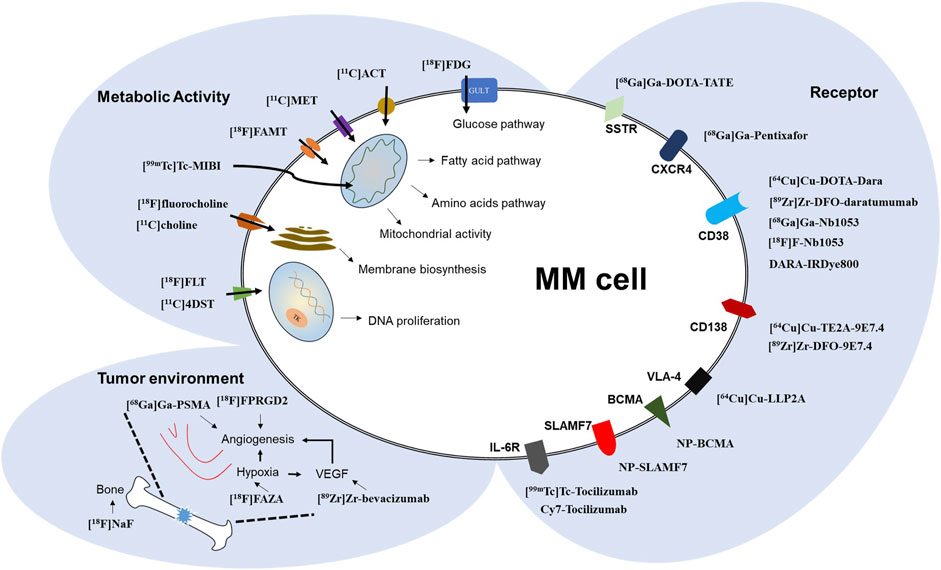
FIGURE 1. Schematic diagram of MM molecular imaging targets and associated imaging probes.
[18F]FDG, a glucose analog, is subjected to the glucose metabolism pathway after intravenous injection. High retention of [18F]FDG is associated with active energy metabolism in myeloma cells and is related to increased numbers of glucose transporters (GLUT), mainly GLUT-1 and GLUT-3 (Pauwels et al., 1998). Subsequent phosphorylation of [18F]FDG by hexokinase makes it unable to escape cells or follow the glucose pathway, trapping it intracellularly (Kanazawa et al., 1986). To obtain optimal imaging qualities, some pre-scan patient preparations, including fasting, confirmation of normal blood glucose levels and a post-injection rest period, are required. In most clinical cases, PET is combined with CT to allow precise anatomical localization of the area of high tracer accumulation, and data is quantified as a standardized uptake value (SUV), traditionally SUVmax, SUVmean, and SUVratio (commonly indexed to liver values) (Huang, 2000). Under normal physiological states, homogeneous uptake of [18F]FDG in the bone marrow is low and less intense than that in the liver. A positive scan result with focal or diffuse active bone marrow uptake indicates that the disease is at an active and advanced stage, while a negative scan means a remission stage (van Lammeren-Venema et al., 2012). Evidence has shown that the number of abnormal avid lesions and associated changes in metabolic uptake after treatment are highly related to patient outcome, and can serve as an independent prognostic factor (Zamagni et al., 2016). For newly diagnosed MM patients receiving therapy, the uptake of bone lesions at levels lower than liver uptake can be thought of as a complete metabolic response, as referenced in the PET response criterion (shown in Table 1), (Zamagni et al., 2021) and is intertwined with MRD negativity.
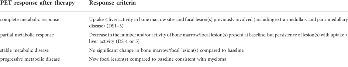
TABLE 1. Proposed refinement of the PET response criteria after therapy.
Many factors can cause false-positive or negative results, including 1) patients lacking the hexokinase enzyme (10%–15%) critical to trapping [18F]FDG in cells (Rasche et al., 2017); 2) changes in bone marrow uptake after therapy (e.g., recent chemotherapeutic drugs or use of cell growth factors) (Sugawara et al., 1998); 3) non-myeloma-associated high uptake (e.g., benign bone inflammation changes). Dynamic tracking can be combined with clinical patient information to reduce misinterpretation. However, more sensitive and specific imaging probes represent preferable alternative approaches for improving MM detection accuracy, which is complementary to the values of [18F]FDG imaging (shown in Tables 2, 3).
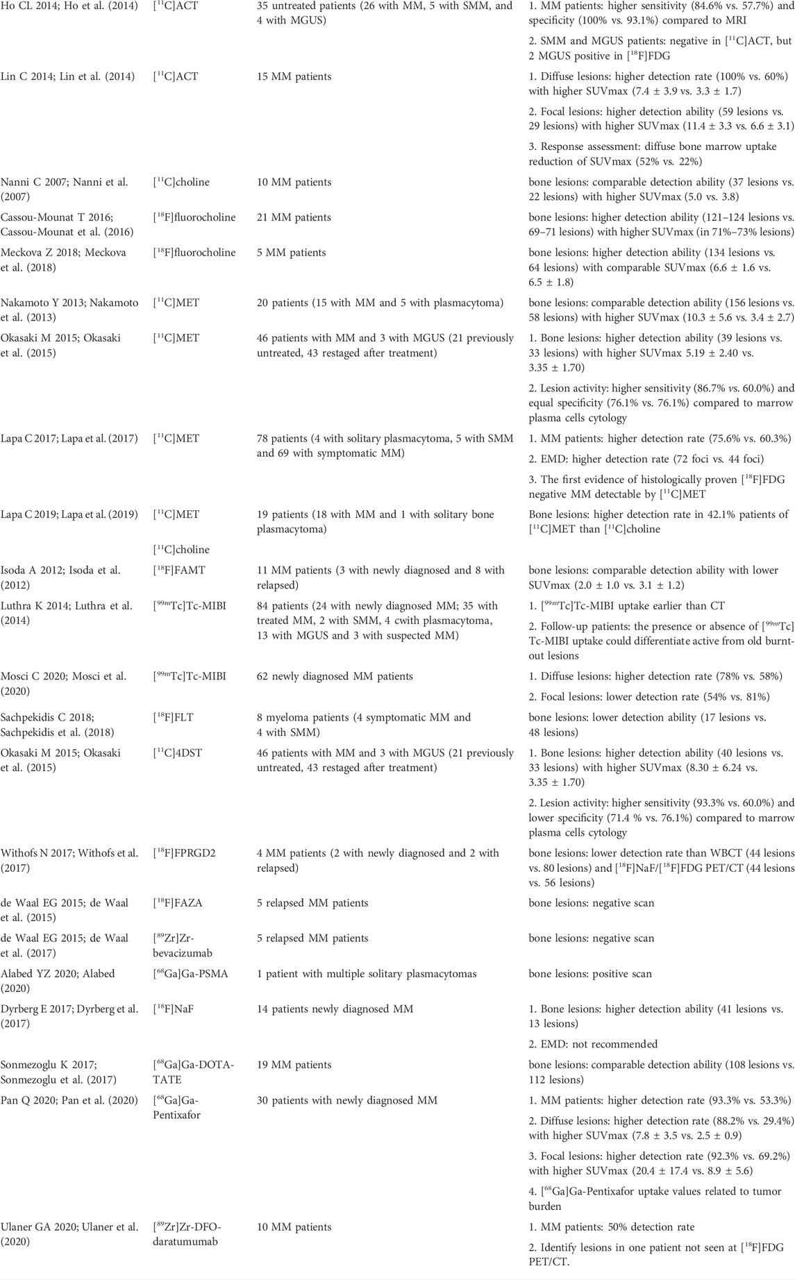
TABLE 2. Reported MM related imaging probe at clinical evaluation level.
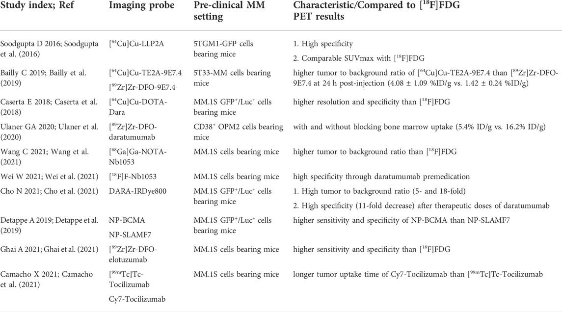
TABLE 3. Reported MM related imaging probe at pre-clinical evaluation level.
Lipogenesis is a shared feature of a variety of malignant cells, and increased fatty acid synthase (FAS) expression has been observed in MM samples and human myeloma cell lines (Wang et al., 2008). [11C]acetate ([11C]ACT), an exogenous acetate, can be rapidly taken up by cells and metabolized to produce acetyl CoA, a carbon source for fatty acid synthesis. In a heterogeneous group of MM patients, [11C]ACT PET/CT exhibited better overall sensitivity and specificity than [18F]FDG. Furthermore, [11C]ACT PET/CT imaging, but not [18F]FDG imaging, was negative for indolent plasma cell neoplasms (SMM and MUGS) (Ho et al., 2014). A similar study also showed that, for newly diagnosed MM patients, [11C]ACT imaging has a higher detection rate for a focal or infiltrated myeloma lesions than [18F]FDG. Moreover, this positive treatment response was visualized via [11C]ACT PET/CT as a significant decrease in SUVmax (listed in Table 2). (Lin et al., 2014)
Choline, which can be phosphorylated by choline kinase into phosphatidylcholine, is involved in cell membrane biosynthesis. The use of [11C]choline for MM patients can be justified based on the increase in proliferating malignant plasma cells with high demands related to membrane metabolism and growth. For MM patients, [11C]choline imaging was performed similarly to [18F]FDG imaging for lesion detection, treatment evaluation, and monitoring (Nanni et al., 2007). Subsequent PET imaging studies of choline were performed with labeled fluorine-18. In relapsing MM patients, [18F]fluorocholine PET/CT indicated that a significantly higher number of lesions were detected compared to [18F]FDG (Cassou-Mounat et al., 2016). Another study that applied [18F]fluorocholine to the detection of skeletal involvement showed that [18F]fluorocholine PET/CT detected about twice as many bone lesions as [18F]FDG, particularly on the skull bone (listed in Table 2). (Meckova et al., 2018) These results are likely due to higher background metabolic level noise for [18F]FDG.
Amino acids are important substrates in the biosynthesis of lipid and protein molecules. [11C]methionine ([11C]MET) possesses a higher specificity than [18F]FDG for the detection of original and recurrent brain tumors and, due to its low physiological background, can also delimit surgical boundaries (Ogawa et al., 1987). Extensive MET is required for the unrestricted proliferation of plasma cells, along with the excessive synthesis of monoclonal immunoglobulins. Cellular transport of [11C]MET is determined by the sodium-independent L-type amino acid transporter (LAT). The physiological uptake of [11C]MET is distributed in the bone marrow and liver. High expression of LAT1 has been identified as a relevant prognostic factor associated with overall poor long-term survival (Isoda et al., 2014). Regarding patients, a study showed that more abnormal lesions were identified by [11C]MET PET/CT than by [18F]FDG, making it useful in grading disease stage (Nakamoto et al., 2013). Another study indicated that [11C]MET PET/CT detected a greater number of positive uptake lesions with more clarity than [18F]FDG, especially with 10–30% plasma cells in the bone marrow (Okasaki et al., 2015). In a study of 78 patients, the largest so far, [11C]MET PET/CT was shown to have higher sensitivity than [18F]FDG in detecting myeloma infiltrated lesions within or outside of bone marrow, as confirmed by histological biopsy. [11C]MET can be potentially applied to disease staging and re-staging with higher accuracy than [18F]FDG (Lapa et al., 2017). In addition, the same group also compared [11C]MET to [11C]choline, and the advantages of [11C]MET were supported by a higher detection rate of MM bone lesions in approximately 40% of patients, as well as higher SUVmax (listed in Table 2). (Lapa et al., 2019)
18F α-methyl tyrosine ([18F]FAMT), a fluorine-18 labeled version of the unnatural amino acid methyltyrosine, can also be transported into cells through LAT-1. Likewise, the uptake of [18F]FAMT by lesions is positively correlated with the expression of LAT-1. In MM patients, a comparable detection rate was observed for [18F]FAMT and [18F]FDG imaging, but uptake discrepancies were evident in several presented lesions (listed in Table 2). (Isoda et al., 2012)
Technetium 99 m sestamibi ([99mTc]Tc-MIBI) is a typical radiotracer used in single-photon emission computed tomography (SPECT) to investigate myocardial perfusion (Alexander and Oberhausen, 1995). Generally, the spatial resolution of SPECT is much lower than that of PET. The high lipid solubility of [99mTc]Tc-MIBI allows it to enter the mitochondria along with the negative membrane potential difference formed by membrane electrophysiological activities. The lesion concentration reflects the high energy metabolism levels found within active malignant plasma cells. A study with 112 [99mTc]Tc-MIBI SPECT was performed in 84 myeloma-associated patients, scan results indicated that the concentration of MIBI in myeloma lesions, corresponds with unchanged and changed radiological changes in CT, could expose earlier ongoing disease activity or old treated lesions (Luthra et al., 2014). In newly diagnosed MM patients, diffuse involvement of bone marrow was better visualized by [99mTc]Tc-MIBI SPECT scan than by [18F]FDG PET/CT scan but was less efficient for focal lesions (listed in Table 2). (Mosci et al., 2020) [99mTc]Tc-MIBI seems to be particularly useful in evaluating the existence of extensive infiltration to avoid underestimation of disease status, meanwhile, the low spatial resolution of SPECT limits the identification of small lesions.
Pyrimidine 3-deoxy-3- (Mulé et al., 2020)F-fluorothymidine ([18F]FLT) and the newer tracer 4′-methyl- (Terao and Matsue, 2022)C-thiothymidine ([11C]4DST) can participate in DNA synthesis as thymidine analogs and have been used to image high DNA proliferation activity in cells (Toyohara et al., 2011; Peck et al., 2015). After being phosphorylated by thymidine kinase 1 (TK1), both compounds become metabolically trapped within cells. Due to the structures, [11C]4DST is more stable than [18F]FLT, and de-phosphorylation occurs relatively rarely. Like [11C]MET, [11C]4DST PET/CT can detect more bone lesions per patient than [18F]FDG in patients with low levels of plasma cell infiltration (10–30%) (Okasaki et al., 2015). However, [11C]4DST also tends to accumulate in active hematopoietic marrow, and has to be associated with the patient background to distinguish from diffuse MM lesions. In contrast, preliminary data indicate that [18F]FLT is not suitable for initial MM diagnostics due to the interference of background bone marrow activity in the cell compartment (Sachpekidis et al., 2018). Otherwise, [18F]FLT can be used to obtain updated information on the distribution of normal bone marrow tissue during therapy (listed in Table 2). (Hayman et al., 2011)
Oxygen consumption is increased with the proliferation of malignant MM cells, resulting in a relatively hypoxic cellular environment that ultimately activates the vascular endothelial growth factor (VEGF) signaling pathway (Apte et al., 2019), leading to tumor angiogenesis. 18F-FB-NH-mini-PEG-E [c (RGDyK)]2 ([18F]FPRGD2) is a standard PET tracer for imaging integrin αvβ3, a type of integrin highly expressed by vascular endothelial cells, and can be used to image tumor angiogenesis (Wu et al., 2007). However, in relapsed MM patients, [18F]FPRGD2 PET/CT is not particularly helpful and was dependent on the presence of obvious lytic bone lesions found by CT (listed in Table 2). (Withofs et al., 2017)
1-α-D:-(5-deoxy-5-18F-fluoroarabinofuranosyl)-2-nitroimidazole ([18F]FAZA) is a PET tracer used to identify hypoxic conditions associated with tumor metabolism (Wuest and Wuest, 2013). When cells are oxygen-deficient, nitroimidazole reduction products will bind to intracellular biomacromolecules and remain in cells. One report indicated that no increased uptake of [18F]FAZA was found for any of five relapsing MM patients, while numerous focal uptakes presented on [18F]FDG PET/CT (de Waal et al., 2015). Likely due to the hypoxic nature of the whole bone marrow compartment, no differences were observed between MM lesions and their surroundings. Bevacizumab, the first humanized nanoantibody (mAb) approved by the Food and Drug Administration (FDA) to inhibit tumor angiogenesis, targets the VEGF receptor. Unfortunately, like [18F]FAZA, PET imaging with zirconium-89 labeled bevacizumab failed to detect significant abnormalities in all patients (listed in Table 2). (de Waal et al., 2017)
Prostate-specific membrane antigen (PSMA) is a characteristic biomarker for prostate cancer cells (Wester and Schottelius, 2019), and enhanced expression has also been observed in tumor vasculature. A case report indicated that 68Ga-prostate-specific membrane antigen-targeted ligand PET imaging can be used to visualize multiple lytic bone lesions throughout the spine (listed in Table 2), (Alabed, 2020) but the definite application in MM is still unclear.
Osteoclastic lesions result from increased plasma cell infiltration in the bone marrow microenvironment, which stimulates bone resorption and impedes bone formation (Mukkamalla and Malipeddi, 2021). The PET tracer [18F]NaF is ‘bone-depositing’, reflecting bone osteoblastic reactions related to regional blood flow. Thus, the typical accumulation of [18F]NaF around lesions can be explained by a secondary osteoblastic reaction, indicating that [18F]NaF is suitable for comprehensive evaluation of bone injury in late stages (listed in Table 2). (Dyrberg et al., 2017) And, [18F]NaF cannot detect EMD logically.
Somatostatin receptor scintigraphy (SRS) using [111mIn]In-pentetreotide has been applied in the workup of neuroendocrine tumors (NETs) for visualizing somatostatin receptors (SSTR), particularly subtypes 2 and 5. Due to the advantages of PET regarding spatial resolution, 68Ga-tetraazacyclododecane-tetraacetic acid-octreotate ([68Ga]Ga-DOTA-TATE) PET/CT has largely replaced SRS for staging NET (Ambrosini et al., 2010). In vitro studies have shown that functional SST is expressed by all MM cell lines, predominantly SSTR5 (Georgii-Hemming et al., 1999). No significant difference was observed between [18F]FDG PET/CT, but diffuse bone marrow uptake can be better shown with [68Ga]Ga-DOTA-TATE (listed in Table 2). (Sonmezoglu et al., 2017)
Very late antigen-4 (VLA-4), a transmembrane adhesion receptor expressed on normal plasma cells, is an important contributor to interactions between plasma cells and the extracellular matrix and bone marrow stromal cells (Schlesinger and Bendas, 2015). Upregulated expression of VLA-4 has been confirmed for myeloma cells and surrounding tissues. With a high binding affinity, N-[[4-[[[(2-ethylphenyl)amino]carbonyl]amino]phenyl]acetyl]-N (epsilon)-6-[(2E)-1-oxo-3-(3-pyridinyl-2-propenyl)]-L-lysyl-L-2-aminohexanedioyl-(1-amino-1-cyclohexane)carboxamide (LLP2A) is a peptidomimetic ligand for VLA-4. LLP2A was conjugated to 1,4,8,11-tetraazacyclotetradecane-1-(methane phosphonic acid)-8-(methane carboxylic acid) (CB-TE1A1P) chelators for cuprum-64 labelling. Favorable pre-clinical results regarding the biodistribution and dosimetry of [64Cu]Cu-LLP2A imaging have been reported for MM mice models, suggesting that this approach is a promising candidate for further imaging of activated VLA-4 in humans (listed in Table 3). (Soodgupta et al., 2016)
A member of the G-protein-coupled chemokine receptor family, chemokine receptor-4 (CXCR4) is mainly expressed in bone marrow primitive hematopoietic cells and is involved in the survival of myeloma cells (Philipp-Abbrederis et al., 2015). Plerixafor (Wang et al., 2020), an exogenous CXCR4 antagonist with high binding affinity, can disrupt adhesive tumor-stroma interactions and achieve treatment goals. [68Ga]Ga-pentixafor PET/CT has been proposed as a theranostics tracer targeting CXCR4 for directed radio-targeted treatment with [177Lu]Lu-pentixafor (or [90Y]Y-pentixafor). In newly diagnosed MM patients, [68Ga]Ga-pentixafor exhibited superior detection ability for myeloma lesions compared to [18F]FDG (93.3% vs. 53.3%). What’s more, quantitative analysis results indicated that the uptake of [68Ga]Ga-pentixafor in bone marrow is a promising biomarker for tumor burden assessment, as it is positively correlated with serum β2-microglobulin and other clinical tumor burden parameters (Pan et al., 2020). A profound therapeutic impact was observed on two heavily pretreated patients following CXCR4-directed lutetium-177 or yttrium-90 endo-radiotherapy, with patients exhibiting a remarkable [18F]FDG uptake reduction in intra and extra-medullary lesions despite the ultimately limited 3–6 months progression-free survival (listed in Table 2). (Herrmann et al., 2016)
Cluster of differentiation (CD) 138, or syndecan-1, is a type of transmembrane proteoglycan found at high levels on the surface of myeloma cells (Sanderson and Yang, 2008). It has been used as a positive sorting marker in the preconcentration of plasma cells for efficient cytogenetic analysis of bone marrow samples. In this respect, CD138 may be an important and potentially beneficial target for imaging and mAbs-based immunotherapy. Cuprum-64 or zirconium-89 labeled anti-CD138 antibodies were realized by incorporating 1,4,8,11-tetraazabicyclo [6.6.2]hexadecane (TE2A) or defetoxamine (DFO) chelator into the antibodies, thus delivering the immuno-PET tracers [64Cu]Cu-TE2A-9E7.4 and [89Zr]Zr-DFO-9E7.4, respectively. In the bone lesions of MM-bearing mice, higher uptake was observed during both PET imaging for [64Cu]Cu-TE2A-9E7.4 and [89Zr]Zr-DFO-9E7.44, but not for [18F]FDG. In addition, the osteophilicity of zirconium-89 resulted in undesired bone background. The higher signal-to-noise ratio of [64Cu]Cu-TE2A-9E7.4 indicated a potential use as a new specific option for MM imaging diagnosis (listed in Table 3). (Bailly et al., 2019) What’s more, mouse models and dosimetry results corroborated the feasibility of radioimmunotherapy for the treatment of advanced-stage MM using anti-CD138 monoclonal antibody namely B-B4, which was radiolabeled with bismuth-213 to promote longer median survival (Chérel et al., 2013).
Daratumumab (Dara), a humanized IgG1K mAb approved by the FDA for use in relapsed MM (Lokhorst et al., 2015), is targeted to the receptor cluster of differentiation 38 (CD38) upregulated in malignant plasma cells but remained low-level expression by surrounding hematopoietic cells. Therefore, CD38-targeted imaging offers a novel approach to the dynamic and invasive assessment of its expression in MM. In preclinical studies, [64Cu]Cu-DOTA-Dara displayed satisfactory potency for CD38 imaging on the surface of MM cell lines, primarily associated with bone infiltration foci. The higher specificity of [64Cu]Cu-DOTA-Dara PET/CT compared to [18F]FDG supports possible clinical applications for MRD detection (listed in Table 3). (Caserta et al., 2018) Recently, zirconium-89 labeled CD38-targeting antibodies for MM were generated using the chelator DFO, providing the first published proof-of-principle for chemical synthesis, preclinical evaluation, and Phase 0 imaging in humans (Ulaner et al., 2020). The results showed that [89Zr]Zr-DFO-daratumumab has a robust ability to visualize CD38 in the murine model (listed in Table 3), and the use of this immuno-PET tracer could be a valuable diagnostic approach due to its high specificity. In addition, full antibody imaging means that a single injection can be sufficient for four PET/CT scans on different days. In humans, the dosimetry of [89Zr]Zr-DFO-daratumumab was found to be acceptable and within safe limits. The Phase 0 clinical trials by [89Zr]Zr-DFO-daratumumab PET/CT imaging study included 10 MM patients and half of them demonstrated avidity on osseous lesions, especially one patient who demonstrated unexpected focal tracer uptake previously undetected by 18F-FDG, consistent with the lack of uptake in low CD38 expression lesions demonstrated by molecular detection approaches (listed in Table 2). By labelling a CD38-specific nanobody (Nb1053) with gallium-68 (Wang et al., 2021) or fluorine-18 (Wei et al., 2021), two preclinical studies indicated that Nb1053-based molecular imaging radiotracers may be useful for MM diagnosis and follow-up (listed in Table 3). Another imaging technique is NIR fluorescence imaging. Preclinical evaluation of DARA-IRDye800, in which DARA conjugated to the NIR fluorophore IRDye800CW, revealed a significant (∼10×) reduction in vivo in fluorescence intensity for the treated group (listed in Table 3). (Cho et al., 2021)
B cell maturation antigen (BCMA) is a member of the tumor necrosis factor receptor superfamily that is found almost exclusively on mature B cells. Its expression level increases significantly in MM cells, and its expression level is positively correlated with MM progression (Shah et al., 2020). In addition, signaling lymphocyte activation molecule 7 (SLAMF 7), which is expressed on immune cells including plasma cells, is a receptor involved in regulating MM cell migration within bone marrow stroma (Malaer and Mathew, 2017). By combining ultra-small sub-5 nm gadolinium-containing nanoparticles (NP) with BCMA and SLAMF 7 targeted antibodies, NP-BCMA and NP-SLAMF7 MR probes were successfully generated. Whole-body imaging of MM tumor-bearing mice showed that BCMA not only had better specificity than SLAMF 7 but also supported clearer imaging of lesions (listed in Table 3). (Detappe et al., 2019)
In terms of radiotracers, elotuzumab, a human monoclonal antibody against SLAMF7 that has been approved by the FDA for use in relapsed MM, has been labeled with zirconium-89. Micro-PET imaging with [89Zr]Zr-DFO-elotuzumab in MM tumor mice indicated that it can specifically identify bone lesions with high expression of SLAMF7. SUVmax was significantly higher than that of [18F]FDG, suggesting that [89Zr]Zr-DFO-elotuzumab can be used to evaluate changes in tumor load after elotuzumab treatment (listed in Table 3). (Ghai et al., 2021) Regarding immunotherapy, BCMA would be a better choice due to its exclusively high expression in malignant plasma cells. Remarkable clinical effects have been witnessed in patients with relapsed/refractory multiple myeloma (RRMM) following antibody-drug conjugate (ADC) treatment, a type of BCMA-targeted therapeutics (Demel et al., 2021). Thus, further incorporation of PET radioisotopes with antibodies targeted to BCMA may be pursued to enhance sensitivity.
Interleukin-6 (IL-6) is a cytokine with broad functions in inflammation and immunity that has been identified as a proliferative factor for MM (Zhang et al., 1992). The results of an early preclinical imaging study using technetium-99 m labeled or fluorophore Cy7-labeled tocilizumab, a humanized Ab that binds to the IL-6 receptor, showed that both [99mTc]Tc-tocilizumab and Cy7-tocilizumab require a long time for uptake into MM engrafted tumors, with up to 72 h required for Cy7-tocilizumab (listed in Table 3), (Camacho et al., 2021) thus hampering further clinical translational application.
[18F]FDG, one of the most common medical probes used in MM functional imaging, has provided valuable guidance for the management of MM patients, like standard WB-MRI. Standardized clinical care and proper imaging evaluation criteria have been promoted for wide distribution. Due to the limitations of [18F]FDG in imaging MM, various probes, especially the radioisotope labeled PET tracers, have been suggested and assessed in clinical patients with related malignancies or during preclinical evaluation. Different imaging agents were used to identify various pathological features of MM; while their values are worth consideration, the primary pursuit in this review is superior performance compared to [18F]FDG (Table 2). Some of these agents, including metabolic tracers such as [11C]ACT, [11C]MET, and [11C]choline, have exhibited promising results in the detection of lesions in MM patients, and tend to have higher SUVmax than [18F]FDG. Based on the data reported so far, it may be suggested that metabolic characterization of lipid and protein metabolism can be more accurate than glucose metabolism in the early diagnosis, disease staging, and treatment response monitoring of MM. Meanwhile, by its short half-life (t1/2 = 20.4 min), carbon-11 makes a complementary PET/CT scan with [18F]FDG on the same day realizable. But their use is somewhat limited by the requirement for an on-site cyclotron and the only very few nuclear medicine centers so far. For [11C]choline, fluorine-18 (t1/2 = 109.8 min) labeled choline ([18F]fluorocholine) could be a good alternative. In terms of the background metabolic level noise, some metabolic PET tracers, such as [18F]FLT, [18F]fluorocholine, and [11C]choline, has unfavorable physiological distribution, characterized by increased uptake in the bone marrow and liver. Further validation of these agents in larger patient cohorts and clinical trials is important. Limited performance of tracers related to the tumor environment is also reflected in the workups of MM patients, and these agents do not appear to be individually useful for clinical evaluation, except [99mTc]Tc-MIBI. [99mTc]Tc-MIBI can be a good alternative for [18F]FDG PET/CT scan with a much lower cost, especially for late-stage MM patients.
Increasingly, MRD assessment has become a critical standard in the clinical assessment of MM, with major efforts to develop methods with sensitive detection and specific exclusion. The traditional treatment of MM has been revolutionized by the progression of immunotherapy. Meanwhile, immuno-PET imaging with radiolabeled antibodies or antibody fragments has potential for MRD assessment and optimization of personalized therapy, [64Cu]Cu-DOTA-Dara might helpful. In the context of theragnostic approaches to MM, the major advantage of the PET tracer [68Ga]Ga-pentixafor is its potential for use in combination with the therapeutic lutetium-177 or yttrium-90 labeled pentixafor in progressive MM patients with CXCR4-positive tumor cells, as confirmed by a [68Ga]Ga-pentixafor PET scan. Likewise, [89Zr]Zr-DFO-daratumumab could be used to identify MM patients who would benefit from daratumumab and thus predict the effectiveness of treatment. Additional research is needed to validate and explore the practical application indications of these novel agents in various MM clinical conditions. Most other reported probes are in very early preclinical development, but some agents, particularly the NP-BCMA have shown promising potential for further prospective studies, which also signifies the possibility and feasibility of a PET tracer for BCMA aimed at immuno-PET imaging. And zirconium-89 (t1/2 = 78.4 h) and cuprum-64 (t1/2 = 12.7 h) are the most common radioisotopes for antibody labelling. The “bone-seeking” nature of zirconium-89 must be considered to understand the intrinsic impact of immuno-PET imaging. Even though routine clinical use of immune-PET imaging is hindered by a lack of proper long-lived radionuclides and the availability of antibodies or corresponding fragments, mAb-based immune-PET holds the potential to maximize patient benefits through MRD detection and the promotion of immunotherapy.
SZ contributed to the writing of the manuscript and constructed the figures and tables. JS and WY and TZ provided analysis and interpretation of data and constructive suggestions. HX contributed to the English language editing and revising. HZ and LW conceived the project and modified the paper for submission and publication.
The authors gratefully acknowledge the support of K.C. Wong Education Foundation (China), and the Project of Innovative Team for the Guangdong Universities (2018KCXTD001, China). This work was financially supported by the National Natural Science Foundation of China (No. 82071974 to LW) and Guangdong Basic and Applied Basic Research Foundation (2020A1515011192, China).
The authors declare that the research was conducted in the absence of any commercial or financial relationships that could be construed as a potential conflict of interest.
All claims expressed in this article are solely those of the authors and do not necessarily represent those of their affiliated organizations, or those of the publisher, the editors and the reviewers. Any product that may be evaluated in this article, or claim that may be made by its manufacturer, is not guaranteed or endorsed by the publisher.
Alabed, Y. Z. (2020). Multiple solitary plasmacytomas with multifocal bone involvement diagnosed with 68Ga-Prostate-Specific membrane antigen PET/CT. Clin. Nucl. Med. 45, e51–e52. doi:10.1097/rlu.0000000000002682
Alexander, C., and Oberhausen, E. (1995). Myocardial scintigraphy. Semin. Nucl. Med. 25, 195–201. doi:10.1016/s0001-2998(95)80026-3
Ambrosini, V., Tomassetti, P., Franchi, R., and Fanti, S. (2010). Imaging of NETs with PET radiopharmaceuticals. Q. J. Nucl. Med. Mol. Imaging. 54, 16–23.
Apte, R. S., Chen, D. S., and Ferrara, N. (2019). VEGF in signaling and disease: beyond discovery and development. Cell 176, 1248–1264. doi:10.1016/j.cell.2019.01.021
Bailly, C., Gouard, S., Guérard, F., Chalopin, B., Carlier, T., Faivre-Chauvet, A., et al. (2019). What is the best radionuclide for immuno-PET of multiple myeloma? A comparison study between 89Zr- and 64Cu-labeled anti-CD138 in a preclinical syngeneic model. Int. J. Mol. Sci. 20, 2564. doi:10.3390/ijms20102564
Camacho, X., Perroni, C., Machado, C. L., de Godoi Carneiro, C., de Souza Junqueira, M., Faria, D., et al. (2021). 99mTechnetium- or Cy7-labeled fab(tocilizumab) as potential multiple myeloma imaging agents. Anticancer. Agents Med. Chem. 21, 1883–1893. doi:10.2174/1871520621999210104181238
Caserta, E., Chea, J., Minnix, M., Poku, E. K., Viola, D., Vonderfecht, S., et al. (2018). Copper 64-labeled daratumumab as a PET/CT imaging tracer for multiple myeloma. Blood 131, 741–745. doi:10.1182/blood-2017-09-807263
Cassou-Mounat, T., Balogova, S., Nataf, V., Calzada, M., Huchet, V., Kerrou, K., et al. (2016). 18F-fluorocholine versus 18F-fluorodeoxyglucose for PET/CT imaging in patients with suspected relapsing or progressive multiple myeloma: a pilot study. Eur. J. Nucl. Med. Mol. Imaging 43, 1995–2004. doi:10.1007/s00259-016-3392-7
Cavo, M., Terpos, E., Nanni, C., Moreau, P., Lentzsch, S., Zweegman, S., et al. (2017). Role of 18F-FDG PET/CT in the diagnosis and management of multiple myeloma and other plasma cell disorders: a consensus statement by the international myeloma working group. Lancet Oncol. 18, e206–e217. doi:10.1016/s1470-2045(17)30189-4
Chérel, M., Gouard, S., Gaschet, J., Sai-Maurel, C., Bruchertseifer, F., Morgenstern, A., et al. (2013). 213Bi radioimmunotherapy with an anti-mCD138 monoclonal antibody in a murine model of multiple myeloma. J. Nucl. Med. 54, 1597–1604. doi:10.2967/jnumed.112.111997
Cho, N., Ko, S., and Shokeen, M. (2021). Preclinical development of near-infrared-labeled CD38-targeted daratumumab for optical imaging of CD38 in multiple myeloma. Mol. Imaging Biol. 23, 186–195. doi:10.1007/s11307-020-01542-4
de Waal, E. G., Slart, R. H., Leene, M. J., Kluin, P. M., and Vellenga, E. (2015). 18F-FDG PET increases visibility of bone lesions in relapsed multiple myeloma: is this hypoxia-driven? Clin. Nucl. Med. 40, 291–296. doi:10.1097/rlu.0000000000000629
de Waal, E. G. M., Glaudemans, A., Schröder, C. P., Vellenga, E., and Slart, R. H. J. A. (2017). Nuclear medicine imaging of multiple myeloma, particularly in the relapsed setting. Eur. J. Nucl. Med. Mol. Imaging 44, 332–341. doi:10.1007/s00259-016-3576-1
Demel, I., Bago, J. R., Hajek, R., and Jelinek, T. (2021). Focus on monoclonal antibodies targeting B-cell maturation antigen (BCMA) in multiple myeloma: update 2021. Br. J. Haematol. 193, 705–722. doi:10.1111/bjh.17235
Detappe, A., Reidy, M., Yu, Y., Mathieu, C., Nguyen, H. V. T., Coroller, T. P., et al. (2019). Antibody-targeting of ultra-small nanoparticles enhances imaging sensitivity and enables longitudinal tracking of multiple myeloma. Nanoscale 11, 20485–20496. doi:10.1039/c9nr06512a
Dimopoulos, M. A., Hillengass, J., Usmani, S., Zamagni, E., Lentzsch, S., Davies, F. E., et al. (2015). Role of magnetic resonance imaging in the management of patients with multiple myeloma: a consensus statement. J. Clin. Oncol. 33, 657–664. doi:10.1200/jco.2014.57.9961
Durie, B. G. (2006). The role of anatomic and functional staging in myeloma: description of durie/salmon plus staging system. Eur. J. Cancer 42, 1539–1543. doi:10.1016/j.ejca.2005.11.037
Dyrberg, E., Hendel, H. W., Al-Farra, G., Balding, L., Logager, V. B., Madsen, C., et al. (2017). A prospective study comparing whole-body skeletal X-ray survey with 18F-FDG-PET/CT, 18F-NaF-PET/CT and whole-body MRI in the detection of bone lesions in multiple myeloma patients. Acta Radiol. Open 6, 2058460117738809. doi:10.1177/2058460117738809
Georgii-Hemming, P., Strömberg, T., Janson, E. T., Stridsberg, M., Wiklund, H. J., and Nilsson, K. (1999). The somatostatin analog octreotide inhibits growth of interleukin-6 (IL-6)-dependent and IL-6-independent human multiple myeloma cell lines. Blood 93, 1724–1731. doi:10.1182/blood.v93.5.1724
Ghai, A., Zheleznyak, A., Mixdorf, M., O’Neal, J., Ritchey, J., Rettig, M., et al. (2021). Development of [89Zr]DFO-elotuzumab for immunoPET imaging of CS1 in multiple myeloma. Eur. J. Nucl. Med. Mol. Imaging 48, 1302–1311. doi:10.1007/s00259-020-05097-y
Hayman, J. A., Callahan, J. W., Herschtal, A., Everitt, S., Binns, D. S., Hicks, R. J., et al. (2011). Distribution of proliferating bone marrow in adult cancer patients determined using FLT-PET imaging. Int. J. Radiat. Oncol. Biol. Phys. 79, 847–852. doi:10.1016/j.ijrobp.2009.11.040
Herrmann, K., Schottelius, M., Lapa, C., Osl, T., Poschenrieder, A., Hanscheid, H., et al. (2016). First-in-Human experience of CXCR4-directed endoradiotherapy with 177Lu- and 90Y-labeled pentixather in advanced-stage multiple myeloma with extensive intra- and extramedullary disease. J. Nucl. Med. 57, 248–251. doi:10.2967/jnumed.115.167361
Hideshima, T., Mitsiades, C., Tonon, G., Richardson, P. G., and Anderson, K. C. (2007). Understanding multiple myeloma pathogenesis in the bone marrow to identify new therapeutic targets. Nat. Rev. Cancer 7, 585–598. doi:10.1038/nrc2189
Ho, C. L., Chen, S., Leung, Y. L., Cheng, T., Wong, K. n., Cheung, S. K., et al. (2014). 11C-acetate PET/CT for metabolic characterization of multiple myeloma: a comparative study with 18F-FDG PET/CT. J. Nucl. Med. 55, 749–752. doi:10.2967/jnumed.113.131169
Huang, S. C. (2000). Anatomy of SUV. Standardized uptake value. Nucl. Med. Biol. 27, 643–646. doi:10.1016/s0969-8051(00)00155-4
Isoda, A., Higuchi, T., Nakano, S., Arisaka, Y., Kaira, K., Kamio, T., et al. (2012). 18F-FAMT in patients with multiple myeloma: clinical utility compared to 18F-FDG. Ann. Nucl. Med. 26, 811–816. doi:10.1007/s12149-012-0645-9
Isoda, A., Kaira, K., Iwashina, M., Oriuchi, N., Tominaga, H., Nagamori, S., et al. (2014). Expression of L-type amino acid transporter 1 (LAT1) as a prognostic and therapeutic indicator in multiple myeloma. Cancer Sci. 105, 1496–1502. doi:10.1111/cas.12529
Kanazawa, Y., Momozono, Y., Ishikawa, M., Yamada, T., Yamane, H., Haradahira, T., et al. (1986). Metabolic pathway of 2-deoxy-2-fluoro-D-glucose studied by F-19 NMR. Life Sci. 39, 737–742. doi:10.1016/0024-3205(86)90022-6
Kumar, S., Paiva, B., Anderson, K. C., Durie, B., Landgren, O., Moreau, P., et al. (2016). International Myeloma Working Group consensus criteria for response and minimal residual disease assessment in multiple myeloma. Lancet Oncol. 17, e328–e346. doi:10.1016/s1470-2045(16)30206-6
Kumar, S. K., Rajkumar, V., Kyle, R. A., van Duin, M., Sonneveld, P., Mateos, M. V., et al. (2017). Multiple myeloma. Nat. Rev. Dis. Prim. 3, 17046. doi:10.1038/nrdp.2017.46
Kyle, R. A., Durie, B. G., Rajkumar, S. V., Landgren, O., Blade, J., Merlini, G., et al. (2010). Monoclonal gammopathy of undetermined significance (MGUS) and smoldering (asymptomatic) multiple myeloma: IMWG consensus perspectives risk factors for progression and guidelines for monitoring and management. Leukemia 24, 1121–1127. doi:10.1038/leu.2010.60
Kyle, R. A., Remstein, E. D., Therneau, T. M., Dispenzieri, A., Kurtin, P. J., Hodnefield, J. M., et al. (2007). Clinical course and prognosis of smoldering (asymptomatic) multiple myeloma. N. Engl. J. Med. Overseas. Ed. 356, 2582–2590. doi:10.1056/nejmoa070389
Lapa, C., Garcia-Velloso, M. J., Lückerath, K., Samnick, S., Schreder, M., Otero, P. R., et al. (2017). 11C-Methionine-PET in multiple myeloma: a combined study from two different institutions. Theranostics 7, 2956–2964. doi:10.7150/thno.20491
Lapa, C., Kircher, M., Da Via, M., Schreder, M., Rasche, L., Kortum, K. M., et al. (2019). Comparison of 11C-choline and 11C-methionine PET/CT in multiple myeloma. Clin. Nucl. Med. 44, 620–624. doi:10.1097/rlu.0000000000002638
Lin, C., Ho, C. L., Ng, S. H., Wang, P. N., Huang, Y., Lin, Y. C., et al. (2014). 11C-acetate as a new biomarker for PET/CT in patients with multiple myeloma: initial staging and postinduction response assessment. Eur. J. Nucl. Med. Mol. Imaging 41, 41–49. doi:10.1007/s00259-013-2520-x
Lokhorst, H. M., Plesner, T., Laubach, J. P., Nahi, H., Gimsing, P., Hansson, M., et al. (2015). Targeting CD38 with daratumumab monotherapy in multiple myeloma. N. Engl. J. Med. Overseas. Ed. 373, 1207–1219. doi:10.1056/nejmoa1506348
Luthra, K., Bhave, A., and Lele, R. D. (2014). Tc-99m sestamibi scanning in multiple myeloma-a new look with SPECT-CT. J. Assoc. Physicians India 62, 801–812.
Malaer, J. D., and Mathew, P. A. (2017). CS1 (SLAMF7, CD319) is an effective immunotherapeutic target for multiple myeloma. Am. J. Cancer Res. 7, 1637–1641.
Meckova, Z., Lambert, L., Spicka, I., Kubinyi, J., and Burgetova, A. (2018). Is fluorine-18-fluorocholine PET/CT suitable for the detection of skeletal involvement of multiple myeloma? Hell. J. Nucl. Med. 21, 167–168. doi:10.1967/s002449910900
Mosci, C., Pericole, F. V., Oliveira, G. B., Delamain, M. T., Takahashi, M. E., Carvalheira, J. B. C., et al. (2020). 99mTc-sestamibi SPECT/CT and 18F-FDG-PET/CT have similar performance but different imaging patterns in newly diagnosed multiple myeloma. Nucl. Med. Commun. 41, 1081–1088. doi:10.1097/mnm.0000000000001259
Mosebach, J., Thierjung, H., Schlemmer, H. P., and Delorme, S. (2019). Multiple myeloma guidelines and their recent updates: implications for imaging. Rofo 191, 998–1009. doi:10.1055/a-0897-3966
Mukkamalla, S. K. R., and Malipeddi, D. (2021). Myeloma bone disease: a comprehensive review. Int. J. Mol. Sci. 22, 6208. doi:10.3390/ijms22126208
Mulé, S., Reizine, E., Blanc-Durand, P., Baranes, L., Zerbib, P., Burns, R., et al. (2020). Whole-body functional MRI and PET/MRI in multiple myeloma. Cancers (Basel) 12 (11), 3155. doi:10.3390/cancers12113155
Munshi, N. C., Avet-Loiseau, H., Anderson, K. C., Neri, P., Paiva, B., Samur, M., et al. (2020). A large meta-analysis establishes the role of MRD negativity in long-term survival outcomes in patients with multiple myeloma. Blood Adv. 4, 5988–5999. doi:10.1182/bloodadvances.2020002827
Nadeem, O., Tai, Y. T., and Anderson, K. C. (2020). Immunotherapeutic and targeted approaches in multiple myeloma. Immunotargets Ther. 9, 201–215. doi:10.2147/itt.s240886
Nakamoto, Y., Kurihara, K., Nishizawa, M., Yamashita, K., Nakatani, K., Kondo, T., et al. (2013). Clinical value of 11C-methionine PET/CT in patients with plasma cell malignancy: comparison with 18F-FDG PET/CT. Eur. J. Nucl. Med. Mol. Imaging 40, 708–715. doi:10.1007/s00259-012-2333-3
Nanni, C., Zamagni, E., Cavo, M., Rubello, D., Tacchetti, P., Pettinato, C., et al. (2007). 11C-choline vs. 18F-FDG PET/CT in assessing bone involvement in patients with multiple myeloma. World J. Surg. Oncol. 5, 68. doi:10.1186/1477-7819-5-68
Ogawa, T., Kanno, I., Shishido, F., Inugami, A., Higano, S., Fujita, H., et al. (1987). Clinical value of PET with 18F-fluorodeoxyglucose and L-methyl-11C-methionine for diagnosis of recurrent brain tumor and radiation injury. Acta Radiol. 32, 197–202. doi:10.3109/02841859109177547
Okasaki, M., Kubota, K., Minamimoto, R., Miyata, Y., Morooka, M., Ito, K., et al. (2015). Comparison of 11C-4'-thiothymidine, 11C-methionine, and 18F-FDG PET/CT for the detection of active lesions of multiple myeloma. Ann. Nucl. Med. 29, 224–232. doi:10.1007/s12149-014-0931-9
Ormond Filho, A. G., Carneiro, B. C., Pastore, D., Silva, I. P., Yamashita, S. R., Consolo, F. D., et al. (2019). Whole-body imaging of multiple myeloma: diagnostic criteria. Radiographics 39, 1077–1097. doi:10.1148/rg.2019180096
Pan, Q., Cao, X., Luo, Y., Li, J., Feng, J., and Li, F. (2020). Chemokine receptor-4 targeted PET/CT with 68Ga-pentixafor in assessment of newly diagnosed multiple myeloma: comparison to 18F-FDG PET/CT. Eur. J. Nucl. Med. Mol. Imaging 47, 537–546. doi:10.1007/s00259-019-04605-z
Pandit-Taskar, N. (2018). Functional imaging methods for assessment of minimal residual disease in multiple myeloma: current status and novel ImmunoPET based methods. Semin. Hematol. 55, 22–32. doi:10.1053/j.seminhematol.2018.02.009
Pauwels, E. K., Ribeiro, M. J., Stoot, J. H., McCready, V., Bourguignon, M., and Maziere, B. (1998). FDG accumulation and tumor biology. Nucl. Med. Biol. 25, 317–322. doi:10.1016/s0969-8051(97)00226-6
Pawlyn, C., and Davies, F. E. (2019). Toward personalized treatment in multiple myeloma based on molecular characteristics. Blood 133, 660–675. doi:10.1182/blood-2018-09-825331
Pawlyn, C., Fowkes, L., Otero, S., Jones, J. R., Boyd, K. D., Davies, F. E., et al. (2016). Whole-body diffusion-weighted MRI: a new gold standard for assessing disease burden in patients with multiple myeloma? Leukemia 30, 1446–1448. doi:10.1038/leu.2015.338
Peck, M., Pollack, H. A., Muzi, M., Friesen, A., Shoner, S. C., Shankland, E. G., et al. (2015). Applications of PET imaging with the proliferation marker 18F-FLT. Q. J. Nucl. Med. Mol. Imaging. 59, 95–104.
Philipp-Abbrederis, K., Herrmann, K., Knop, S., Schottelius, M., Eiber, M., Luckerath, K., et al. (2015). In vivo molecular imaging of chemokine receptor CXCR4 expression in patients with advanced multiple myeloma. EMBO Mol. Med. 7, 477–487. doi:10.15252/emmm.201404698
Rajkumar, S. V., Dimopoulos, M. A., Palumbo, A., Blade, J., Merlini, G., Mateos, M. V., et al. (2014). International Myeloma Working Group updated criteria for the diagnosis of multiple myeloma. Lancet Oncol. 15, e538–548. doi:10.1016/s1470-2045(14)70442-5
Rama, S., Suh, C. H., Kim, K. W., Durieux, J. C., Ramaiya, N. H., and Tirumani, S. H. (2022). Comparative performance of whole-body MRI and FDG PET/CT in evaluation of multiple myeloma treatment response: systematic review and meta-analysis. Am. J. Roentgenol. 218, 602–613. doi:10.2214/ajr.21.26381
Rasche, L., Angtuaco, E., McDonald, J. E., Buros, A., Stein, C., Pawlyn, C., et al. (2017). Low expression of hexokinase-2 is associated with false-negative FDG-positron emission tomography in multiple myeloma. Blood 130, 30–34. doi:10.1182/blood-2017-03-774422
Sachpekidis, C., Goldschmidt, H., and Dimitrakopoulou-Strauss, A. (2019). Positron emission tomography (PET) radiopharmaceuticals in multiple myeloma. Molecules 25, 134. doi:10.3390/molecules25010134
Sachpekidis, C., Goldschmidt, H., Kopka, K., Kopp-Schneider, A., and Dimitrakopoulou-Strauss, A. (2018). Assessment of glucose metabolism and cellular proliferation in multiple myeloma: a first report on combined 18F-FDG and 18F-FLT PET/CT imaging. EJNMMI Res. 8, 28. doi:10.1186/s13550-018-0383-7
Sanderson, R. D., and Yang, Y. (2008). Syndecan-1: a dynamic regulator of the myeloma microenvironment. Clin. Exp. Metastasis 25, 149–159. doi:10.1007/s10585-007-9125-3
Schlesinger, M., and Bendas, G. (2015). Contribution of very late antigen-4 (VLA-4) integrin to cancer progression and metastasis. Cancer Metastasis Rev. 34, 575–591. doi:10.1007/s10555-014-9545-x
Shah, N., Chari, A., Scott, E., Mezzi, K., and Usmani, S. Z. (2020). B-cell maturation antigen (BCMA) in multiple myeloma: rationale for targeting and current therapeutic approaches. Leukemia 34, 985–1005. doi:10.1038/s41375-020-0734-z
Sonmezoglu, K., Vatankulu, B., Elverdi, T., Akyel, R., Erkan, M. E., Halac, M., et al. (2017). The role of 68Ga-DOTA-TATE PET/CT scanning in the evaluation of patients with multiple myeloma: preliminary results. Nucl. Med. Commun. 38, 76–83. doi:10.1097/mnm.0000000000000610
Soodgupta, D., Zhou, H., Beaino, W., Lu, L., Rettig, M., Snee, M., et al. (2016). Ex vivo and in vivo evaluation of overexpressed VLA-4 in multiple myeloma using LLP2A imaging agents. J. Nucl. Med. 57, 640–645. doi:10.2967/jnumed.115.164624
Sugawara, Y., Fisher, S. J., Zasadny, K. R., Kison, P. V., Baker, L. H., and Wahl, R. L. (1998). Preclinical and clinical studies of bone marrow uptake of fluorine-1-fluorodeoxyglucose with or without granulocyte colony-stimulating factor during chemotherapy. J. Clin. Oncol. 16, 173–180. doi:10.1200/jco.1998.16.1.173
Terao, T., and Matsue, K. (2022). Progress of modern imaging modalities in multiple myeloma. Int. J. Hematol. 115, 778–789. doi:10.1007/s12185-022-03360-6
Toyohara, J., Nariai, T., Sakata, M., Oda, K., Ishii, K., Kawabe, T., et al. (2011). Whole-body distribution and brain tumor imaging with 11C-4DST: a pilot study. J. Nucl. Med. 52, 1322–1328. doi:10.2967/jnumed.111.088435
Ulaner, G. A., Sobol, N. B., O'Donoghue, J. A., Kirov, A. S., Riedl, C. C., Min, R., et al. (2020). CD38-targeted immuno-PET of multiple myeloma: from xenograft models to first-in-human imaging. Radiology 295, 606–615. doi:10.1148/radiol.2020192621
van Lammeren-Venema, D., Regelink, J. C., Riphagen, , Zweegman, S., Hoekstra, O. S., and Zijlstra, J. M. (2012). 18F-fluoro-deoxyglucose positron emission tomography in assessment of myeloma-related bone disease: a systematic review. Cancer 118, 1971–1981. doi:10.1002/cncr.26467
Wang, C., Chen, Y., Hou, Y. N., Liu, Q., Zhang, D., Zhao, H., et al. (2021). ImmunoPET imaging of multiple myeloma with [68Ga]Ga-NOTA-Nb1053. Eur. J. Nucl. Med. Mol. Imaging 48, 2749–2760. doi:10.1007/s00259-021-05218-1
Wang, J., Tannous, B. A., Poznansky, M. C., and Chen, H. (2020). CXCR4 antagonist AMD3100 (plerixafor): From an impurity to a therapeutic agent. Pharmacol. Res. 159, 105010. doi:10.1016/j.phrs.2020.105010
Wang, W. Q., Zhao, X. Y., Wang, H. Y., and Liang, Y. (2008). Increased fatty acid synthase as a potential therapeutic target in multiple myeloma. J. Zhejiang Univ. Sci. B 9, 441–447. doi:10.1631/jzus.b0740640
Wei, W., Zhang, D., Wang, C., Zhang, Y., An, S., Chen, Y., et al. (2021). Annotating CD38 expression in multiple myeloma with [18F]F-Nb1053. Mol. Pharm. doi:10.1021/acs.molpharmaceut.1c00733
Wester, H. J., and Schottelius, M. (2019). PSMA-targeted radiopharmaceuticals for imaging and therapy. Semin. Nucl. Med. 49, 302–312. doi:10.1053/j.semnuclmed.2019.02.008
Withofs, N., Cousin, F., De Prijck, B., Bonnet, C., Hustinx, R., Gambhir, S. S., et al. (2017). A first report on 18F-FPRGD(2) PET/CT imaging in multiple myeloma. Contrast Media Mol. Imaging 2017, 6162845. doi:10.1155/2017/6162845
Wu, Z., Li, Z. B., Cai, W., He, L., Chin, F. T., Li, F., et al. (2007). 18F-labeled mini-PEG spacered RGD dimer (18F-FPRGD2): synthesis and microPET imaging of alphavbeta3 integrin expression. Eur. J. Nucl. Med. Mol. Imaging 34, 1823–1831. doi:10.1007/s00259-007-0427-0
Wuest, M., and Wuest, F. (2013). Positron emission tomography radiotracers for imaging hypoxia. J. Label. Comp. Radiopharm. 56, 244–250. doi:10.1002/jlcr.2997
Yang, Y., Li, Y., Gu, H., Dong, M., and Cai, Z. (2020). Emerging agents and regimens for multiple myeloma. J. Hematol. Oncol. 13, 150. doi:10.1186/s13045-020-00980-5
Zamagni, E., Nanni, C., Dozza, L., Carlier, T., Bailly, C., Tacchetti, P., et al. (2021). Standardization of 18F-FDG-PET/CT according to deauville criteria for metabolic complete response definition in newly diagnosed multiple myeloma. J. Clin. Oncol. 39, 116–125. doi:10.1200/jco.20.00386
Zamagni, E., Nanni, C., Gay, F., Pezzi, A., Patriarca, F., Bello, M., et al. (2016). 18F-FDG PET/CT focal, but not osteolytic, lesions predict the progression of smoldering myeloma to active disease. Leukemia 30, 417–422. doi:10.1038/leu.2015.291
Zamagni, E., Tacchetti, P., Barbato, S., and Cavo, M. (2020). Role of imaging in the evaluation of minimal residual disease in multiple myeloma patients. J. Clin. Med. 9, 3519. doi:10.3390/jcm9113519
Zamagni, E., Tacchetti, P., and Cavo, M. (2019). Imaging in multiple myeloma: how? when? Blood 133, 644–651. doi:10.1182/blood-2018-08-825356
Zhang, X. G., Bataille, R., Widjenes, J., and Klein, B. (1992). Interleukin-6 dependence of advanced malignant plasma cell dyscrasias. Cancer 69, 1373–1376. doi:10.1002/1097-0142(19920315)69:6<1373::aid-cncr2820690612>3.0.co;2-1
[11C]4DST 4′-methyl-11C-thiothymidine
[11C]ACT [11C]acetate
[11C]MET [11C]methionine
[18F]FAMT 18F α-methyl-tyrosine
[18F]FAZA 1-α-D: -(5-deoxy-5-18F-fluoroarabinofuranosyl)-2-nitroimidazole
[18F]FDG [18F]fluorodeoxyglucose
[18F]FLT Pyrimidine 3-deoxy-3-18F-fluorothymidine
[18F]FPRGD2 18F-FB-NH-mini-PEG-E[c(RGDyK)]2
[64Cu]Cu-LLP2A 64Cu-CB-TE1A1P-PEG4-LLP2A
[68Ga]Ga-DOTA-TATE 68Ga-tetraazacyclododecane-tetraacetic acid-octreotate
[99mTc]Tc-MIBI Technetium 99 m sestamibi
ADC antibody-drug conjugate
BCMA B cell maturation antigen
CB-TE1A1P 1,4,8,11-tetraazacyclotetradecane-1-[methane phosphonic acid]-8-[methane carboxylic acid]
CD cluster of differentiation
CXCR4 chemokine receptor-4
Dara Daratumumab
DCE dynamic contrast-enhanced
DFO Defetoxamine
DWI diffusion weight imaging
FAS fatty acid synthase
FDA Food and Drug Administration
GLUT glucose transporters
IL-6 Interleukin-6
IMWG International Myeloma Working Group
LAT L-type amino acid transporter
LLP2A N-[[4-[[[(2-ethylphenyl)amino]carbonyl]amino]phenyl]acetyl]-N(epsilon)-6-[(2E)-1-oxo-3-(3-pyridinyl-2-propenyl)]-L-lysyl-L-2-aminohexanedioyl-(1-amino-1-cyclohexane)carboxamide
mAb humanized nanoantibody
MGUS monoclonal gammopathy of undetermined significance
MM multiple myeloma
MRD minimal residual disease
NETs neuroendocrine tumors
NIR near-infrared
NOTA 1,4,7-triazacyclononane-N,N′,N″-triacetic acid
NP nanoparticles
PET/CT positron emission tomography/computed tomography
PSMA prostate-specific membrane antigen
RRMM relapsed/refractory multiple myeloma
SLAMF 7 signaling lymphocyte activation molecule 7
SMM smoldering multiple myeloma
SRS somatostatin receptor scintigraphy
SSTR somatostatin receptors
SUV standardized uptake value
TE2A 1,4,8,11-tetraazabicyclo[6.6.2]hexadecane
TK1 thymidine kinase 1
VEGF vascular endothelial growth factor
VLA-4 very late antigen-4
WBLWCT whole-body low-dose computed tomography
WB-MRI whole-body magnetic resonance imaging
EMD extramedullary disease
Keywords: multiple myeloma, biomarkers, molecular functional imaging, positron emission tomography (PET), radiotracers
Citation: Zhang S, Shang J, Ye W, Zhao T, Xu H, Zeng H and Wang L (2022) Recent developments on the application of molecular probes in multiple myeloma: Beyond [18F]FDG. Front. Bioeng. Biotechnol. 10:920882. doi: 10.3389/fbioe.2022.920882
Received: 15 April 2022; Accepted: 29 July 2022;
Published: 26 August 2022.
Edited by:
Michele Iafisco, National Research Council (CNR), ItalyCopyright © 2022 Zhang, Shang, Ye, Zhao, Xu, Zeng and Wang. This is an open-access article distributed under the terms of the Creative Commons Attribution License (CC BY). The use, distribution or reproduction in other forums is permitted, provided the original author(s) and the copyright owner(s) are credited and that the original publication in this journal is cited, in accordance with accepted academic practice. No use, distribution or reproduction is permitted which does not comply with these terms.
*Correspondence: Hui Zeng, YW5kcm9wczIwMTFAaG90bWFpbC5jb20=; Lu Wang, bF93YW5nMTAwOUBqbnUuZWR1LmNu
Disclaimer: All claims expressed in this article are solely those of the authors and do not necessarily represent those of their affiliated organizations, or those of the publisher, the editors and the reviewers. Any product that may be evaluated in this article or claim that may be made by its manufacturer is not guaranteed or endorsed by the publisher.
Research integrity at Frontiers
Learn more about the work of our research integrity team to safeguard the quality of each article we publish.