94% of researchers rate our articles as excellent or good
Learn more about the work of our research integrity team to safeguard the quality of each article we publish.
Find out more
ORIGINAL RESEARCH article
Front. Aging Neurosci., 11 April 2023
Sec. Neurocognitive Aging and Behavior
Volume 15 - 2023 | https://doi.org/10.3389/fnagi.2023.1114426
This article is part of the Research TopicInsights in Neurocognitive Aging and Behavior: 2022View all 33 articles
Background: The clinical features and pathological process of cerebral microbleed (CMB)-related cognitive impairment are hot topics of cerebral small vessel disease (CSVD). However, how to choose a more suitable cognitive assessment battery for CMB patients is still an urgent issue to be solved. This study aimed to analyze the performance of CMB patients on different cognitive tests.
Methods: This study was designed as a cross-sectional study. The five main markers of CSVD (including the CMB, white matter hyperintensities, perivascular spaces, lacunes and brain atrophy) were assessed according to magnetic resonance imaging. The burden of CMB was categorized into four grades based on the total number of lesions. Cognitive function was assessed by Mini-Mental State Examination (MMSE), Trail-Making Test (TMT, Part A and Part B), Stroop color-word test (Stroop test, Part A, B and C), Verbal Fluency Test (VF, animal), Digit-Symbol Substitution Test (DSST), Digit Cancellation Test (DCT) and Maze. Multiple linear regression analysis was conducted to analyze the association between CMB and cognitive findings.
Results: A total of 563 participants (median age of 69 years) were enrolled in this study, including 218 (38.7%) CMB patients. CMB patients showed worse performance than non-CMB subjects in each cognitive test. Correlation analysis indicated the total number of CMB lesions had positive correlations with the time of TMT, Maze and Stroop test, and negative correlations with the performance of MMSE, VF, DSST, and DCT. After the adjustment for all the potential confounders by linear regression, the CMB burden grade was correlated with the performance of VF, Stroop test C, Maze and DCT.
Conclusion: The presence of CMB lesions was associated with much worse cognitive performances. In VF, Stroop test C, Maze and DCT, the correlations between CMB severity and assessment results were more significant. Our study further confirmed that the attention/executive function domain was the most commonly evaluated in CMB, which provided a picture of the most utilized tools to analyze the prognostic and diagnostic value in CMB.
Increased life expectancy poses a huge public health challenge to society, and advanced age is also associated with an increased risk of dementia. The cerebral microbleed (CMB) is one of the important neuroimaging markers for cerebral small vessel disease (CSVD), and its’ prevalence increases with age (Poels et al., 2011). Besides, more and more studies have found that CMB can lead to cognitive impairment and is an independent risk factor for dementia (Poels et al., 2012; Akoudad et al., 2016; Ding et al., 2017).
Studies have found that CMB lesions can coexist with some neurodegenerative diseases such as Parkinson’s disease and Alzheimer’s disease (AD), and may be related to cognitive and motor dysfunction (Sparacia et al., 2017; Chiu et al., 2018; Tsai et al., 2021). CMB lesions have similar pathological manifestations (β-amyloid accumulation) as AD, while the feature of the cognitive impairment may be different. CMB may be useful to expound the overlap between cerebrovascular and neurodegenerative mechanisms underlying cognitive decline and dementia (Akoudad et al., 2016).
To date, it is still a hot issue now for the specific characteristic of the damage to different cognitive domains about CMB numbers and location. Previous studies have found CMB patients have more serious impairments in special cognitive domains such as attention, executive function, processing speed and memory (van Norden et al., 2011; Poels et al., 2012; Meier et al., 2014; Qian et al., 2021; Nannoni et al., 2022). However, the performance of CMB patients on different cognitive domain tests has been poorly studied.
The purpose of this study was to describe the performance of CMB patients in different cognitive domains and find out which tests are closely related to the CMB burden independent of other CSVD markers. Our study may be helpful to design a battery of neuropsychology assessments, which is more suitable for the early identification of CMB-related cognitive impairment and the observation of CMB burden in clinical practice.
This was a cross-sectional study. Subjects for the physical examination were recruited from the department of Neurology at Beijing Chaoyang Hospital, Capital Medical University from January 2018 to December 2021. This study was reviewed and approved by the Ethics Committee of Beijing Chaoyang Hospital, Capital Medical University. Written informed consent was obtained from each eligible subject.
Participants aged 40 years or older with available clinical data and brain magnetic resonance imaging (MRI) were included in the study. The exclusion criteria included: (1) acute cerebrovascular diseases; (2) history of the massive relevant cerebral infarction or cerebral hemorrhage with obvious neurological sequelae; (3) brain damage caused by neurodegeneration, infection, inflammation, trauma, tumor, poisoning and metabolic disease; (4) severe psychiatric disorders; (5) severe organ insufficiency; (6) severe visual or hearing impairment; (7) taking cognitive-affecting drugs within 24 h, such as sedative-hypnotic drugs, acetylcholine-esterase inhibitors and so on (van Norden et al., 2011; Wang et al., 2017); and (8) incomplete data or brain MRI with poor quality.
Magnetic resonance imaging was performed on the 3.0-T MRI scanner (Prisma; Siemens AG, Erlangen, Germany) in the department of Radiology at Beijing Chaoyang Hospital. The standardized sequences included T1-weighted imaging (repetition time = 2,000.0 ms, echo time = 9.2 ms, slice thickness = 5.0 mm, and field of view = 220 × 220 mm2), T2-weighted imaging (repetition time = 4,500.0 ms, echo time = 84.0 ms, slice thickness = 5.0 mm, and field of view = 220 × 220 mm2), fluid-attenuated inversion recovery (repetition time = 8,000.0 ms, echo time = 86.0 ms, slice thickness = 5.0 mm, and field of view = 199 × 220 mm2), diffusion weighted imaging (repetition time = 3,300.0 ms, echo time = 91.0 ms, slice thickness = 5.0 mm, field of view = 230 × 230 mm2, and b = 0 and 1,000 s/mm2), and susceptibility weighted imaging (repetition time = 27.0 ms, echo time = 20.0 ms, slice thickness = 3.2 mm, and field of view = 172 × 230 mm2).
Imaging markers of CSVD were defined according to STandards for ReportIng Vascular changes on nEuroimaging criteria described previously (Wardlaw et al., 2013). CMB was evaluated according to Microbleed Anatomical Rating Scale (Gregoire et al., 2009). The total number of CMB lesions was recorded. And, the burden of CMB was categorized into four grades: non-CMB, 1 CMB, 2–4 CMBs, 5–9 CMBs, and ≥10 CMBs. The white matter hyperintensity (WMH) in periventricular and deep areas was graded using the Fazekas scale (range 0 to 3, respectively) (Fazekas et al., 1987). The severity of perivascular space (PVS) in basal ganglia (BG) and centrum semiovale was divided into five grades (range 0 to 4, respectively) (Maclullich et al., 2004). Brain atrophy was evaluated using the visual rating scale for posterior atrophy ranging from 0 to 3 (Koedam et al., 2011). The number of subjects with lacune was recorded.
All participants underwent the face-to-face assessment of cognitive function. The selection strategy of cognitive scales was based on the characteristics of vascular cognitive impairment and prior large clinical studies on CSVD-related cognitive dysfunction (Benisty et al., 2009; Akoudad et al., 2016). The seven tests in this study included Mini-Mental State Examination (MMSE), Trail-Making Test (TMT, including Part A and Part B), Stroop Color-Word Test (Stroop test, including Part A, B and C), Verbal Fluency Test (VF, animal), Digit-Symbol Substitution Test (DSST), Digit Cancellation Test (DCT) and Maze. We recorded the time taken to complete the TMT, Stroop test and Maze, as well as the number of correct responses in the VF, DSST, and DCT within the specified time, respectively. The major cognitive domains assessed by these tests include global cognitive function, executive function, processing speed, attention, language, and visuospatial function.
Basic clinical information of each participant was collected according to medical records and questionnaires, including age, gender, years of education, body mass index, smoking and drinking status, medication use (hypotensive drugs, statin, antiplatelet drugs/anticoagulants, hypoglycemic drugs), serological test (total cholesterol, high-density lipoprotein cholesterol, low-density lipoprotein cholesterol, triglyceride, glycosylated hemoglobin, homocysteine), and medical history (hypertension, diabetes, hyperlipidemia, stroke, transient ischemic attack, and cardiovascular diseases).
Mann–Whitney U test and chi-square test were used to compare the differences between CMB patients and non-CMB subjects. The Kruskal–Wallis H test was used to compare the differences in cognition among subjects with different CMB burden grades. Spearman’s correlation analysis was used for the association between the CMB number (abnormal distribution) and the results of cognitive tests. Multiple linear regression analysis (stepwise method) was used for the relationship between CMB burden grades/CMB lesion number and cognitive function after the confounder adjustment. We performed tests for linear trends based on the variable containing a median value of each CMB burden grade. Statistical analysis was performed using Statistical Product and Service Solutions 26.0. A p-value of less than 0.05 was considered significant.
A total of 563 subjects were included in this study with a median age of 69 years, including 324 (57.5%) males and 218 (38.7%) CMB patients. Compared with non-CMB subjects, the CMB group had more male patients (p < 0.001), fewer years of education (p = 0.018), higher smoking rate (p = 0.001), more people with hypertension (p = 0.003) and stroke/TIA (p < 0.001), higher rates of taking antihypertensive drugs (p = 0.020) and antithrombotic drugs (p < 0.001), and higher serum levels of triglyceride (p = 0.008) and homocysteine (p = 0.001). As for other imaging markers of CSVD, patients with CMB had a higher prevalence of the lacune (p < 0.001), more severe WMH (p < 0.001) and BG-PVS (p < 0.001) than non-CMB subjects. Table 1 shows the clinical characteristics of all enrolled participants.
Comparing the results of cognitive tests between the two groups, CMB patients showed significantly worse global cognitive function (MMSE) than the non-CMB group (p < 0.001). CMB patients took more time in Maze (p = 0.001), TMT (Part A and B, p < 0.001) and Stroop test (Part A, B and C; p < 0.001), and had fewer correct responses in VF (p < 0.001), DSST (p < 0.001) and DCT (p < 0.001). These results are shown in Table 2.
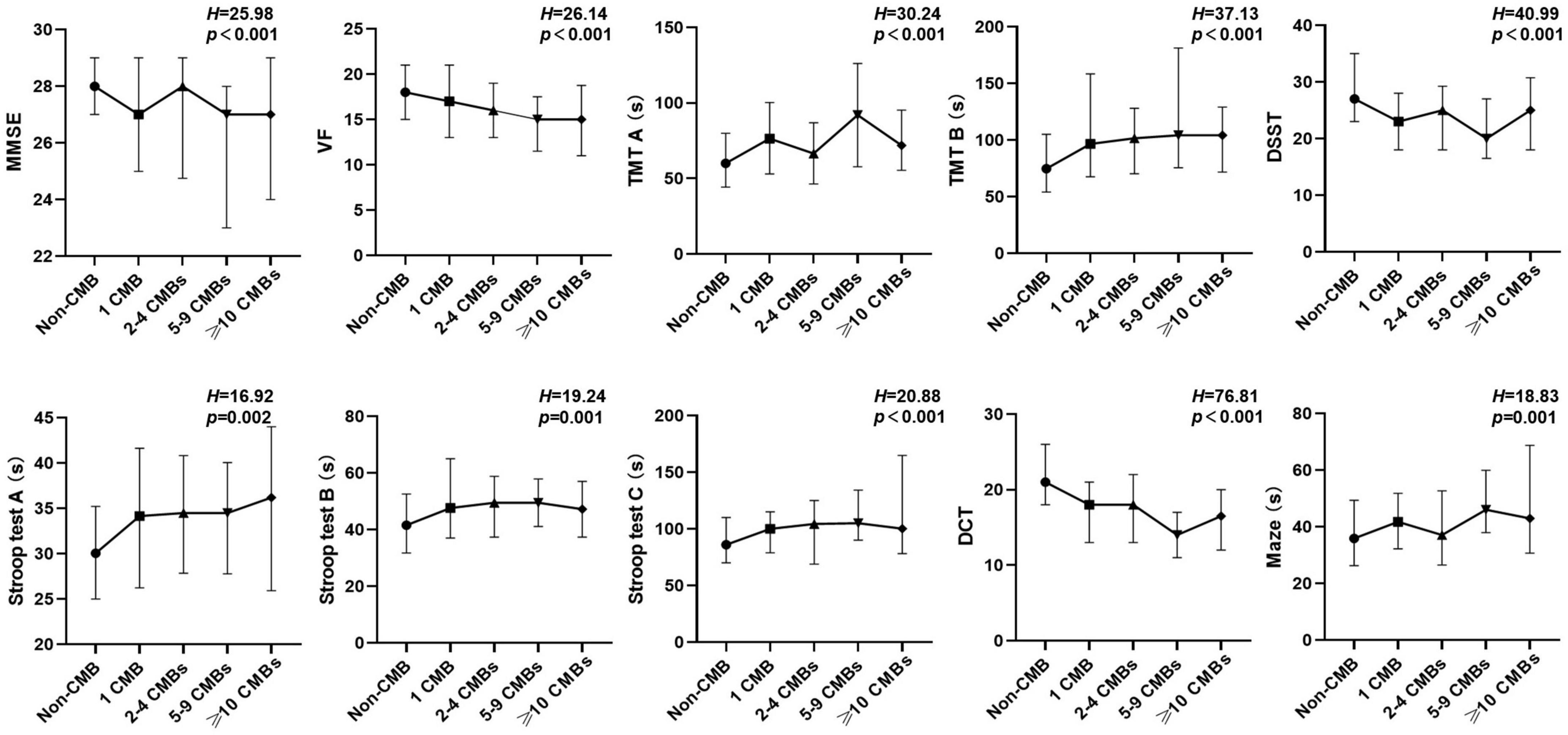
All subjects were divided into five CMB burden grades according to the total number of CMB lesions. There were 345 non-CMB subjects, 67 patients with 1 CMB, 66 patients with 2–4 CMBs, 41 patients with 5–9 CMBs, and 44 patients with ≥10 CMBs. We also found the results of all cognitive function tests were significantly different among five CMB burden grades (MMSE, p < 0.001; VF, p < 0.001; TMT A and B, p < 0.001; Stroop test A, p = 0.002; Stroop test B, p = 0.001; Stroop test C, p < 0.001; Maze, p = 0.001; DSST, p < 0.001; DCT, p < 0.001). These comparisons are shown in Figure 1.

Figure 1. Comparison of cognitive function among subjects with different CMB burden grades (Kruskal–Wallis H test). CMB, cerebral microbleed; DCT, digit cancellation test; DSST, digit-symbol substitution test; MMSE, mini-mental state examination; TMT, trail making test; VF, verbal fluency.
The results of Spearman correlation analysis showed the CMB number had positive correlations with the results of TMT (A, rho = 0.20; B, rho = 0.25; p < 0.001), Maze (rho = 0.16, p < 0.001) and Stroop test (A, rho = 0.17; B, rho = 0.18; C, rho = 0.18; p < 0.001). And, there were negative correlations between the CMB number and the performance of MMSE (rho = −0.21, p < 0.001), VF (rho = −0.21, p < 0.001), DSST (rho = −0.25, p < 0.001) and DCT (rho = −0.36, p < 0.001).
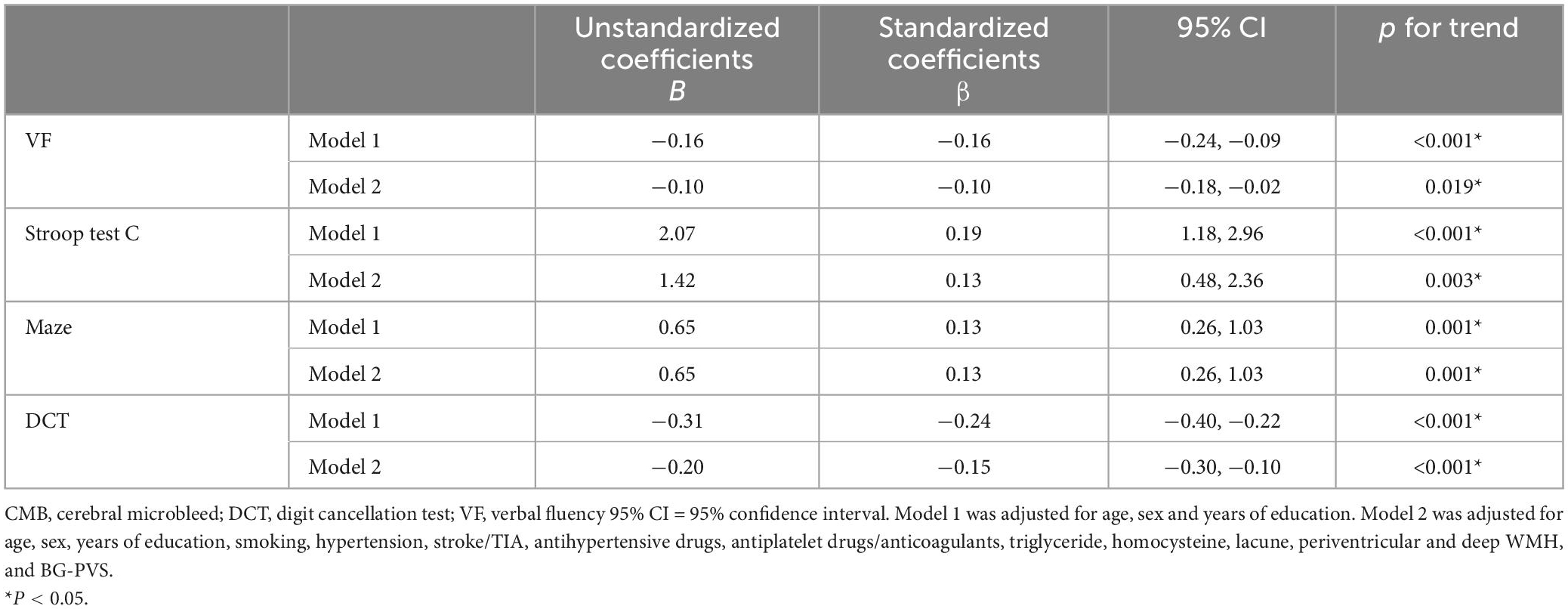
The multiple linear regression analysis showed after adjusting for age, gender and years of education (Model 1), there were significant positive correlations between the CMB burden grade and Stroop test (A, β = 0.10, p = 0.012; B, β = 0.09, p = 0.031; C, β = 0.19, p < 0.001), TMT (A, β = 0.13, p = 0.001; B, β = 0.09, p = 0.015) and Maze (β = 0.13, p = 0.001), and negative correlations between CMB burden and MMSE (β = −0.12, p = 0.001), VF (β = −0.16, p < 0.001), DSST (β = −0.12, p < 0.001) and DCT (β = −0.24, p < 0.001). Based on Model 1, smoking, hypertension, stroke/TIA, antihypertensive drugs, antithrombotic drugs, triglyceride, homocysteine, lacune, periventricular and deep WMH, and BG-PVS were added into the regression equation as Model 2. There were still significant correlations between the CMB burden grade and VF, Stroop test C, Maze and DCT (Table 3). However, the relationships between CMB burden and MMSE, Stroop test A, Stroop test B, TMT A, TMT B, and DSST were no longer significant (p > 0.05). There was no statistical difference in the incidence of posterior atrophy between CMB subjects and non-CMB subjects (Table 1). So, brain atrophy was not a covariate in the multifactor linear regression. However, when it was added to the regression equation, the results were consistent with Model 2. Brain atrophy was an excluded variable in the stepwise method.

Table 3. Linear regression analysis of the relationship between CMB burden grades and cognitive test results.
As for the association between the results of cognitive tests and the number of CMB lesions, we found the CMB number is correlated positively with TMT (A, β = 0.10, p = 0.016; B, β = 0.13, p = 0.001) and Maze (β = 0.11, p = 0.006), and negatively with MMSE (β = −0.08, p = 0.040), VF (β = −0.10, p = 0.018), DSST (β = −0.12, p = 0.002) and DCT (β = −0.19, p < 0.001), after adjusting for all confounding factors (age, gender, years of education, smoking, hypertension, stroke/TIA, antihypertensive drugs, antithrombotic drugs, triglyceride, homocysteine, lacune, periventricular and deep WMH, and BG-PVS). There was no significant relationship between CMB number and Stroop test (p > 0.05).
In our study, CMB patients showed significantly worse performance in global cognitive function (MMSE) compared with the non-CMB group. CMB patients took much more time in Maze, TMT and Stroop test, and had fewer correct responses in VF, DSST, and DCT. Spearman correlation analysis showed the total number of CMB lesions had positive correlations with the TMT, Maze and Stroop test, and negative correlations with MMSE, VF, DSST, and DCT. After adjusting for age, sex and education, the CMB burden was significantly associated with the performances of all seven cognitive function tests. After further adjustment for all confounders, only the relationship of CMB burden with VF, Stroop test C, Maze, and DCT remained significant.
A growing number of studies have found that CMB is associated with vascular cognitive impairment. A meta-analysis of prospective studies showed that CMB patients were connected with overall 1.84 times increased risk of developing dementia than individuals without CMB, and CMB lesions increased the risk of progressing to incident dementia over time (Hussein et al., 2023). Over a mean follow-up of around 5 years, 1.5−4.5% of participants developed all-cause dementia, and 3.5−4.6% of CMB patients progressed to dementia (Akoudad et al., 2016; Ding et al., 2017).
Mini-Mental State Examination is the most well-known and widely utilized global cognitive function scale (Anderson, 2019). However, this scale is easily affected by age, education level and cultural background, and it is short of the evaluation item for executive function. Therefore, we used other tests focusing on different cognitive domains to conduct a more comprehensive assessment of cognitive performance. The Stroop test is commonly used as an indicator of attentional and executive measures. Stroop test A is related to the speed of visual search, Stroop test B is related to the working memory and visual search, and Stroop test C reflects working memory, conflict monitoring and visual search (Periáñez et al., 2021). The TMT is considered one sensitive standard of visual scanning, graphomotor speed and executive function. The score of TMT A can reflect the performance of visual scanning, graphomotor speed and visuomotor processing speed, while the TMT B and derived TMT scores are mainly related to working memory and inhibition control (Llinàs-Reglà et al., 2017). The VF is not only associated with language function but also describes the model of cognitive processes involved in task performance mainly: semantic memory access and executive function (Piskunowicz et al., 2013). Besides, good performances on the DSST, DCT, and Maze require the intact function of associative learning, motor control, complex attention, and visuoperceptual function (Jaeger, 2018; Canfora et al., 2021).
Some researchers combined the findings of several cognitive tests to represent the function of a specific cognitive domain, and supported the results that mixed CMB lesions and higher CMB burden mostly correlated with the dysfunction in global cognition, executive function, processing speed and memory (Akoudad et al., 2016; Ding et al., 2017; Gyanwali et al., 2021; Li et al., 2021). In this study, we described the performance of CMB patients on seven cognitive tests targeting different cognition domains. We found CMB burden grades were closely related to the results of VF, Stroop test C, Maze and DCT, but not MMSE, TMT (A and B), Stroop test A, Stroop test B and DSST, after adjusting for all covariates. These four tests had a better correlation with the severity of CMB, which may help clinical observation of the progression of CMB.
Some other studies also evaluated the performance of CSVD patients on different cognitive tests, but inconsistent results were found. A recent Chinese study used quantitative susceptibility mapping (QSM) to quantitatively measure the brain iron deposition burden of CMB lesions, which showed the iron deposition burden was one of the influencing factors for TMT (TMT A + TMT B) and MoCA scores (Li et al., 2022). A high-resolution MRI study found subjects with more than 10 CMBs showed significantly lower scores of VF than the non-CMB group, while there was no difference in TMT (Part A and B) between the two groups (Ueda et al., 2016). A community-based longitudinal study suggested strictly lobar CMBs (not deep or infratentorial) were related to MMSE and visuospatial executive function (Taylor complex figure test and clock drawing test) (Chung et al., 2016). However, another study demonstrated the number of lacunes was the main predictive factor of cognitive dysfunction in patients with cerebral autosomal-dominant arteriopathy with subcortical infarcts and leukoencephalopathy disease, while there was no significant association between CMB number and MMSE, TMT B, and Stroop test C (Lee et al., 2011). Differences among the findings may be caused by heterogeneities in the population, study design, operation procedure of cognitive tests and covariates in the regression equation.
The location of CMB lesions may have different effects on cognitive domains, and it has become one of the focus topics of CMB-related cognitive impairment. Some studies found only strictly lobar or lobar CMB was associated with cognitive impairment (Poels et al., 2012; Chung et al., 2016; Li et al., 2020), however, some different studies found a close relationship between deep, infratentorial or mixed CMB lesions and cognitive dysfunction (Qiu et al., 2010; Wang et al., 2019). Those inconsistencies may be due to the heterogeneities in study population, different types of cognitive tests, different grouping method of CMB locations. In our present study, the number of CMB patients in each location group is not large enough, especially in the strictly infratentorial sub-group. As a result, we did not analyze the relationship between the CMB location and cognitive function. More studies with large sample sizes will be needed urgently to quantify the CMB burden in different brain sub-regions.
The mechanism of cognitive dysfunction caused by CMB is still the theoretical speculation, which may be the direct damage of CMB to the surrounding brain tissue, disruption of white matter tracts and cortical-subcortical circuits, as well as effects on brain structure and functional networks (Ter Telgte et al., 2018; Jo et al., 2022; Nannoni et al., 2022). An animal experiment of inflammatory response after a cortical microhemorrhage has found CMB may impact the adjacent brain microenvironment through the migration and proliferation of brain-resident microglia and the activation of astrocytes (Ahn et al., 2018).
As for the management of CSVD-related cognitive impairment, it is centered on preventing and controlling vascular risk factors such as hypertension, diabetes, smoking and obesity. Symptomatic treatment includes cholinesterase inhibitors, N-methyl D-aspartate antagonist and ginkgo biloba. The other protective factors include higher education, occupation, social networks, good sleep quality, cognitive, and physical exercise (Zanon Zotin et al., 2021). Some studies have found that antiplatelet and anticoagulant drugs may increase the risk of CMB prevalence and intracerebral hemorrhage in CMB patients (Darweesh et al., 2013; Tsivgoulis et al., 2016). However, these drugs are crucial for the treatment of ischemic cerebrovascular disease. A recent study has found that cilostazol versus aspirin may be a better option in ischemic stroke with a high CMB burden (Park et al., 2021). More clinical trials are needed to provide more evidence for antithrombotic therapy in patients with CMB.
The highlight of this study was the use of multiple cognitive function tests to compare the cognitive characteristics between CMB patients and non-CMB subjects from multiple cognitive domains, and also describe the changes in cognitive performances with the severity of CMB in Chinese CMB patients. This study may provide a more theoretical basis for the selection of cognitive assessment scales for CMB patients in clinical practice.
There were some limitations in our study. Firstly, this was a single-center study based on one hospital, and there might be some selection, information or confounding bias. We recruited subjects strictly, enhanced standardized training of raters, and used multiple regression analysis to control potential confounders. However, the extrapolation of these conclusions still needed to be cautious. Secondly, according to previous studies, CMB lesions had more serious damage to the executive function, processing speed, attention and memory (Qiu et al., 2010; Poels et al., 2012; Akoudad et al., 2016; Ding et al., 2017; Li et al., 2021). So, we selected the commonly used scales for these cognitive domains. However, not all subjects completed the auditory vocabulary learning test (15 words), as a result, memory function was not included in our analysis. Thirdly, the number of CMB patients in different location groups is relatively small, and we did not analyze the relationship between the CMB distribution and cognitive scale results. Finally, the volume of CMB lesions has not been quantitatively evaluated, which can be improved using the QSM and other post-processing methods for the magnetic resonance image in the future.
In our present study, subjects with CMB showed obvious abnormalities in global cognitive function, executive function, processing speed, attention and language. The cognitive battery we used, especially VF, Maze, DCT, and Stroop test, may be helpful to reflect the severity of CMB and play an auxiliary role in clinical practice. In future studies, quantitative analysis of CMB lesions and assessments for other cognitive domains should be carried out. Moreover, it’s essential to conduct more research on the underlying pathophysiology of CMB and the mechanism for CMB-related cognitive dysfunction.
The original contributions presented in this study are included in the article/supplementary material, further inquiries can be directed to the corresponding authors.
The studies involving human participants were reviewed and approved by the Ethics Committee of Beijing Chaoyang Hospital, Capital Medical University. The patients/participants provided their written informed consent to participate in this study.
WH and JY contributed to the conception and design of the study. XL, SY, YL, and JY contributed to the data collection and database organization. XL performed the statistical analysis and wrote the first draft of the manuscript. JY wrote sections of the manuscript. LY and WQ helped with important intellectual content. All authors contributed to the manuscript revision, read, and approved the submitted version.
This study was supported by the National Natural Science Foundation of China (Grant No. 82071552).
The authors declare that the research was conducted in the absence of any commercial or financial relationships that could be construed as a potential conflict of interest.
All claims expressed in this article are solely those of the authors and do not necessarily represent those of their affiliated organizations, or those of the publisher, the editors and the reviewers. Any product that may be evaluated in this article, or claim that may be made by its manufacturer, is not guaranteed or endorsed by the publisher.
Ahn, S., Anrather, J., Nishimura, N., and Schaffer, C. (2018). Diverse inflammatory response after cerebral microbleeds includes coordinated microglial migration and proliferation. Stroke 49, 1719–1726. doi: 10.1161/strokeaha.117.020461
Akoudad, S., Wolters, F., Viswanathan, A., de Bruijn, R., van der Lugt, A., Hofman, A., et al. (2016). Association of cerebral microbleeds with cognitive decline and dementia. JAMA Neurol. 73, 934–943. doi: 10.1001/jamaneurol.2016.1017
Anderson, N. (2019). State of the science on mild cognitive impairment (MCI). CNS Spectr. 24, 78–87. doi: 10.1017/s1092852918001347
Benisty, S., Gouw, A., Porcher, R., Madureira, S., Hernandez, K., Poggesi, A., et al. (2009). Location of lacunar infarcts correlates with cognition in a sample of non-disabled subjects with age-related white-matter changes: the LADIS study. J. Neurol. Neurosurg. Psychiatry 80, 478–483. doi: 10.1136/jnnp.2008.160440
Canfora, F., Calabria, E., Cuocolo, R., Ugga, L., Buono, G., Marenzi, G., et al. (2021). Burning fog: cognitive impairment in burning mouth syndrome. Front. Aging Neurosci. 13:727417. doi: 10.3389/fnagi.2021.727417
Chiu, W., Chan, L., Wu, D., Ko, T., Chen, D., and Hong, C. (2018). Cerebral microbleeds are associated with postural instability and gait disturbance subtype in people with Parkinson’s disease. Eur. Neurol. 80, 335–340. doi: 10.1159/000499378
Chung, C., Chou, K., Chen, W., Liu, L., Lee, W., Chen, L., et al. (2016). Strictly lobar cerebral microbleeds are associated with cognitive impairment. Stroke 47, 2497–2502. doi: 10.1161/strokeaha.116.014166
Darweesh, S., Leening, M., Akoudad, S., Loth, D., Hofman, A., Ikram, M., et al. (2013). Clopidogrel use is associated with an increased prevalence of cerebral microbleeds in a stroke-free population: the rotterdam study. J. Am. Heart Assoc. 2:e000359. doi: 10.1161/jaha.113.000359
Ding, J., Sigurðsson, S., Jónsson, P., Eiriksdottir, G., Meirelles, O., Kjartansson, O., et al. (2017). Space and location of cerebral microbleeds, cognitive decline, and dementia in the community. Neurology 88, 2089–2097. doi: 10.1212/wnl.0000000000003983
Fazekas, F., Chawluk, J., Alavi, A., Hurtig, H., and Zimmerman, R. (1987). MR signal abnormalities at 1.5 T in Alzheimer’s dementia and normal aging. AJR Am. J. Roentgenol. 149, 351–356. doi: 10.2214/ajr.149.2.351
Gregoire, S., Chaudhary, U., Brown, M., Yousry, T., Kallis, C., Jäger, H., et al. (2009). The microbleed anatomical rating scale (MARS): reliability of a tool to map brain microbleeds. Neurology 73, 1759–1766. doi: 10.1212/WNL.0b013e3181c34a7d
Gyanwali, B., Lui, B., Tan, C., Chong, E., Vrooman, H., Chen, C., et al. (2021). Cerebral microbleeds and white matter hyperintensities are associated with cognitive decline in an asian memory clinic study. Curr. Alzheimer Res. 18, 399–413. doi: 10.2174/1567205018666210820125543
Hussein, A., Shawqi, M., Bahbah, E., Ragab, B., Sunoqrot, M., Gadallah, A., et al. (2023). Do cerebral microbleeds increase the risk of dementia? A systematic review and meta-analysis. IBRO Neurosci. Rep. 2023, 86–94. doi: 10.1016/j.ibneur.2022.12.009
Jaeger, J. (2018). Digit symbol substitution test: the case for sensitivity over specificity in neuropsychological testing. J. Clin. Psychopharmacol. 38, 513–519. doi: 10.1097/jcp.0000000000000941
Jo, S., Cheong, E., Kim, N., Oh, J., Shim, W., Kim, H., et al. (2022). Role of white matter abnormalities in the relationship between microbleed burden and cognitive impairment in cerebral amyloid angiopathy. J. Alzheimer’s Dis. 86, 667–678. doi: 10.3233/jad-215094
Koedam, E., Lehmann, M., van der Flier, W., Scheltens, P., Pijnenburg, Y., Fox, N., et al. (2011). Visual assessment of posterior atrophy development of a MRI rating scale. Eur. Radiol. 21, 2618–2625. doi: 10.1007/s00330-011-2205-4
Lee, J., Choi, J., Kang, S., Kang, J., Na, H., and Park, J. (2011). Effects of lacunar infarctions on cognitive impairment in patients with cerebral autosomal-dominant arteriopathy with subcortical infarcts and leukoencephalopathy. J. Clin. Neurol. 7, 210–214. doi: 10.3988/jcn.2011.7.4.210
Li, J., Nguyen, T., Zhang, Q., Guo, L., and Wang, Y. (2022). Cerebral Microbleeds are associated with increased brain iron and cognitive impairment in patients with cerebral small vessel disease: a quantitative susceptibility mapping study. J. Magn. Reson. Imaging 56, 904–914. doi: 10.1002/jmri.28092
Li, L., Wu, D., Li, H., Tan, L., Xu, W., Dong, Q., et al. (2020). Association of cerebral microbleeds with cognitive decline: a longitudinal study. J. Alzheimer’s Dis. 75, 571–579. doi: 10.3233/jad-191257
Li, X., Yuan, J., Qin, W., Yang, L., Yang, S., Li, Y., et al. (2021). Cerebral microbleeds are associated with impairments in executive function and processing speed. J. Alzheimer’s Dis. 81, 255–262. doi: 10.3233/jad-201202
Llinàs-Reglà, J., Vilalta-Franch, J., López-Pousa, S., Calvó-Perxas, L., Torrents Rodas, D., and Garre-Olmo, J. (2017). The trail making test. Assessment 24, 183–196. doi: 10.1177/1073191115602552
Maclullich, A., Wardlaw, J., Ferguson, K., Starr, J., Seckl, J., and Deary, I. (2004). Enlarged perivascular spaces are associated with cognitive function in healthy elderly men. J. Neurol. Neurosurg. Psychiatry 75, 1519–1523. doi: 10.1136/jnnp.2003.030858
Meier, I., Gu, Y., Guzaman, V., Wiegman, A., Schupf, N., Manly, J., et al. (2014). Lobar microbleeds are associated with a decline in executive functioning in older adults. Cerebrovasc. Dis. 38, 377–383. doi: 10.1159/000368998
Nannoni, S., Ohlmeier, L., Brown, R., Morris, R., MacKinnon, A., and Markus, H. (2022). Cognitive impact of cerebral microbleeds in patients with symptomatic small vessel disease. Int. J. Stroke 17, 415–424. doi: 10.1177/17474930211012837
Park, H., Lee, J., Kim, B., Park, J., Kim, Y., Yu, S., et al. (2021). Cilostazol versus aspirin in ischemic stroke with cerebral microbleeds versus prior intracerebral hemorrhage. Int. J. Stroke 16, 1019–1030. doi: 10.1177/1747493020941273
Periáñez, J., Lubrini, G., García-Gutiérrez, A., and Ríos-Lago, M. (2021). Construct validity of the stroop color-word test: influence of speed of visual search, verbal fluency, working memory, cognitive flexibility, and conflict monitoring. Arch. Clin. Neuropsychol. 36, 99–111. doi: 10.1093/arclin/acaa034
Piskunowicz, M., Bieliński, M., Zgliński, A., and Borkowska, A. (2013). [Verbal fluency tests–application in neuropsychological assessment]. Psychiatr. Pol. 47, 475–485.
Poels, M., Ikram, M., van der Lugt, A., Hofman, A., Krestin, G., Breteler, M., et al. (2011). Incidence of cerebral microbleeds in the general population: the Rotterdam scan study. Stroke 42, 656–661. doi: 10.1161/strokeaha.110.607184
Poels, M., Ikram, M., van der Lugt, A., Hofman, A., Niessen, W., Krestin, G., et al. (2012). Cerebral microbleeds are associated with worse cognitive function: the Rotterdam scan study. Neurology 78, 326–333. doi: 10.1212/WNL.0b013e3182452928
Qian, Y., Zheng, K., Wang, H., You, H., Han, F., Ni, J., et al. (2021). Cerebral microbleeds and their influence on cognitive impairment in dialysis patients. BrainImaging Behav. 15, 85–95. doi: 10.1007/s11682-019-00235-z
Qiu, C., Cotch, M., Sigurdsson, S., Jonsson, P., Jonsdottir, M., Sveinbjrnsdottir, S., et al. (2010). Cerebral microbleeds, retinopathy, and dementia: the AGES-reykjavik study. Neurology 75, 2221–2228. doi: 10.1212/WNL.0b013e3182020349
Sparacia, G., Agnello, F., La Tona, G., Iaia, A., Midiri, F., and Sparacia, B. (2017). Assessment of cerebral microbleeds by susceptibility-weighted imaging in Alzheimer’s disease patients: a neuroimaging biomarker of the disease. Neuroradiol. J. 30, 330–335. doi: 10.1177/1971400916689483
Ter Telgte, A., van Leijsen, E., Wiegertjes, K., Klijn, C., Tuladhar, A., and de Leeuw, F. (2018). Cerebral small vessel disease: from a focal to a global perspective. Nat. Rev. Neurol. 14, 387–398. doi: 10.1038/s41582-018-0014-y
Tsai, H., Tsai, L., Lo, Y., and Lin, C. (2021). Amyloid related cerebral microbleed and plasma Aβ40 are associated with cognitive decline in Parkinson’s disease. Sci. Rep. 11:7115. doi: 10.1038/s41598-021-86617-0
Tsivgoulis, G., Zand, R., Katsanos, A., Turc, G., Nolte, C., Jung, S., et al. (2016). Risk of symptomatic intracerebral hemorrhage after intravenous thrombolysis in patients with acute ischemic stroke and high cerebral microbleed burden: a meta-analysis. JAMA Neurol. 73, 675–683. doi: 10.1001/jamaneurol.2016.0292
Ueda, Y., Satoh, M., Tabei, K., Kida, H., Ii, Y., Asahi, M., et al. (2016). Neuropsychological features of microbleeds and cortical microinfarct detected by high resolution magnetic resonance imaging. J. Alzheimer’s Dis. 53, 315–325. doi: 10.3233/jad-151008
van Norden, A., van den Berg, H., de Laat, K., Gons, R., van Dijk, E., and de Leeuw, F. (2011). Frontal and temporal microbleeds are related to cognitive function: the Radboud university nijmegen diffusion tensor and magnetic resonance cohort (RUN DMC) study. Stroke 42, 3382–3386. doi: 10.1161/strokeaha.111.629634
Wang, S., Yuan, J., Guo, X., Teng, L., Jiang, H., Gu, H., et al. (2017). Correlation between prefrontal-striatal pathway impairment and cognitive impairment in patients with leukoaraiosis. Medicine 96:e6703. doi: 10.1097/md.0000000000006703
Wang, Y., Jiang, Y., Suo, C., Yuan, Z., Xu, K., Yang, Q., et al. (2019). Deep/mixed cerebral microbleeds are associated with cognitive dysfunction through thalamocortical connectivity disruption: the taizhou imaging study. Neuroi. Clin. 22:101749. doi: 10.1016/j.nicl.2019.101749
Wardlaw, J., Smith, E., Biessels, G., Cordonnier, C., Fazekas, F., Frayne, R., et al. (2013). Neuroimaging standards for research into small vessel disease and its contribution to ageing and neurodegeneration. Lancet Neurol. 12, 822–838. doi: 10.1016/s1474-4422(13)70124-8
Keywords: cerebral microbleed, cerebral small vessel disease, cognitive impairment, cognitive test, neuroimaging
Citation: Li X, Yang S, Li Y, Qin W, Yang L, Yuan J and Hu W (2023) The performance of patients with cerebral microbleeds in different cognitive tests: A cross-sectional study. Front. Aging Neurosci. 15:1114426. doi: 10.3389/fnagi.2023.1114426
Received: 02 December 2022; Accepted: 13 March 2023;
Published: 11 April 2023.
Edited by:
Kristy A. Nielson, Marquette University, United StatesReviewed by:
Hongkai Wang, Northwestern University, United StatesCopyright © 2023 Li, Yang, Li, Qin, Yang, Yuan and Hu. This is an open-access article distributed under the terms of the Creative Commons Attribution License (CC BY). The use, distribution or reproduction in other forums is permitted, provided the original author(s) and the copyright owner(s) are credited and that the original publication in this journal is cited, in accordance with accepted academic practice. No use, distribution or reproduction is permitted which does not comply with these terms.
*Correspondence: Wenli Hu, d2VubGlodTMzNjZAMTI2LmNvbQ==; Junliang Yuan, anVubGlhbmd5dWFuQGJqbXUuZWR1LmNu
†These authors have contributed equally to this work
Disclaimer: All claims expressed in this article are solely those of the authors and do not necessarily represent those of their affiliated organizations, or those of the publisher, the editors and the reviewers. Any product that may be evaluated in this article or claim that may be made by its manufacturer is not guaranteed or endorsed by the publisher.
Research integrity at Frontiers

Learn more about the work of our research integrity team to safeguard the quality of each article we publish.