 Yu-Qian Wu1†
 Yi-Ning Wang1†Li-Juan Zhang1†Li-Qi Liu1Yi-Cong Pan1Ting Su2Xu-Lin Liao3Hui-Ye Shu1Min Kang1Ping Ying1San-Hua Xu1Yi Shao1*
- 1Department of Ophthalmology and Neurology, The First Affiliated Hospital of Nanchang University, Nanchang, China
- 2Department of Ophthalmology, Massachusetts Eye and Ear, Harvard Medical School, Boston, MA, United States
- 3Department of Ophthalmology and Visual Sciences, The Chinese University of Hong Kong, Sha Tin, Hong Kong SAR, China
A corrigendum on
Regional homogeneity in patients with mild cognitive impairment: A resting-state functional magnetic resonance imaging study
by Wu, Y. -Q., Wang, Y. -N., Zhang, L. -J., Liu, L. -Q., Pan, Y. -C., Su, T., Liao, X. -L., Shu, H. -Y., Kang, M., Ying, P., Xu, S. -H., and Shao, Y. (2022). Front. Aging Neurosci. 14:877281. doi: 10.3389/fnagi.2022.877281
In the published article, there was an error in Table 2 as published. The data of subject three was included in the previous data processing of demographics. Due to the excessive head movement of subject three in the fMRI data acquisition, the data of subject three needs to be deleted, so the following contents need to be corrected. The corrected Table 2 and its caption appear below.
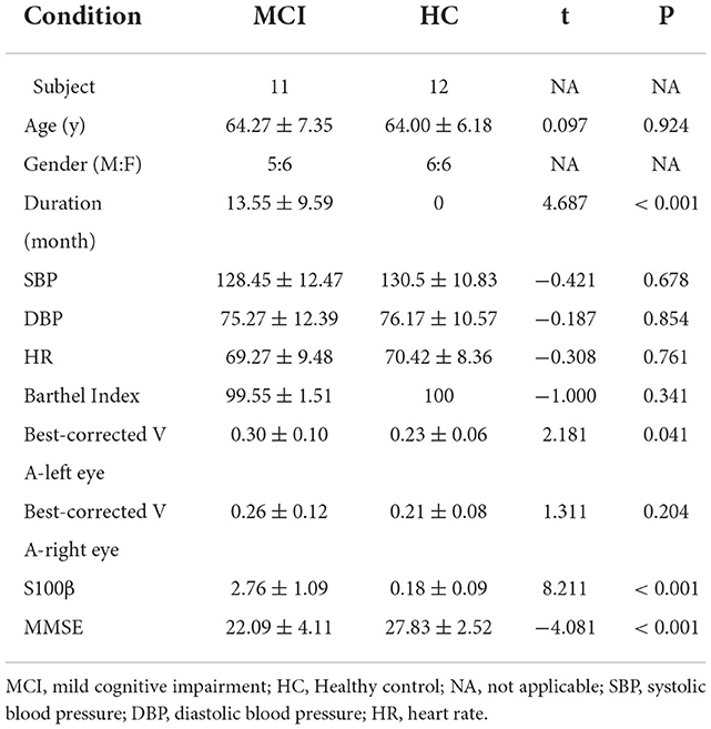
Table 2. Demographic characteristic of the enrolled subjects.
In the published article, there was an error. The data of subject three was included in the previous data processing of demographics. Due to the excessive head movement of subject three in the fMRI data acquisition, the data of subject three needs to be deleted. A correction has been made to Results, “Demographics,” Paragraph 1:
“Subject 3's data have been deleted due to excessive head movement. There was no significant difference in mean age between the MCI and HC groups (64.27 ± 7.35 and 64.00 ± 6.18 yr, respectively; P = 0.924). There was no significant difference in the male to female ratio between the MCI and HC groups. The average MMSE scores of the MCI group was 22.09 ± 4.11 (P < 0.001). The average duration of the MCI was 13.55 ± 9.59 months (P < 0.001). The average S100β of MCI was 2.76 ± 1.09 (P < 0.001). A detailed summary of the data is presented in Table 2.”
In the published article, there was an error. There was a mistake in the description of the signals in the MCI group. A correction has been made to Discussion, Paragraph 6:
“A major function of the superior temporal gyrus is extracting meaningful linguistic features from speech inputs, and is strongly modulated by learning knowledge and perceived goals (Zhongwei et al., 2017). There is some evidence that the right STG functions in allocentric neglect deficits (Mao et al., 2021). The significantly higher signal in this region in the MCI group in the present study may be related to the altered perception of language in patients with MCI.”
The authors apologize for this error and state that this does not change the scientific conclusions of the article in any way. The original article has been updated.
Publisher's note
All claims expressed in this article are solely those of the authors and do not necessarily represent those of their affiliated organizations, or those of the publisher, the editors and the reviewers. Any product that may be evaluated in this article, or claim that may be made by its manufacturer, is not guaranteed or endorsed by the publisher.
References
Mao, Y., Liao, Z., Liu, X., Li, T., Hu, J., Le, D., et al. (2021). Disrupted balance of long and short-range functional connectivity density in Alzheimer's disease (AD) and mild cognitive impairment (MCI) patients: a resting-state fMRI study. Ann. Transl. Med. 9:65. doi: 10.21037/atm-20-7019
Keywords: rs-fMRI, mild cognitive impairment, ReHo, spontaneous brain activity, Alzheimer's disease
Citation: Wu Y-Q, Wang Y-N, Zhang L-J, Liu L-Q, Pan Y-C, Su T, Liao X-L, Shu H-Y, Kang M, Ying P, Xu S-H and Shao Y (2022) Corrigendum: Regional homogeneity in patients with mild cognitive impairment: A resting-state functional magnetic resonance imaging study. Front. Aging Neurosci. 14:1098983. doi: 10.3389/fnagi.2022.1098983
Received: 15 November 2022; Accepted: 18 November 2022;
Published: 13 December 2022.
Edited and reviewed by: Yuzhen Xu, Tongji University, China
Copyright © 2022 Wu, Wang, Zhang, Liu, Pan, Su, Liao, Shu, Kang, Ying, Xu and Shao. This is an open-access article distributed under the terms of the Creative Commons Attribution License (CC BY). The use, distribution or reproduction in other forums is permitted, provided the original author(s) and the copyright owner(s) are credited and that the original publication in this journal is cited, in accordance with accepted academic practice. No use, distribution or reproduction is permitted which does not comply with these terms.
*Correspondence: Yi Shao, ZnJlZWJlZTk5QDE2My5jb20=
†These authors have contributed equally to this work