Grupo de Neurobiología del Desarrollo-GNDe, Hospital Nacional de Parapléjicos, Toledo, Spain
The carotid body or glomus caroticum is a chemosensory organ bilaterally located between the external and internal carotid arteries. Although known by anatomists since the report included by Von Haller and Taube in the mid XVIII century, its detailed study started the first quarter of the XX. The Austro-German physiologist Heinrich E. Hering studied the cardio-respiratory reflexes searched for the anatomical basis of this reflex in the carotid sinus, while the Ghent School leaded by the physio-pharmacologists Jean-François Heymans and his son Corneille focussed in the cardio-aortic reflexogenic region. In 1925, Fernando De Castro, one of the youngest and more brilliant disciples of Santiago Ramón y Cajal at the Laboratorio de Investigaciones Biológicas (Madrid, Spain), profited from some original novelties in histological procedures to study the fine structure and innervation of the carotid body. De Castro unravelled them in a series of scientific papers published between 1926 and 1929, which became the basis to consider the carotid body as a sensory receptor (or chemoreceptor) to detect the chemical changes in the composition of the blood. Indeed, this was the first description of arterial chemoreceptors. Impressed by the novelty and implications of the work of De Castro, Corneille Heymans invited the Spanish neurologist to visit Ghent on two occasions (1929 and 1932), where both performed experiences together. Shortly after, Heymans visited De Castro at the Instituto Cajal (Madrid). From 1932 to 1933, Corneille Heymans focused all his attention on the carotid body his physiological demonstration of De Castro’s hypothesis regarding chemoreceptors was awarded with the Nobel Prize in Physiology or Medicine in 1938, just when Spain was immersed in its catastrophic Civil War.
The carotid body (also known as the glomus caroticum, carotid corpuscule, carotid ganglion and carotid gland) is a very small anatomical structure situated at the bifurcation of the internal and external carotid branches from the primitive carotid artery. Although it was initially described in the XVIII century, its function remained elusive to scientists for centuries. In the mid XIX century, the Weber brothers
1
modulated cardiac frequency by electrical stimulation of the vagal nerve (even stopping the heart) in frogs (Weber and Weber, 1845
). This was reproduced in 1852 by Jacob Henle in a decapitated man. Both experiments represented the first step in this line of research into the cardio-aortic-carotid region. The studies that followed explored the physiology of the cardio-respiratory system, including its reflexes. In 1859, the developer of sphygmograph, Ettienne-Jules Marey (1859
), showed the direct and opposing relationship between arterial pressure and cardiac frequency: if one increases the other falls, and vice versa. Although Ludwig and Cyon confirmed that the vagal nerves were implicated in the origin of bradycardia and hypotension also in the frog (Cyon and Ludwig , 1866
), the studies of François-Franck (1876
, 1877
, 1878
) localised the origin of this reflex in the brainstem (that is, in the central nervous system–CNS).
In 1900, the Italian scientists Pagano and Siciliano proposed that the cardio-respiratory reflexes originated in the carotid region, independent of the CNS (Pagano, 1900
; Siciliano, 1900
). For more than 50 years, this contradiction with François-Franck’s postulate was ignored by the scientists studying this problem, until Heinrich Hering (1923
, 1924
, 1927
) elegantly demonstrated that the electrical or mechanical stimulus of the carotid sinus (a dilatation of the bifurcation of both carotid arteries from the primitive one) triggers a reflex (the “sinus reflex”) that provokes bradycardia and arterial hypotension. Indeed, this German physiologist also discovered that this region was innervated by a branch of the glossopharyngeal nerve, named the “sinus nerve” or “Hering’s nerve”.
In parallel, and not far from Hering’s laboratory at Köln, the group lead by Jean-François Heymans and his son Corneille
2
, employed the famous technique in which two living dogs are maintained in parabiosis to propose that hypertensive bradycardia is a reflex mechanism mediated by the vagal nerves, and it can not be triggered by cephalic or bulbar circulatory hypertension alone (Heymans and Ladon, 1925
). They concluded that the “main regulation of respiration depends on the cardio-aortic region, and it is conditioned by the pressure and composition of circulating blood”
3
(Heymans and Heymans, 1927
). While this was clarified, the study of cardio-respiratory reflexes moved its centre of gravity to Madrid, where one of the youngest and the last direct disciples of the renowned Santiago Ramón y Cajal
4
, Fernando de Castro, began to publish his anatomo-histological observations that were to transform the research in this field.
The rich blood supply and sympathetic innervation of the glomus caroticum had become evident by the middle of the 1920’s, but the fine details behind the organization of this anatomical structure, as well as its physiological implications, remained completely unknown. After his remarkable studies on the structure and organization of the sensory and sympathetic ganglia (mainly: De Castro, 1921
, 1922
), Fernando de Castro decided to study the aorto-carotid region. He found that the entire heads of animals could be fixed by adding nitric acid (3–4%) to the classic fixatives (urethane, chloral hydrate, formol or especially somnifene), perfectly preserving the nervous structures within their skeletal casing, as well as the peripheral innervations, permitting the use of Cajal’s famous reduced silver impregnation method (De Castro, 1925
). The technique De Castro developed is especially effective in small animals, such as rodents, and it reduces the staining of the connective tissue, thereby increasing the contrast of peripheral nerve structures like the glomus caroticum. In his own words, this technique was crucial to study the detailed innervation of the carotid sinus and the carotid body (De Castro, 1926
). In fact, the first microphotographs of the rich vascular supply and the innervation of the glomus caroticum were published by De Castro in that methodological paper (De Castro, 1925
).
The first detailed study of the innervation associated with the carotid region was published by De Castro in 1926, using tissue from both adults and embryos of different mammalian species (from small rodents to humans). Through microphotography, he performed an exhaustive and intensive study of this area, enabling him to make the first fundamental discoveries regarding the glomus caroticum. Accordingly, De Castro showed that the nerve fibres do not form a closed plexus around the carotid body but rather, they project into this structure from the superior cervical ganglion (sympathetic system) and, to a lesser extent, from the glossopharyngeal nerve (the intercarotid branch of the glossopharyngeal nerve that exclusively innervates the carotid body). Years before, Hering had identified this intercarotid branch as the “sinus nerve”, which innervates the carotid sinus and that is responsible for the “sinus reflex” (see above). Although initially De Castro thought that these fibres should be part of the vagal contingent of the intercarotid nerve (De Castro, 1926
), he balanced till consider the sinus nerve as a branch of the glossopharyngeal nerve, and he proposes to call it as intercarotid nerve, as a more appropriated designation (De Castro, 1926
, 1928
).
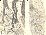
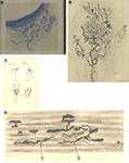
De Castro also described in detail the complex structure of the glomus caroticum: a tangle of small blood vessels, sympathetic axons and glandular cells that which may form small glomeruli within the carotid body, as well as a minuscule and complicated plexuses of glossopharyngeal fibres that surround these glomeruli (Figures 1
A and 2
A,B). The complexity of these nervous plexuses within the carotid body is even greater in humans than in the other mammals studied, although conversely, there is less complexity in the structures that surround the human carotid body (Figures 1
A,B).
Figure 1. De Castro describes fine structure of the carotid body in detail (1926). Original drawings from Fernando De Castro included in his first publication on the innervation of the carotid region (De Castro, 1926
). (A) Arrival of the inter-carotid nerve (branch of the glossopharyngeal nerve; (c) to the carotid body (d) of a young human. Within the carotid body, different glomeruli with fine innervation can be identified, as well as sympathetic microganglia (e). (B) Illustration of the glomus caroticum of the young human. Glomic cells (coloured nuclei) are surrounded by sensitive innervation surrounds, but these fibres do not form a closed plexus is not formed around the carotid body.
Figure 2. De Castro describes fine structure of the carotid body in detail-II (1926). As in the previous figure, these are Fernando De Castro’s original drawings from his first publication on the innervation of the carotid region (De Castro, 1926
). (A) Illustration of the carotid body, where different glomeruli are close to the carotid artery (A). Incoming sympathetic nerve from the superior cervical ganglion (E) is a minor contribution to the innervation of the carotid body. The same can be said about the vagus nerve (LX) in the vicinity of the carotid body. By contrast, the most relevant contingent of afferents comes from the intercarotid nerve (C), branch of the glossopharyngeal nerve (IX). A sympathetic microganglion can be seen within the latter nerve (cg). (B) Detailed illustration of one of the sympathetic microganglia observed within the intercarotid nerve [see (A) and Figure 1
A].
Following this description in1926, the young Spanish neuro-histologist continued his detailed study of the innervation of the glomus caroticum and its physiological implications, which he continued in his second paper on this subject (De Castro, 1928
) and intermittently, in successive papers until the end of his scientific career. At that time, there was much controversy among the physiologists interested in the function of the carotid body and the anatomical basis of the Hering’s reflex. Hering attributed the profuse innervation of the carotid sinus to the fact that, when mechanically or electrically stimulated, it provokes bradychardia and a drop in arterial pressure (Hering, 1924
). One year later, through his intuition and without the support of any experimental data, Drüner hypothesized that the intercarotid gland (the carotid body) was responsible for Hering’s reflex (Drüner, 1925
). Soon after, Hering responded that the mechanical stimulation of the intercarotid territory, as well as that of the carotid body, does not fire the “sinus reflex”, confirming that this must be due to the excitation of the arterial wall in the sinus (Hering, 1925
).
Although his first paper directly touched on the controversy between Hering and Drüner, De Castro finally resolved this scientific joust in his second article on the subject, published in 1928. Once more, De Castro fell back on the methylene blue reaction of Erlich-Dogiel and Bielschowsky’s method (the protocol modified by Boecke) to complement the observations obtained with Cajal’s technique in material from various species, including rodents, cows, monkeys and humans. This paper was essentially divided into two parts: (i) the confirmation of the physiological existence of the carotid sinus in all these species and the study of its innervation (baroreceptors); (ii) the detailed study of the fine innervation of the carotid body and the description of a different kind of receptors (chemoreceptors). Although the present work focuses on the chemoreceptors, we must briefly review the results obtained by De Castro on the innervation of the carotid sinus.
In agreement with Hering’s earlier description (1927), De Castro confirmed the existence of the carotid sinus in all the species, and at all ages, he had studied, except in the human foetus in which the sinus is not macroscopically evident. Clear proof was provided by the cow, which has no internal carotid artery but still, the carotid sinus exists at the bifurcation of the primitive carotid and occipital arteries. Through this study, De Castro ruled out other hypothesis about the nature of the carotid sinus, such as that of a pathological malformation raised by Binswanger and other anatomists at the end of the XIX century, or the existence of enlargements in every vascular bifurcation suggested by Henle, Luschka and other prestigious anatomists.
In this 1928 paper, De Castro described the sensory innervation of the carotid region in detail. He showed that the fibres concentrate at the end of the carotid sinus, immediately prior to the origin of the internal carotid artery. This is the thinnest part of the vessel’s wall and it is surrounded by two concentric bands, one with little sensory innervation and the second with almost no such terminals, a kind of “sensory penumbra” (Figure 3
C). This part of the carotid sinus virtually has the densest innervation of the entire circulatory tree. Moreover, the fact that this distribution exists in all the species studied (including the cow), coupled with the specific location of the carotid sinus (see above), confirms that it this finding is not merely anecdotic. Rather, in all animals the sensory fibres are specifically situated at the origin of the artery that irrigates the brain. Having defined their morphology and distribution, De Castro identified the different types of sensory receptors: either “disperse” or “circumscribed” receptors (Figures 3
A,B). He also noted that the fibres are terminals that extend and ramify through all the different planes of the adventitial layer of the artery, even the deepest one,. In addition, he described them as nude terminals, devoid of any kind of coat, so that they could directly sense the changes in the volume of the vessel due to the changes in arterial pressure (Figure 3
D). Unfortunately, the silver method used for this study does not stain the elastic fibres of the arterial vessels, so De Castro could not study the intimate relationship between the baroreceptors and the elastic fibres that determine the movement of the artery. In none of the thousands of sensory terminals studied by De Castro was any kind of cellular anastomosis observed. In the text, he highlights that this observation was not due to the esprit d’ecole, in clear reference to Cajal’s “neuronal theory”, even though he and the Master himself were the most ardent defenders of this theory against the continued attacks of the “reticularists”. However, these observations confirmed those published earlier, in 1924 and 1926, when De Castro showed that the innervation the carotid sinus is more profuse in big mammals and simpler in the smaller rodents (De Castro, 1928
).
Figure 3. De Castro’s detailed description of the baroreceptors in the carotid sinus (1928). Fernando De Castro’s original drawings from his second publication on the innervation of the carotid region (De Castro, 1928
). (A) Illustration of a type I (diffuse) baroreceptor close to the adventitia of the artery from a young human, stained with methylene blue. (B) Detailed illustration of a type II (circumscribed) baroreceptor of the human carotid sinus, stained by silver impregnation. The myelin trunk is marked by A. (C) Schematic distribution of the baro-receptors in the human carotid sinus. The symbols identify dense terminals in an increasing scale (o,+,>). The section of the artery (B) illustrates how the sensitive terminals are situated in the thinner part of the arterial wall. (D) Distribution of baroreceptor fine terminals (b,c) intermingled with the collagen fibres [in diffuse grey, (a)]; this allows to detect the changes in volume of the blood vessels. In some cases, these nerve terminals form a meniscus.
The ingenuity of Fernando De Castro can be clearly appreciated by how he studied the nature of carotid sinus innervation. To disprove the hypothesis of Boecke (proposed in ca. 1918) regarding the sympathetic origin of these nerve terminals, De Castro ablated the sympathetic ganglia of the cervical chain and he failed to detect any sign of degeneration in the terminals innervating the carotid sinus. Identical results were obtained when the glossopharyngeal, vagus and spinal nerves were sectioned proximally to the respective sensory ganglia
5
. From these results on trophic deprivation, De Castro concluded that the fibres innervating the carotid sinus must necessarily be sensory fibres, neurons projecting towards the CNS that in turn provides them with sufficient trophic support to survive. At that point, he proposed that these observations reflect the morphological basis of the “sinus reflex” described by Hering (1927)
, and thus, the hypothesis proposed by Drüner was definitively discarded by the physiological studies ofHeinrich Hering (1927
) combined with the anatomo-histological studies of De Castro (1926 and mainly, 1928).
After reviewing the studies of the carotid sinus and the baroreceptors, we now move our focus to the second block of results published by De Castro (1928)
regarding the glomus caroticum and the chemoreceptors. When his first observations on this subject were published (1926), De Castro proposed that the cells of the carotid body were probably cells with a paracrine or autocrine function, in agreement with the generalized opinion of many contemporary scientists that the glomus caroticum was a real gland (the intercarotid gland). However, with the elegant ablation experiments described above, De Castro observed that a few motile sympathetic fibres within the glomus did not degenerate once the sympathetic ganglia of the cervical chain were extirpated, indicating that they came from the sympathetic neurons of the microganglia present within the glomus (see above: De Castro, 1926
). By contrast, sectioning of the glossopharyngeal nerve just where it exits the skull produced a rapid (5- to 6-days post lesion) and almost total degeneration of the fibres forming the nervous plexus in the carotid body and the terminals connecting with glomic cells. This degeneration affected the cell cytoplasm and mitochondria, but the feochrome reaction of Henle and Vulpian was not observed, ruling out the possibility that the glomus might be a paraganglion as suggested by Kohn (1900
, 1903
). In this sense, De Castro wrote that one organ as exquisitely innervated as the carotid body could not be residual or involutive.
Accordingly, he painted the surface of the carotid artery of adult cats with phenol to kill the terminals innervating the carotid sinus, and to study the possibility that the glomus caroticum might regulate arterial pressure. However, he only detected minimal changes in the latter, which suggested that the carotid body contributed little to the “sinus reflex”. This minimal contribution to the control of blood pressure might be due to motile fibres from the sympathetic microganglia within the carotid body (or even to a minimal branch coming from the superior cervical ganglion). But, this did not seem to be the main function of the carotid body, since electrical stimulation of the distal segment of the intercarotid nerve (which remained intact in the ablation experiments, because, in the cat, it emerges before the point where the glossopharyngeal nerve was sectioned) did not provoke any relevant vasoconstriction of the artery. By that time, a Rumanian group was insisting that the glomus caroticum regulated arterial pressure (Jacobovici et al., 1928
), as suggested previously by Drüner 3 years before (Drüner, 1925
), even though De Castro’s experiments completely ruled out this possibility.
To clarify the function of the glomus, De Castro performed a second series of experiments in cats and dogs, sectioning the glossopharyngeal and vagus nerves and studying the effects of this procedure on the carotid body. The degeneration of the fibres was fast and almost total, from which it is deduced that the terminals innervating the glomus belong to sensory neurons from the nuclei of both nerves (glossopharyngeal and vagus). Given that this sensory nature is different to that which determines arterial pressure through Hering’s “sinus reflex”, De Castro hypothesised that these nerve receptors within the glomus would detect changes in the chemical composition of the blood. Indeed, blood pressure needs more urgent control than that exerted by the baroreceptors in the carotid sinus, while qualitative changes in the composition of the blood should be detected by a second system, not far from the first: the chemoreceptors of the glomus caroticum. In his proposal, De Castro added that the nerve terminals would not directly detect the changes in the composition of blood because they were not in direct contact with the circulating blood. Rather, he stated that the glomic epithelial cells should perform this task via a protruding “active protoplasmic process” and then this information would be centripetally transmitted to the nerve terminals.
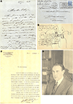
As stated above, in the decade from 1920 to 1930 the two most important researchers in cardio-respiratory physiology were Heinrich Hering (at the Köln University, Germany) and Jean-François Heymans, along with his son Corneille (both working at the University of Ghent, Belgium). Fernando de Castro presented his own results at successive meetings of the Association d’Anatomistes held at Liege (Belgium) in 1926, London (Great Britain) in 1927, and at Bourdeaux (France) in 1929, and together with the aforementioned publications, his work raised the attention of the entire scientific community interested in the field. During the last of these meetings, Professor Goormaghtigh, the chair of Pathology at the University of Ghent, offered him an invitation from Corneille Heymans to visit Ghent: the physio-pharmacologist was deeply interested in discussing De Castro’s work with him. Just after the meeting, Fernando De Castro visited his sister who lived in La Roche-Chalais (French Périgord, not far from Bourdeaux), where he received a letter from Heymans again inviting him to visit Ghent (Figure 4
A). De Castro accepted and his own description of the visit is better than any other:
Figure 4. Corneille Heymans reflects on the work of Fernando De Castro on the innervation of the carotid sinus and carotid body. (A) Letter dated the 28th March 1929, in which Heymans invites Fernando De Castro to share experiences at his laboratory at Ghent University. The letter was sent to “Chez Lagoubie, La Roche-Chalais, Dordogne” (see envelop in upper-right corner), the family name of De Castro’s French brother-in-law where the Spanish histologist was staying for a few days after the meeting of the Association d’Anatomistes held in Bourdeaux (France; see text, for details). (B) De Castro’s sketch showing two dogs in parabiosis (the famous physiological technique in which the Heymans –father and son- were consumed masters). The hand-written notes (in Spanish) complete the information about this experiments. (C) Letter from Heymans, dated 12th May 1930, in which he asks Fernando De Castro whether he can visit him at his laboratory in the Cajal Institute (Madrid) during his stay in Barcelona along the entire month of June, where he had been invited to teach. (D) Photographic portrait of Fernando De Castro (circa 1926), reflecting the aspect of the Spanish neurohistologist at the time of his studies on the innervation of the carotid body and sinus.
“I went to Ghent and I was with him [Heymans] for 2 or 3 days, during which we performed some experiments on the carotid sinus of dogs. At that time, he was not interested in the carotid body. Rather, he had studied the vasoconstriction phenomena on the cardio-aortic region and he had just started to study the carotid region. His work on the carotid body came afterwards, and he said to me “I’m deeply interested in your idea about this. It would be great if I could demonstrate it!”
6
This was Fernando De Castro’s first visit to the laboratory of Corneille Heymans in Ghent (April 1929), although there was to be a second one. However, the Belgian group of physiologists did not directly focus their studies on the function of the carotid body and they took years to abandon their hypothesis that the respiratory reflex originated in the carotid sinus. For example, although in his first work about the physiological reflexes originated in the carotid sinus, Heymans recognised that the anatomy and the histology of the sinus region had been described “recently, in a precise and detailed manner by De Castro”
7
, he did not pay attention to the glomus caroticum, and even less so to De Castro’s hypothesis on the function of this small structure in chemoreception (Heymans, 1929
). In their detailed studies the following year, Heymans and collaborators consecrated the primacy of the carotid sinus to detect changes in arterial pressure and in the concentration of H+, CO2 and O2 in the blood, even ahead of the respiratory centre in the brainstem. However, while they cited the work ofDe Castro (1928)
among the reference, they did not discuss his studies in the text (Heymans and Bouckaert, 1930
; Heymans et al., 1930
). This is likely to be the origin (entirely or in part) of the persistent failure to differentiate between the anatomical sites at which blood pressure and its composition are detected (Gallego, 1967
). In 1930, Corneille Heymans was invited by Professor Puche to give some conferences in Barcelona and to perform different exhibitions of his experiments at the University. To take advantage of this trip, Heymans wrote to De Castro to arrange a visit to the Cajal Institute in Madrid as he wanted to complete their scientific discussion held at Ghent the year before (Figure 4
C). Thus, it is not by chance that Heymans and his group published a study in 1931 (devoted to the study of bradycardia induced by different drugs) in which they explicitly accepted the hypothesis of De Castro on the chemoreceptor nature of the carotid body:
“…our experiments indicate that the starting point of the reflexes triggered by the injection of chemical substances may be localised in the region of the carotid bifurcation and, more exactly, in the glomus caroticum, as proposed by De Castro”
8
Perhaps even more explicit is the reference published in a second paper on the same subject a year later:
“…the substances injected in the common carotid [artery] abandon this slowly through the small arterial branches which have not been sutured, and then they can act for a relatively long period of time on the region of the carotid ganglion which is, as De Castro and we have demonstrated, the starting point of the reflexes triggered by the chemical excitants”
9
(Heymans et al., 1932
).
Coinciding with the second visit of De Castro to the Pharmacological Institute at the University of Ghent (where he repeated experiments on dogs together with Heymans; Figure 4
B), the Belgian physio-pharmacologists once and for all adopted the hypothesis of De Castro about the existence of chemoreceptors in the carotid body to detect chemical variations in the composition of blood. Accordingly, they then began to study the region of the glomus caroticum with greater impetus and the physiological importance of its chemoreceptors:
“The reflexogenic hypothesis of vascular sensitivity in the regions of the carotid sinus was already postulated by De Castro in 1928 based on experimental morphological observations”
10
Shortly after, Heymans brought together all the discoveries published by his group in a book (Heymans et al., 1933
). In this text, a complete scenario of the different types of cardio-respiratory reflexes was profiled and the authors reflected that changes in arterial pressure (detected by the baroreceptors from the carotid sinus) or in the chemical composition of blood (detected by the chemoreceptors of the glomus caroticum or carotid body) control respiratory frequency, heart rate and arterial pressure. From this year on, Heymans and his collaborators will cite this compilation and less frequently their initial publications on the subject (1929, 1930, 1931, 1932).
At this time, De Castro was using complex (and sometimes indirect) surgical approaches to experimentally demonstrate that the neurons of the glomus respond to changes in the chemical composition of blood. For this purpose, he developed different anastomosis between the glossopharyngeal, vagus and hypoglossal nerves to create artificial reflex arches, and they required the prior and detailed study of the regeneration of the sectioned preganglionic branches. These studies (De Castro, 1930
, 1933
, 1934
, 1937
), necessary to demonstrate the physiological role of arterial chemoreceptors, distracted Fernando de Castro from his original goal by raising other scientific questions, which led him to work with the world famous and reputed biologist Giusseppe Levi at Turin (Italy), both in 1932 and particularly in 1934. One of De Castro’s last collaborators, Prof. Jaime Merchán, can not understand why De Castro “did not try to put two living animals in parabiosis, a classical physiological technique and certainly easy enough for someone with the surgical skills of your grand-father [Fernando De Castro]” (personal communication). Is it possible that these parabiosis experiments, similar to those performed by the Heymans and by De Castro and Heymans, in Ghent (see above), were those he referred to at the end of his work published in 1928 (see above).
At this time, the octogenarian Santiago Ramón y Cajal was worried about the poor international repercussion of the studies of his disciples, who mostly published in Spanish (Penfield, 1977
), and in particular of the works of De Castro on the carotid sinus and carotid body (for example see the letters from Cajal to De Castro, dated February 18th, 1932 and March 15th, 1933–both conserved in the Fernando de Castro Archive). This led Cajal to take two significant steps: (i) from 1923 onwards, the journal published by the Cajal Institute (“Trabajos del Laboratorio de Investigaciones Biológicas”) was published in French as “Travaux du Laboratoire de Recherches Biologiques”; (ii) he selected Fernando De Castro to compile all the different Histological protocols and experimental procedures that made up the technical corpus of the Spanish School of Neurologists (or Cajal’s School), and to publish them in a book (Ramón y Cajal and De Castro, 1933
).
However, another young disciple of Cajal and a close friend of De Castro, Rafael Lorente de Nó, tried to draw the attention of their Maestro and of his colleague and friend to the error in their ways. In Lorente’s opinion, they should not forsake the study of the carotid region and of its physiological implications in order to start working on the regeneration of the nervous system in vitro, which seems clear after the letter written by Cajal on May 19th 1934, to De Castro who was ill in Turin (Italy) at that time:
“I received a letter from Lorente regretting, that after many years of work in a difficult histological speciality, with an excellent orientation and dominion of scientific bibliography, you have changed your bearing to work in a field that, if not exhausted, does not initially offer to the researcher unexpected fruits. When I answer him, I will inform him that both paths converge and not only will no harm be done, but it is favourable to air one’s intelligence in other scientific domains.”
(de Castro, 1972
).
This episode suggests that the detour in Fernando De Castro’s scientific career was adopted with Cajal’s agreement. Although there are additional events that may help understand the stance adopted by Cajal, as indicated in the letter above and including events associated with the research on arterial chemoreceptors, it seems clear that Lorente de Nó was not entirely wrong.
In Febraury1934, De Castro went to Turin to work with Levi, and there his life ran a serious risk due to a rare gastric haemorrhage. Cajal died in October 1934, when De Castro had returned to Madrid, provoking organizational changes at the Cajal Institute. However, at this time the political and social events in Spain (and other European countries) became more and more complicated, leading to the start of the Spanish Civil War in July 1936.
In 1938, the Nobel Committee evaluated over a hundred of proposals for the Nobel Prize in Physiology or Medicine; among them, and to cite only some of the most relevant names that of Hess, Houssay, Stanley, Sasaki, Lapique, Erlanger, Gasser, Cushing… and Cornelius Heymans
11
. Two direct collaborators of the Belgian physio-pharmacologists, Professor Liljestrand (member of the committee) and Ulf von Euler (a Nobel Prize winner in 1970) evaluated the candidature of Heymans
12
, and they left this testimony in the records of the Nobel Foundation regarding the prize for Physiology or Medicine:
“Hering’s work was twice submitted to special investigation. In 1932, the reviewer exposed some doubts about his qualification for a prize like this, taken into account the obvious analogy with previous research performed on the aortic nerves and those from their predecessors mentioned above [Pagano, Siciliano, Sollmann y Brown]; afterwards, it was considered the division of the prize between Hering and Heymans, but the Committee was still sceptic about the merits of the first one. Pagano was also nominated by his compatriots, but only in 1943”
In fact, when consulting the Nobel Foundation database, it can be seen that the Austro-German physiologist Heinrich Hering was proposed for the prize in 1932, 1933 and 1937, as well as sharing a nomination with the Louis Lapique and Corneille Heymans in 1934. But in 1938, no one nominated the physiologist from Köln for the Nobel Prize.
The case of Heymans merits certain attention because he was nominated for the Nobel Prize in 1934, 1936, 1938 (when it was awarded to him) and also in 1939. But, only in the proposals from 1938 and 1939 is there an explicit mention of his studies on the baro- and chemoreceptors of the blood, while on the previous occasions he was nominated for his studies on blood circulation in general. It also seems interesting that while he was nominated by the Hungarian professor Mansfeld in 1938, on all the other occasions he was supported by Belgian professors and physicians, including the deluge of proposals supporting his candidature in 1939 (either alone or sharing nomination with Louis Lapique and Walter Hess).
So, what about De Castro? A legend circulates that during the deliberation of the Nobel Committee over lunch, one of those present argued in favour of De Castro’s nomination and that another committee member, remembering the war that was desolating Spain at that time, replied: “But, does someone know if De Castro is still alive?”
13
. Once again, the database of the Nobel Foundation is clear (which opens all its documents to the public 50 years after the concession of each Prize): nobody, either from Spain or from any other country, nominated Fernando De Castro for the Nobel Prize in those years
14
. By contrast, the Spaniard Pío del Río-Hortega was nominated in 1929 by a professor at the University of Valladolid and in 1937 (in the middle of the Spanish Civil War) by two professors from the University of Valencia
15
. After carefully studying the database of the Nobel Foundation, it can be seen that Cajal supported only one nomination during his lifetime (together with another eight Spanish scientists and medical doctors) and that he did not lead the proposal: that of the French immunologist Richet in 1912
16
. It remains a mystery that during the 28 years after he was awarded the Nobel prize and as active and influential as he was until close to his death in 1934, the Spanish genius did not participate in these scientific jousts.
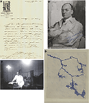
The draft written by De Castro to congratulate Heymans in receiving the Nobel prize, dated December 15th 1939, is conserved in his archives, as is the hand-written reply from the Nobel Prize dated December 29th of the same year (Figure 5
A). In the trail of the physiological studies of the Ghent group, Comroe discovered the chemoreceptors in the so called glomus aorticum (innervated by the depressor nerves), which fulfilled a minor role in the respiratory reflexes because they only respond to extreme cases of hypoxemia (Comroe, 1939
). Fernando De Castro recommenced his research into the fine structure and physiology of the carotid body at the end of the Spanish Civil War (April, 1939), despite the paucity of technical support, and the difficult personal and economical circumstances (Figures 5
C,D), which did not really change until the 1950’s
17
.
Figure 5. The long friendship of Corneille Heymans and Fernando De Castro. (A) Letter from Heymans, dated 29th December 1939, in response to a previous letter from Fernando De Castro (dated 15th December 1939) where he congratulated the Belgian physio-pharmacologist for the award of the Nobel Prize in Physiology or Medicine. (B) Picture of the 1939 portrait of Corneille Heymans, dedicated to De Castro (see hand-writing in blue at the bottom-right corner of the image); Fernando De Castro kept this dedicated picture on his bureau until he died in 1967. (C) De Castro prepared to perform one of his famous and complicated nerve anastomosis in a cat; note the precarious conditions of his laboratory in Madrid (circa 1941). (D) Part of a big type II baro-receptor (stained with methylene blue), original drawing from De Castro published in his first paper once the Spanish Civil War finished (De Castro, 1940
).
In the presentation of the Nobel Prize in Physiology or Medicine to professor Corneille Heymans at Ghent on January 1940
18
, the Swedish professor G. Liljestrand recognised the work of Fernando De Castro as fundamental in the path that Heymans followed towards his final success:
“Since the end of the 18th century we know of the existence of a curious structure in the region of the sinus, the glomus caroticum or carotid body which, in man, extends over only a few millimetres. The glomus consists of a small mass of very fine intertwining vessels arising from the internal carotid and enclosing various different types of cells. It has been considered by some as being a sort of endocrine gland similar to the medulla of the suprarenal glands. De Castro, however, in 1927 demonstrated that the anatomy of the glomus could in no way be compared to that of the suprarenal medulla. De Castro suggested rather that the glomus was an organ whose function was to react to variations in the composition of the blood, in other words an internal gustatory organ with special «chemo-receptors». In 1931, Bouckaert, Dautrebande, and Heymans undertook to find out whether these supposed chemo-receptors were responsible for the respiratory reflexes produced by modifications in the composition of the blood. By localized destruction in the sinus area they had been able to stop reflexes initiated by pressure changes, but respiratory reflexes could still continue to occur in answer to changes in the composition of the blood. Other experiments showed that Heymans’s concepts on the important role played by the glomus in the reflex control of respiration by the chemical composition of the blood were undoubtedly correct”
19
.
Due to the II World War, Heymans did not offer his Nobel lecture until December 12th, 1945 (when he received the prize de facto). It was then surprising that he barely cited the previous work of Fernando De Castro:
“Histological research carried out by de Castro, Meyling and Gosses, and our own experimental findings, obtained with J. J. Bouckaert and L. Dautrebande in particular, has led to the locating of the carotid sinus chemo-receptors in the glomus caroticum and of the presso-receptors in the walls of the large arteries arising from the carotid artery”
20
.
When asked both indirectly and directly by the interviewer if he could explain why Cornelius Heymans did not share the Nobel Prize he received in 1938 with him, Fernando De Castro explained:
“He performed the physiological demonstration, not the anatomical one. Obviously, there was a tremendous loss of time from 1936 to 1938 due to the war and because at that time I was performing the experiments on nervous anastomosis. This was to automatically register the phenomenon of the carotid reflexes on the eye, with the nervous anastomosis detecting chemical changes in the blood. Years after, I presented this work in a symposium held at Stockholm, in which the opening conference was entrusted to Heymans and the second to me. There, I presented the work that I couldn’t finish during the war. I had no more cats during that difficult time, they died of hungry or I was forced to sacrifice them. For this reason, I could not complete my experiments at that time” (Gómez-Santos, 1968
).
In this respect, the Chilean scientist Juan de Dios Vial, who worked with De Castro in the fifties, published interesting comments on this chapter of the life of the Spanish neuroscientis:
“The period of intense activity around 1930, which had ended by the proposal of the idea that the glomus was a chemoreceptor, was followed by a lull. This may be due to the fact that his contribution was widely ignored as coming from Spain. He opened the way to Heymans’ discoveries but did not receive due credit, even at the moment when the latter was awarded the Nobel Prize. 1 never heard De Castro himself refer to that circumstance, but his disciples and friends often did, and were somewhat bitter about it. These were also the years of Cajal’s death, and of organizational changes in his Institute”. (Vial, 1996
).
Dozens of letters sent by Corneille Heymans and his closest collaborators are conserved in Fernando De Castro’s archives, as well as several drafts of the correspondence maintained by De Castro with the different members of the Ghent School: the first letters sent by Heymans date from the early 1920’s, although most of the correspondence conserved corresponds to the period between 1930 and 1960. Some of these documents are especially significant, like: (i) the draft of the felicitation for the award of the Nobel Prize sent by De Castro to Heymans (mentioned above); (ii) the dedicated photograph of Heymans’ that was permanently in De Castro’s office (Figure 5
B); (iii) the correspondence associated with the invitation sent by De Castro to Corneille Heymans to take part in the symposium in honour of Cajal (held in Madrid in 1952 to commemorate the centenary of the birth of the founder of modern Neuroscience); (iv) the two invitations sent in 1952 by Bouckaert and Heymans himself inviting Fernando De Castro to take part in the symposium held in the honour of the Belgian Nobel prize winner; and even (v) a letter from Heymans’ wife (dated on February 17th, 1953) in which she personally welcomes De Castro to the aforementioned homage or inviting him to the wedding of Heymans’ daughter in 1957. All these are examples of the friendship between Corneille Heymans and Fernando De Castro, as published by the latter’s son: “based strongly on mutual admiration, their friendship persisted until April 15th 1967, the date of my father’s death”
21
.
How can we define the situation of the subject today? Different terminologies are used, indistinctly, for the same nerve: Hering’s nerve, sinus nerve, carotid sinus nerve and, in a minor extent, De Castro’s intercarotid nerve and the Latin one, ramus sinus carotici. It is generally accepted that this nerve is a branch of the glossopharyngeal nerve, single or double bundled, composed by fibres from the baroreceptors located in the wall of the carotid sinus and the fibres from the chemoreceptors placed in the carotid body. Minor contingents of fibres of this nerve belong to the sympathetic trunk and vagus nerve in variable number (Williams and Warwick, 1989
). A significant degree of variability in the innervation of the carotid body has been described with modern histological techniques and in different species (McDonald, 1983
; Ichikawa, 2002
; Milsom and Burleson, 2007
). A recent study on the innervation of the human carotid body confirms this heterogeneity and variability (Toorop et al., 2009
), but, in general, corroborates the observations published by Fernando De Castro 80 years ago and reviewed in this work.
The author declares that the research was conducted in the absence of any commercial or financial relationships that could be construed as a potential conflict of interest.
The work of our group is supported with grants from the following Spanish institutions: Ministerio de Ciencia e Innovación (SAF2007-65845), Consejerías de Sanidad (ICS06024-00) y de Educación (PAI08-0242-3822) de la Junta de Comunidades de Castilla-La Mancha, FISCAM (G-2008-C8 and PI2007-66), and Instituto de Salud Carlos III-FIS (RD07-0060-2007). Fernando de Castro is a researcher contracted by SESCAM. Fernando de Castro is the only grandson of Fernando De Castro, one of the characters of the present work. I am in debt with Dr. Javier Arenzana by his help on preparing the high-resolution figures for this paper. All the original drawings, letters and photographs used in this article belong to the Fernando De Castro Archives.
- ^ Ernst Heinrich Weber (1795) and Eduard Friedrich Wilhelm Weber (1806–1871) were born in Wittenberg (Germany). Ernst became professor of anatomy and physiology in Leipzig University in 1821 and worked in the nervous system and special senses. Eduard Weber became also professor in 1847 at Leipzig, and his most relevant contribution was the experiments on the vagus nerve referred here.
- ^ Together, all the physio-pharmacologists who worked with J.-F. and C. Heymans are collectively known as the “Ghent school” since the Institute for Pharmacology was situated in this Belgian city.
- ^ Translated into English by the author of this article.
- ^ Santiago Ramón y Cajal (1852–1934) and Camillo Golgi (1843–1926) shared the Nobel Prize in Physiology or Medicine in 1906. This was the first Nobel prize devoted to Neurosciences.
- ^ The sensitive ganglia of the IX craneal nerve are the Ehrenritter’s and Andersch’s ganglia, those from the X nerve are the jugular, the nodose and the petrose ganglia.
- ^ Translated into English by the author of this article.
- ^ 7Translated into English by the author of this article.
- ^ Translated into English by the author of this article.
- ^ 9Translated into English by the author of this article.
- ^ 10Translated into English by the author of this article.
- ^ Consulting the database of the Nobel Foundation, there were a total of 96 scientists nominated for the Nobel Prize in Physiology or Medicine in 1938.
- ^ Database of the Nobel Foundation: http://nobelprize.org/nomination/medicine/nomination.php?action=simplesearch&string=Heymans&start=11.
- ^ Professor Gunnar Grant’s personal communication (meeting of the Cajal Club; Stockholm, May 2006). This commentary referred to the fact that Madrid was sieged and in the front line most of the duration of the Spanish Civil War (July 1936-April 1939).
- ^ This information was corroborated by a letter dated April 3rd, 2007 written by the Administrator of the Nobel Committee in answer to the author of the present paper.
- ^ While his 1929 nomination was evaluated, that in 1937 was not..
- ^ 16Charles Richet was awarded the Nobel Prize in Physiology or Medicine in 1913 “in recognition of his work on anaphylaxis”.
- ^ The research on chemoreceptors was De Castro’s main research line till he died in April, 1967.
- ^ Although the II World War broke out in September 1939, it was not until May 10th 1940 that the German troops invaded Belgium, The Netherlands and Luxembourg in their race towards France. Thus, life in these countries would have been relatively normal in January 1940, which permitted this ceremony to be celebrated. However, these circumstances delayed holding the Nobel ceremony in Stockholm for 6 years.
- ^ Prof. G. Liljestrand (Karolinska Institutet); presentation of the 1938 Nobel Prize in Physiology or Medicine to Prof. C. Heymans (Ghent, Belgium; January 16th, 1940).
- ^ Nobel Lecture, Prof. C. Heymans (Stockholm, Sweden; December 12th, 1945).
- ^ F.-G. de Castro in letter to the Director of the newspaper “ABC” (Madrid) published on November 20th, 1974.
Heymans, C., Bouckaert, J. J., and Dautrebande, L. (1930). Sinus carotidien et réflexes respiratoires. II. Influences respiratoires réflexes de l’acidôse de l’alcalose, de l’anhydride carbonique, de l’ion hydrogéne et de l’anoxémie: Sinus carotidiens et échanges respiratoires dans le poumons et au delá des poumons. Arch. Int. Pharmacodyn. 39, 400–448.


