 Mohamed A. A. Mahdy1,2*†
 Mohammed S. Nasr Eldeen1
- 1Department of Anatomy and Histology, Faculty of Veterinary Medicine, King Salman International University, Ras Sudr, Egypt
- 2Department of Anatomy and Embryology, Faculty of Veterinary Medicine, South Valley University, Qena, Egypt
Background: Uterus didelphys is a rare congenital anomaly of the female reproductive tract characterized by a divided uterine cervix and body. It occurs due to abnormal development of the paramesonephric (Müllerian) duct. Different forms of uterus didelphys have been reported in several animal species, including bovine, equine, ewe, goat, swine, and bitch. However, there is no previous report that has documented a completely divided female genital tract in she-camel. Moreover, there is a lack of literature regarding this anomaly in animals. Therefore, the present study reports, for the first time, a rare case of a completely divided female genital tract in a she-camel. In addition, the existing relevant literature on uterus didelphys in different animal species is reviewed.
Case presentation: A female reproductive tract of she-camel, approximately 10 years old, with a history of previous successful pregnancy, was brought to the anatomy department following the slaughtering of the animal. Initial examination revealed a normal reproductive tract consisting of two ovaries, two fallopian tubes, a uterus, and a vagina. A closer examination revealed a completely divided vagina, with an external os opened into each part of the vagina, as well as a divided uterine body and cervix. Intrauterine infusion of saline through one external os confirmed complete separation of uterine body and cervix.
Conclusion: To the authors’ knowledge, this is the first reported case of a completely divided female genital tract in a she-camel. This review summarizes the previous reports about uterus didelphys in farm animals.
Introduction
Uterus didelphys is a rare congenital anatomical defect of the female reproductive tract of different animal species, including bovine, equine, sheep, goat, and pig (1–6). It happens due to the failure of fusion of the two paramesonephric (Müllerian) ducts, the primordium of the female reproductive tract. This congenital anomaly varies according to the degree of fusion failure of the two Müllerian ducts (7). A complete fusion failure of the two ducts results in uterus didelphys (true uterus didelphys), which is characterized by a completely divided genital tract including a double uterine body, two separated crevices, and a longitudinal vaginal septum (7–9). Partial fusion failure results in varying degrees of division affecting either the caudal part of the uterine body, cervix, or vagina, according to the area affected (7).
Abusineina (10) classified cervical abnormalities into four main types: uterus didelphys, complete double cervix, incomplete double cervix, and double external uterine orifices.
Uterus didelphys
Uterus didelphys (completely divided female genital tract) is caused by the persistence of the median walls of the Müllerian ducts along their entire length, resulting in two cervices and two separate uterine bodies.
Complete double cervix
It is caused by the persistence of the median walls of the Müllerian ducts along the whole length of the cervix, resulting in two cervices and one uterine body.
Incomplete double cervix
It is caused by the persistence of the median walls of the Müllerian ducts at the posterior part of the cervix, resulting in one cervical canal cranially and two cervical canals caudally.
Double external uterine orifices
It is caused by the persistence of the median walls of the Müllerian ducts at the external uterine orifice, resulting in one cervix with a band of tissue at the external os.
Different types of cervical abnormalities reported in different animal species are listed in Table 1. Clinical studies reported that congenital anomalies due to fusion failure of the Müllerian ducts are associated with reproductive difficulties (35). Ishiyama (36) reported that severe cases of incomplete fusion, such as double external os with blind diverticulum, complete double cervix with blind diverticulum, and uterus didelphys, are associated with infertility in cattle, while double cervix and external cervical os increase the incidence of dystocia in animals (33).
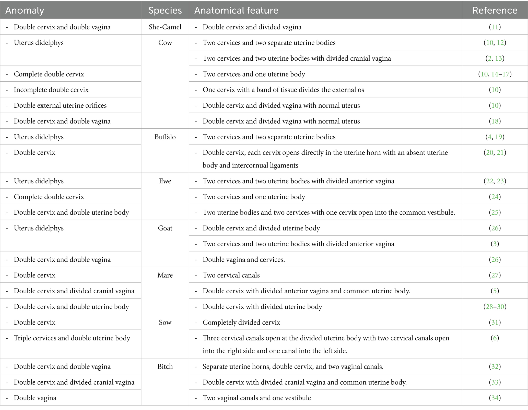
Table 1. Different reported types of cervical abnormalities in different animal species.
Although different forms of uterus didelphys have been reported in several animal species, only a case of double cervix and divided vagina has been reported in she-camel (11). There is no previous report that has documented a case of true uterus didelphys (completely divided female genital tract) in she-camel. Moreover, there is a lack of literature regarding this anomaly in animals. Therefore, the present study reports, for the first time, a rare case of a completely divided female genital tract in a she-camel. In addition to reviewing the existing relevant literature on uterus didelphys in different animal species, the current paper provides a brief summary of the anatomy, physiology, and development of the female reproductive tract in she-camels.
This condition is thought to be hereditary and associated with a recessive gene of unknown etiology. Cases of cervical duplication are detected incidentally at the time of breeding (33). The absence of Müllerian inhibiting substance (MIS) induces the differentiation of the Müllerian duct, forming the female reproductive tract (37). The cranial part that runs parallel to the mesonephric ducts and the transverse part that crosses the mesonephric duct develop, forming the epithelium of the fallopian tubes. The medial walls of the caudal fused part degenerate, forming a single canal (38) which develops into the epithelium of the uterus and cranial vagina (39). The species-specific morphological characteristics of the uterus (either simplex, bicornuate, or duplex) depend on the degree of fusion of Müllerian ducts being either complete, partial, or incomplete (38). The differentiation of Müllerian ducts is regulated by the members of Hoxa genes, specifically Hoxa9, Hoxa10, Hoxa11, and Hoxa13 (8, 37). The morphological diversity in the uterus among different animal species is due to different expression levels of Hoxa13 and Hoxd13gene (38). Other genes involved in the development of the Müllerian ducts include Emx2, Pax2, Lim1, and members of Wnt family 4 (Wnt4, Wnt5a, and Wnt7a) (8, 37, 40, 41).
Case presentation
A female reproductive tract of she-camel was brought to the anatomy department, Faculty of Veterinary Medicine, King Salman International University, Egypt, following slaughter at an abattoir in Giza governorate. Consultation with the owner revealed that the animal’s weight was approximately 270 kg and aged approximately 10 years old, with a history of previous successful pregnancy. Initial gross examination revealed a normal reproductive tract consisting of two ovaries and two flexuous fallopian tubes; each tube opened into a uterine horn. The two ovaries were normal and functional, as evidenced by the presence of some growing follicles on the left ovary and corpus luteum on the right one. The two horns were attached to a uterine body, followed by a cervix and vagina with no oblivious marks externally. The vulva and the caudal part of the vagina had been cut away. Closer examination of the reproductive tract revealed the presence of a completely divided vagina. Each division of the vagina had an external os opened into it (Figure 1). Morphometric measurements of the different parts of the female reproductive tract in this case were taken using a caliper. The data are listed in Table 2.
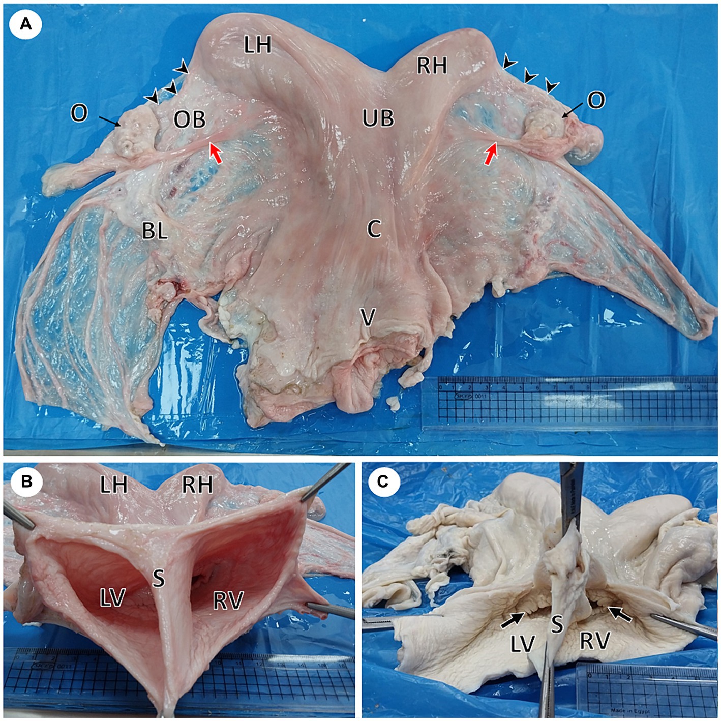
Figure 1. Gross photographs of female reproductive system of a she-camel show: (A) Apparently normal reproductive system consisted of right and left ovaries (O), each ovary was located inside an ovarian bursa (OB), the ovary attached to the uterine horn by the round ligament (red arrow), fallopian tubes (arrowheads), right (RH) and left (LH) horns, uterine body (UB), cervix (C), and vagina (V). Note the broad ligament of the uterus (BL). (B) Per vaginal view shows a completely divided vagina into the left (LV) and right (RV) vagina by a median septum (S). (C) The dorsal wall of the vagina was opened, showing each vagina had its external os (arrow).
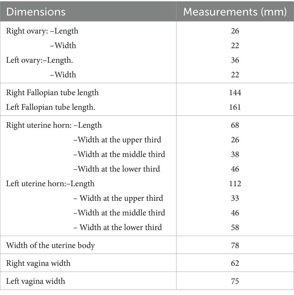
Table 2. Morphometric measurements of the female reproductive tract of the reported case.
Intrauterine infusion of saline solution through the left external os revealed complete separation of uterine body and cervix. The uterus was then preserved in 10% neutrally buffered formalin. Later, a longitudinal incision was performed through each uterine horn, passing through the uterine body, cervix, and vagina. A complete longitudinal septum extending from the fundus to the vagina was observed. Each horn was connected to a separate uterine body that had its own internal and external os (Figure 2).
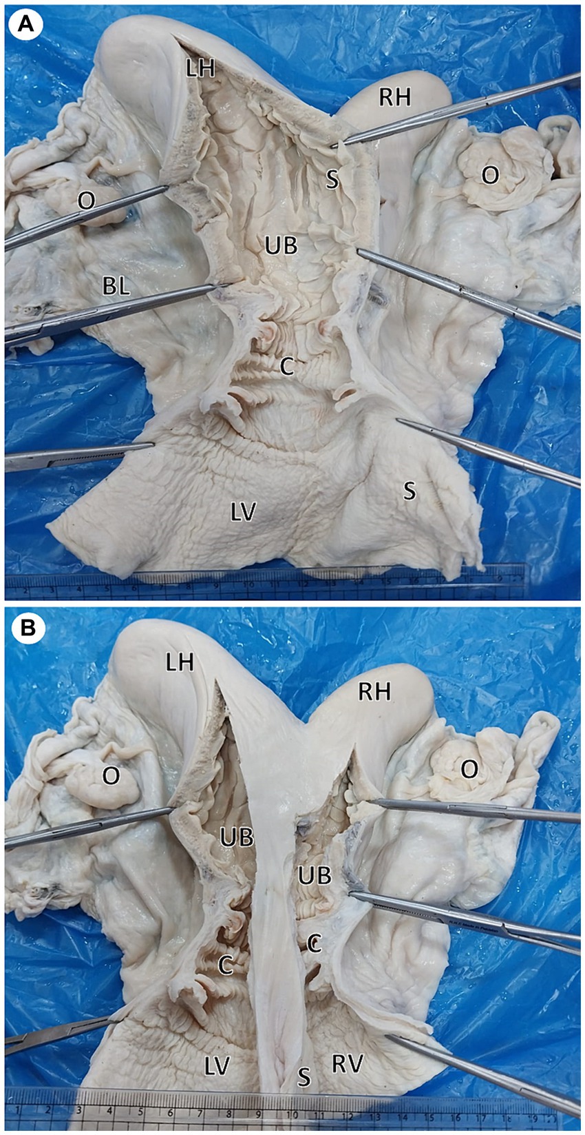
Figure 2. Gross photographs showing a dorsal view of uterus didelphys: (A) An incision was performed in the left horn (LH), body (UB), cervix (C), and vagina (LV). Note the complete septum extending from the fundus to the vagina. (B) Incised right (RH) and left (LH) horns show each horn connected to a separate uterine body that had its own internal and external os.
Discussion
The female reproductive tract develops from the paramesonephric (Müllerian) ducts. The Müllerian duct appears as a longitudinal invagination of the genital ridge lateral to the mesonephric (Wolffian) ducts. This invagination deepens and then separates from the peritoneal lining, forming a solid cord, which canalizes later (37, 39). The Müllerian ducts run lateral and parallel to the Wolffian ducts, with their cranial ends opening into the coelomic cavity with a funnel-like structure. The ducts pass caudomedially and ventrally, crossing the mesonephric ducts to fuse, forming the uterovaginal duct, with its caudal end projecting into the urogenital sinus, forming the Müllerian tubercle (8, 39).
Uterus didelphys is a rare congenital anomaly of the female reproductive tract that has been reported in different animal species (3, 4, 19, 20, 24, 25). Abusineina (10) classified cervical abnormalities into four main types: uterus didelphys, complete double cervix, incomplete double cervix, and double external uterine orifices. In the present case, the uterus was completely divided by a longitudinal septum, resulting in two uterine bodies, two separated cervices with their own internal and external os, and a completely divided vagina, indicating a case of uterus didelphys.
The condition has been attributed to the failure of fusion of the two paramesonephric (Müllerian) ducts. This congenital anomaly varies according to the degree of fusion failure of the two ducts (7). A complete fusion failure of the two ducts results in a double uterine body, the uterus didelphys. In this case, each uterine horn opens into a separate uterine body that leads to two separate crevices with a longitudinal vaginal septum (7–9).
The present case had functional ovaries, as indicated by the presence of growing follicles on the left ovary and a corpus luteum on the right one, which indicated normal cyclic activity of the ovary. Clinical studies have reported that congenital anomalies due to the fusion failure of the Müllerian ducts are associated with reproductive difficulties (35). Ishiyama, Nakamura (36) reported that severe cases of incomplete fusion, such as double external os with a blind diverticulum, complete double cervix with blind diverticulum, and uterus didelphys, are associated with infertility in cattle, while the double cervix and external cervical os increase the incidence of dystocia in animals (9, 33). Uterine anomalies restrict uterine space, which either alters or decreases the efficiency of the functional placenta, causing fetal growth deficiency (42). Moreover, artificial insemination is considered a challenge as the semen might be deposited in the cervix on the side opposite to the ovary from which ovulation has occurred. However, a cow with uterus didelphys had a normal birth after semen was introduced into each cervical canal (14). The present case had a history of previous successful pregnancies following natural mating and normal parturition. In accordance with the present data, reports claim that animals with uterus didelphys undergo a normal conception rate following natural mating (9, 20). Moreover, pregnancy has been reported in a cow (12) and an ewe (24) with uterus didelphys. Chethan and Singh (20) claimed that the condition is hereditary and associated with recessive genes of unknown etiology. Therefore, such animals should be excluded from breeding after diagnosis.
Uterus didelphys can be diagnosed during the pre-breeding examination, through a variety of methods, including physical examination using vaginoscopy, endoscopy, transrectal ultrasonography, and intrauterine injection of saline through one cervix to visualize the two separate uterine bodies with the median septum in between (28).
Molecular (DNA) marker-assisted selection and cytogenetic analyses linked to genes of interest with significant impacts on reproduction, such as WNT and HOXA genes, can be reinforced for the early selection of breeding females. This would aid in the development of cytogenetic profiles and molecular markers for diagnosing reproductive diseases with a genetic or physiological origin. This, in turn, would allow for the detection of animals with reproductive disorders and enable culling at an early stage (43).
In conclusion, the present study reported, for the first time, a rare case of uterus didelphys in a she-camel. The uterine body, cervix, and vagina were divided completely by a longitudinal septum extended from the fundus to the vagina. This rare case may have an educational role, either for the students studying the congenital abnormalities of the reproductive tract or for the practitioner/technician in the field, who should be alert for the pre-breeding diagnosis of conditions that may hinder female fertility. Moreover, the current study provides an overview of uterus didelphys in farm animals.
Data availability statement
The original contributions presented in the study are included in the article/supplementary material, further inquiries can be directed to the corresponding author.
Ethics statement
Ethical approval was not required for the study involving animals in accordance with the local legislation and institutional requirements because the study was done on slaughterhouse materials, so no need for ethical approval. Written informed consent was obtained from the owners of the animals for the publication of this case report.
Author contributions
MM: Writing – review & editing, Writing – original draft, Visualization, Validation, Supervision, Software, Project administration, Methodology, Investigation, Formal analysis, Data curation, Conceptualization. MN: Writing – review & editing, Writing – original draft, Visualization, Methodology, Investigation, Formal analysis.
Funding
The author(s) declare that no financial support was received for the research, authorship, and/or publication of this article.
Acknowledgments
The authors would like to thank Gamal Eldin A. Salem and Noha H. Hassan for providing the female reproductive tract of she-camel to the Anatomy Department, Faculty of Veterinary Medicine, King Salman International University.
Conflict of interest
The authors declare that the research was conducted in the absence of any commercial or financial relationships that could be construed as a potential conflict of interest.
Publisher’s note
All claims expressed in this article are solely those of the authors and do not necessarily represent those of their affiliated organizations, or those of the publisher, the editors and the reviewers. Any product that may be evaluated in this article, or claim that may be made by its manufacturer, is not guaranteed or endorsed by the publisher.
References
1. Ladds, PW. Congenital abnormalities of the genitalia of cattle, sheep, goats, and pigs. Vet Clin N Am Food Anim Pract. (1993) 9:127–44.
2. Raggio, I, Lefebvre, RC, Poitras, P, Vaillancourt, D, and Champagne, JP. Clinical diagnosis of uterine didelphia in Ayrshire heifer. Can Vet J. (2006) 47:899–901.
3. Timurkaan, N, and Ozer, H. Uterus didelphys in a goat. Vet Rec. (2002) 151:217. doi: 10.1136/vr.151.7.217
4. T Noaman, U, J Ali, A, I Azawi, O, and H Lazim, E. Uterus didelphys in a buffalo heifer: a case report. Iraqi J Vet Sci. (2009) 23:63–3. doi: 10.33899/ijvs.2009.5729
5. Volkmann, DH, and Gilbert, RO. Uterus bicollis in a Clydesdale mare. Equine Vet J. (1989) 21:71. doi: 10.1111/j.2042-3306.1989.tb02093.x
6. Morris, P. An unusual case in swine of uterus didelphys. Br Vet J. (1954) 110:205–7. doi: 10.1016/S0007-1935(17)50380-2
7. Immegart, H. CHAPTER 19 - infertility due to noninflammatory abnormalities of the tubular reproductive tract In: RS Youngquist and WR Threlfall, editors. Current therapy in large animal Theriogenology. Saint Louis: W.B. Saunders (2007). 153–7.
8. Wilson, D, and Bordoni, B. Embryology, Mullerian ducts (paramesonephric ducts). Treasure Island (FL): StatPearls Publishing (2023).
9. Noakes, D., Parkinson, T., and England, G., Infertility in the cow: Structural and functional abnormalities, management deficiencies and non-specific infections, in Arthur's veterinary reproduction and obstetrics. (2001), W. B. Saunders, Elsevier: China. p. 381–472.
10. Abusineina, ME. Anomalies of the cervix uteri of cattle. Br Vet J. (1970) 126:347–56. doi: 10.1016/S0007-1935(17)48297-2
11. Ali, A, Derar, DR, and Almundarij, TI. Infertility in female dromedary camels. J Camel Pract Res. (2021) 28:267–76. doi: 10.5958/2277-8934.2021.00042.4
12. Fathalla, M. Pregnancy in a cow with uterus didelphys. Aust Vet J. (2000) 78:616–6. doi: 10.1111/j.1751-0813.2000.tb11934.x
13. Moritomo, Y. Morphological observations of uterine and vaginal duplexes with a developmental anomaly at the vaginovestibular junction in a Japanese brown calf. J Vet Med Sci. (2004) 66:1165–6. doi: 10.1292/jvms.66.1165
14. Ajala, O, Oyeyemi, M, and Aderoju, O. Double cervix in a five-year-old white Fulami cow. Afr J Biomed Res. (2000) 3:201–2.
15. Millward, S, Mueller, K, Smith, R, and Higgins, HM. A post-mortem survey of bovine female reproductive tracts in the UK. Front Vet Sci. (2019) 6:451. doi: 10.3389/fvets.2019.00451
16. Hatipoglu, F, Ortatatli, M, Kıran, MM, Erer, H, and Çiftçi, MK. An abattoir study of genital pathology in cows: II. Uterus, cervix and vagina. Revue Méd Vét. (2002) 153:93–100.
17. Mimoune, N, Kaidi, R, Yassine, AM, Keddour, R, Belarbi, A, and Derdour, S-Y. Genital tract pathologies of cows slaughtered at El-Harrach abattoir in Algeria. Kafkas Üniversitesi Veteriner Fakültesi Dergisi. (2016) 22:639–646. doi: 10.9775/kvfd.2015.14737
19. Iqbal, U, Ijaz, M, Awan, F, Nawaz, M, Ali, S, and Mubeen, M. Uterine didelphys in Nili Ravi buffalo-a case report. Buffalo Bull. (2017) 36:713–5.
20. Chethan, SG, Singh, SK, Mathesh, K, Krishnaswamy, N, Behera, BK, and Kumar, H. Congenital anomalies of the uterus in riverine buffalo (Bubalus bubalis). Buffalo Bull. (2017) 36:581–7.
21. Herenda, D. An abattoir survey of reproductive organ abnormalities in beef heifers. Can Vet J. (1987) 28:33–7.
22. Smith, KC, Long, SE, and Parkinson, TJ. Congenital abnormalities of the ovine paramesonephric ducts. Br Vet J. (1995) 151:443–52. doi: 10.1016/S0007-1935(95)80133-2
23. Smith, KC, Long, SE, and Parkinson, TJ. Abattoir survey of congenital reproductive abnormalities in ewes. Vet Rec. (1998) 143:679–85.
24. Carr, D, Aitken, RP, Milne, JS, David, AL, and Wallace, JM. A case of successful pregnancy in a ewe with uterus didelphys. Reprod Domest Anim. (2013) 48:e78–80. doi: 10.1111/rda.12191
25. Nazem, MN, and Kheirandish, R. Ectopic cross ureter with uterus didelphys in a lamb. Comp Clin Pathol. (2016) 25:251–5. doi: 10.1007/s00580-015-2200-2
26. Beena, V, Pawaiya, RVS, Gururaj, K, Shivasharanappa, N, Singh, DD, Gangwar, NK, et al. Pathological studies of female reproductive tract in goats. Indian J Vet Pathol. (2016) 40:27–34. doi: 10.5958/0973-970X.2016.00005.5
27. Kania, J, Wilson, K, Raudsepp, T, Castaneda, C, Jevit, M, Horteloup, M, et al. Double cervices in a gypsy Vanner mare. Clinic Theriogenol. (2023) 15:9395. doi: 10.58292/ct.v15.9395
28. Kelly, GM, and Newcombe, JR. Uterus bicorpora bicollis as a possible cause of infertility in a mare. Vet Rec. (2009) 164:20–1. doi: 10.1136/vr.164.1.20
29. Blue, MG. A uterocervical anomaly (uterus bicorpor bicollis) in a mare, and the manual disruption of early bilateral pregnancies. N Z Vet J. (1985) 33:17–9. doi: 10.1080/00480169.1985.35137
30. Murcia-Robayo, R, Diaw, M, and Raggio, I. An unusual case of uterus didelphys in an infertile mare with mosaic X-chromosome aneuploidy. J Reprod Biol Endocrinol. (2018) 2:22–5.
31. Carvalho, LF, Oliveira, CJ, and Franceschini, PH. Malformations of the sexual organs of female pigs in a Brazilian abattoir. Vet Rec. (2004) 155:710–1. doi: 10.1136/vr.155.22.710
32. Coffman, JM, McConkey, M, Ellison, G, Huynh, E, and Dark, MJ. Complete urogenital and anorectal duplication in a dog. Case Rep Vet Med. (2019) 2019:3696978. doi: 10.1155/2019/3696978
33. Mahoney, D, Nebel, A, Whitacre, M, and Lyle, S. Cervical duplication in dogs. Clinic Theriogenol. (2022) 14:366–9. doi: 10.58292/ct.v14i4.9173
34. Vasiu, I, Moraru, MC, Cosma, C, Taulescu, M, McCartney, W, Yiapanis, C, et al. A rare case of a double vagina in a young American Staffordshire terrier bitch (Canis lupus familiaris). A case report. Res Vet Sci. (2022) 153:23–6. doi: 10.1016/j.rvsc.2022.10.014
35. Robertson, L, Ng, IH, Bonniwell, M, Ferguson, D, and Harvey, MJA. Developmental abnormalities of the reproductive tract associated with infertility in Highland heifers. Vet Rec. (1996) 138:396–7. doi: 10.1136/vr.138.16.396
36. Ishiyama, D, Nakamura, Y, Tanaka, T, Magata, F, Matsuda, F, and Maeda, KI. Severe incomplete fusion of the Müllerian ducts influences reproduction in Holstein cattle. Theriogenology. (2019) 123:209–15. doi: 10.1016/j.theriogenology.2018.09.035
37. Roly, ZY, Backhouse, B, Cutting, A, Tan, TY, Sinclair, AH, Ayers, KL, et al. The cell biology and molecular genetics of Müllerian duct development. WIREs Dev Biol. (2018) 7:e310. doi: 10.1002/wdev.310
38. Spencer, TE, Dunlap, KA, and Filant, J. Comparative developmental biology of the uterus: insights into mechanisms and developmental disruption. Mol Cell Endocrinol. (2012) 354:34–53. doi: 10.1016/j.mce.2011.09.035
39. Sinowatz, F In: P Hyttel, F Sinowatz, and M Vejlsted, editors. Development of the urogenital system in essentials of domestic animal embryology. New York: Elsevier: Edinburgh (2010). 252–85.
40. Robbins, JB, Broadwell, C, Chow, LC, Parry, JP, and Sadowski, EA. Müllerian duct anomalies: embryological development, classification, and MRI assessment. J Magn Reson Imaging. (2015) 41:1–12. doi: 10.1002/jmri.24771
41. Backhouse, B, Hanna, C, Robevska, G, van den Bergen, J, Pelosi, E, Simons, C, et al. Identification of candidate genes for Mayer-Rokitansky-Küster-Hauser syndrome using genomic approaches. Sex Dev. (2019) 13:26–34. doi: 10.1159/000494896
42. Meyer, KM, Koch, JM, Ramadoss, J, Kling, PJ, and Magness, RR. Ovine surgical model of uterine space restriction: interactive effects of uterine anomalies and multifetal gestations on fetal and placental growth. Biol Reprod. (2010) 83:799–806. doi: 10.1095/biolreprod.110.085381
Keywords: congenital abnormalities, Müllerian duct anomalies, she-camel, uterus didelphys, case report
Citation: Mahdy MAA and Nasr Eldeen MS (2024) True uterus didelphys in she-camel: a case report and review of literature. Front. Vet. Sci. 11:1419234. doi: 10.3389/fvets.2024.1419234
Edited by:
Khalid El Allali, Agronomic and Veterinary Institute Hassan II, MoroccoReviewed by:
Mohamed Rachid Achaaban, Agronomic and Veterinary Institute Hassan II, MoroccoMohammed Piro, Agronomic and Veterinary Institute Hassan II, Morocco
Copyright © 2024 Mahdy and Nasr Eldeen. This is an open-access article distributed under the terms of the Creative Commons Attribution License (CC BY). The use, distribution or reproduction in other forums is permitted, provided the original author(s) and the copyright owner(s) are credited and that the original publication in this journal is cited, in accordance with accepted academic practice. No use, distribution or reproduction is permitted which does not comply with these terms.
*Correspondence: Mohamed A. A. Mahdy, bW9oYW1lZC5lbG1haGR5QGtzaXUuZWR1LmVn
†ORCID: Mohamed A. A. Mahdy, https://orcid.org/0000-0001-6402-2944