 Yutaro Ito
 Miho Nagasawa
 Kahori Koyama2
 Kohei Ito
 Takefumi Kikusui
- 1BIOTA Inc., Tokyo, Japan
- 2Laboratory of Human-Animal Interaction and Reciprocity, Department of Animal Science and Biotechnology, Azabu University, Kanagawa, Japan
Introduction: The One Health concept is a comprehensive understanding of the interaction between humans, animals, and the environment. The cohabitation of humans and pets positively affects their physical, mental, and social well-being. It is recognized as an essential factor from the One Health perspective. Furthermore, a healthy balance in the gut microbiome is essential for good health, and the changes in the gut microbiome associated with cohabitation between humans and pets could potentially affect various aspects of the health of both hosts. Therefore, elucidating the sharing of gut bacteria between humans and pets associated with cohabitation is important for understanding One Health. However, most studies have examined sharing at the taxonomic level, and it remains unclear whether the same bacteria are transferred between humans and pets, and whether they mutually influence each other.
Methods: Here, microbiome analysis and shared 16S rRNA gene amplicon sequence variant (ASV) analysis were conducted before the start of cohabitation between humans and dogs, as well as at 2 weeks, 1 month, and 3 months after cohabitation.
Results: 16S rRNA gene ASVs analysis indicated that gut microbes have been transferred between humans and dogs. The overall structure of the gut microbiota within human–dog pairs remained unchanged after 3 months of adaptation. However, 11ASVs were shared within human–dog pairs. Many shared ASVs were highly abundant within each host, and this high abundance may be considered a factor that influences bacterial transfer between hosts.
Discussion: Our results provide important insights into the potential for the transfer of gut bacteria between humans and dogs. These findings are considered crucial for understanding the impact of human–dog cohabitation on various aspects of health.
1 Introduction
The concept of “One Health” is based on the comprehensive understanding that humans, animals, and the environment surrounding them are interconnected. It is a cross-disciplinary approach to solving problems through collaboration among people involved in human, animal, and environmental health. Therefore, discussions are underway to promote the comprehensive health of pets, who spend considerable time in the same environment as humans (1, 2). One of the central issues is the sharing of microbes and infectious diseases; issues related to zoonoses, in particular, have consistently received high attention.
Human residential microbiomes coexist in various body sites, such as the gut, skin, lungs, and oral cavity. It is estimated that the total number of bacteria in the 70 kg “reference man” is 38 trillion cells (3). The gut microbiome is the primary factor maintaining health. An imbalance due to external changes can lead to the development of cardiovascular diseases, cancer, respiratory diseases, diabetes, inflammatory bowel disease (IBD), brain diseases, chronic kidney diseases, and liver diseases (4, 5).
Human residential bacteria are substantially affected by multiple factors in the external environment, including living spaces (6, 7). Pets sharing living environments with humans have been reported to be a considerable factor influencing the taxonomic composition and phylogenetic diversity of the human gut and skin microbiomes via direct or indirect microbial transfer (8–14). Contact between humans and pets alters the composition of gut bacteria and potentially reduces the risk of allergic diseases in infants (9, 15, 16), and metabolic syndromes (17). The dog is regarded as the first domesticated animal (18). Domestic dogs are in daily contact with their owners and share their living environments. Regarding mental health, some studies have shown that dog ownership has an impact on improving human well-being through changes in physiological functions, such as endocrine regulation (19–21). Another study reported that the modification of dog microbiota by specific probiotics was reflected in the gut microbiome of children (22). Therefore, we must understand the impact of ecological interactions on microbial structures and how they affect human and dog health.
Although the effect of dogs on the human microbiome is considered substantial, most studies have discussed this at the taxonomic level. The direct transfer of gut microbes from dogs or coincidental sharing of the same taxa between humans and dogs is unclear. The dog gut microbiome was similar to the human gut microbiome, with 63% mapping to the human gene catalog (23), suggesting a possible interaction. In this study, we hypothesized that spending time with owners leads to microbial sharing between humans and dogs, resulting in similar gut microbiomes. To test this hypothesis, we analyzed microbial sharing at the amplicon sequence level.
2 Materials and methods
2.1 Study design
We examined 28 individual families with dogs and human participants between the ages of 20 and 72 years (48.5 ± 15.7; 13 males and 15 females), and dogs between the ages of 1 and 10 years old (4.4 ± 2.6; 18 males and 10 females; 5 pure breed, 23 mix breed; 16 stray, 8 breeder, 4 surrendered). The dogs were originally obtained from shelters and breeders and were adopted to Azabu University. These dogs were kept in a dog training facility in the university for 6 months, and then adopted to new families as part of an educational program at Azabu University that ran from 2015 to 2022 (24). All dogs were kept indoors, disease-free, not on medication, and were fed commercial dog food (including small amounts of treats). Detailed characteristics of the study are summarized in Table 1.
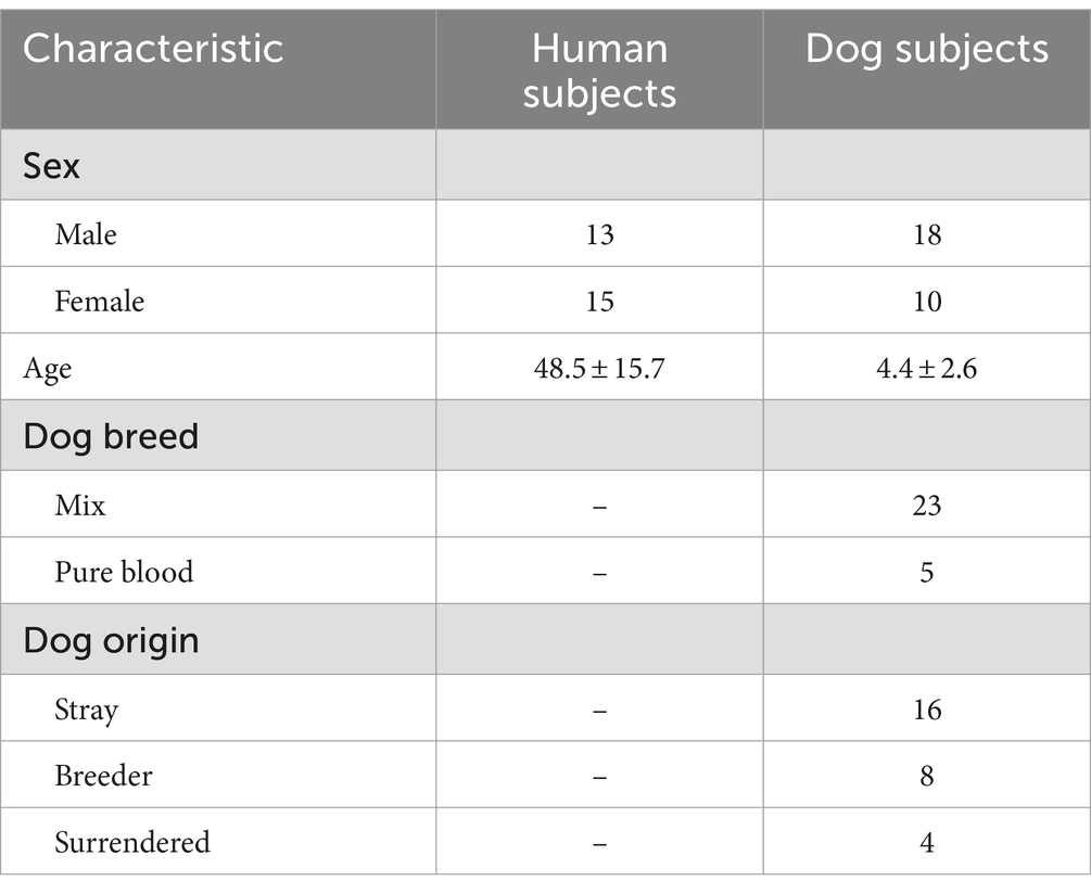
Table 1. Characteristics of human and dog subjects participating in this study (Kanagawa Japan, from 2015 to 2022).
2.2 Sample collection
Fecal samples were collected from both humans and dogs. Fecal samples were collected from the dogs at the facility where they were kept for 2–3 months prior to adoption and from the owners 1 week prior to adoption, as well as from both the owners and dogs at 2 weeks, 1 month, and 3 months after adoption. Regarding human samples, dog owners defecated on a fecal inspection sheet (Nagasale 0-9761-01, AS ONE Co. Ltd., Osaka, Japan) placed in the toilet. A portion of the fecal sample was scooped out without contact with water using disposable chopsticks, placed in a tube (CELL reactor filter cap centrifuge tube, 227245, Greiner Bio-one, Tokyo, Japan), and covered with a lid. Samples from dogs were collected indoors or outdoors using the same type of sheet when the dog defecated. When dogs defecated, such as during a walk, an uncontaminated portion of feces without soil or sand was collected using disposable chopsticks and placed in a tube under the same anaerobic conditions as those used for humans. The tube containing the fecal sample was placed in a pouch bag (A-58, Mitsubishi Chemical Corporation, Tokyo, Japan) together with AnaeroPack™-Anaero (A-03, Mitsubishi Gas Chemical Corporation, Tokyo, Japan) and made anaerobic. After collection, the samples were placed in a cooler box with frozen refrigerant, sealed, and refrigerated until the following day. Samples from three of the 28 pairs were stored in preservation solutions (RNAlater™ Stabilization Solution, AM7022, Invitrogen, Thermo Fisher Scientific Inc.) due to changes in transportation methods. This method is comparable to the immediate freezing (25). Immediately after defecation, a small amount of feces was removed with a disposable microspatula (1-9404-02, AS ONE Co. Ltd., Osaka, Japan), placed in a 1.5 mL tube containing RNAlater and sealed. These samples were stored in a freezer at −80°C in the laboratory until analysis.
2.3 Ethics
The study protocol was approved by the Animal Ethics Committee of Azabu University (#210325-12) and the Ethical Committee for Medical and Health Research Involving Human Subjects of Azabu University (#097). All procedures were conducted in accordance with the guidelines and regulations of the Ethics Committee. Informed consent was obtained from all participants, who were provided with detailed information about the study’s objectives, procedures, potential risks, and their right to withdraw at any time without penalty. To protect the privacy of participants, all personal identifiers were removed, and data were coded to maintain confidentiality and anonymity.
2.4 Total DNA extraction and high-throughput sequencing
Samples were treated with Lysis Solution F (NIPPON GENE Co., Ltd., Tokyo, Japan) and homogenized for 2 min at 1,500 rpm using a Shake Master Neo (Biomedical Science, Japan). The suspension was heat-treated at 65°C for 10 min, and centrifuged for 2 min at 12,000×g. DNA was extracted from the separated supernatant using a Lab-Aid824s DNA Extraction Kit (Zeesan Biotech Co., China) according to the manufacturer’s protocol. In addition, PCR reactions were conducted with the bacterial universal primers 1st-341f_MIX (5′-ACACTCTTTCCCTACACGACGCTCTTCCGATCT-NNNNN-CCTACGGGNGGCWGCAG-3′) and 1st-805r_MIX (5′-GTGACTGGAGTTCAGACGTGTGCTCTTCCGATCT-NNNNN-GACTACHVGGGTATCTAATCC-3′), to amplify the V3–V4 of the 16S rRNA gene. The thermal conditions were 94°C for 2 min, followed by 98°C for 10 s, 55°C for 30 s, and 68°C for 30 s, with a final extension at 68°C for 7 min. DNA samples, library preparation, and amplicon sequencing were performed using 300-bp paired-end sequencing on the MiSeq Reagent Kit v3 (Illumina Inc., San Diego, CA, USA) and Illumina MiSeq platform (Illumina Inc., San Diego, CA, USA) at the Bioengineering Lab. Co., Ltd. (Kanagawa, Japan).
2.5 Microbiome analysis
Microbiome analysis was performed as previously described (26). Briefly, the raw FASTQ data were imported into the QIIME2 platform (version 2023.5) as qza files (27). Denoising sequences and quality control were performed using QIIME dada2, which was shown to identify real variation at the finest scales in amplicon sequence data while outputting few false positives without constructing Operational Taxonomic Units (OTUs) (28). Sequences were then converted into amplicon sequence variants (ASVs). ASVs were assigned to the SILVA database SSU 138.1 using the QIIME feature-classifier classification scikit-learn package (29, 30). Subsequent analyses excluded ASVs classified as mitochondrial, chloroplast, or unassigned. To evaluate the effect of sequence read counts on microbiome diversity, we plotted changes in the Shannon diversity index, a measure of alpha diversity that accounts for both species richness and evenness, over a range of read counts from 0 to 10,000 using rarefaction curves. In the rarefaction curves, the number of ASVs leveled off when the number of reads reached approximately 4,000 (Supplementary Figure S1). Beta diversity indices weighted by UniFrac distances, which evaluate differences in microbial community structure based on distances, were visualized using principal coordinate analysis (PCoA). Data were visualized using ggplot2 (version 3.4.4) (31) and ggprism (version 1.0.4)1 (Creators Charlotte Dawson1 Show affiliations 1. University of Cambridge, no date; Wickham, no date) software.
2.6 Calculation of shared ASVs
Shared ASV analyses were performed as described previously (32). In the shared ASV analysis, to exclude noisy ASVs, we targeted ASVs with an abundance of more than 1% and defined shared ASVs as those that were shared between human and dog pairs at the same time point. The calculation was conducted using custom Python code2 with 0.01 for p-percentage.
2.7 Statistical analysis
Mann–Whitney U tests were used to compare Shannon diversity index and pairwise UniFrac distances to account for comparisons across sampling time courses. All multiple testing corrections were performed by computing False Discovery Rate using the Benjamini–Hochberg method, and Q-values (adjusted p-values) < 0.05 were considered statistically significant. Statistical tests were performed using SciPy (version 1.9.3) (33) and Scikit-bio (version 0.5.9).3 To compare differences in beta diversity (Weighted UniFrac distance) between species, for all PERMANOVA analyses, 5,000 trials were performed to assess statistical significance. To validate the abundance of genera in humans and dogs, we used the analysis of composition of microbiomes (ANCOM) (34).
3 Results
3.1 The taxonomic composition of gut microbiomes in humans and dogs
We analyzed the composition of the gut microbiota in humans and dogs. The most abundant genera in the human gut were Bifidobacterium, Blautia, Streptococcus, Bacteroides, and Faecalibacterium (Figure 1). Fusicatenibacter was significantly abundant only in humans, as determined by ANCOM (Supplementary Table S1). The most abundant genera in dog gut were Streptococcus, Blautia, Peptoclostridium, Fusobacterium, and Ruminococcus gnavus (Figure 1). Peptocrostridium and Blautia were significantly more abundant in dogs, compared to that in humans, using ANCOM (Supplementary Table S1). Blautia and Streptococcus were abundant in humans and dogs. The top five dominant genera in each host collectively represented 51.6% (Interquartile range [IQR] 42.0–63.7) in the human gut and 46.2% (IQR 33.0–63.7) in the dog gut, based on the median relative abundance (Supplementary Figure S2A). The Shannon diversity index, the most commonly used index to measure the alpha diversity of the gut microbiome (35), did not change throughout the three-month cohabitation period (Supplementary Figure S2B).
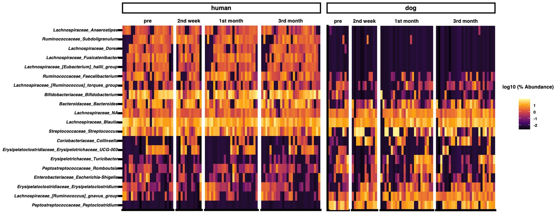
Figure 1. Taxon abundance heatmap at the genus level. Heatmap depicts the relative abundance (log10 scale) of the top 20 genera in humans and dogs.
3.2 The changes in microbial diversity and structures through the cohabitation of humans and dogs
We analyzed beta diversity to investigate the influence of cohabitation between humans and dogs on bacterial communities. According to PCoA using weighted UniFrac distances, two distinct clusters were formed based on host species (p = 0.00020 based on PERMANOVA), whereas no clusters were formed within species based on the duration of cohabitation (Figure 2A). The weighted UniFrac distances for each human–dog cohabitation pair were calculated and compared across different cohabitation periods, and no variations were observed based on the duration of cohabitation (Figure 2B). Calculations of changes in weighted UniFrac distances over the cohabitation period for the same individuals revealed that while the human gut microbiome did not change over time after cohabitation, the dog gut microbiome showed significant alterations during the first month of cohabitation (Figure 2C).
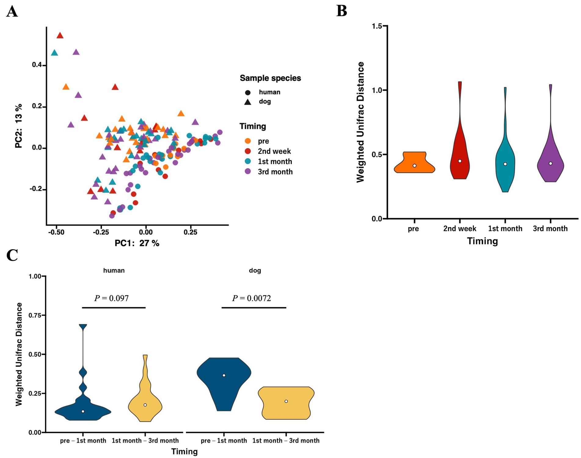
Figure 2. The gut microbiota structures in humans and dogs during each cohabitation period. Microbial profiles of gut microbiomes within human–dog pairs. (A) Principal coordinate analysis (PCoA) plot of human and dog gut microbiomes at each time point based on weighted UniFrac distance. (B) Violin plots of weighted UniFrac distance within each human–dog cohabitation pair at pre (human: n = 24, dog: n = 10), 2nd week (n = 12), 1st month (n = 25), and 3rd month (n = 28). Significance was calculated using the Mann–Whitney U tests, and the False Discovery Rate (FDR) was adjusted using the Benjamini–Hochberg method. (C) Violin plots of weighted UniFrac distance between pre-and 1st month (n = 10) and between 1st month and 3rd month (n = 25) for each individual human and dog. Significance was calculated using the Mann–Whitney U test.
3.3 Time-series changes in shared ASVs of the gut microbiota between humans and dogs
Although the overall gut microbiomes within human–dog pairs were not influenced by shared living conditions, the possibility of sharing the same taxon at the ASV level within each pair was considered. A total of 5,709 ASVs were obtained from all samples. Shared ASV analysis revealed that only 11 ASVs were shared within human–dog pairs during the 1st and 3rd month of cohabitation, but not the 2nd week of cohabitation (Table 2). Multiple ASVs were shared in some pairs, such as Pair_C, D and E. ASV001 and ASV002 were assigned to the R. gnavas group, the major bacterial genus in dog guts, and were shared across multiple pairs (Figure 3A). In one pair, these ASVs were exclusively identified in a dog sample at the first month and were later shared between humans and dogs at the third month. The other pairs shared ASVs at the same time point. ASV007, assigned to Faecalibacterium, was shared during the first month (Figure 3B). ASV was not detected at any other time points in either host strain. ASV010, assigned to Streptococcus, was detected only in the third month, with a relative abundance of 56.8% in the dog sample and was shared at this time point (Figure 3C). ASV005, which was assigned to Blautia, appeared simultaneously and was shared during the first month (Figure 3D). ASVs assigned to Prevotella_9, Erysipelactoclostridium, Fusobacterium, Lachnospiraceae, and Sutterella were also shared among several cohabitating pairs (Supplementary Table S2).
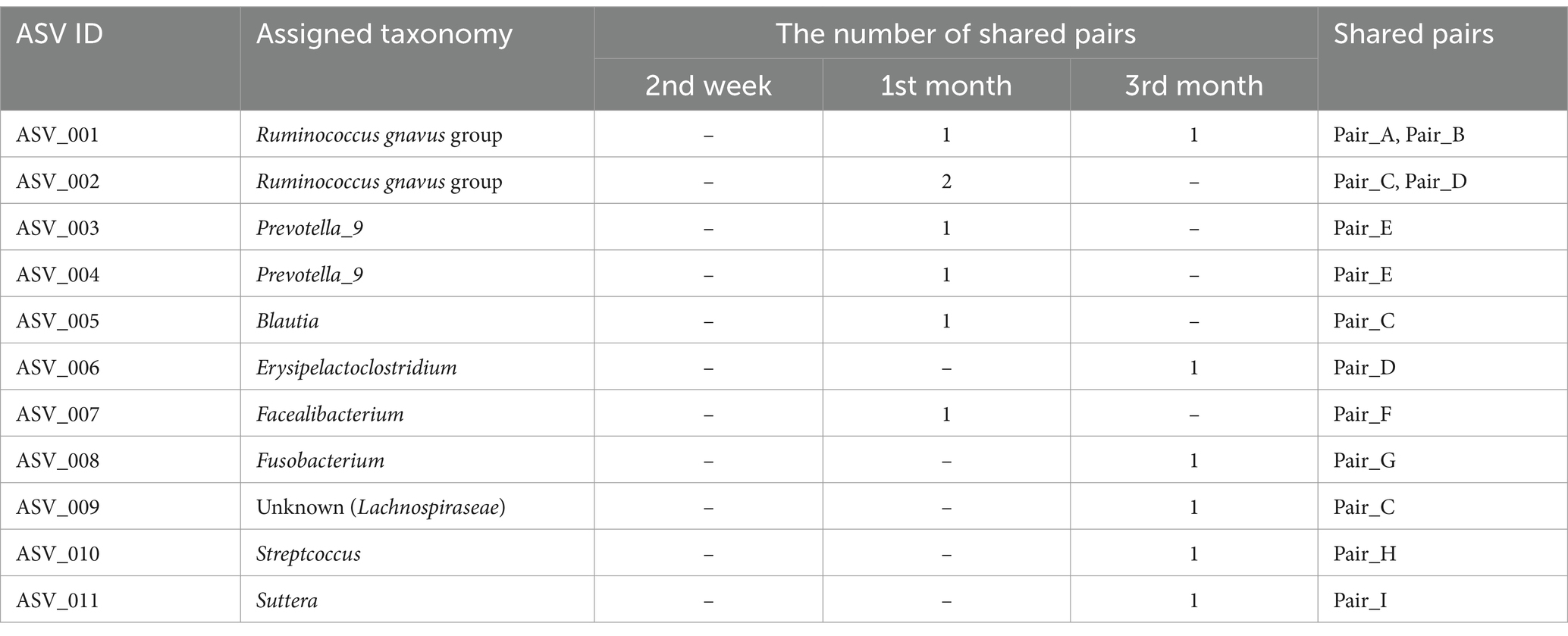
Table 2. Genera of shared ASVs and the number of pairs of samples sharing ASVs within human–dog cohabitation at each time point.
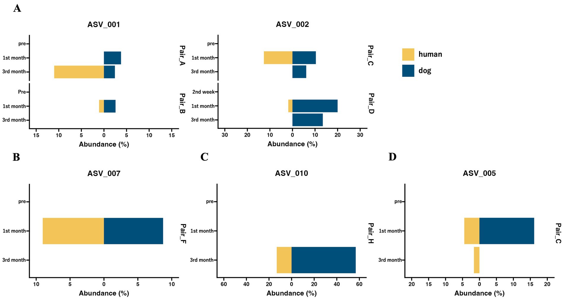
Figure 3. The abundance of shared ASVs within human–dog pairs at each time point. Butterfly chart of shared ASVs within cohabitating human–dog pairs based on relative abundance. The pair IDs represent cohabitating pairs of individuals, including both humans and dogs, that share ASVs. (A) ASV_001 and ASV_002 were assigned to the Ruminococcus gnavas group. (B) ASV_007 was assigned to Faecalibacterium. (C) ASV_010, which was assigned to Streptococcus. (D) ASV_005 is assigned to Blautia.
4 Discussion
In the present study, we showed that the five dominant genera in each host collectively constituted approximately half of the relative abundance in their respective hosts. The top five dominant genera in the human gut microbiome, Bifidobacterium, Blautia, Streptococcus, Bacteroides, and Faecalibacterium have been reported as major components of the Japanese human gut microbiome (36). The genera abundant in the gut microbiome of dogs, including Streptococcus, Blautia, Peptoclostridium, and Fusobacterium, have also been identified as major components of the dog gut microbiome (37). R. gnavas has been reported as the most abundant species in the dog gut (23), which was also identified in this study. The human and dog gut microbiota in this study were considered to have no significant differences compared to those in previous reports.
We evaluated the Shannon diversity index to investigate the impact of human–dog cohabitation on community diversity. However, no variation was observed in cohabitation duration. Previous studies have reported that alpha diversities in the human gut microbiome do not show substantial differences (10, 38), consistent with the current results. Beta diversity based on weighted UniFrac distances was compared over time, revealing no substantial changes in the overall microbial structures between pairs within the three-month cohabitation period. Similar to previous studies, in our study, the hosts (humans or dogs) were the main factors in explaining gut microbiota differences, and cohabitation did not seem to be one of the main factors affecting the overall gut microbiome composition (12, 22). Finally, we analyzed the temporal variations in beta diversity to elucidate the changes in within-species microbial structures due to cohabitation. The weighted UniFrac distance of dog gut microbiomes between the pretest period and first month was significantly different from the distance between the 1st and 3rd month. Changes in living conditions, such as diet and residence, when rescued dogs begin living with humans result in significant alterations in their gut microbiota during the early stages of cohabitation (39, 40). However, no temporal changes were observed in the human gut microbiota due to cohabitation, suggesting that compared to dogs, there is a limited impact of environmental changes caused by cohabitation.
Although a correlation within human–dog pairs was not observed when considering the entire microbiome, we considered the possibility of shared individual bacteria between humans and dogs. Previous studies have compared changes in the gut microbiota due to cohabitation at the level of bacterial genera or OTUs (10, 22, 38). However, these analyses did not determine whether the same bacteria were transferred or shared. To precisely evaluate the sharing of the gut microbiota between humans and dogs, we conducted a shared ASV analysis, which is an approach for inferring the sharing ratio of the microbiome at the ASV level between samples and has been used previously (32, 41, 42). While the sharing of ASVs was not detectable after 2 weeks of cohabitation, it was observed in the 1st and 3rd month. These results suggest that a cohabitation period of at least 1 month may be important for microbial sharing. On the other hand, the number of shared ASVs was similar at 1 month and 3 months of cohabitation. This suggests that, beyond the first month, the number of shared ASVs does not continue to increase over time but rather stabilizes at a certain level. Among the nine pairs where ASV sharing was confirmed, three pairs shared multiple ASVs, suggesting the possibility of conditions that facilitate the sharing of ASVs. The presence or absence of most of the shared ASVs fluctuated over time. It is known that gut bacteria fluctuate under various conditions, such as external environment, diet, and health status, and it is considered necessary to establish individual baselines through long-term observation for detailed analysis.
Among the 11 ASVs, six were classified as the dominant bacterial genera in the top five in each host. ASV001 and ASV002, assigned to the R. gnavas group, the common bacterial genera in the dog gut, were present in the dog samples of the shared pairs at more time points and were abundantly detected in the dog samples (Supplementary Table S3). These results suggested that the two ASVs were transferred from dogs to humans. Previous studies have demonstrated that Ruminococcus group 2 is more abundant in the guts of children with dogs, suggesting that Ruminococcus is easily transferred from dogs to humans (22). The R. gnavus group, found in abundance in the feces of human patients with IBD, produces polysaccharides and triggers the secretion of TNF-α from dendritic cells (43). The transfer of the R. gnavus group from dogs to humans is speculated to negatively affect human health. ASV007, assigned to Faecalibacterium, the major bacterial genus in the human gut, was shared in one pair and mainly detected in human samples (Supplementary Table S3). These results imply that the ASVs are shared from humans to dogs. Faecalibacterium is known as a beneficial bacterium in the human gut, where it is reduced in various diseases, including inflammatory bowel disease (IBD) (44). It may have beneficial effects when transferred to dogs. ASV010, assigned to Streptococcus mainly existed in dog samples (Supplementary Table S3) and was detected in the third month, with a relative abundance of 56.8% in the dog sample. Negative effects, such as an increase in the proportion of Streptococcus in the gut due to chronic inflammatory enteropathy in dogs, have been reported, and this may be one reason why ASV010 became dominant (45). It is possible that this temporary increase in proportion in dogs was associated with its transmission to humans, where it could also potentially cause negative effects. ASV005, assigned to Blautia, was significantly detected in the dog gut based on ANCOM, while Blautia is a genus abundantly present in both hosts. These results suggested that this ASV is shared between dogs and humans. Notably, this ASV was detected exclusively in the human gut 2 months after sharing. Blautia is the second most abundant genus in humans, and it is possible that Blautia transferred from dogs to readily colonize humans. OTUs classified as Blautia become more abundant in the human gut due to cohabitation with pets, supporting this hypothesis (38). Blautia is recognized for its potential probiotic function in the human gut and it is speculated that transferring Blautia from dogs to humans may have beneficial effects (46). Some anaerobic bacteria, such as Ruminococcus and Blautia, are transferred from dogs to humans. These results suggest that the transfer of bacteria may be triggered by cleaning feces containing a high abundance of anaerobic bacteria, similar to zoonotic infections (47). In conclusion, the pattern of this shared ASV suggests that the mutual sharing of bacteria between humans and dogs and high abundance are critical factors for interhost microbial transfer.
In this study, we aimed to analyze the interactions between the gut microbiota of adult humans and dogs. Consequently, there were very few (only 11) shared ASVs. Infants exposed to dogs at an early age have altered gut microbiota, which supports a potential mechanism explaining reduced atopy and asthma risk (48). The effect of microbial transfer may depend on the host age. Additionally, dog ownership increases the similarity of the skin microbiota between humans and dogs, rather than the gut microbiota (12). The closed nature of the intestinal environment may reduce the probability of bacterial transfer between hosts. In the future, it may be important to evaluate shared ASVs in various age groups and locations to better understand the interactions between humans and dogs.
This study has three limitations. First, we used amplicon sequences of the V3–V4 region of the 16S rRNA gene, which limited bacteria identification to the genus level (30). PCR amplification bias and differences in DNA extraction methods affect the accuracy of the relative composition of the gut microbiome (49, 50). Shared ASV analysis is a convenient way to track microbial sources, and the full-length 16S rRNA gene can be used to predict microbial sharing between samples more rigorously. The lineages of bacterial strains between the two groups can be compared using metagenome-assembled genomes, which show more precision in tracking at the whole-genome level, not just the restricted specific hypervariable regions of the 16S rRNA gene. The second limitation is an imbalance in the sample size at each time point. Unlike the first and third month, where 25 and 28 samples were collected, respectively, we conducted the analysis using only 10 samples from the dog’s pretest and 12 samples collected during the 2nd week. It cannot be ruled out that the lack of shared ASVs in the 2nd week samples may be attributed to the small sample size. Finally, we still need to demonstrate the impact of shared ASVs on each host. The shared ASVs analysis revealed the possibility of bacterial transfer. The influence of bacterial sharing on the health of each host could not be clarified. We believe that additional experiments, such as animal experiments using isolated bacteria demonstrated in this study, are necessary.
In conclusion, this study combined 16S rRNA gene amplicon and shared ASV analyzes to provide high-resolution evidence of gut microbiome transfer during cohabitation between humans and dogs. ASVs shared in the gut exhibited a high relative abundance in each host, suggesting that ASV sharing is more likely to occur in the dominant taxon. Many ASVs that were confirmed to be shared were the dominant taxa in each host. A larger sample size is needed in future studies to differentiate the effects of different living environments, dog breeds, host sex, host age, and time spent with dogs. Further analysis is required to determine the relevance of ASVs specifically shared by each individual to subsequent health risks.
Data availability statement
The datasets presented in this study can be found in online repositories. The names of the repository/repositories and accession number(s) can be found at: https://www.ncbi.nlm.nih.gov/, DRR542122–DRR542285.
Ethics statement
The studies involving humans were approved by Ethical Committee for Medical and Health Research Involving Human Subjects of Azabu University. The studies were conducted in accordance with the local legislation and institutional requirements. Written informed consent for participation in this study was provided by the participants’ legal guardians/next of kin. The animal studies were approved by Animal Ethics Committee of Azabu University. The studies were conducted in accordance with the local legislation and institutional requirements. Written informed consent was obtained from the owners for the participation of their animals in this study.
Author contributions
YI: Data curation, Formal analysis, Validation, Visualization, Writing – original draft, Writing – review & editing. MN: Conceptualization, Funding acquisition, Methodology, Supervision, Validation, Visualization, Writing – review & editing. KK: Data curation, Investigation, Writing – review & editing. KI: Data curation, Formal analysis, Investigation, Methodology, Writing – original draft, Writing – review & editing. TK: Conceptualization, Data curation, Formal analysis, Funding acquisition, Investigation, Methodology, Supervision, Validation, Writing – review & editing.
Funding
The author(s) declare that financial support was received for the research, authorship, and/or publication of this article. This work was supported by JSPS KAKENHI (Grant Numbers 21H05173 and 21H03333), for which MN was the principal investigator, and JSPS KAKENHI (Grant Number 23H05472) and JST (Grant NumberJPMJMI21J3), for which TK is the Principal Investigator.
Acknowledgments
All authors thank Morgenrot Inc. for providing the computational environment for the analysis. We would like to thank Editage (www.editage.jp) for English language editing.
Conflict of interest
KI is a board member at BIOTA Inc., Tokyo, Japan. YI is employed by BIOTA Inc. as a part-time developer.
The remaining authors declare that the research was conducted in the absence of any commercial or financial relationships that could be construed as a potential conflict of interest.
Publisher’s note
All claims expressed in this article are solely those of the authors and do not necessarily represent those of their affiliated organizations, or those of the publisher, the editors and the reviewers. Any product that may be evaluated in this article, or claim that may be made by its manufacturer, is not guaranteed or endorsed by the publisher.
Supplementary material
The Supplementary material for this article can be found online at: https://www.frontiersin.org/articles/10.3389/fvets.2024.1417461/full#supplementary-material
Footnotes
1. ^https://csdaw.github.io/ggprism/
2. ^q2-shared_asv, https://github.com/biota-inc/q2-shared_asv.
References
1. Overgaauw, PAM, Vinke, CM, van Hagen, MAE, and Lipman, LJA. A One Health perspective on the human-companion animal relationship with emphasis on zoonotic aspects. Int J Environ Res Public Health. (2020) 17:3789. doi: 10.3390/ijerph17113789
2. Zeigler, MK, and Vander Wyst, KB. Microbial associations and transfers across the One Health triad effects on human and animal adiposity and temperament: a protocol for an observational pilot study. Front Public Health. (2023) 11:1225188. doi: 10.3389/fpubh.2023.1225188
3. Sender, R, Fuchs, S, and Milo, R. Revised estimates for the number of human and bacteria cells in the body. PLoS Biol. (2016) 14:e1002533. doi: 10.1371/journal.pbio.1002533
4. Shreiner, AB, Kao, JY, and Young, VB. The gut microbiome in health and in disease. Curr Opin Gastroenterol. (2015) 31:69–75. doi: 10.1097/MOG.0000000000000139
5. Hou, K, Wu, Z-X, Chen, X-Y, Wang, J-Q, Zhang, D, Xiao, C, et al. Microbiota in health and diseases. Signal Transduct Target Ther. (2022) 7:135. doi: 10.1038/s41392-022-00974-4
6. Ito, K. Understanding the characteristics of the microbiomes of the built environment and their effects on the human body. Indoor Environ. (2023) 26:29–42. doi: 10.7879/siej.26.29
7. Gilbert, JA, and Stephens, B. Microbiology of the built environment. Nat Rev Microbiol. (2018) 16:661–70. doi: 10.1038/s41579-018-0065-5
8. Sitarik, AR, Havstad, S, Levin, AM, Lynch, SV, Fujimura, KE, Ownby, DR, et al. Dog introduction alters the home dust microbiota. Indoor Air. (2018) 28:539–47. doi: 10.1111/ina.12456
9. Tun, HM, Konya, T, Takaro, TK, Brook, JR, Chari, R, Field, CJ, et al. Exposure to household furry pets influences the gut microbiota of infant at 3-4 months following various birth scenarios. Microbiome. (2017) 5:40. doi: 10.1186/s40168-017-0254-x
10. Jiang, C, Cui, Z, Fan, P, and Du, G. Effects of dog ownership on the gut microbiota of elderly owners. PLoS One. (2022) 17:e0278105. doi: 10.1371/journal.pone.0278105
11. Torres, S, Clayton, JB, Danzeisen, JL, Ward, T, Huang, H, Knights, D, et al. Diverse bacterial communities exist on canine skin and are impacted by cohabitation and time. PeerJ. (2017) 5:e3075. doi: 10.7717/peerj.3075
12. Song, SJ, Lauber, C, Costello, EK, Lozupone, CA, Humphrey, G, Berg-Lyons, D, et al. Cohabiting family members share microbiota with one another and with their dogs. eLife. (2013) 2:e00458. doi: 10.7554/eLife.00458
13. Wetzels, SU, Strachan, CR, Conrady, B, Wagner, M, Burgener, IA, Virányi, Z, et al. Wolves, dogs and humans in regular contact can mutually impact each other’s skin microbiota. Sci Rep. (2021) 11:17106. doi: 10.1038/s41598-021-96160-7
14. Kuthyar, S, and Reese, AT. Variation in microbial exposure at the human-animal interface and the implications for microbiome-mediated health outcome. mSystems. (2021) 6:e0056721. doi: 10.1128/mSystems.00567-21
15. Azad, MB, Konya, T, Maughan, H, Guttman, DS, Field, CJ, Sears, MR, et al. Infant gut microbiota and the hygiene hypothesis of allergic disease: impact of household pets and siblings on microbiota composition and diversity. Allergy Asthma Clin Immunol. (2013) 9:15. doi: 10.1186/1710-1492-9-15
16. Nermes, M, Endo, A, Aarnio, J, Salminen, S, and Isolauri, E. Furry pets modulate gut microbiota composition in infants at risk for allergic disease. J Allergy Clin Immunol. (2015) 136:1688–90.e1. doi: 10.1016/j.jaci.2015.07.029
17. Arenas-Montes, J, Perez-Martinez, P, Vals-Delgado, C, Romero-Cabrera, JL, Cardelo, MP, Leon-Acuña, A, et al. Owning a pet is associated with changes in the composition of gut microbiota and could influence the risk of metabolic disorders in humans. Animals (Basel). (2021) 11:2347. doi: 10.3390/ani11082347
18. Wang, G-D, Zhai, W, Yang, H-C, Wang, L, Zhong, L, Liu, Y-H, et al. Out of southern East Asia: the natural history of domestic dogs across the world. Cell Res. (2016) 26:21–33. doi: 10.1038/cr.2015.147
19. Koyasu, H, Ogasawara, S, Kikusui, T, and Nagasawa, M. Ownership of dogs and cats leads to higher levels of well-being and general trust through family involvement in late adolescence. Front Vet Sci. (2023) 10:1220265. doi: 10.3389/fvets.2023.1220265
20. Nagasawa, M, Kikusui, T, Onaka, T, and Ohta, M. Dog’s gaze at its owner increases owner’s urinary oxytocin during social interaction. Horm Behav. (2009) 55:434–41. doi: 10.1016/j.yhbeh.2008.12.002
21. Endo, K, Yamasaki, S, Ando, S, Kikusui, T, Mogi, K, Nagasawa, M, et al. Dog and cat ownership predicts adolescents’ mental well-being: a population-based longitudinal study. Int J Environ Res Public Health. (2020) 17:884. doi: 10.3390/ijerph17030884
22. Gómez-Gallego, C, Forsgren, M, Selma-Royo, M, Nermes, M, Collado, MC, Salminen, S, et al. The composition and diversity of the gut microbiota in children is modifiable by the household dogs: impact of a canine-specific probiotic. Microorganisms. (2021) 9:557. doi: 10.3390/microorganisms9030557
23. Coelho, LP, Kultima, JR, Costea, PI, Fournier, C, Pan, Y, Czarnecki-Maulden, G, et al. Similarity of the dog and human gut microbiomes in gene content and response to diet. Microbiome. (2018) 6:72. doi: 10.1186/s40168-018-0450-3
24. Nagasawa, M, Mogi, K, and Kikusui, T. Continued distress among abandoned dogs in Fukushima. Sci Rep. (2012) 2:724. doi: 10.1038/srep00724
25. Wang, Z, Zolnik, CP, Qiu, Y, Usyk, M, Wang, T, Strickler, HD, et al. Comparison of fecal collection methods for microbiome and metabolomics studies. Front Cell Infect Microbiol. (2018) 8:301. doi: 10.3389/fcimb.2018.00301
26. Ito, K, Niwa, R, Yamagishi, Y, Kobayashi, K, Tsuchida, Y, Hoshino, G, et al. A unique case in which Kimoto-style fermentation was completed with leuconostoc as the dominant genus without transitioning to lactobacillus. J Biosci Bioeng. (2023) 135:451–7. doi: 10.1016/j.jbiosc.2023.03.007
27. Bolyen, E, Rideout, JR, Dillon, MR, Bokulich, NA, Abnet, CC, Al-Ghalith, GA, et al. Reproducible, interactive, scalable and extensible microbiome data science using QIIME 2. Nat Biotechnol. (2019) 37:852–7. doi: 10.1038/s41587-019-0209-9
28. Callahan, BJ, McMurdie, PJ, Rosen, MJ, Han, AW, Johnson, AJA, and Holmes, SP. DADA2: high-resolution sample inference from Illumina amplicon data. Nat Methods. (2016) 13:581–3. doi: 10.1038/nmeth.3869
29. Quast, C, Pruesse, E, Yilmaz, P, Gerken, J, Schweer, T, Yarza, P, et al. The SILVA ribosomal RNA gene database project: improved data processing and web-based tools. Nucleic Acids Res. (2013) 41:D590–6. doi: 10.1093/nar/gks1219
30. Bokulich, NA, Kaehler, BD, Rideout, JR, Dillon, M, Bolyen, E, Knight, R, et al. Optimizing taxonomic classification of marker-gene amplicon sequences with QIIME 2’s q2-feature-classifier plugin. Microbiome. (2018) 6:90. doi: 10.1186/s40168-018-0470-z
31. Villanueva, RAM, and Chen, ZJ. ggplot2: elegant graphics for data analysis (2nd ed.). Measurement. (2019) 17:160–7. doi: 10.1080/15366367.2019.1565254
32. Niwa, R, Chen, D, Seong, YA, Jo, K, and Ito, K. Direct contact of fermented rice bran beds promotes food-to-hand transmission of lactic acid bacteria. FEMS Microbiol Lett. (2023) 370:fnad120. doi: 10.1093/femsle/fnad120
33. Virtanen, P, Gommers, R, Oliphant, TE, Haberland, M, Reddy, T, Cournapeau, D, et al. SciPy 1.0: fundamental algorithms for scientific computing in Python. Nat Methods. (2020) 17:261–72. doi: 10.1038/s41592-019-0686-2
34. Mandal, S, Van Treuren, W, White, RA, Eggesbø, M, Knight, R, and Peddada, SD. Analysis of composition of microbiomes: a novel method for studying microbial composition. Microb Ecol Health Dis. (2015) 26:27663. doi: 10.3402/mehd.v26.27663
35. Samuthpongtorn, C, Nopsopon, T, and Pongpirul, K. Gut microbiome diversity measures for metabolic conditions: a systematic scoping review. bioRxiv. (2021). doi: 10.1101/2021.06.25.21259549
36. Takagi, T, Inoue, R, Oshima, A, Sakazume, H, Ogawa, K, Tominaga, T, et al. Typing of the gut microbiota community in Japanese subjects. Microorganisms. (2022) 10:664. doi: 10.3390/microorganisms10030664
37. Kubinyi, E, Bel Rhali, S, Sándor, S, Szabó, A, and Felföldi, T. Gut microbiome composition is associated with age and memory performance in pet dogs. Animals (Basel). (2020) 10:1488. doi: 10.3390/ani10091488
38. Kates, AE, Jarrett, O, Skarlupka, JH, Sethi, A, Duster, M, Watson, L, et al. Household pet ownership and the microbial diversity of the human gut microbiota. Front Cell Infect Microbiol. (2020) 10:73. doi: 10.3389/fcimb.2020.00073
39. Kim, J, An, J-U, Kim, W, Lee, S, and Cho, S. Differences in the gut microbiota of dogs (Canis lupus familiaris) fed a natural diet or a commercial feed revealed by the Illumina MiSeq platform. Gut Pathog. (2017) 9:68. doi: 10.1186/s13099-017-0218-5
40. Pilla, R, and Suchodolski, JS. The gut microbiome of dogs and cats, and the influence of diet. Vet Clin North Am Small Anim Pract. (2021) 51:605–21. doi: 10.1016/j.cvsm.2021.01.002
41. Kageyama, S, Furuta, M, Takeshita, T, Ma, J, Asakawa, M, and Yamashita, Y. High-level acquisition of maternal oral bacteria in formula-fed infant oral microbiota. MBio. (2022) 13:e0345221. doi: 10.1128/mbio.03452-21
42. Ito, K, Niwa, R, Kobayashi, K, Nakagawa, T, Hoshino, G, and Tsuchida, Y. A dark matter in sake brewing: origin of microbes producing a Kimoto-style fermentation starter. Front Microbiol. (2023) 14:1112638. doi: 10.3389/fmicb.2023.1112638
43. Henke, MT, Kenny, DJ, Cassilly, CD, Vlamakis, H, Xavier, RJ, and Clardy, J. Ruminococcus gnavus, a member of the human gut microbiome associated with Crohn’s disease, produces an inflammatory polysaccharide. Proc Natl Acad Sci USA. (2019) 116:12672–7. doi: 10.1073/pnas.1904099116
44. Martín, R, Rios-Covian, D, Huillet, E, Auger, S, Khazaal, S, Bermúdez-Humarán, LG, et al. Faecalibacterium: a bacterial genus with promising human health applications. FEMS Microbiol Rev. (2023) 47:fuad039. doi: 10.1093/femsre/fuad039
45. AlShawaqfeh, MK, Wajid, B, Minamoto, Y, Markel, M, Lidbury, JA, Steiner, JM, et al. A dysbiosis index to assess microbial changes in fecal samples of dogs with chronic inflammatory enteropathy. FEMS Microbiol Ecol. (2017) 93. doi: 10.1093/femsec/fix136
46. Liu, X, Mao, B, Gu, J, Wu, J, Cui, S, Wang, G, et al. Blautia-a new functional genus with potential probiotic properties? Gut Microbes. (2021) 13:1–21. doi: 10.1080/19490976.2021.1875796
47. Ghasemzadeh, I, and Namazi, SH. Review of bacterial and viral zoonotic infections transmitted by dogs. J Med Life. (2015) 8:1–5.
48. Panzer, AR, Sitarik, AR, Fadrosh, D, Havstad, SL, Jones, K, Davidson, B, et al. The impact of prenatal dog keeping on infant gut microbiota development. Clin Exp Allergy. (2023) 53:833–45. doi: 10.1111/cea.14303
49. Bonk, F, Popp, D, Harms, H, and Centler, F. PCR-based quantification of taxa-specific abundances in microbial communities: quantifying and avoiding common pitfalls. J Microbiol Methods. (2018) 153:139–47. doi: 10.1016/j.mimet.2018.09.015
Keywords: dogs, gut microbiome, human-pet microbial interaction, shared amplicon sequence variants, One Health
Citation: Ito Y, Nagasawa M, Koyama K, Ito K and Kikusui T (2024) Comparative analysis based on shared amplicon sequence variants reveals that cohabitation influences gut microbiota sharing between humans and dogs. Front. Vet. Sci. 11:1417461. doi: 10.3389/fvets.2024.1417461
Edited by:
Kate Worthing, The University of Sydney, AustraliaReviewed by:
Stephanie Salyer, Centers for Disease Control and Prevention (CDC), United StatesGeorge Golovko, University of Texas Medical Branch at Galveston, United States
Copyright © 2024 Ito, Nagasawa, Koyama, Ito and Kikusui. This is an open-access article distributed under the terms of the Creative Commons Attribution License (CC BY). The use, distribution or reproduction in other forums is permitted, provided the original author(s) and the copyright owner(s) are credited and that the original publication in this journal is cited, in accordance with accepted academic practice. No use, distribution or reproduction is permitted which does not comply with these terms.
*Correspondence: Takefumi Kikusui, dGFra2lrdUBjYXJhemFidS5jb20=; Kohei Ito, a29oZWlAYmlvdGEubmUuanA=
†These authors have contributed equally to this work and share first authorship