
94% of researchers rate our articles as excellent or good
Learn more about the work of our research integrity team to safeguard the quality of each article we publish.
Find out more
BRIEF RESEARCH REPORT article
Front. Vet. Sci. , 07 June 2024
Sec. Veterinary Epidemiology and Economics
Volume 11 - 2024 | https://doi.org/10.3389/fvets.2024.1411624
This article is part of the Research Topic Sentinels of Health: Advancements in Monitoring and Surveillance of Vector-Borne Diseases in Domestic and Wild Animals and Vectors View all 13 articles
 Gina Zanella1*
 Cécile Beck2José-Carlos Valle-Casuso3,4Madeline Anthony3,4Marilyn Cruz5Alberto Vélez5Rommel Lenin Vinueza6
 Gaëlle Gonzalez2
Domestic species, including equids, were introduced in the Galapagos Islands in the XIX century. Equine vector-borne diseases are circulating in South America but their occurrence in the Galapagos Island was unknown. The objective of this study was to detect the occurrence of West Nile virus (WNV), Usutu virus (USUV) and equine infectious anemia virus (EIAV) in the four Galapagos Islands raising equids if they were present at a prevalence >1%. Serum samples were collected from 411 equids belonging to 124 owners from April to July 2019. All the results were negative to the ELISA tests used suggesting that WNV, USUV and EIAV are not circulating in the equine population of the Galapagos Islands.
The Galapagos Islands are a set of insular territories 960 km off the coast of Ecuador and an Ecuadorian province. They have been listed as a UNESCO World Heritage Site. Only 3% of their territory is colonized while the remaining 97% belongs to the Galapagos National Park. The introduction of livestock into the islands dates back from the beginning of the XIX century. In 1832, there was a massive introduction of productive domestic species, including equids, which may have carried parasites or pathogens (1). In 2003, animal importation from mainland Ecuador was banned, except for day-old chicks. Equids are present among the four colonized islands: Santa Cruz, Isabela, San Cristóbal and Floreana. Equid movements are allowed between the Galapagos Islands for animal genetic improvement. Surveillance of viral equid disease through laboratory diagnosis had never been conducted. Considering the movement of animals between mainland Ecuador and the Galapagos Islands in the past, the likelihood of the occurrence of viruses affecting the equids into the islands could not be excluded. Vector-borne viruses could have also been introduced by vectors coming from mainland.
West Nile virus (WNV) and Usutu virus (USUV) are vector–borne viruses that can affect equids and humans (2). They belong to the family Flaviviridae, genus Flavivirus, and are responsible for multiple outbreaks of disease in different countries of the world. WNV and USUV transmission cycles include wild birds as amplifying hosts and ornithophilic mosquitoes as vectors but can also infect and cause disease in horses and humans, which serve as incidental dead-end hosts (3, 4). WNV is one of the pathogenic agents that can lead to equine neurological clinical signs, although the infection is not usually accompanied by presentation of clinical illness (5). WNV is endemic in parts of Africa, Europe, the Middle East, and Asia, and since 1999 has spread to North America, Mexico, South America, and the Caribbean (6). WNV has been detected in mainland Ecuador through serological surveys in equids (7) and also in Colombia, an Ecuadorian neighboring country (8). USUV is particularly pathogenic in a few species of birds and has been detected in many mammalian species, including equids, considered to be dead-end hosts (9). USUV has not been reported in Latin America but in view of the emergence of different arboviral diseases, such as chikungunya and Zika, it can be considered as a new emerging threat (10).
Equine infectious anemia (EIA) is caused by a lentivirus and can be transmitted mechanically by biting flies (the virus does not replicate in the vector) (11). EIA virus (EIAV) infection leads to recurring episodes of fever in equids, thrombocytopenia, and wasting symptoms. EIA has been detected in different regions in Brazil (12–14) and Argentina (15) and several outbreaks were reported in different regions in mainland Ecuador (16).
The present study aimed to detect the occurrence of WNV, USUV and EIAV in the Galapagos Islands where equids are raised.
The Galapagos veterinary services had estimated that around 650 equids are present in the islands of Santa Cruz (~ 230 equids), Isabela (~ 270 equids), San Cristóbal (~ 100 equids) and Floreana (~ 30 equids). The equids often belong to farmers who raise other species, mainly cattle, and are used for farm work, transportation or leisure purposes. There are approximately 132 equid owners in the four islands (63 in Santa Cruz, 34 in Isabela, 26 in San Cristobal, 9 in Floreana) and between one and 15 equids per farm.
For each island, the sample size was calculated to detect a 1% seroprevalence with a 95% level of confidence at the level of the equid population using Epitools.1 This yielded animal sample sizes of 144 in Santa Cruz, 171 in Isabela, 79 in San Cristobal and 24 in Floreana. A mean number of three equids per farm was used to estimate the total number of farms to be included in the study. A minimum of 10 animals were sampled per farm and in farms with less than 10 animals, all animals were tested. The farms and the animals were randomly selected.
Sera were tested using the commercial ID Screen West Nile competition multi-species ELISA kit (Innovative Diagnostics, Montpellier, France) according to manufacturer’s instructions. This method, that uses pre-coated plates with the envelop (E) protein of WNV, estimates the competition between antibodies present in the animal serum and monoclonal anti-WNV E antibody conjugated to horseradish peroxidase (HRP). This ELISA, through cross-reactions, allows the detection of other flaviviruses belonging to the Japanese encephalitis virus serocomplex as USUV (17). Assays were interpreted according to Beck et al. (18).
To detect antibodies against EIAV, serum samples were screened using a commercial ELISA, Equine Infectious Anemia Virus Antibody Test Kit, ELISA v2 (VMRD, Pullman, USA). Assays were performed according to the manufacturer’s instructions.
Serum samples were collected from 411 equids (367 horses, 20 donkeys and 24 mules) in 124 farms in the four islands (Table 1; Figure 1) from April to July 2019. The sampled animals were between 3 months and 30 years old.
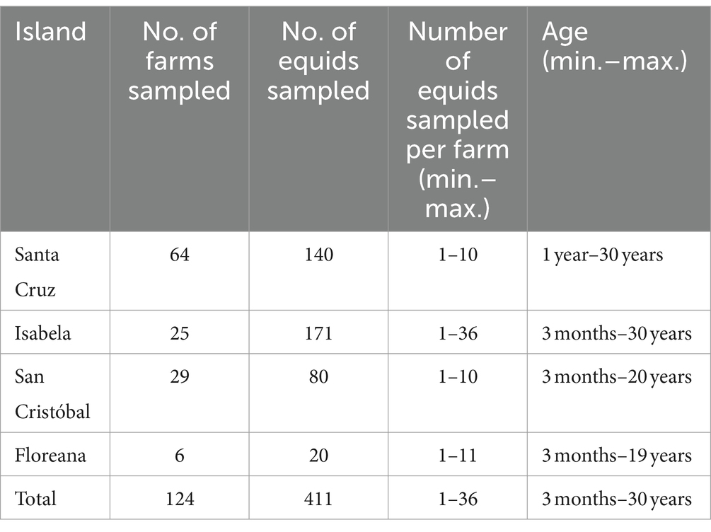
Table 1. Number of farms and equids sampled in the Galapagos Islands.
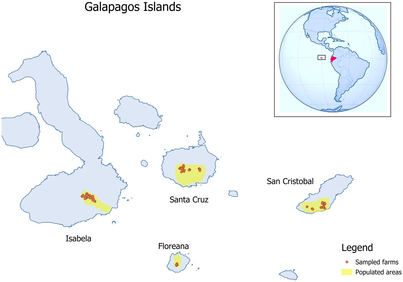
Figure 1. Location of the farms where equids where sampled to detect West Nile virus, Usutu virus and equine infectious anemia virus in 2019 in the Galapagos Islands. The georeferenced map was obtained in shape file format from the Geoportal of the Ecuadorian Military Geographic Institute and processed in Qgis 3.6.0 Noosa (QGIS.org, 2020).
All animals had negative results in the serological tests for the Flavivirus genus and EIAV.
The negative serological results obtained in this study suggest that the flavivirus that could affect equids, such WNV or USUV, and EIAV were not circulating in the Galapagos Islands above the prevalence value used to calculate the sample size for this study. This absence could go up to 30 years considering the age of some of the sampled equids.
The Galapagos Islands fill the climatic conditions favorable for the circulation of flaviviruses or EIAV provided the presence of competent vectors. The islands has three mosquito vectors capable of transmitting WNV and USUV (Culex quinquefasciatus, Aedes aegypti, and Aedes taeniorhynchus) (19, 20), bird species related to competent avian hosts (Dendroica petechia, Mimus spp) (21) and horseflies that can transmit EIAV (20). Since the climatic and vector conditions are filled, it could be argued that the prevalence viruses could be <1% in the equid population. However, in territories where those viruses are circulating, the prevalence values were higher than 1%. For example, WNV has been reported to be at seroprevalence values of 11% in horses in equids in Algeria (serums collected from 2015 to 2017) (22), 44.9% in Spain further to an outbreak in 2020 (23) or 31.6% in asymptomatic horses in Mexico (24). Analysis of 28,089 equids included in a systematic review and meta-analysis of seroprevalence studies of WNV in equids from 16 European countries between 2001 and 2018 revealed a pooled seroprevalence of 8% (25). In Morocco, prevalence of USUV was estimated to be 4% in a survey conducted in military working horses (26). In Spain, of 341 feral horses tested from 2010 to 2020, 9% were found seropositive to USUV (27). Regarding EIAV, in countries where this virus is circulating, prevalence values can be as high as 44% [survey conducted in Argentina (28)] but has also be found at lower values [2% in horses of the Para State in Brazil (14)]. On the other hand, according to the local veterinary services, there had been no reports of disease in equids in the Galapagos Islands consistent with WNV, USUV or EIAV or mortalities linked with WNV in birds.
The fact that the Galapagos Islands have semi-isolated conditions and the ban on animal importation do not provide sufficient guarantees to prevent the introduction of these viruses. Their introduction could arise by windborne transportation of infected vectors, infected migratory birds or infected vectors carried by airplanes or ships. These introduction routes were explored by Kilpatrick et al. (29) through a quantitative risk assessment to predict the WNV introduction into Galapagos Islands. These authors also included day-old chicks imports and mosquitoes in the larval stage present in shipment of tires as likely sources of WNV infection. They found that mosquitoes transported on airplanes carrying tourists represented the highest risk of WNV reaching the Galapagos Islands by a vector pathway and that the risk of introduction through migratory birds was lower but non-negligible. Moreover, it has been estimated that 107 Diptera species have arrived on the Galapagos Islands through human introductions and that 42 species have naturally colonized the islands from mainland Americas (20). Bataille et al. (30) showed from the monitoring of aeroplanes and genetic analysis that C. quinquefasciatus was regularly introduced via aircraft into the Galapagos islands. Therefore, effective control methods should be implemented by the national authorities to minimize the likelihood of introduction of insects through these routes that could put in danger endemic wildlife of Galapagos as well as domestic animal populations. Those measures could be combined with surveillance measures to detect any possible introduction. As the equid population is naïve to WNV and EIAV it is possible that that they could display clinical signs and, hence, equid owners should be made aware of the importance to report the occurrence of unexpected clinical cases. Future serum sampling carried out for other diseases in equids could also be used to detect flavivirus or EIAV. Hence, equids could act as sentinels to provide early warning of virus circulation.
The raw data supporting the conclusions of this article will be made available by the authors, without undue reservation.
The animal studies were approved by Agencia de Regulación y Control de la Bioseguridad y Cuarentena para Galápagos (ABG), Puerto Ayora, Ecuador. The studies were conducted in accordance with the local legislation and institutional requirements. Written informed consent was obtained from the owners for the participation of their animals in this study.
GZ: Conceptualization, Data curation, Formal analysis, Investigation, Methodology, Validation, Writing – original draft, Writing – review & editing. CB: Formal analysis, Resources, Writing – review & editing. J-CV-C: Formal analysis, Resources, Writing – review & editing. MA: Formal analysis, Writing – review & editing. MC: Resources, Supervision, Validation, Writing – review & editing. AV: Resources, Supervision, Validation, Writing – review & editing. RV: Data curation, Validation, Writing – review & editing. GG: Formal analysis, Resources, Validation, Writing – review & editing.
The author(s) declare that no financial support was received for the research, authorship, and/or publication of this article.
We would like to thank the equid owners who accepted to participate in the study. Thanks also go to technicians from Agencia de Regulación y Control de la Bioseguridad y Cuarentena para Galápagos (ABG).
The authors declare that the research was conducted in the absence of any commercial or financial relationships that could be construed as a potential conflict of interest.
All claims expressed in this article are solely those of the authors and do not necessarily represent those of their affiliated organizations, or those of the publisher, the editors and the reviewers. Any product that may be evaluated in this article, or claim that may be made by its manufacturer, is not guaranteed or endorsed by the publisher.
1. Toral-Granda, MV, Causton, CE, Jager, H, Trueman, M, Izurieta, JC, Araujo, E, et al. Alien species pathways to the Galapagos Islands, Ecuador. PLoS One. (2017) 12:e0184379. doi: 10.1371/journal.pone.0184379
2. Agliani, G, Giglia, G, Marshall, EM, Grone, A, Rockx, BHG, and van den Brand, JMA. Pathological features of West Nile and Usutu virus natural infections in wild and domestic animals and in humans: A comparative review. One Health. (2023) 16:100525. doi: 10.1016/j.onehlt.2023.100525
3. Lim, SM, Koraka, P, Osterhaus, AD, and Martina, BE. West Nile virus: immunity and pathogenesis. Viruses. (2011) 3:811–28. doi: 10.3390/v3060811
4. Golding, M, Seechurn, N, Baylis, M, and Johnson, N. JMM Profile: Usutu virus. J Med Microbiol. (2023) 72:001652. doi: 10.1099/jmm.0.001652
5. Castillo-Olivares, J, and Wood, J. West Nile virus infection of horses. Vet Res. (2004) 35:467–83. doi: 10.1051/vetres:2004022
6. Charrel, RN, and de Lamballerie, X. West Nile virus, an emerging arbovirus. Presse Med. (2004) 33:1521–6. doi: 10.1016/S0755-4982(04)98977-4
7. Coello-Peralta, R. Serological test for West Nile virus in horses of Los Ríos, Ecuador. Revista Ciencia UNEMI. (2016) 9:59–62. doi: 10.29076/issn.2528-7737vol9iss20.2016pp59-62p
8. Mattar, S, Komar, N, Young, G, Alvarez, J, and Gonzalez, M. Seroconversion for West Nile and St. Louis encephalitis viruses among sentinel horses in Colombia. Mem Inst Oswaldo Cruz. (2011) 106:976–9. doi: 10.1590/S0074-02762011000800012
9. Roesch, F, Fajardo, A, Moratorio, G, and Vignuzzi, M. Usutu virus: an arbovirus on the rise. Viruses. (2019) 11:640. doi: 10.3390/v11070640
10. Paniz-Mondolfi, AE, Villamil-Gomez, WE, and Rodriguez-Morales, AJ. Usutu virus infection in Latin America: A new emerging threat. Travel Med Infect Dis. (2016) 14:641–3. doi: 10.1016/j.tmaid.2016.08.004
11. Cook, RF, Leroux, C, and Issel, CJ. Equine infectious anemia and equine infectious anemia virus in 2013: a review. Vet Microbiol. (2013) 167:181–204. doi: 10.1016/j.vetmic.2013.09.031
12. Barros, ML, Borges, AMC, Oliveira De, ACS, Lacerda, W, Souza De, A, and Aguiar, DM. Spatial distribution and risk factors for equine infectious anaemia in the state of Mato Grosso. Brazil Rev Sci Tech. (2018) 37:971–83. doi: 10.20506/rst.37.3.2900
13. Cursino, AE, Vilela, APP, Franco-Luiz, APM, de Oliveira, JG, Nogueira, MF, Junior, JPA, et al. Equine infectious anemia virus in naturally infected horses from the Brazilian Pantanal. Arch Virol. (2018) 163:2385–94. doi: 10.1007/s00705-018-3877-8
14. de Padua, BR, Dias, RA, Fioravanti, MCS, and Borsanelli, AC. Seroprevalence and risk factors associated with equine infectious anemia in the state of Goias, Brazil. Prev Vet Med. (2022) 209:105781. doi: 10.1016/j.prevetmed.2022.105781
15. Hebert, L, Polledo, G, Lecouturier, F, Giorgi, M, Beck, C, Lowenski, S, et al. Serological evidence of equine infectious anaemia, West Nile fever, Surra and equine piroplasmosis in a herd of horses in northern Argentina. Vet Parasitol Reg Stud Reports. (2021) 24:100566. doi: 10.1016/j.vprsr.2021.100566
16. Agrocalidad. Programa nacional sanitario equino. Ministerio de agricultura, ganaderia acuacultura y pesca del Ecuador (2016).
17. Chevalier, V, Marsot, M, Molia, S, Rasamoelina, H, Rakotondravao, R, Pedrono, M, et al. Serological evidence of West Nile and Usutu viruses circulation in domestic and wild birds in wetlands of Mali and Madagascar in 2008. Int J Environ Res Public Health. (2020) 17:1998. doi: 10.3390/ijerph17061998
18. Beck, C, Leparc-Goffart, I, Desoutter, D, Deberge, E, Bichet, H, Lowenski, S, et al. Serological evidence of infection with dengue and Zika viruses in horses on French Pacific Islands. PLoS Negl Trop Dis. (2019) 13:e0007162. doi: 10.1371/journal.pntd.0007162
19. Peck, SB, Heraty, J, Landry, B, and Sinclair, BJ. Introduced insect fauna of an oceanic archipelago: the Galápagos Islands, Ecuador. Am Entomol. (1998) 44:218–37. doi: 10.1093/ae/44.4.218
20. Sinclair, BJ. An annotated checklist of the Diptera of the Galapagos archipelago (Ecuador). Zootaxa. (2023) 5283:1–102. doi: 10.11646/zootaxa.5283.1.1
21. Eastwood, G, Goodman, SJ, Hilgert, N, Cruz, M, Kramer, LD, and Cunningham, AA. Using avian surveillance in Ecuador to assess the imminence of West Nile virus incursion to Galapagos. EcoHealth. (2014) 11:53–62. doi: 10.1007/s10393-014-0911-5
22. Laabassi, F, Dheilly, N, Beck, C, Amaral, R, Gonzalez, G, Gaudaire, D, et al. Serological evidence of circulation of West Nile virus in equids in Algerian eastern drylands and its epidemiological risk factors. Comp Immunol Microbiol Infect Dis. (2023) 94:101947. doi: 10.1016/j.cimid.2023.101947
23. Garcia-Bocanegra, I, Franco, JJ, Leon, CI, Barbero-Moyano, J, Garcia-Mina, MV, Fernandez-Molera, V, et al. High exposure of West Nile virus in equid and wild bird populations in Spain following the epidemic outbreak in 2020. Transbound Emerg Dis. (2022) 69:3624–36. doi: 10.1111/tbed.14733
24. Alonso-Padilla, J, Loza-Rubio, E, Escribano-Romero, E, Cordoba, L, Cuevas, S, Mejia, F, et al. The continuous spread of West Nile virus (WNV): seroprevalence in asymptomatic horses. Epidemiol Infect. (2009) 137:1163–8. doi: 10.1017/S0950268809002325
25. Metz, MBC, Olufemi, OT, Daly, JM, and Barba, M. Systematic review and meta-analysis of seroprevalence studies of West Nile virus in equids in Europe between 2001 and 2018. Transbound Emerg Dis. (2021) 68:1814–23. doi: 10.1111/tbed.13866
26. Durand, B, Haskouri, H, Lowenski, S, Vachiery, N, Beck, C, and Lecollinet, S. Seroprevalence of West Nile and Usutu viruses in military working horses and dogs, Morocco, 2012: dog as an alternative WNV sentinel species? Epidemiol Infect. (2016) 144:1857–64. doi: 10.1017/S095026881600011X
27. Magallanes, S, Llorente, F, Ruiz-Lopez, MJ, Martinez-de la Puente, J, Soriguer, R, Calderon, J, et al. Long-term serological surveillance for West Nile and Usutu virus in horses in south-West Spain. One Health. (2023) 17:100578. doi: 10.1016/j.onehlt.2023.100578
28. Ricotti, S, Garcia, MI, Veaute, C, Bailat, A, Lucca, E, Cook, RF, et al. Serologically silent, occult equine infectious anemia virus (EIAV) infections in horses. Vet Microbiol. (2016) 187:41–9. doi: 10.1016/j.vetmic.2016.03.007
29. Kilpatrick, AM, Daszak, P, Goodman, SJ, Rogg, H, Kramer, LD, Cedeno, V, et al. Predicting pathogen introduction: West Nile virus spread to Galaipagos. Conserv Biol. (2006) 20:1224–31. doi: 10.1111/j.1523-1739.2006.00423.x
Keywords: WNV, USUV, EIAV, West Nile, Usutu, equine infectious anemia, Galapagos Islands, Ecuador
Citation: Zanella G, Beck C, Valle-Casuso J-C, Anthony M, Cruz M, Vélez A, Vinueza RL and Gonzalez G (2024) Undetection of vector-borne viruses in equids of Galapagos Islands. Front. Vet. Sci. 11:1411624. doi: 10.3389/fvets.2024.1411624
Edited by:
Heinzpeter Schwermer, Federal Food Safety and Veterinary Office (FSVO), SwitzerlandReviewed by:
Serafeim Christos Chaintoutis, Aristotle University of Thessaloniki, GreeceCopyright © 2024 Zanella, Beck, Valle-Casuso, Anthony, Cruz, Vélez, Vinueza and Gonzalez. This is an open-access article distributed under the terms of the Creative Commons Attribution License (CC BY). The use, distribution or reproduction in other forums is permitted, provided the original author(s) and the copyright owner(s) are credited and that the original publication in this journal is cited, in accordance with accepted academic practice. No use, distribution or reproduction is permitted which does not comply with these terms.
*Correspondence: Gina Zanella, Z2luYS56YW5lbGxhQGFuc2VzLmZy
Disclaimer: All claims expressed in this article are solely those of the authors and do not necessarily represent those of their affiliated organizations, or those of the publisher, the editors and the reviewers. Any product that may be evaluated in this article or claim that may be made by its manufacturer is not guaranteed or endorsed by the publisher.
Research integrity at Frontiers
Learn more about the work of our research integrity team to safeguard the quality of each article we publish.