 Halie K. Miller1*
 Matthew Branan2
 Rachael A. Priestley1
 Raquel Álvarez-Alonso1,3
 Cara Cherry1Cody Smith1
 Natalie J. Urie2Alyson Wiedenheft2,4Clayton Bliss2
 Katherine Marshall2Gilbert J. Kersh1
- 1Rickettsial Zoonoses Branch, Centers for Disease Control and Prevention, Atlanta, GA, United States
- 2United States Department of Agriculture, Animal and Plant Health Inspection Service, Center for Epidemiology and Animal Health, National Animal Health Monitoring System, Fort Collins, CO, United States
- 3Animal Health Department, NEIKER-Instituto Vasco de Investigación y Desarrollo Agrario, Derio, Spain
- 4Department of Clinical Sciences, Colorado State University, Fort Collins, CO, United States
Coxiella burnetii is a bacterial pathogen capable of causing serious disease in humans and abortions in goats. Infected goats can shed C. burnetii through urine, feces, and parturient byproducts, which can lead to infections in humans when the bacteria are inhaled. Goats are important C. burnetii reservoirs as evidenced by goat-related outbreaks across the world. To better understand the current landscape of C. burnetii infection in the domestic goat population, 4,121 vaginal swabs from 388 operations across the United States were analyzed for the presence of C. burnetii by IS1111 PCR as part of the United States Department of Agriculture, Animal Plant Health Inspection Service, Veterinary Services’ National Animal Health Monitoring System Goats 2019 Study. In total, 1.5% (61/4121) of swabs representing 10.3% (40/388) (weighted estimate of 7.8, 95% CI 4.4–13.5) of operations were positive for C. burnetii DNA. The quantity of C. burnetii on positive swabs was low with an average Ct of 37.9. Factors associated with greater odds of testing positive included suspected Q fever in the herd in the previous 3 years, the presence of wild deer or elk on the operation, and the utilization of hormones for estrus synchronization. Factors associated with reduced odds of testing positive include the presence of kittens and treatment of herds with high tannin concentrate plants, diatomaceous earth, and tetrahydropyrimidines. In vitro analysis demonstrated an inhibitory effect of the tetrahydropyrimidine, pyrantel pamoate, on the growth of C. burnetii in axenic media as low as 1 μg per mL. The final multivariable logistic regression modeling identified the presence of wild predators on the operation or adjacent property (OR = 9.0, 95% CI 1.3–61.6, p value = 0.0248) as a risk factor for C. burnetii infection.
Introduction
Coxiella burnetii is a zoonotic pathogen and the causative agent of Q fever. Reservoir hosts play an important role in C. burnetii transmission to humans and animals as copious amounts of the organism can be shed into the environment through contaminated feces, urine, milk, and parturition byproducts (1). Inhalation is the primary route of infection and ruminant livestock such as cattle, sheep, and goats are believed to be the major reservoirs, although a wide range of animals can become infected (1). Infected herds may go unnoticed due to minimal clinical signs, particularly for cattle; however, abortion storms are often the first sign of C. burnetii infection for sheep and goat herds (1). Infected placentas may contain as many as 109 C. burnetii per gram of tissue, allowing for the introduction of high levels of infectious aerosols into the environment, not only following abortions, but also from live births as well (2–4). With an infectious dose for humans of less than 10 organisms, infected herds have the potential for widespread impact on public health (1). Environmental contamination with C. burnetii can be extensive and long lasting due to its ability to be carried long distances in the wind and withstand heat, desiccation, and UV exposure (1). In the United States there are no vaccines approved for human or animal use, nor are there approved therapies for the prevention and treatment of Q fever in domestic livestock. Furthermore, effective methods for eradication of C. burnetii from the environment are also lacking. Understanding how widespread C. burnetii is and how vulnerable the United States goat population is to C. burnetii infection is important for public health.
Infected goat herds were responsible for the largest Q fever outbreak in humans on record, which occurred in the Netherlands from 2007 to 2010 and resulted in over 4,000 acute infections (5). Acute Q fever is often marked by fever and symptoms frequently overlap with other respiratory pathogens such as influenza or severe acute respiratory syndrome coronavirus 2, which may lead to an underestimation of disease burden (1). Indeed, serological analysis following the Netherlands outbreak revealed an estimated 40,600 infections between 2007 and 2010 and impacted an estimated 15% of the population in the worst affected areas (5–7). Acute infections are typically self-limiting and a substantial number may be asymptomatic, although serious complications such as pneumonia and hepatitis can occur (1). Coxiella burnetii can persist in the human host for an extended period following the initial infection to erupt months to years later as a devastating chronic illness (1). Cases of chronic Q fever resulting from the 2007 to 2010 Netherlands outbreaks have been diagnosed at least 8 years after the initial outbreak (8). Chronic Q fever is a serious manifestation of C. burnetii infection that can present as fatal endocarditis (1). Other common manifestations of chronic Q fever include granulomatous hepatitis, osteomyelitis, and endovascular infection with atypical manifestations ranging from granulomatous lymphadenitis to abdominal aortic aneurysm (1).
Human cases of Q fever have resulted from domestic goat-associated outbreaks in the United States in Colorado, Washington, and Montana (9, 10). Despite the importance of goats as a reservoir for C. burnetii and the potential for widespread dissemination, the burden of disease in the United States domestic goat population is not well understood and testing is typically limited to outbreak events. To gain a better understanding of the current landscape of C. burnetii infection across goat herds in the United States, and to better inform public health measures, vaginal swabs were collected from goat does as part of the United States Department of Agriculture (USDA), Animal Plant Health Inspection Service, Veterinary Services’ National Animal Health Monitoring System (NAHMS) Goat 2019 Study (11). NAHMS conducts national studies on animal health, management, and productivity and the prior study in goats was carried out in 2009. NAHMS conducts studies on goats approximately every 10 years. Through this cross-sectional survey, 4,121 vaginal swabs were tested for the presence of C. burnetii DNA, an indicator of current infection. A risk factor analysis of herds positive for shedding of C. burnetii was carried out to assess operation characteristics and management practices associated with increased infection risk.
Methods
Study design and sampling strategy
Overview
This study was conducted as part of the USDA NAHMS Goat 2019 study, which occurred in the top 24 goat producing states. These states represented 75.8 percent of United States goat operations with five or more adult goats and 80.4 percent of goats on operations with five or more adult goats. Participating states were categorized into two regions: west [California, Colorado, Oregon, Washington, as well as portions of Oklahoma and Texas located west of interstate 35 (I-35)] and east (Alabama, Connecticut, Florida, Georgia, Indiana, Iowa, Kentucky, Michigan, Minnesota, Missouri, New York, North Carolina, Ohio, Pennsylvania, Tennessee, Vermont, Virginia, Wisconsin, as well as portions of Oklahoma and Texas located east of I-35). A map depicting the participating states and region descriptions can be found in the NAHMS Goat 2019 Descriptive Report I page 2 (11). Generally, NAHMS studies are designed with the prespecified precision criterion of achieving a coefficient of variation of 20% or less with 95% confidence for each of the sampling stratification variables for estimated proportions of 0.10 for the Phase I and Phase II questionnaire estimates. The number of operations sampled from the USDA National Agricultural Statistics Service (NASS) list frame was chosen to meet this criterion using a stratified random sample, with strata defined by region, herd size (5–19, 20–99, and 100 or more adult goats), and operation type (dairy, meat and other, and unknown), where large operations and dairy operations were selected at a higher rate than they appeared in the population to ensure that they were adequately represented in the sample. The sample sizes met or exceeded coefficient of variation design goal at each phase of the study. More information about the design of the NAHMS Goat 2019 study can be found in the NAHMS Goat 2019 Descriptive Report I (11).
Data collection
A total of 4,770 operations were sampled from the NASS list frame. NASS enumerators contacted sampled operations and asked producers to complete the Phase I questionnaire via personal interviews. Those who completed the Phase I questionnaire were asked if they would like to consent to the opportunity to be contacted by an APHIS-Veterinary Services (VS) veterinarian and/or animal health technician (AHT) to participate in Phase II of the study. Those who consented were contacted by APHIS-VS veterinarians and/or AHTs and were asked to complete the Phase II questionnaire via personal interviews. Those who completed the Phase II questionnaire had the opportunity to complete the vaginal swab biologics collection activities. The general goat management questionnaire and veterinary services questionnaires along with other material associated with the NAHMS Goat 2019 study can be accessed online (12). Samples from three operations that did not submit complete questionnaire data and 10 that were out of scope of the sample design were excluded from the analysis.
Sample procurement
Operations completing both the Phase I and Phase II questionnaires were eligible for biologics collection and testing. Because the NAHMS studies are voluntary, the number of operations participating in biologics collection is unknown at the design phase of the study. To make the most efficient use of biologics field data collection resources, the number of animals to sample from each operation was standardized across biologics specimen type and was estimated using projected response rates for previous phases of the study, estimated herd sizes of those operations, and estimated prevalence of the pathogens, diseases, and conditions of interest to meet the prespecified precision criteria. All does on participating operations were able to be sampled for the vaginal swab collection, up to 15 does, though three operations sampled up to 20 does.
Samples were collected from does on goat operations from September 2019 through January 2020, with most samples collected in October and November. Vaginal swabs were collected from does at least 15 months of age using sterile media-free rayon swabs (Becton, Dickinson). Veterinary medical officers and animal health technicians performed the sampling by inserting the swab gently at least halfway into the vagina and rotating 180 degrees, 4–5 times. Any swabs dropped on the ground were discarded. Pregnant does were included at the owner’s discretion.
Sample size
More vaginal swab samples were collected than were needed to achieve the prespecified precision criteria and the number of samples collected exceeded lab capacity. Therefore, a subset of vaginal swabs was chosen and sent to the lab for testing, where representativeness was enforced by herd size, region, primary production of the operation, state, and whether the operation experienced any abortions in the previous 12 months, ensuring that the subset sample size still met the precision criteria. Calculations based on the equation for simple random sampling of a binary population to obtain a confidence level of 95% and a relative error of 0.20 deemed a sample size of 1,276 goats sufficient to detect a rate of 7% in a population size of 2,698,636 goats (13). Population data are from the 2017 U.S. census of agriculture report (14). The rate of 7% is based on C. burnetii shedding in milk from a study in Indiana (15). The minimum sample size is further adjusted to account for clustering within herds based on sampling of 15 goats per herd according to the equation D = 1 + (b−1)p, where b is the number of goats sampled per herd and p is the intracluster correlation coefficient (13). The intracluster correlation coefficient is set at 0.06 based on data from C. burnetii in goats in Moretele (16). After accounting for C. burnetii-specific clustering an estimated 2,348 goats (1,276 × 1.84) across 157 operations (2,348/15) is deemed sufficient to obtain a confidence level of 95% and a relative error of 0.20. The subset of vaginal swabs tested (4,121 from 388 operations) exceeded this minimum.
Sample storage conditions
Swabs were maintained at 4°C for a median time of 7 days (range: 1–169) and shipped to the Centers for Disease Control and Prevention (CDC) on ice packs.
Analysis of Coxiella burnetii DNA from vaginal swabs
Sample processing
Upon arrival at the CDC, swabs were placed in 800 μL of sterile PBS and vortexed for 30–60 s, followed by incubation at 35°C with shaking at 200 rpm for 1 h.
DNA extraction
DNA was extracted from a 200 μL aliquot and remaining volumes were stored at −80°C. DNA extractions were automated using the KingFisher Flex System in conjunction with the KingFisher Cell and Tissue DNA Kit (Thermo Fisher Scientific) according to the manufacturer’s instructions.
PCR analysis
Resulting eluates were analyzed for the presence of C. burnetii DNA using a quantitative TaqMan PCR assay specific for the multi-copy IS1111 sequence as previously described (17). A total of 320 (7.5%) DNA eluates were randomly selected for testing for the presence of PCR inhibitors as previously described (17). Analysis of 234 of the 4,121 samples was deemed sufficient to obtain a confidence level of 95% with a 1% margin of error based on an expected frequency of 0.65% (unpublished observations) (Epi Info v7.2.3.1). The subset of eluates tested for inhibitors exceeded this minimum and no inhibition was evident in any of the tested samples. Samples with at least one replicate Ct value equal to or less than 40 were considered positive for C. burnetii DNA, while samples with both replicate Ct values greater than 40 were considered negative. Genotyping analysis using a rapid PCR-based method to identify single nucleotide polymorphisms (SNP) was conducted on the four samples with the highest concentration of C. burnetii DNA, based on IS1111 Ct values; however, only one sample had sufficient C. burnetii DNA to allow for successful analysis. This analysis identifies the C. burnetii sequence type as defined by multispacer sequence typing as previously described (18).
Statistical analysis
Animal-level data and operation-level responses were aggregated into data sets for analysis using SAS software version 9.4 (19). Mode-based imputation was used to infer values of factors of interest where item-level non-response existed in the survey data (20, 21).
Descriptive analyses included computation of frequencies, proportions, means, and standard errors. Estimates of proportions of positive operations and goats on positive operations were computed, and univariate and multiple logistic regression models were fit using R version 4.1.1 (22), implemented within R Studio version 2022.12.0.4353 (23). Estimation was performed using the R survey package (24), which accounts for survey sampling design and weighting. These adjustments allow inference to the population of domestic goat operations with five or more goats in the states included in the study and to the goats on those operations, specifically to those does 15 months of age or older. In addition to the survey package, other R packages were used to import and manipulate data, perform estimation and modeling, and create plots (24–31).
The svyglm function from the survey package in R was used to fit weighted logistic regression models. The svyglm function fits binary response regression models to survey data, which accounts for the survey sampling design (including stratification and clustering) and survey weighting and estimates standard errors using Taylor series linearization. Sample selection weights were adjusted for nonresponse and calibrated to known population totals at Phase I, Phase II, and the vaginal swab biologics collection phase of the study at the levels of the stratification variables (region, herd size, and operation type). Univariate operation-level prevalence models were fit using a binary response variable that took on a value of 1 if the operation had a positive swab for C. burnetii and a 0 if the operation was not positive. Univariate animal-level prevalence models were fit on data from does that were on positive operations using a binary response variable that took on a value of 1 if the doe had a positive swab and 0 if the doe did not have a positive swab. Multiple logistic regression models were fit using the svyglm function as well. Risk factors that had a univariate p value of 0.20 or less, had sufficient cell sizes, and were biologically plausible were considered for inclusion in multiple regression model selection. Confounding relationships between potential risk factors were assessed using tests of significance of potential confounding effects on other potential risk factors, of potential confounding effects and potential risk factors on C. burnetii presence, and changes in odds ratios in multiple regression models by more than 10% (32). All possible models were ranked, and the final model was selected according to AIC (33) and were further assessed using the precision-recall F1 statistic (34), which estimates the ability of the model to accurately estimate the presence or absence of C. burnetii on operations in the population, as well as McFadden’s adjusted pseudo-R2 value (35), which attempts to quantify the improvement of the model over the intercept-only model. Estimated odds ratios along with their estimated 95% confidence intervals were calculated using these logistic regression models. Overall statistical significance of factors in the weighted logistic regression model were assessed using Type III Wald test p values (26, 36). Pairwise comparisons were made using Tukey’s Honestly Significant Difference p values in order to account for multiple comparisons (37) and pairwise comparison results were displayed using a capital letter coding (38). Statistical significance was assessed at the 0.05 significance level.
Screening for Coxiella burnetii growth inhibition
The select agent exempt strain of C. burnetii, Nine Mile phase II clone 4, was propagated and grown in ACCM-2 as previously described (39). Drugs were resuspended in DMSO or sterile water (gallic acid and tannic acid) at 10 mg per mL and added to the growth media at final concentrations of 10.0, 1.0, 0.1, and 0.01 μg per mL alongside a no drug control. Samples were collected on days 0 and 7, and DNA quantified by com1 PCR as described previously (40). The quantity of C. burnetii was expressed as genome equivalents (GE) and the mean ± SEM were determined at days 0 and 7. Data are reported as fold change in the mean GE between days 0 and 7. The upper and lower bounds for fold change were calculated based on the mean ± SEM as described previously (39). Data are from two independent experiments.
Results
Sample profile
Active C. burnetii infections in the United States domestic doe goat population were analyzed by testing for vaginal shedding. A total of 4,121 vaginal swabs were tested from does across 388 operations. The proportion of samples positive for C. burnetii by PCR analysis was 1.5% (61/4121) of does and 10.3% (40/388) of operations. The mean Ct value among positives was 37.9 (SEM = 0.1919) and positive values ranged from 32.1 to 39.8. Genotyping of the most concentrated sample identified the strain as an ST8. DNA quantities of all other samples were insufficient for genotype analysis. Overall, an average of 63.9% of the does on the operation were sampled. At least half of the does were tested by PCR on 66.0% of the operations and 100.0% of the does were tested on 31.4% of the operations, while the lower and upper quartiles of the distribution of does tested on the operation were 33.3 and 100.0%, respectively. The number of does tested per operation ranged from 1 to 20 with a mean of 10.6, and lower and upper quartiles of 7 and 15 does, respectively.
Univariate analysis of Coxiella burnetii animal-level prevalence among United States goat does
Univariate logistic regression modeling of factors associated with C. burnetii animal-level prevalence was conducted on goat operations positive for C. burnetii. There was no item nonresponse for the animal-level variables for animals on positive operations. This analysis included operations with at least one positive vaginal swab resulting in a total of 479 does analyzed (Table 1). Overall, an estimated 15.9% (95% CI 11.4–21.8) of does across all positive operations had a positive sample for C. burnetii while an estimated 1.8% (95% CI 1.0–3.3) of does across all operations were positive. No significant differences were observed by month of sample collection (p = 0.3763) or by breed (p = 0.3523). Pregnant does had proportionally reduced odds of shedding C. burnetii relative to both nursing does (OR = 4.5, 95% CI 1.8–11.1) and open does (not pregnant or nursing) (OR = 3.3, 95% CI 1.5–7.3). Multiple logistic regression modeling was not conducted for animal-level prevalence as the only variable in the animal-level univariate analysis to meet the threshold for inclusion (p < 0.2) is unlikely to accurately inform infection risk. Specifically, shedding of C. burnetii in vaginal secretions often occurs only after parturition or abortion; therefore, pregnant does may be infected but not actively shedding (2, 41).
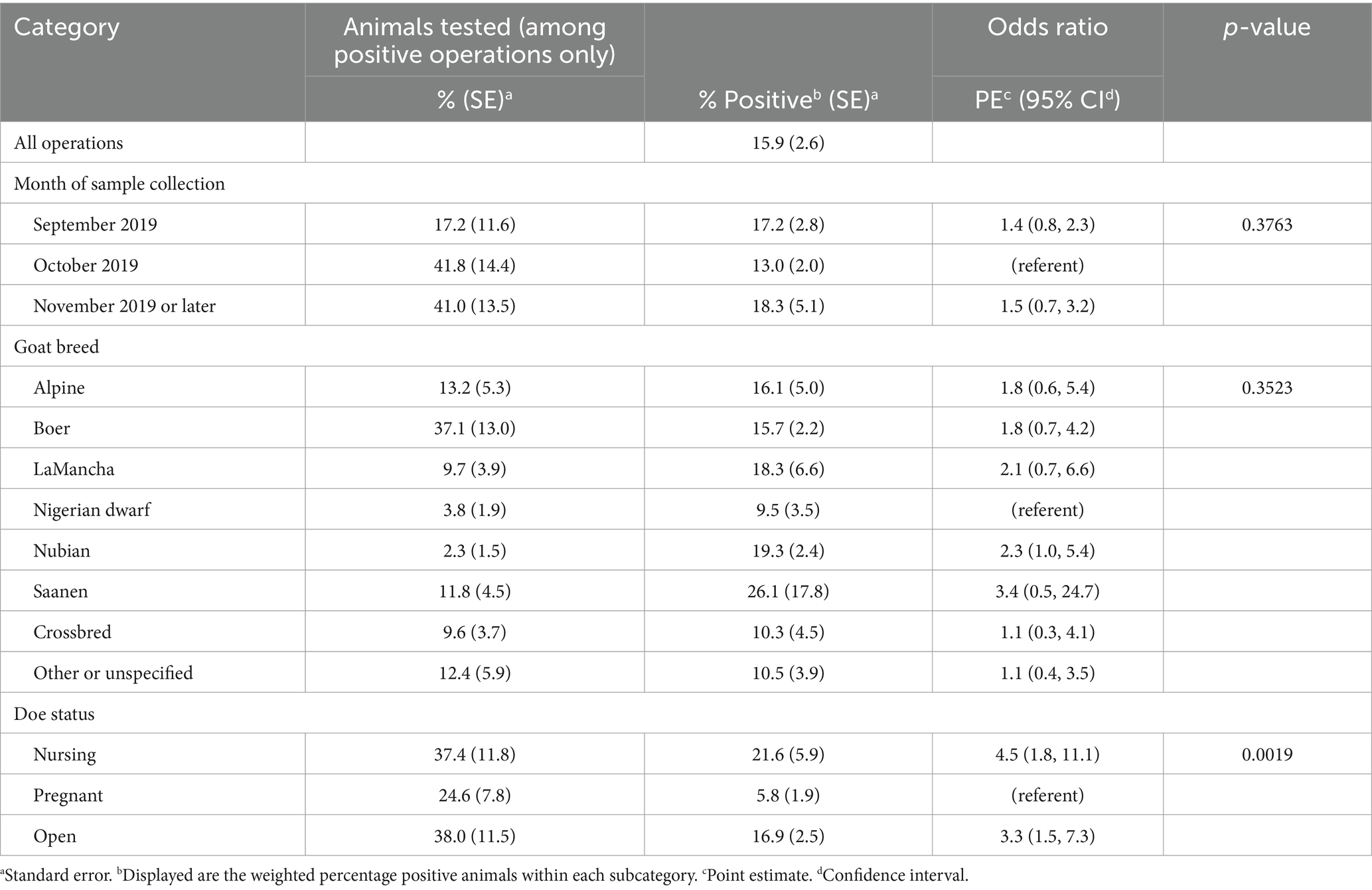
Table 1. Univariate logistic regression model results for the animal-level prevalence among animals on operations positive for Coxiella burnetii.
Univariate analysis of Coxiella burnetii operation-level prevalence among United States goat herds
Univariate logistic regression modeling was performed to assess management practices associated with operation-level shedding of C. burnetii across 388 operations (Table 2; Supplementary Table S1). The weighted estimate of the percentage of operations with one or more positive does was 7.8% (95% CI 4.4–13.5). No significant difference was observed by region, herd size, primary production of the operation, and primary land/facility management type between positive and negative operations. Operations reporting suspected Q fever in the herd within the previous 3 years had 119.5 times higher odds (95% CI 16.8–851.9) of testing positive for C. burnetii. Operations with wild deer, elk, or other hoof stock on the operation or adjacent property had 10.9 times higher odds (95% CI 2.8–42.9) of testing positive and operations with wild predators such as coyotes, bears, mountain lions, or wolves on the operation or adjacent property had 11.3 times higher odds (95% CI 2.5–51.7) of having a doe test positive. There was not a significant association between operations reporting other animals such as sheep, cattle, horses, chickens, dogs, cats, or raccoons on the operation or adjacent property and operation status. Operations that utilized hormones for estrus synchronization had an 8.7 (95% CI 1.3–59.0) times higher odds of being positive than operations that did not. Never or sometimes requiring handwashing after handling goats with scabs around the mouth, feet, or udder, was associated with a 26.8 (95% CI 2.2–328.4) times higher odds of being positive compared to operations that always required hand washing. Operations that had goats that temporarily left the operation and returned and isolated those goats only for a specific reason had a 24.4 (95% CI 2.7–225.4) times higher odds of being positive with C. burnetii compared to operations that routinely isolated returning goats. Operations reporting litters of kittens had 10.6 (95% CI 2.3–49.6) times lower odds of testing positive for C. burnetii. Finally, treatment for worms during the previous 12 months using tetrahydropyrimidines, high tannin concentrate plants, or diatomaceous earth had proportionally lower odds of testing positive by factors of 23.4 (95% CI 3.5–157.6), 42.5 (95% CI 7.2–250.1), and 69.1 (95% CI 11.9–401.2), respectively, compared to operations that dewormed but not with the given product.
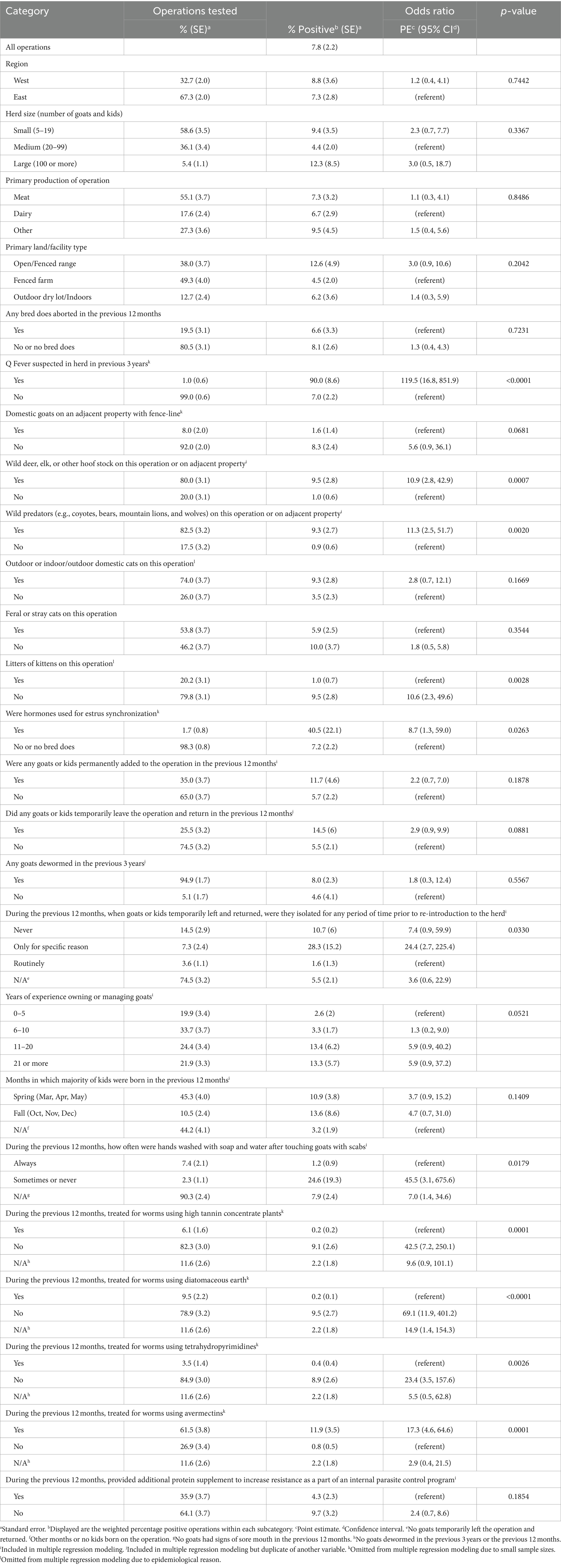
Table 2. Univariate logistic regression model results for the operation-level prevalence of Coxiella burnetii.
Effect of select tetrahydropyrimidines and tannins on growth of Coxiella burnetii in vitro
As operations that treated for worms during the previous 12 months using tetrahydropyrimidines or high tannin concentrate plants were associated with decreased shedding of C. burnetii, we sought to examine the effect of these compounds on the growth of C. burnetii in vitro. To this end, we cultured C. burnetii in the presence of tetrahydropyrimidines (pyrantel pamoate, pyrantel tartrate, and morantel tartrate) as well as the hydrolysable tannins, tannic acid, and gallic acid, at concentrations up to 10 μg per mL. Pyrantel pamoate demonstrated an inhibitory effect on the growth of C. burnetii as low as 1 μg per mL (1.68 μM) (Figure 1). For comparison, the MIC for doxycycline, the recommended treatment option for humans, is 0.01 μg per mL (22.5 nM).
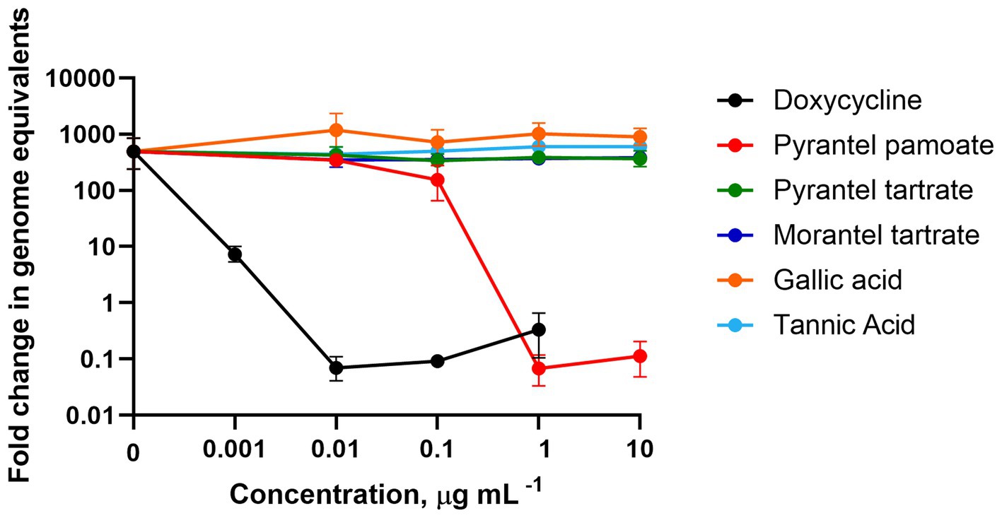
Figure 1. Susceptibility of Coxiella burnetii to hydrolysable tannins and tetrahydropyrimidines. Coxiella burnetii was grown for 7 days in ACCM-2. Data demonstrate the fold change of the mean genome equivalents for day 7 relative to day 0. Error bars represent the upper and lower bounds of fold change, which were calculated based on the SEM of the genome equivalents. Data are from two independent experiments.
Multiple regression analysis of Coxiella burnetii operation-level prevalence among United States goat herds
The effects of imputation on the final results were minimal. There were 36 observations that had mode-imputed values across 11 of the 48 operation-level variables considered in Supplementary Table S1. The range in the number of missing values for those 11 variables was 1–12. None of the percentages of operations changed by more than 5.1% (2.2 percentage points), the greatest change being in the majority of kidding that occurred in the spring (Mar, Apr, May), which changed from 45.3% (SE = 4.0) to 43.1% (SE = 4.0). None of the percent of positive operations changed by more than 7.8% (0.9 percentage points), the largest being in the majority of kidding in spring (Mar, Apr, May), which changed from 10.9% (SE = 3.8) to 11.8% (SE = 4.2). Only one of the p values changed significance at the 0.05 level, for years of experience raising goats, which changed from 0.0521 to 0.0465.
A model summary for the multiple logistic regression model fit to model the operation-level prevalence of C. burnetii is included in Table 3. The only risk factor that was statistically significant at the 0.05 significance level after accounting for the other factors included in the model was the presence of wild predators (e.g., coyotes, bears, mountain lions, and wolves) on the operation or adjacent property (OR = 9.0, 95% CI 1.3–61.6, p value = 0.0248). Five other variables were statistically significant at the 0.10 significance level but not the 0.05 significance level. These included the presence of wild deer, elk, or other hoof stock on the operation or adjacent property (OR = 8.1, 95% CI 0.9–72.8, p value = 0.0608), frequency of hand washing after touching goats with scabs (OR = 1.9, 95% CI 0.1–33.9 and OR = 6.0, 95% CI 0.5–36.4 for operations that sometimes or never did so and for operations with no goats with signs of sore mouth, respectively, compared to operations that always washed hands, p value = 0.0884), whether goats and kids that temporarily left the operation and returned were isolated after returning to the operation (OR = 3.7, 95% CI 0.7–20.4 and OR = 10.3, 95% CI 1.2–85.6 for operations that never isolated and for operations that isolated only for a specific reason, respectively, compared to operations that isolated routinely or did not have any goats temporarily leave and return, p value = 0.0891), the length of time that goats had been managed (OR = 1.6, 95% CI 0.1–23.3, OR = 6.5, 95% CI 0.5–80.2, and OR = 11.3, 95% CI 0.7–196.0, for operations that owned or managed goats for 6–10, 11–20, and 21 or more years, respectively, compared to operations that did so for 0–5 years, p value = 0.0990), and the months in which the majority of kids were born (OR = 3.5, 95% CI 0.8–14.5 and OR = 6.5, 95% CI 0.5–80.2 for operations that primarily kidded in spring and fall, respectively, compared to operations that kidded in other months or had no kids born on the operation, p value = 0.0550). The McFadden’s adjusted pseudo-R2 value for this model was 0.397, approximately, and the F1 score was 0.286.
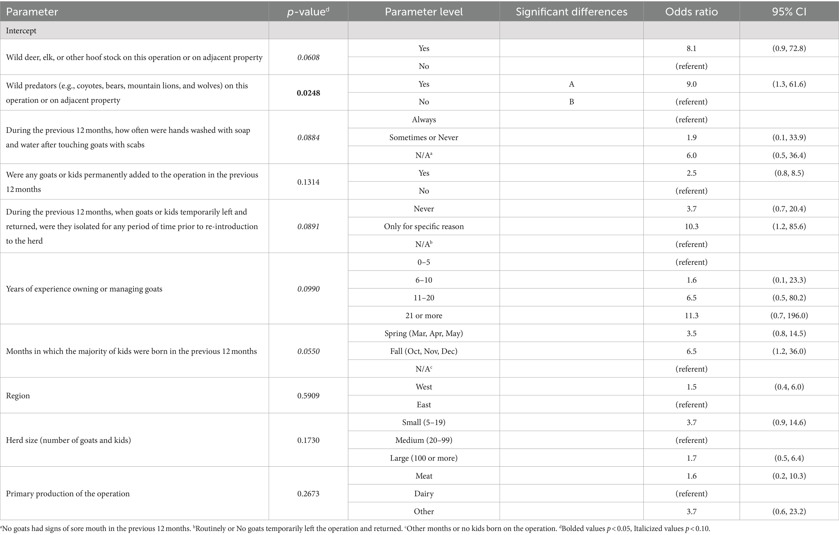
Table 3. Multiple logistic regression model results for the operation-level prevalence of Coxiella burnetii.
Discussion
To our knowledge, this is the first large scale report of C. burnetii infection across the United States goat population. Active shedding of C. burnetii was investigated for 4,121 doe goats, which demonstrated 1.5% positivity at the time of sampling. The quantity of C. burnetii being shed was low, which is likely influenced by the timing of sample collection. The bulk of samples were collected in October and November and positivity and bacterial loads would likely have been higher in the spring, when most operations have kid goats born. In this study, animal-level univariate analysis found pregnant does had reduced odds of testing positive for C. burnetii. Shedding in vaginal secretions often only occurs after parturition or abortion and the bacterial load is typically highest immediately post-kidding (2, 41). The lower percentage of pregnant does shedding the bacteria is likely highlighting the shedding dynamics of C. burnetii rather than a lower infection rate in pregnant does but underscores the fact that targeted sampling following the kidding seasons may increase positivity. Given the fact that testing occurred outside of kidding season and did not target high risk goats, the data suggest widespread infection of goat herds across the United States at any given time underscoring the importance of the disease and the potential risk to goat producers and the public.
Coxiella burnetii seroprevalence has been reported in goats in the United States with herd-level prevalence ranging from 11.5, 21.0, and 41.6% (15, 42, 43); however, given that seropositivity can persist for years after infection and infected animals may be seronegative, evaluation of active infections by molecular analysis of C. burnetii shedding is important (44). Studies investigating C. burnetii shedding prevalence are limited, particularly for goats in the United States. Small, localized studies found shedding prevalence ranging from 0% in fecal samples from agricultural fairs in Iowa to 7% in milk from Indiana (15, 45). Herein, we identified 61 does positive for shedding C. burnetii across 10.3% of the tested operations (7.8% weighted, SE = 2.2), which is in line with the observations in Indiana, but is lower than that observed in goats worldwide. Shedding was observed in 60% of sheep and goat herds in Algeria, 51.16% of goat herds in Poland, and between 16.6 and 56.4% in Iran (46–49). Shedding in dairy goat herds in Belgium decreased from 12% in 2009 to 6.3% in 2012 and 6% in 2019 only after the introduction of mandatory vaccination in 2011 (50, 51). Curiously, the shedding prevalence of goat herds in the United States observed in the current study is better aligned with that of vaccinated herds despite the inability to vaccinate goats against C. burnetii in the United States. These differences are likely multifactorial but may involve the presence of different C. burnetii genotypes. The ST61 and ST18 genotypes have been reported in goat herds in Poland, ST61 and ST62 genotypes have been reported in Iran, ST61 and ST33 genotypes have been reported in goats in Belgium, while the ST8 genotype is consistently associated with goats in the United States (47, 52–55). Only one sample was concentrated enough in the present study to conduct genotyping analysis and indeed it belonged to the ST8 genogroup. Murine in vivo studies have found strains from the ST8 genogroup to be less virulent than other genotypes which may account for the reduced shedding prevalence observed in United States goat herds (52, 56–58). Despite this, ST8 strains can cause abortions in goats and chronic Q fever in humans (52, 53).
Operations reporting the presence of wild elk, deer, and other hoof stock were more likely to test positive for C. burnetii by univariate analysis, while the presence of wild predators on the operation or adjacent property was significantly associated with greater odds of testing positive by both univariate and multiple logistic regression modeling. These findings highlight the possible role for wild animals as C. burnetii reservoirs. Antibodies against C. burnetii have been detected in coyotes at 78% and foxes at 55% and viable C. burnetii has been isolated from tissues of both species (59). Coxiella burnetii seroprevalence of wild deer in parts of the United States has been documented as high as 22% (59, 60). Coxiella burnetii infection in red deer can lead to abortions, shedding, and transmission to humans (61). The proximity of wild predators and deer to domestic animals and humans highlights the need to further examine their role in the maintenance and transmission of C. burnetii.
Another factor associated with greater odds of C. burnetii infection in this study was operations utilizing hormones for estrus synchronization. In experimental settings, sex hormones have been shown to influence the host response to C. burnetii infection (62, 63). Progesterone, which is commonly administered for estrus synchronization in goats has been shown to inhibit replication of C. burnetii (64). The findings herein, demonstrate that administration of hormones for estrus synchronization does not have a protective effect in this setting, rather C. burnetii may be more likely to spread due to large quantities of C. burnetii being introduced into the environment at once from a more concentrated kidding season. Furthermore, shared equipment during the estrus synchronization process could promote the spread of C. burnetii throughout the herd. The influence of other management practices among operations that utilize estrus synchronization cannot be ruled out.
Herds treated with high tannin concentration plants, diatomaceous earth, or tetrahydropyrimidines were less likely to test positive for C. burnetii. The antimicrobial properties of tannins are well documented and the tannin containing forage, sainfoin, has been shown to decrease fecal shedding of Escherichia coli in cattle (65–69). Although the hydrolysable tannins, tannic acid and gallic acid, had no direct effect on growth of C. burnetii in vitro (Figure 1), the activity of condensed tannins remains to be determined. Diatomaceous earth possesses electromagnetic properties that bind bacteria (70). Although inhalation is the major route of infection by C. burnetii, infection via the oral route is possible (56, 58). Therefore, treatment with diatomaceous earth may serve to reduce C. burnetii in the gastrointestinal tract and possibly a reduction in the aerosol burden as C. burnetii complexed with the diatomaceous earth are excreted into the environment. Pyrantel pamoate, a tetrahydropyrimidine anthelmintic, can inhibit growth of C. burnetii in vitro as demonstrated in Figure 1 (71). This effect has only been characterized for the lab attenuated Nine Mile Phase II strain (ST16) of C. burnetii. As such, the efficacy of pyrantel pamoate on virulent or goat-associated strains of C. burnetii remains to be determined. The usefulness of these potential treatments for C. burnetii infection control in ruminants should be further explored; however, in the absence of further research, off-label use of anthelmintics should be restricted to avoid exacerbating resistance.
This study highlighted two biosecurity measures that goat producers may consider employing to reduce C. burnetii shedding on the operation. Operations requiring handwashing after handling goats with scabs, were less likely to test positive for C. burnetii. While C. burnetii does not cause scabs, frequent handwashing likely reduces the spread of a variety of pathogens. Secondly, routine isolation of goats returning to the operation was associated with reduced shedding. Therefore, goat producers should consider incorporating these strategies, along with other biosecurity measures into their management practices.
Operations reporting litters of kittens were associated with decreased shedding of C. burnetii, which is surprising given that exposure to parturient cats has been associated with outbreaks of Q fever (72–75). Furthermore, C. burnetii positivity in farm cats has been shown to be associated with infected ruminant farms (76). Interestingly, the presence of cats (domestic or feral) on the operation was not significantly associated with C. burnetii shedding in goats (Table 2). Coxiella burnetii has been detected in feline reproductive tissues and is suspected of causing feline reproductive health issues (75, 77, 78). Perhaps cats are less likely to give birth to healthy kittens on operations where C. burnetii is present; however, additional research is needed to draw any conclusions regarding the effect of C. burnetii on feline reproductive health. It is not clear if parturition in other animals such as dogs would be associated with C. burnetii shedding in goats as this information was not captured in this study.
No association was found with regard to location, herd size, and primary production of the operation, which highlights the widespread vulnerability of the goat industry to this disease. Given the rise in popularity of goats in the United States and the number of operations experiencing infection, the development and implementation of prevention and treatment strategies for C. burnetii infected goat farms is important to protect goat producers and public health. In the United States, there are currently no approved Q fever vaccines or therapies to prevent or eradicate C. burnetii from goat herds or their environment. However, this study highlights potential practices that can be implemented to identify high risk goats and mitigate exposure and spread, such as limiting visitor contact with kidding does and preweaned kids, ensuring handwashing protocols for visitors, routine isolation of goats returning to the operation, and minimizing contact with wildlife when feasible. However, just as important, this study highlighted that producers are aware of C. burnetii in their herds as shedding was associated with operations that suspected Q fever; therefore, this could be capitalized on should effective therapies be identified. However, the percentage of operations that suspected C. burnetii in their herds in the previous 3 years (1.0%, SE = 0.6) was much lower than the estimated percentage of operations that tested positive at the time of the study (7.8%, SE = 2.2) suggesting that raising awareness of this important disease among goat producers is needed.
In conclusion, C. burnetii infected goat herds are more common across the United States than reported outbreaks would suggest, highlighting the need for effective prevention and treatment strategies for these operations and continued surveillance to reduce public health risk. This study highlights that some producers are aware of C. burnetii infection in the herd, however there is still room for improvement. Therefore, future public health prevention measures should focus on the development of mitigation strategies alongside awareness campaigns. This study also highlights C. burnetii potential therapies that should be further investigated as they are already approved for use in goats. However, caution should be taken, as using anthelmintics off-label or inappropriately may promote anthelmintic resistance. Any treatment should be performed under the advisement of a veterinarian with valid veterinary-client-patient-relationship (VCPR). Finally, the analysis demonstrates a need to investigate the role of potential wildlife reservoirs, particularly wild predators, in the maintenance and transmission of C. burnetii.
Data availability statement
The data that support the findings of this study are available upon reasonable request from the corresponding author; however, restrictions apply as some data are not available due to the sensitive nature of the research. Data availability will be at the discretion of the USDA Animal and Plant Health Inspection Service (APHIS). Requests to access the datasets should be directed to HM, SGFsaWUuTWlsbGVyQGNkYy5oaHMuZ292.
Ethics statement
Ethical approval was not required for the studies involving animals in accordance with the local legislation and institutional requirements because as a federal facility conducting research on owned livestock, the USDA Animal and Plant Health Inspection Service (APHIS) is not required to register under the U.S. Animal Welfare Act. However, as the agency that administers the Animal Welfare Act, USDA APHIS staff ensure that all animal treatment that is part of the National Animal Health Monitoring System is in compliance with the Animal Welfare Act. Written informed consent was obtained from the owners for the participation of their animals in this study.
Author contributions
HM: Conceptualization, Formal analysis, Investigation, Methodology, Writing – original draft, Writing – review & editing. MB: Formal analysis, Methodology, Writing – review & editing. RP: Investigation, Writing – review & editing. RÁ-A: Investigation, Writing – review & editing. CC: Conceptualization, Writing – review & editing. CS: Investigation, Writing – review & editing. NU: Resources, Writing – review & editing. AW: Resources, Writing – review & editing. CB: Formal analysis, Methodology, Writing – review & editing. KM: Conceptualization, Resources, Supervision, Writing – review & editing. GK: Conceptualization, Methodology, Project administration, Resources, Supervision, Writing – review & editing.
Funding
The author(s) declare that no financial support was received for the research, authorship, and/or publication of this article.
Acknowledgments
The authors would like to acknowledge the goat producers for their participation in the NAHMS Goat 2019 study and the field collection teams comprised of veterinary medical officers and animal health technicians for their sample collection efforts.
Conflict of interest
The authors declare that the research was conducted in the absence of any commercial or financial relationships that could be construed as a potential conflict of interest.
Publisher’s note
All claims expressed in this article are solely those of the authors and do not necessarily represent those of their affiliated organizations, or those of the publisher, the editors and the reviewers. Any product that may be evaluated in this article, or claim that may be made by its manufacturer, is not guaranteed or endorsed by the publisher.
Author disclaimer
The findings and conclusions in this report are those of the authors and do not necessarily represent the official position of the Centers for Disease Control and Prevention or the United States Department of Agriculture.
Supplementary material
The Supplementary material for this article can be found online at: https://www.frontiersin.org/articles/10.3389/fvets.2024.1393296/full#supplementary-material
SUPPLEMENTARY TABLE S1 | Univariate logistic regression model results for all variables considered for analysis.
References
1. Miller, HK, Priestley, RA, and Kersh, GJ. Q fever: a troubling disease and a challenging diagnosis. Clin Microbiol Newsl. (2021) 43:109–18. doi: 10.1016/j.clinmicnews.2021.06.003
2. Roest, HJ, van Gelderen, B, Dinkla, A, Frangoulidis, D, van Zijderveld, F, Rebel, J, et al. Q fever in pregnant goats: pathogenesis and excretion of Coxiella Burnetii. PLoS One. (2012) 7:e48949. doi: 10.1371/journal.pone.0048949
3. Rousset, E, Berri, M, Durand, B, Dufour, P, Prigent, M, Delcroix, T, et al. Coxiella Burnetii shedding routes and antibody response after outbreaks of Q fever-induced abortion in dairy goat herds. Appl Environ Microbiol. (2009) 75:428–33. doi: 10.1128/AEM.00690-08
4. Mandell, B, Bennett, J R D, and Dolin, Mandell B. (2005). Principles and Practice of Infectious Diseases. Philadelphia, PA USA: Churchill Livingstone An Imprint of Elsevier, 1864–1890
5. Schimmer, B, Dijkstra, F, Vellema, P, Schneeberger, PM, Hackert, V, ter Schegget, R, et al. Sustained intensive transmission of Q fever in the south of the Netherlands, 2009. Euro Surveill. (2009) 14. doi: 10.2807/ese.14.19.19210-en
6. Kampschreur, LM, Hagenaars, JC, Wielders, CC, Elsman, P, Lestrade, PJ, Koning, OH, et al. Screening for Coxiella Burnetii Seroprevalence in chronic Q fever high-risk groups reveals the magnitude of the Dutch Q fever outbreak. Epidemiol Infect. (2013) 141:847–51. doi: 10.1017/S0950268812001203
7. Van der Hoek, W, Morroy, G, Renders, NH, Wever, PC, Hermans, MH, Leenders, AC, et al. Epidemic Q fever in humans in the Netherlands. Adv Exp Med Biol. (2012) 984:329–64. doi: 10.1007/978-94-007-4315-1_17
8. Buijs, SB, Bleeker-Rovers, CP, van Roeden, SE, Kampschreur, LM, Hoepelman, AIM, Wever, PC, et al. Still new chronic Q fever cases diagnosed eight years after a large Q fever outbreak. Clin Infect Dis. (2021) 73:1476–83. doi: 10.1093/cid/ciab476
9. Bamberg, WM, Pape, WJ, Beebe, JL, Nevin-Woods, C, Ray, W, Maguire, H, et al. Outbreak of Q fever associated with a horse-boarding ranch, Colorado, 2005. Vect Borne Zoo Dis. (2007) 7:394–402. doi: 10.1089/vbz.2007.0104
10. Centers for Disease Control and Prevention. Notes from the field: Q fever outbreak associated with goat farms--Washington and Montana, 2011. MMWR Morb Mortal Wkly Rep. (2011) 60:1393. Available at: https://www.cdc.gov/mmwr/preview/mmwrhtml/mm6040a5.htm
11. USDA-APHIS. Goat (2022). "Part I: Reference of Goat Management Practices in the United States, 2019". USDA-APHIS-VS-CEAH-NAHMS. Fort Collins, Co. Available at: https://www.aphis.usda.gov/animal_health/nahms/goats/downloads/goat19/goat2019-pt1-final.pdf
12. USDA-APHIS (2019). Nahms Studies Table. Animal and Plant Health Inspection Service U.S. Department of Agriculture. Available at: https://www.aphis.usda.gov/aphis/ourfocus/animalhealth/monitoring-and-surveillance/nahms/nahms-studies/nahms-studies-table?cat=goat
13. Stevenson, MA. Sample size estimation in veterinary epidemiologic research. Front Vet Sci. (2020) 7:539573. doi: 10.3389/fvets.2020.539573
14. NASS (2017). Census of Agriculture United States Summary and State Data Volume 1 Geographic Area Series Part 51 Ac-17-a-51. (2017) April 2019. Report No.
15. Bauer, AE, Hubbard, KR, Johnson, AJ, Messick, JB, Weng, HY, and Pogranichniy, RM. A cross sectional study evaluating the prevalence of Coxiella Burnetii, potential risk factors for infection, and agreement between diagnostic methods in goats in Indiana. Prev Vet Med. (2016) 126:131–7. doi: 10.1016/j.prevetmed.2016.01.026
16. Magadu, R, and Thompson, PN. Seroprevalence and factors associated with Coxiella Burnetii exposure in goats in Moretele. Onderstepoort J Vet Res. (2023) 90:e1–7. doi: 10.4102/ojvr.v90i1.2071
17. Kersh, GJ, Wolfe, TM, Fitzpatrick, KA, Candee, AJ, Oliver, LD, Patterson, NE, et al. Presence of Coxiella Burnetii DNA in the environment of the United States, 2006 to 2008. Appl Environ Microbiol. (2010) 76:4469–75. doi: 10.1128/AEM.00042-10
18. Hornstra, HM, Priestley, RA, Georgia, SM, Kachur, S, Birdsell, DN, Hilsabeck, R, et al. Rapid typing of Coxiella Burnetii. PLoS One. (2011) 6:e26201. doi: 10.1371/journal.pone.0026201
20. Särndal, C-E, Swensson, B, and Wretman, J. Model Assisted Survey Sampling. New York, NY USA: Springer Science & Business Media (2003).
21. Donders, AR, van der Heijden, GJ, Stijnen, T, and Moons, KG. Review: a gentle introduction to imputation of missing values. J Clin Epidemiol. (2006) 59:1087–91. doi: 10.1016/j.jclinepi.2006.01.014
22. R Core Team (2022). R: A language and environment for statistical computing. R Foundation for Statistical Computing, Vienna, Austria. Available at: https://www.r-project.org/
23. Posit Team (2022). Rstudio: Integrated development environment for R. Posit Software, Pbc, Boston, MA. Available at: http://www.posit.co/
25. Hothorn, T, Bretz, F, and Westfall, P. Simultaneous inference in general parametric models. Biom J. (2008) 50:346–63. doi: 10.1002/bimj.200810425
26. Fox, J, and Weisberg, S. An R Companion to Applied Regression. 3rd ed. Los Angeles: SAGE (2019). 577 p.
27. Wickham, H, Averick, M, Bryan, J, Chang, W, McGowan, LD, François, R, et al. Welcome to the Tidyverse. J Open Source Softw. (2019) 4:1686. doi: 10.21105/joss.01686
28. Wilke, C. O. (2020). Cowplot: Streamlined plot theme and plot annotations for Ggplot2. R package version 1.1.1. Available at: https://cran.R-project.org/package=cowplot
29. Freedman Ellis, G, and Srvyr, Schneider B. (2023). 'Dplyr'-like syntax for summary statistics of survey data. R Package Version 1.2.0. Available at: https://cran.R-project.org/package=Srvyr
30. Ggpubr, K. A. (2023). 'Ggplot2' based publication ready plots. R Package Version 0.6.0. Available at: https://cran.R-project.org/package=Ggpubr
31. Wickham, H, Miller, E, and Smith, D (2023). Haven: Import and Export 'Spss', 'Stata' and 'Sas' Files. R Package Version 2.5.3. w: https://cran.R-project.org/package=haven
32. Hernan, MA, Hernandez-Diaz, S, Werler, MM, and Mitchell, AA. Causal knowledge as a prerequisite for confounding evaluation: an application to birth defects epidemiology. Am J Epidemiol. (2002) 155:176–84. doi: 10.1093/aje/155.2.176
33. Akaike, H. A new look at the statistical model identification. IEEE Trans Autom Control. (1974) 19:716–23. doi: 10.1109/TAC.1974.1100705
34. Tharwat, A. Classification assessment methods. Appl Comput Inform. (2018) 17:168–92. doi: 10.1016/j.aci.2018.08.003
35. McFadden, D. Conditional Logit Analysis of Qualitative Choice Behavior. Berkeley: Academic Press (1974).
36. Wald, A. Tests of statistical hypotheses concerning several parameters when the number of observations is large. Trans Am Math Soc. (1943) 54:426–82. doi: 10.1090/S0002-9947-1943-0012401-3
37. Kutner, MH, Nachtsheim, CJ, Neter, J, and Li, W. Applied Linear Statistical Models. 5th ed. Blue Ridge, IL: McGraw-Hill/Irwin (2005).
38. Piepho, HP. An algorithm for a letter-based representation of all-pairwise comparisons. J Comput Graph Stat. (2004) 13:456–66. doi: 10.1198/1061860043515
39. Smith, CB, Evavold, C, and Kersh, GJ. The effect of Ph on antibiotic efficacy against Coxiella Burnetii in axenic media. Sci Rep. (2019) 9:18132. doi: 10.1038/s41598-019-54556-6
40. Kersh, GJ, Lambourn, DM, Self, JS, Akmajian, AM, Stanton, JB, Baszler, TV, et al. Coxiella Burnetii infection of a Steller Sea lion (Eumetopias Jubatus) found in Washington state. J Clin Microbiol. (2010) 48:3428–31. doi: 10.1128/JCM.00758-10
41. Arricau Bouvery, N, Souriau, A, Lechopier, P, and Rodolakis, A. Experimental Coxiella Burnetii infection in pregnant goats: excretion routes. Vet Res. (2003) 34:423–33. doi: 10.1051/vetres:2003017
42. McQuiston, JH, and Childs, JE. Q fever in humans and animals in the United States. Vect Borne Zoo Dis. (2002) 2:179–91. doi: 10.1089/15303660260613747
43. Welch, JL, Branan, M, Urie, N, Shrestha, R, Wiedenheft, A, Marshall, K, et al. Coxiella Burnetii Seroprevalence in domestic goat does in the United States: prevalence, distribution, and associated risk factors. Prev Vet Med. (2024) 223:106114. doi: 10.1016/j.prevetmed.2023.106114
44. Plummer, PJ, McClure, JT, Menzies, P, Morley, PS, Van den Brom, R, and Van Metre, DC. Management of Coxiella Burnetii infection in livestock populations and the associated zoonotic risk: a consensus statement. J Vet Intern Med. (2018) 32:1481–94. doi: 10.1111/jvim.15229
45. Still Brooks, KM, Stensland, WR, Harmon, KM, O'Connor, AM, and Plummer, PJ. Risk of exposure to Coxiella Burnetii from ruminant livestock exhibited at Iowa agricultural fairs. Zoonoses Public Health. (2018) 65:334–8. doi: 10.1111/zph.12439
46. Khaled, H, Sidi-Boumedine, K, Merdja, S, Dufour, P, Dahmani, A, Thiery, R, et al. Serological and molecular evidence of Q fever among small ruminant flocks in Algeria. Comp Immunol Microbiol Infect Dis. (2016) 47:19–25. doi: 10.1016/j.cimid.2016.05.002
47. Jodelko, A, Szymanska-Czerwinska, M, Rola, JG, and Niemczuk, K. Molecular detection of Coxiella Burnetii in small ruminants and genotyping of specimens collected from goats in Poland. BMC Vet Res. (2021) 17:341. doi: 10.1186/s12917-021-03051-0
48. Khademi, P, Ownagh, A, Ataei, B, Kazemnia, A, Enferadi, A, Khalili, M, et al. Prevalence of C. burnetii DNA in sheep and goats Milk in the northwest of Iran. Int J Food Microbiol. (2020) 331:108716. doi: 10.1016/j.ijfoodmicro.2020.108716
49. Abiri, Z, Khalili, M, Kostoulas, P, Sharifi, H, Rad, M, and Babaei, H. Bayesian estimation of sensitivity and specificity of a Pcr method to detect Coxiella Burnetii in Milk and vaginal secretions in sheep and goat samples. J Dairy Sci. (2019) 102:4954–9. doi: 10.3168/jds.2018-15233
50. Boarbi, S, Mori, M, Rousset, E, Sidi-Boumedine, K, Van Esbroeck, M, and Fretin, D. Prevalence and molecular typing of Coxiella Burnetii in bulk tank Milk in Belgian dairy goats, 2009-2013. Vet Microbiol. (2014) 170:117–24. doi: 10.1016/j.vetmic.2014.01.025
51. Jansen, W, Cargnel, M, Boarbi, S, Mertens, I, Van Esbroeck, M, Fretin, D, et al. Belgian bulk tank Milk surveillance program reveals the impact of a continuous vaccination protocol for small ruminants against Coxiella Burnetii. Transbound Emerg Dis. (2022) 69:e141–52. doi: 10.1111/tbed.14273
52. Kersh, GJ, Fitzpatrick, KA, Self, JS, Priestley, RA, Kelly, AJ, Lash, RR, et al. Presence and persistence of Coxiella Burnetii in the environments of goat farms associated with a Q fever outbreak. Appl Environ Microbiol. (2013) 79:1697–703. doi: 10.1128/AEM.03472-12
53. Kersh, GJ, Priestley, RA, Hornstra, HM, Self, JS, Fitzpatrick, KA, Biggerstaff, BJ, et al. Genotyping and axenic growth of Coxiella Burnetii isolates found in the United States environment. Vect Borne Zoo Dis. (2016) 16:588–94. doi: 10.1089/vbz.2016.1972
54. Anastacio, S, de Sousa, SR, Saavedra, MJ, and da Silva, GJ. Role of goats in the epidemiology of Coxiella Burnetii. Biology. (2022) 11. doi: 10.3390/biology11121703
55. Tomaiuolo, S, Boarbi, S, Fancello, T, Michel, P, Desqueper, D, Gregoire, F, et al. Phylogeography of human and animal Coxiella Burnetii strains: genetic fingerprinting of Q fever in Belgium. Front Cell Infect Microbiol. (2020) 10:625576. doi: 10.3389/fcimb.2020.625576
56. Miller, HK, Priestley, RA, and Kersh, GJ. Comparison of three Coxiella Burnetii infectious routes in mice. Virulence. (2021) 12:2562–70. doi: 10.1080/21505594.2021.1980179
57. Priestley, RA, Smith, CB, Miller, HK, and Kersh, GJ. Coxiella Burnetii infections in mice: immunological responses to contemporary genotypes found in the us. Virulence. (2021) 12:2461–73. doi: 10.1080/21505594.2021.1975527
58. Miller, HK, Priestley, RA, and Kersh, GJ. Transmission of Coxiella Burnetii by ingestion in mice. Epidemiol Infect. (2020) 148:e21. doi: 10.1017/S0950268820000059
59. Enright, JB, Franti, CE, Behymer, DE, Longhurst, WM, Dutson, VJ, and Wright, ME. Coxiella Burneti in a wildlife-livestock environment. Distribution of Q fever in wild mammals. Am J Epidemiol. (1971) 94:79–90. doi: 10.1093/oxfordjournals.aje.a121298
60. Kirchgessner, MS, Dubovi, EJ, and Whipps, CM. Disease risk surface for Coxiella Burnetii Seroprevalence in white-tailed deer. Zoonoses Public Health. (2013) 60:457–60. doi: 10.1111/zph.12023
61. Gonzalez-Barrio, D, Almeria, S, Caro, MR, Salinas, J, Ortiz, JA, Gortazar, C, et al. Coxiella Burnetii shedding by farmed Red Deer (Cervus Elaphus). Transbound Emerg Dis. (2015) 62:572–4. doi: 10.1111/tbed.12179
62. Leone, M, Honstettre, A, Lepidi, H, Capo, C, Bayard, F, Raoult, D, et al. Effect of sex on Coxiella Burnetii infection: protective role of 17beta-estradiol. J Infect Dis. (2004) 189:339–45. doi: 10.1086/380798
63. Textoris, J, Ban, LH, Capo, C, Raoult, D, Leone, M, and Mege, JL. Sex-related differences in gene expression following Coxiella Burnetii infection in mice: potential role of circadian rhythm. PLoS One. (2010) 5:e12190. doi: 10.1371/journal.pone.0012190
64. Howard, ZP, and Omsland, A. Selective inhibition of Coxiella Burnetii replication by the steroid hormone progesterone. Infect Immun. (2020) 88. doi: 10.1128/IAI.00894-19
65. Berard, NC, Holley, RA, McAllister, TA, Ominski, KH, Wittenberg, KM, Bouchard, KS, et al. Potential to reduce Escherichia Coli shedding in cattle feces by using Sainfoin (Onobrychis Viciifolia) forage, tested in vitro and in vivo. Appl Environ Microbiol. (2009) 75:1074–9. doi: 10.1128/AEM.00983-08
66. Ekambaram, SP, Perumal, SS, and Balakrishnan, A. Scope of hydrolysable tannins as possible antimicrobial agent. Phytother Res. (2016) 30:1035–45. doi: 10.1002/ptr.5616
67. Scalbert, A. Antimicrobial properties of tannins. Phytochemistry. (1991) 30:3875–83. doi: 10.1016/0031-9422(91)83426-L
68. Huang, Q, Liu, X, Zhao, G, Hu, T, and Wang, Y. Potential and challenges of tannins as an alternative to in-feed antibiotics for farm animal production. Anim Nutr. (2018) 4:137–50. doi: 10.1016/j.aninu.2017.09.004
69. Arakkaveettil Kabeer Farha, Q-QY, Kim, G, Li, H-B, Zhu, F, Liu, H-Y, Gan, R-Y, et al. Tannins as an alternative to antibiotics. Food Biosci. (2020):38. doi: 10.1016/j.fbio.2020.100751
70. Johnson, T, Brineman, R, Schultze, C, and Barkovskii, AL. Efficient removal of Bacteria from aqueous media with kaolinite and diatomaceous earth products. J Appl Microbiol. (2020) 129:466–73. doi: 10.1111/jam.14642
71. Czyz, DM, Potluri, LP, Jain-Gupta, N, Riley, SP, Martinez, JJ, Steck, TL, et al. Host-directed antimicrobial drugs with broad-Spectrum efficacy against intracellular bacterial pathogens. MBio. (2014) 5:e01534. doi: 10.1128/mBio.01534-14
72. Marrie, TJ, MacDonald, A, Durant, H, Yates, L, and McCormick, L. An outbreak of Q fever probably due to contact with a parturient cat. Chest. (1988) 93:98–103. doi: 10.1378/chest.93.1.98
73. Kosatsky, T. Household outbreak of Q-fever pneumonia related to a parturient cat. Lancet. (1984) 324:1447–9. doi: 10.1016/s0140-6736(84)91633-7
74. Pinsky, RL, Fishbein, DB, Greene, CR, and Gensheimer, KF. An outbreak of cat-associated Q fever in the United States. J Infect Dis. (1991) 164:202–4. doi: 10.1093/infdis/164.1.202
75. Marrie, TJ, Durant, H, Williams, JC, Mintz, E, and Waag, DM. Exposure to parturient cats: a risk factor for Acquisition of Q Fever in maritime Canada. J Infect Dis. (1988) 158:101–8. doi: 10.1093/infdis/158.1.101
76. Cyr, J, Turcotte, ME, Desrosiers, A, Belanger, D, Harel, J, Tremblay, D, et al. Prevalence of Coxiella Burnetii Seropositivity and shedding in farm, pet and feral cats and associated risk factors in farm cats in Quebec, Canada. Epidemiol Infect. (2021) 149:e57. doi: 10.1017/S0950268821000364
77. Fujishiro, MA, Scorza, AV, Gookin, JL, and Lappin, MR. Evaluation of associations among Coxiella Burnetii and reproductive abnormalities in cats. J Feline Med Surg. (2016) 18:344–7. doi: 10.1177/1098612X15584693
Keywords: coxiellosis, Q fever, caprine, infectious disease, zoonosis
Citation: Miller HK, Branan M, Priestley RA, Álvarez-Alonso R, Cherry C, Smith C, Urie NJ, Wiedenheft A, Bliss C, Marshall K and Kersh GJ (2024) Coxiella burnetii in domestic doe goats in the United States, 2019–2020. Front. Vet. Sci. 11:1393296. doi: 10.3389/fvets.2024.1393296
Edited by:
Fabrizio Bertelloni, University of Pisa, ItalyReviewed by:
Sofia Anastácio, University of Coimbra, PortugalLelde Grantina-Ievina, Animal Health and Environment “BIOR”, Latvia
Aivars Bērziņš, Animal Health and Environment “BIOR”, Latvia
Emmanouil Kalaitzakis, Aristotle University of Thessaloniki, Greece
Copyright © 2024 Miller, Branan, Priestley, Álvarez-Alonso, Cherry, Smith, Urie, Wiedenheft, Bliss, Marshall and Kersh. This is an open-access article distributed under the terms of the Creative Commons Attribution License (CC BY). The use, distribution or reproduction in other forums is permitted, provided the original author(s) and the copyright owner(s) are credited and that the original publication in this journal is cited, in accordance with accepted academic practice. No use, distribution or reproduction is permitted which does not comply with these terms.
*Correspondence: Halie K. Miller, SGFsaWUuTWlsbGVyQGNkYy5oaHMuZ292