
94% of researchers rate our articles as excellent or good
Learn more about the work of our research integrity team to safeguard the quality of each article we publish.
Find out more
ORIGINAL RESEARCH article
Front. Vet. Sci., 14 February 2024
Sec. Veterinary Epidemiology and Economics
Volume 11 - 2024 | https://doi.org/10.3389/fvets.2024.1346514
This article is part of the Research TopicNontuberculous Mycobacterial Infections in Animals And Humans: Pathogenesis, Diagnosis, Prevention, Treatment, And EpidemiologyView all 13 articles
 Soledad Barandiaran1,2
 Loreana Ponce1,2
 Indiana Piras2
 Ana Carolina Rosas3Jorge Peña Martinez3
 María Jimena Marfil1,2*
Introduction: Non-tuberculous Mycobacteria (NTM) are mainly environmental but can cause opportunistic infections and diseases in humans and animals. Livestock and wild animals can be infected with NTM. In Argentina, there are native wild species facing conservation risks, and they are the focus of protection and reintroduction projects designed to preserve biodiversity in various ecoregions. The aim of this study was to report the presence of NTM in samples collected from four endangered native wild species from nine Argentine provinces, as part of their pre-release health assessment.
Methods: A total of 165 samples from giant anteater, peccary, tapir and pampas deer were obtained, these included either bronchoalveolar or endotracheal lavages, or oropharyngeal, nasopharyngeal or tracheal swabs. Bacteriological culture followed by molecular identification and sequencing were performed.
Results: A total of 27 NTM were detected, including Mycobacterium avium subsp. hominissuis, M. intracellulare, M. terrae, M. gordonense, M. kumamotonense, M. fortuitum, M. saskatchewanense, and M. genavense. Results revealed a 16,36% NTM recovery rate, with the giant anteater showing the highest prevalence among the mammals under study.
Discussion: In Argentina, due to extensive production systems, the interaction between domestic and wild species sharing the same environment is frequent, increasing the exposure of all the species to these NTM. In this way, the transmission of infectious agents from one to another is feasible. Moreover, NTMs might interfere with the diagnosis of bovine tuberculosis and paratuberculosis. These findings emphasize the importance of active health surveillance in conservation programs. It highlights the need to address NTM epidemiology in wildlife and its impact on conservation and public health.
The term “non-tuberculous mycobacteria” (NTM) is the most commonly used expression to refer to species of the genus Mycobacterium other than Mycobacterium tuberculosis (MTB) and Mycobacterium leprae (1). NTM encompasses saprophytic and opportunistic mycobacteria. Within this group, there is the Mycobacterium avium -intracellulare complex (MAC), which includes mycobacteria that cause disease in various animal species, while behaving as an opportunistic pathogen in others (2). While mycobacteria within the MTB complex (MTBC) are primarily associated with clinical signs, the role of NTM causing diseases, mainly related with immunocompromised individuals, is increasingly being reported in both humans and animals (3–5).
Wild mammals are susceptible to pathogenic mycobacteria such as Mycobacterium bovis (6, 7). When mammalian tuberculosis (mTB) is endemic in the region, M. bovis is the most frequently identified mycobacteria in wildlife specimens. Nonetheless, in situations of low or nonexistent prevalence, the identification of NTM becomes more significant (4). Free-ranging wildlife can potentially encounter these environmental mycobacteria within their natural habitat, particularly during foraging and water consumption (4).
In Argentina, mTB is endemic in cattle in almost every region of the country (8, 9). Research efforts directed toward the surveillance of this disease in the local wildlife populations, particularly focusing on invasive alien species, are a recent development. Furthermore, besides the detection of M. bovis in both exotic and native species in Argentina, NTMs with relevance in public health and veterinary contexts were identified as well (10–13).
In Argentina, there are programs aimed for the reintroduction and protection of threatened native species in the region (14). Within these programs, there are instances where sample collections are feasible, in activities such as health check-ups prior to the release or translocation of the specimens, and during the capture of individuals for the placement of monitoring collars. Among the species encompassed within such conservation initiatives is the Giant Anteater (Myrmecophaga tridactyla). This species is actively engaged in both conservation and reintroduction efforts, as documented in studies by Jiménez-Pérez et al. (15) and Zamboni et al. (14). Furthermore, it holds a threatened status according to the Ministry of Environment and Sustainable Development (16). For this species, poaching is one of the main threats, and many babies are rescued and raised within these conservation programs (14, 15). Another threatened wildlife species involved in conservation programs is the Pampas Deer (Ozotoceros bezoarticus), which is considered endangered (16). The reasons for its decline include intense commercial exploitation (for skins and meat), poaching, habitat destruction and alteration, predation by dogs, competition with livestock, and disease transmission by introduced wildlife species (17). Similarly, the tapir (Tapirus terrestris) benefits from a Conservation Action Program (18) and holds a threatened status (16). Uncontrolled sport hunting and the reduction of forested areas are among the leading causes of its disappearance. Lastly, the collared peccary (Pecari tajacu) is involved in reintroduction programs (14, 19) and is also classified as a threatened species, with its primary threat being hunting (17).
Few reports are available where epidemiological surveillance of mycobacteriosis is conducted on samples taken from alive native wildlife, especially those with conservation risk, as in this study. Usually, sampling is carried out on tissue from deceased animals within surveillance programs, roadkill, or as part of population control efforts (4, 20). When sampling alive animals, especial conditions are needed, as the collection must be fast as the animals are anesthetized and also, sampling is not invasive most of the times.
Investigating the health condition of native wildlife in a certain region would help protect biodiversity in that ecoregion. This research aims to evidence the presence of NTM in free-ranging native wild animals with different degrees of conservation concern in Argentina.
Between the years 2016 to 2021, the Laboratory of Tuberculosis Diagnosis of the Infectious Diseases Department in the Faculty of Veterinary Science at the University of Buenos Aires received 165 (each corresponding to only one animal: 104 Collared peccary, 31 Pampa's deer, 19 Tapir and 11 Giant anteaters) samples from anesthetized native mammals from 9 provinces of Argentina, including Buenos Aires, Chaco, Córdoba, Corrientes, La Rioja, Mendoza, Salta, Santiago del Estero y Tucumán as part of the health checks within conservation programs. The sampled animal species present in each province is shown in Figure 1. The anesthetic protocol adhered to the standard procedures of each institution during these procedures and was carried out by the institution's responsible veterinarian group, following guidelines that ensure animal's welfare (21). The received samples included bronchoalveolar lavages, endotracheal lavages, oropharyngeal swabs, nasopharyngeal swabs, and tracheal swabs. The conservation categories of each native wildlife species were determined in accordance with Resolution 316/2021 from the Ministry of Environment and Sustainable Development. The samples were kept frozen at−20°C until shipment and processing in the Laboratory of Tuberculosis Diagnosis (FCV-UBA).
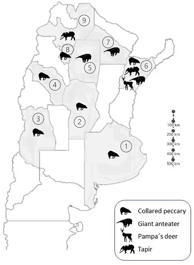
Figure 1. Partial Argentinean map showing the provinces where the native wildlife species are present. The different species are represented with the animal silhouette in each province. References: Provinces: 1. Buenos Aires; 2. Córdoba; 3. Mendoza; 4. La Rioja; 5. Santiago del Estero; 6. Corrientes; 7. Chaco; 8. Tucumán; 9. Salta.
The bacteriological culture of the submitted samples was performed using the Löwenstein-Jensen medium. The Petroff decontamination technique was applied beforehand, treating rhe sample with NaOH (4%) and the sediment obtained is hen neutralized with HCl and placed in the sterile medium, as specified bu Jorge et al. (22). Cultures were incubated at 37°C for up to 12 weeks, and those with no bacterial growth were discarded as negative (22). Colonies compatible with mycobacteria growth were stained using the Ziehl-Neelsen technique for acid-fast bacilli (AFB) observation.
For the detection of the genre Mycobacterium in the isolates, DNA from the bacteriological cultures were obtained by thermal lysis. For this purpose, a colony was taken with a sterile 1 μL loop and suspended in 300 μL of sterile pyrogen-free distilled water in 1.5 mL RNase-free microtubes. Subsequently, the microtubes were subjected to 95°C for 40 min and centrifuged for 10 min at 12,000 rpm. DNA was kept frozen at −20°C until processing. This DNA was subjected to PCR amplification of the heat shock protein 65 kD (hsp65) using the primers TB11 (ACCAACGATGGTGTGTCCAT) and TB12 (CTTGTCGAACCGCATACCCT) and the cycling described by Telenti et al. (23). All PCR products were included in a 2% agarose gel stained with Ethidium bromide (0.5 μL/mL) and observed under a UV light. Those isolates shielding a band at 440 bp were considered positive. These isolates were further studied by sequencing and analyses. Some isolates from different species were selected for sequencing due to economic considerations, aiming to represent each species of wild animal and considering the quality of the band observed in the hsp65 PCR. The selected PCR products were purified using one of the following purification kits “Illustra DNA and Gel Band Purification Kit” (GE Healthcare, UK) or “GFX™ PCR DNA and Gel Band Purification Kit” (Cytiva, USA) following manufacturer's specifications. Purified products' quality was confirmed by quantification in a spectrophotometer at a wavelength of 260 nm (Nanodrop 2000, Thermo Scientific™, Thermo Fisher Scientific, USA).
For the detection of the Mycobacterium avium complex (MAC) members, PCR amplification of the insertion sequence 1245 was performed. Differentiation between species within the complex was achieved through amplification of insertion sequence 901. For IS1245, amplification was performed using primers P1 (GCCGCCGAAACGATCTAC) and P2 (AGGTGGCGTCGAGGAAGAC) and the cycling described by Guerrero et al. (24). A band weighing 427 bp was considered a positive sample for MAC. All MAC-positive isolates were further studied for subspecies detection amplifying the insertion sequence 901, using primers P1 (GGATTGCTAACCACGTGGTG) and P2 (GCGAGTTGCTTGATGAGCG) and the cycling described by Moravkova et al. (25). When a PCR product of 577 bp was observed, Mycobacterium avium subsp. avium was identified, while when no bands were observed Mycobacterium avium subsp. hominissuis was identified.
Sequencing was carried out in the Genomics Unit of the Institute of Biotechnology, in the Institute of Agrobiotechnology and Molecular Biology (IABIMO INTA-CONICET) for the Giant anteater, Tapir and some Pampas deer, and for the collared peccary by Macrogen sequencing service (Korea). The institution that provided sequencing changed during the years these animals were sampled, but both institutions performed the same sequencing, using a 16-capillary sequencer ABI3130xl (Applied Biosystems, Thermo Fisher Scientific, USA), using “Big Dye Terminator v3.1” (Cycle Sequencing Kit). The sequences that shielded good quality were compared to those in the National Center of Biotechnology Information using the Basic Local Alignment Search Tool (BLAST). Identification was based on similarities between our isolates and those in the database, identifying the best match considering the percentage of coverage and identity.
A total of 27 NTM were detected from the 165 (16,36%) investigated samples. The frequency and identity of the Mycobacterium and the frequency in each animal species can be observed in Table 1. Twenty-seven hsp65 positive isolates were subjected to IS1245 PCR and in 4 samples MAC was detected, all of them were IS901 negative, being identified as M. avium subsp. hominissuis. From the remaining IS1245 negative samples, 20 samples were sent for sequencing. Not all the Mycobacterium revealed a clear species identity and coverage when compared to those in the BLAST online database, being identified with the same percentage of identity and coverage as more than two species or only being identified as “Mycobacterium spp.”. Those with no clear identification were kept as “Mycobacterium spp.” for this work. In regards to the giant anteater, the species identified were: M. avium subsp. hominissuis (1/5), M. terrae (1/5), M. gordonae (1/5) and the rest were identified as Mycobacterium. spp (2/5). In regards to the tapir, M. genavense (1/6), M. saskatchewanense (1/6), M. intracellulare (1/6) and three other Mycobacterium spp were identified. Regarding the collared peccary, M. avium subsp. hominissuis (2/10), M. terrae (1/10), M. kumamotonense (1/10), M. fortuitum (1/10) and five other Mycobacterium spp. (5/10) were identified. Lastly, regarding pampas deer, M. avium subsp. hominissuis (1/4), M. intracellulare (1/4) and two other Mycobacterium spp. (2/4) were detected. The most frequent Mycobacterium species detected were M. avium subsp. hominissuis and M. terrae. The animal species with the highest Mycobacteria detection was the giant anteater.
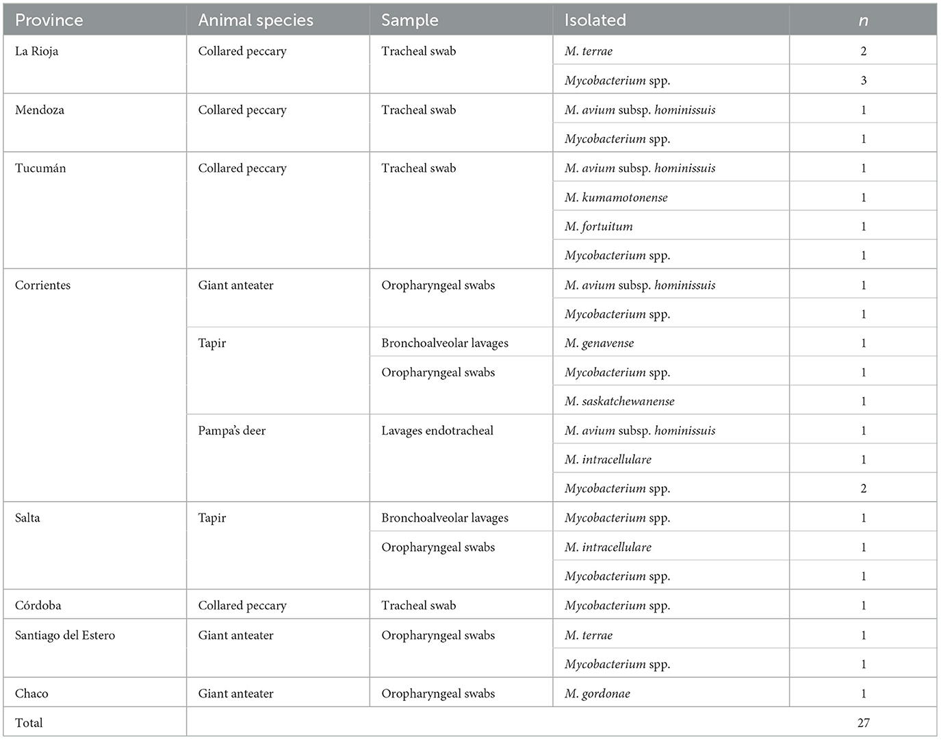
Table 1. Frequency of the identified Mycobacteria in the native wildlife species.
Our study reportsa high NTM recovery rate (16,36%; 27/166) in samples from native wildlife species from different regions of Argentina. None of the sampled animals exhibited clinical signs associated with chronic disease prior to sample collection. Furthermore, from the time of sample collection until the writing of this manuscript, no health data were obtained from these animals. Therefore, it remains unknown whether they developed clinical signs or lesions at any point in their lives.
The animal species with the highest presence of NTM was the giant anteater (45,5%; 5/11). This high recovery of mycobacteria in the oral mucosa of this species might be associated with the way this animal feeds and the distinctive characteristics of its tongue, which is softer, wetter, and rougher, allowing it to adhere to objects before ingestion (26). The presence of NTM such as Mycobacterium fortuitum in this species has been reported previously (27). We did not find any literature describing MTBC infection in this species. This could be due to various reasons, such as limited research on the species, potential resistance of the species to pathogenic mycobacteria, or the possibility that environmental mycobacteria colonize the oropharyngeal mucosa, and potentially regulate or interfere with the colonization of pathogenic mycobacteria, directing the mucosal immune response as has been suggested by other authors in human medicine (28–30). More research is required to corroborate this statement for this species.
Tapirs showed a 32% (6/19) prevalence of NTM. This species is reported to be highly susceptible to both M. bovis and M. tuberculosis (31–34). Given the tapir's high susceptibility to M. bovis and the fact that they were moved from regions where mTB is endemic (7), a comparative intradermal tuberculin test (SICCT) was additionally performed, using both Purified Protein Derivatives (PPD) (bovine and avian) applied on the edge of the ear. This test was negative for both PPDs in all the cases. These results imply a higher sensitivity in detecting NTM from pharyngeal swabs and bronchoalveolar lavage samples through bacteriological culture compared to the SICCT. With these results, we support the assertion that the comparative SICCT, although it is recommended to detect M. bovis, is definitely an inadequate test for the detection of NTM infections in tapirs, as observed by Marcordes et al. (32) in his study.
In the collared peccary, an incidence of 11,5% (12/104) of NTM was detected. In a study conducted in the same region, Brazil, on 330 samples of peccary lymph nodes, a 3% NTM was detected (35). There are reports describing susceptibility to M. bovis infection in this species, and they have been suggested as a possible reservoir for free-ranging animals in some areas of Brazil (35, 36).
The pampas deer is a threatened species in our territory and declines in its wild populations are reported annually (37). In this study, 13% (4/31) of NTM were identified. The Cervidae family is highly susceptible to mTB, both in free-ranging and captive animals (38). Several species of deer have been officially recognized as reservoirs of mTB in several countries across the globe (39–42). There are also reports that demonstrate the presence of NTM in this family (43).
Although the presence of NTM might interfere with the routine mTB diagnostic tests, this is more frequent in areas where TB prevalence in cattle is low. Other authors from Spain had reported M. avium subsp. avium and hominissuis and M. nonchromogenicum as the most common NTM identified in TST-reactors cattle (4). Another author reports that sensitization with M. nonchromogenicum, M. intracellulare, M. avium subsp. paratuberculosis and M. avium subsp. hominissuis, within others, could make animals react to the SITT (44). In Argentina, a study conducted by Oriani et al. (12), in which cattle were inoculated with NTM isolated from soils and wetlands, this NTM included M. kansasii, M. nonchromogenicum, M. gordonae, M. arupense, M. phlei, M. fortuitum and M. peregrinum, and showed that they may cause unspecific reactions, but that these reactions are not maintained over time (12).
As a limitation of this study, we mention the type of samples collected and analyzed for diagnosis. Given that the animals under study were alive, obtaining tissue samples was not feasible. The samples analyzed were restricted to those that could be collected during the examination of the oral and respiratory cavities, knowing that in the literature, the most representative samples for NTM and M. bovis detection are head, mediastinal, and mesenteric lymph nodes from deceased animals (37, 39).
The 16S ribosomal RNA and hsp65 sequencing are both effective for identifying bacteria, particularly Mycobacterium species. Hsp65 sequencing yields comparable results to the widely used 16S ribosomal RNA, as reported by various studies, including one conducted in our laboratory on NTM) in animal samples (10). Other authors also confirm that the use of either 16S ribosomal ARN or hsp65 would allow the identification of Mycobacterium spp. And, in many cases, to the species level (45). Moreover, a combination of more than one sequence could strengthen the identification of the species, using a combination of at least three different sequences. Also, the use of multilocus sequence typing would improve the identification of mycobacterial species (46, 47). Additionally, poor quality or incorrectly identified sequences could limit the identification when compared against the BLAST database (4, 48).
The studied animals came from different provinces to Corrientes province, where they were relocated. These provinces are located far from each other and have different soil and climate conditions, but no significant clustering of NTM species in each region was observed. Among the Mycobacteria detected in this study, ubiquitous environmental bacteria were isolated, such as M. avium, M. gordonae, M. terrae, M. fortuitum, M. kumamotonense. These Mycobacteria can be isolated from soil, water and occasionally have been associated with disease in animals and humans (3, 49–55). M. avium has been isolated from several wild species (4) and MAH has already been reported in wild and domestic animals in Argentina (56). M.intrcellulare has been reported causing disease in a capybara subjected to stressful conditions and causing lesions similar to other pathogenic Mycobacteria (5). M. gordonae and M. terrae have been isolated from sputum samples in human patients with respiratory disease (3, 57), but there are no reports of these agents' causing disease in domestic and wild animals. M. kumamotonense has been documented in immunocompetent individuals with latent tuberculosis and patients with multiple spiculated pulmonary nodules without respiratory symptoms (58, 59). Other NTM such as M. saskatchewanense and M. genavense are found in clinical samples from humans in North America and Europe, acting as opportunistic pathogens in immunocompromised patients (60, 61). M. genavense has been reported in various wild and domestic animals, including birds, rabbits, cats, ferrets, snakes, and dogs (62–64). According to most authors, transmission to humans occurs through oral ingestion from contaminated water or close contact with infected animals. Additionally, a study in the Serengeti ecosystem, focusing on NTM, found M. fortuitum to be a prevalent species. This Mycobacterium was identified in cattle tissues and in the sputum of humans showing clinical signs suggestive of tuberculosis (20). NTM detected in this study have been previously reported in soil, water and cattle and wildlife in Argentina (12, 13, 65).
Emphasizing the importance of infections caused by NTM in human medicine is crucial. The prevalence of NTM in humans is increasing, and there is a belief that in certain industrialized countries, it might exceed the incidence of tuberculosis caused by MTBC (66). Additionally, it is known that NTM has developed resistance to most conventional antibiotics, making treatments ineffective and underscoring their profound impact (67). Although the interaction between human and wildlife is occasional in developed countries, in developing countries the human-wildlife interface is becoming increasingly frequent. Therefore, it is important to understand the distribution of mycobacteria in wildlife from different regions, since the information is very scarce. The active surveillance of wildlife reflects what is happening in the environment, which is the primary source of infection for both humans and coexisting animals (68).
The transmission of NTM between domestic and wild species can occur through direct contact but is largely mediated by shared environments (68, 69). The presence of NTM in free-ranging animals that share their environment with livestock highlights the need to differentiate mycobacteria species, because of the potential interference in diagnostic tests, to control mTB (4, 68, 70). In Argentina, there is a particular scenario where extensive livestock farming is the most frequent strategy, allowing domestic and wild animals to interact in the same environment, increasing the likelihood of disease transmission between them, compared to more confined and intensive farming scenarios (56, 69). The adverse consequences associated with the introduction of livestock into habitats occupied by native fauna have been extensively documented, primarily due to the spread of infectious and parasitic diseases (71–78). The same scenario was observed in tapir (79), giant anteaters (80, 81), and peccaries (35). In regards to the peccaries, efficient transmission is also described between different wild species, such as the invasive exotic wild boar and the vulnerable native peccary (69–82).
Our study provides valuable insights into the presence and diversity of NTM in Argentina's native wildlife. This emphasizes the importance of active surveillance, highlighting potential risks to native species and advocating for conservation strategies to mitigate infectious diseases' impacts in shared environments.
The datasets presented in this study can be found in online repositories. The names of the repository/repositories and accession number(s) can be found below: https://www.ncbi.nlm.nih.gov/genbank/, MW043443; https://www.ncbi.nlm.nih.gov/genbank/, MW043444.
Ethical approval was not required for the studies involving animals in accordance with the local legislation and institutional requirements because animals are wildlife in the context of Conservation and Translocation Programs. Fundación Rewilding Argentina has permits for all the activities they control. The Laboratory of Tuberculosis Diagnosis received the samples and no Ethics Committee is required. Written informed consent was not obtained from the owners for the participation of their animals in this study because this animals have no owners or where translocated from zoos or rescue facilities. Permits are not required as quarantine is mandatory and this tests are in the context of health check-ups.
SB: Conceptualization, Formal analysis, Funding acquisition, Investigation, Project administration, Supervision, Writing—original draft, Writing—review & editing. LP: Methodology, Writing—original draft, Writing—review & editing. IP: Methodology, Writing—original draft, Writing—review & editing. AR: Methodology, Visualization, Writing—review & editing. JP: Methodology, Visualization, Writing—review & editing. MM: Conceptualization, Formal analysis, Investigation, Methodology, Writing—original draft, Writing—review & editing.
The author(s) declare financial support was received for the research, authorship, and/or publication of this article. Research was supported with UBACyT Projects (20020170200311BA; 20020130100082BA 20020190200309BA) and Fundación Rewilding Argentina own projects.
We thank veterinaries and volunteers of Fundación Rewilding Argentina. SB is career member of CONICET, Argentina.
The authors declare that the research was conducted in the absence of any commercial or financial relationships that could be construed as a potential conflict of interest.
All claims expressed in this article are solely those of the authors and do not necessarily represent those of their affiliated organizations, or those of the publisher, the editors and the reviewers. Any product that may be evaluated in this article, or claim that may be made by its manufacturer, is not guaranteed or endorsed by the publisher.
1. Fedrizzi T, Meehan CJ, Grottola A, Giacobazzi E, Fregni Serpini G, Tagliazucchi S, et al. Genomic characterization of Nontuberculous Mycobacteria. Sci Rep. (2017) 7:1–14. doi: 10.1038/srep45258
2. Stanford J, Stanford C. Mycobacteria and their world. Int J Mycobacteriol. (2012) 1:3–12. doi: 10.1016/j.ijmyco.2012.01.001
3. Hoza AS, Mfinanga SGM, Rodloff AC, Moser I, König B. Increased isolation of nontuberculous mycobacteria among TB suspects in Northeastern, Tanzania: Public health and diagnostic implications for control programmes. BMC Res Notes. (2016) 9:1–9. doi: 10.1186/s13104-016-1928-3
4. Varela-Castro L, Barral M, Arnal MC, Fernández de Luco D, Gortázar C, Garrido JM, et al. Beyond tuberculosis: Diversity and implications of non-tuberculous mycobacteria at the wildlife–livestock interface. Transbound Emerg Dis. (2022) 69:e2978–93. doi: 10.1111/tbed.14649
5. Pezzone N, Eberhardt AT, Fernández A, Garbaccio S, Zumárraga M, Gioffré A, et al. Mycobacterium intracellulare infection in a capybara (hydrochoerus hydrochaeris). J Zoo Wildlife Med. (2013) 44:1098–101. doi: 10.1638/2013-0017R1.1
6. Allen AR, Ford T, Skuce RA. Does Mycobacterium tuberculosis var. bovis survival in the environment confound bovine tuberculosis control and eradication? A literature review. Vet Med Int. (2021) 2021:8812898. doi: 10.1155/2021/8812898
7. Abdala AA, Garbaccio S, Zumárraga M, Tarabla HD. Mycobacterium bovis en fauna silvestre de la cuenca lechera de Santa Fe, Argentina. Rev Argent Microbiol. (2015) 47:174–82. doi: 10.1016/j.ram.2015.04.005
8. SENASA. Servicio Nacional de Sanidad y Calidad Agroalimentaria. (2023). Available online at: https://www.argentina.gob.ar/senasa/programas-sanitarios/cadena-animal/bovinos-y-bubalinos/bovinos-y-bubalinos-producci%C3%B3n-primaria/tuberculosis-bovina (accessed February 7, 2024).
9. Torres PM. SITUACION DE LA TUBERCULOSIS BOVINA EN LA REPUBLICA ARGENTINA. Buenos Aires: Programa Nacional de Control y Erradicación de la tuberculosis, SENASA (Servicio Nacional de Sanidad Animal), Secretaría de Agricultura (2016). p. 1–37.
10. Marfil MJ, Garbaccio SG, Barandiaran S, Huertas PS, Vivot MM, Eirin ME, et al. Isolation of Nontuberculous Mycobacteria from Bovine Raw Lungs Bought in Butchers' Shops. Foodborne Pathogens Dis. (2021) 18:805–11. doi: 10.1089/fpd.2021.0026
11. Imperiale B, Zumárraga M, Gioffré A, Di Giulio B, Cataldi A, Morcillo N. Disease caused by non-tuberculous mycobacteria: Diagnostic procedures and treatment evaluation in the north of Buenos Aires province [Enfermedad causada por micobacterias no tuberculosas: Diagnostico y evaluacion del tratamiento en el norte del Gran Bueno. Rev Argent Microbiol. (2012) 44:3–9. doi: 10.1590/S0325-75412012000100002
12. Oriani DS, Gastaldo MF, Tortone CA, Staskevich AS, Remirez P, Valle H, et al. Micobacterias no tuberculosas autóctonas de La Pampa (Argentina) y su capacidad de reacción cruzada en el diagnóstico de tuberculosis bovina. Native non-tuberculous mycobacteria from La Pampa (Argentina) and their ability to cross-reaction in the bov. Vetec Revista Académica de Investigación, Docencia y Extensión de las Ciencias Veterinarias. (2022) 4:10–8. Available online at: https://cerac.unlpam.edu.ar/index.php/Vetec/article/view/7229
13. Traversa MJ, Jorge MC, Garbaccio D, Draghi MG, Abdala A, Tarabla H, et al. Report of mycobacteria isolated from domestic and wildlife species during 2004-2008. Analecta Vet. (2011) 31:10–4. Available online at: https://revistas.unlp.edu.ar/analecta/article/view/12202
14. Zamboni T, Di Martino S, Jiménez-Pérez I, A. review of a multispecies reintroduction to restore a large ecosystem: The Iberá Rewilding Program (Argentina). Perspect Ecol Conserv. (2017) 15:248–56. doi: 10.1016/j.pecon.2017.10.001
15. Jiménez-Pérez I, Abuin R, Antúnez B, Delgado A, Massat M, Pereda I, et al. Re-introduction of the pampas deer in Iberá Nature Reserve, Corrientes, Argentin. In: Soorae PS, , editor. Global Re-introductionPerspectives: 2016: Case studies from around the globe. (2016). p. 221–7.
16. Ministerio de. Ambiente y Desarrollo Sustentable (MAD). Categorización de los Mamíferos de Argentina según su riesgo de extinción. (2021). Available online at: https://www.argentina.gob.ar/normativa/nacional/resolución-316-2021-354496 (accessed February 7, 2024).
18. Chalukian SC, Bustos S De, Lizarraga L, Varela D, Paviolo A, Quse V. PLAN DE ACCIÓN PARA LA CONSERVACIÓN DEL TAPIR (Tapirus terrestris) EN ARGENTINA 2009. (2018):72. Available online at: https://www.researchgate.net/profile/Diego-Varela/publication/327273239_Plan_de_Accion_para_la_Conservacion_del_Tapir_Tapirus_terrestr (accessed February 7, 2024).
19. Hurtado Martínez C. Reintroduction success and ecological aspects of reintroduced peccaries (Pecari tajacu) in the Ibera Natural Reserve, Corrientes, Argentina. Towson, MD: Towson University (2017).
20. Katale BZ, Mbugi E V, Botha L, Keyyu JD, Kendall S, Dockrell HM, et al. Species diversity of non-tuberculous mycobacteria isolated from humans, livestock and wildlife in the Serengeti ecosystem, Tanzania. BMC Infect Dis. (2014) 14:1–8. doi: 10.1186/s12879-014-0616-y
21. Di Martino S, Heinonen S, Donadío E. Rewilding en la Argentina. In: López L, , editor. Ciudad Autónoma de Buenos Aires: The Conservation Land Trust Argentina (2022). p. 264.
22. Jorge MC, Alito A, Bernardelli A, Canal AM, Cataldi A, Cicuta ME, et al. Manual de diagnóstico de micobacterias de importancia en medicina veterinaria. In: Diagnóstico CC, de M de la AA, de L de, , editors. Manual de diagnóstico de micobacterias de importancia en medicina veterinaria. 1st ed. Buenos Aires: Acosta, Imprenta (2005). p. 20–46.
23. Telenti A, Marchesi F, Balz M, Bally F, Böttger EC, Bodmer T. Rapid Identification of Mycobacteria to the Species Level by Polymerase Chain Reaction and Restriction Enzyme Analysis. J Clin Microbiol. (1993) 31:175–8. doi: 10.1128/jcm.31.2.175-178.1993
24. Guerrero C, Bernasconi C, Burki D, Bodmer T, Telenti A. A novel insertion element from Mycobacterium avium, IS1245, is a specific target for analysis of strain relatedness. J Clin Microbiol. (1995) 33:304–7. doi: 10.1128/jcm.33.2.304-307.1995
25. Moravkova M, Hlozek P, Beran V, Pavlik I, Preziuso S, Cuteri V, et al. Strategy for the detection and differentiation of Mycobacterium avium species in isolates and heavily infected tissues. Res Vet Sci. (2008) 85:257–64. doi: 10.1016/j.rvsc.2007.10.006
26. Noel AC, Hu DL. The tongue as a gripper. J Exp Biol. (2018) 221:jeb176289. doi: 10.1242/jeb.176289
27. Terrell SP, Weber MA, Neiffer DL, Miller MA, Mangold BJ, Fontenot DK, et al. Cutaneous Pox Virus Infection in Two Giant Anteaters (Myrmecophaga tridactyla) with Clinical and Pathologic Features Similar to Human Molluscum Contagiosum. In: Wildlife Health Sciences. Bronx, NY: Wildlife Conservation Society (2001). p. 127–30.
28. Belkaid Y, Harrison OJ. Homeostatic immunity and the microbiota - sciencedirect. Immunity. (2017) 46:562–76. doi: 10.1016/j.immuni.2017.04.008
29. Tarashi S, Ahmadi Badi S, Moshiri A, Nasehi M, Fateh A, Vaziri F, et al. The human microbiota in pulmonary tuberculosis: not so innocent bystanders. Tuberculosis. (2018) 113:215–21. doi: 10.1016/j.tube.2018.10.010
30. Shah T, Shah Z, Baloch Z, Cui XM. The role of microbiota in respiratory health and diseases, particularly in tuberculosis. Biomed Pharm. (2021) 143:112108. doi: 10.1016/j.biopha.2021.112108
31. Murakami PS, Monego F, Ho JL, Gibson A, Javorouski ML, Bonat M, et al. Detection of RDRIO strain of mycobacterium tuberculosis in tapirs (tapirus terrestris) from a zoo in Brazil. J Zoo Wildlife Med. (2012) 43:872–5. doi: 10.1638/2010-0108R.1
32. Marcordes S, Lueders I, Grund L, Sliwa A, Maurer FP, Hillemann D, et al. Clinical outcome and diagnostic methods of atypical mycobacteriosis due to Mycobacterium avium ssp. hominissuis in a group of captive lowland tapirs (Tapirus terrestris). Transboundary Emerg Dis. (2021) 68:1305–13. doi: 10.1111/tbed.13786
33. Chaney SB, McAloose D, Greenwald R, Lyashchenko KP, Calle PP. Assessment of multiantigen print immunoassay and rapid lateral-flow test for the detection of mycobacterium bovis infection in Malayan tapir. (Tapirus indicus). J Zoo Wildlife Med. (2021) 16:52. doi: 10.1638/2021-0054
34. Sternberg S, Bernodt K, Holmström A, Röken B. Survey of tuberculin testing in Swedish zoos. J Zoo Wildlife Med Official Publ Am Assoc Zoo Vet. (2002) 33:378–80. doi: 10.1638/1042-7260(2002)033[0378:SOTTIS]2.0.CO;2
35. de Morais ABC, Bolaños CAD, Alves AC, Ikuta CY, Lara GHB, Heinemann MB, et al. Identification of Mycobacterium species and Rhodococcus equi in peccary lymph nodes. Trop Anim Health Prod. (2018) 50:1319–26. doi: 10.1007/s11250-018-1562-2
36. Mayer FQ, Cerva C, Driemeier D, Cruz CEF da, Loiko MR, Coppola M de M, et al. Mycobacterium bovis infection in a collared peccary (Tayassu tajacu): insights on tuberculosis wild reservoirs. Vet Microbiol. (2012) 160:549–51. doi: 10.1016/j.vetmic.2012.06.033
37. National Parks Administration. Biodiversity Information System. (2023). Available online at: sib.gob.ar (accessed February 7, 2024).
38. Vercauteren KC, Gortázar C, Beltrán-Alcrudo D, Vicente J. Diseases at the Wildlife - Livestock Interface. Vicente J, Vercauteren KC, Gortázar C, editors. Cham: Springer International Publishing (2021). p. 3–32.
39. Mackintosh CG, De Lisle GW, Collins DM, Griffin JFT. Mycobacterial diseases of deer. N Z Vet J. (2004) 52:163–74. doi: 10.1080/00480169.2004.36424
40. Fitzgerald SD, Kaneene JB. Wildlife reservoirs of bovine tuberculosis worldwide: hosts, pathology, surveillance, and control. Vet Pathol. (2013) 50:488–99. doi: 10.1177/0300985812467472
41. Nugent G. Maintenance, spillover and spillback transmission of bovine tuberculosis in multi-host wildlife complexes: a New Zealand case study. Vet Microbiol. (2011) 151:34–42. doi: 10.1016/j.vetmic.2011.02.023
42. Santos N, Almeida V, Gortázar C, Correia-Neves M. Patterns of Mycobacterium tuberculosis-complex excretion and characterization of super-shedders in naturally-infected wild boar and red deer. Vet Res. (2015) 46:1–10. doi: 10.1186/s13567-015-0270-4
43. Radulski Ł, Kalicki M, Krajewska-Wedzina M, Lipiec M, Szulowski K. Pulmonary mycobacteriosis of sitatunga antelope caused by M. avium ssp hominissuis. Annals Agri Environ Med. (2022) 29:220–3. doi: 10.26444/aaem/145158
44. Fernández-Veiga L, Fuertes M, Geijo M V, Pérez de Val B, Vidal E, Michelet L, et al. Differences in skin test reactions to official and defined antigens in guinea pigs exposed to non-tuberculous and tuberculous bacteria. Sci Rep. (2023) 13:1–12. doi: 10.1038/s41598-023-30147-4
45. Kim SH, Shin JH. Identification of nontuberculous mycobacteria using multilocous sequence analysis of 16S rRNA, hsp65, and rpoB. J Clin Lab Anal. (2018) 32:1–6. doi: 10.1002/jcla.22184
46. Liu H, Lian L, Jiang Y, Huang M, Tan Y, Zhao X, et al. Identification of Species of Nontuberculous Mycobacteria Clinical Isolates from 8 Provinces of China. Biomed Res Int. (2016) 2016:1–10. doi: 10.1155/2016/2153910
47. Adékambi T, Drancourt M. Dissection of phylogenetic relationships among 19 rapidly growing Mycobacterium species by 16S rRNA, hsp65, sodA, recA and rpoB gene sequencing. Int J Syst Evol Microbiol. (2004) 54:2095–105. doi: 10.1099/ijs.0.63094-0
48. Böddinghaus B, Rogall T, Flohr T, Blöcker H, Böttger EC. Detection and identification of mycobacteria by amplification of rRNA. J Clin Microbiol. (1990) 28:1751–9. doi: 10.1128/jcm.28.8.1751-1759.1990
49. Lande L, Alexander DC, Wallace RJ, Kwait R, Iakhiaeva E, Williams M, et al. Mycobacterium avium in community and household water, suburban Philadelphia, Pennsylvania, USA, 2010-2012. Emerg Infect Dis. (2019) 25:473–81. doi: 10.3201/eid2503.180336
50. Eaton T, Falkinham JO, Aisu TO, Daniel TM. Isolation and characteristics of Mycobacterium avium complex from water and soil samples in Uganda. Tubercle Lung Dis. (1995) 76:570–4. doi: 10.1016/0962-8479(95)90536-7
51. Pavlik I, Matlova L, Dvorska L, Shitaye JE, Parmova I. Mycobacterial infections in cattle and pigs caused by Mycobacterium aviumcomplex members and atypical mycobacteria in theCzech Republicduring 2000-2004. Veterinární medicína. (2005) 50:281–90. doi: 10.17221/5625-VETMED
52. Klanicova-Zalewska B, Slana I. Presence and persistence of Mycobacterium avium and other nontuberculous mycobacteria in animal tissues and derived foods: A review. Meat Sci. (2014) 98:835–41. doi: 10.1016/j.meatsci.2014.08.001
53. Tortoli E. Microbiological features and clinical relevance of new species of the genus Mycobacterium. Clin Microbiol Rev. (2014) 27:727–52. doi: 10.1128/CMR.00035-14
54. Ghielmetti G, Giger U. Mycobacterium avium: an Emerging Pathogen for Dog Breeds with Hereditary Immunodeficiencies. Current Clin Microbiol Rep. (2020) 7:67–80. doi: 10.1007/s40588-020-00145-5
55. Tortone CA, Oriani DS, Staskevich AS, Oriani AS, Gino LM, Marfil MJ, et al. Species diversity of non-tuberculous mycobacteria isolated from aquatic environments of General Pico city, Province of La Pampa (Argentina). Rev Argent Microbiol. (2019) 51:259–67. doi: 10.1016/j.ram.2018.08.005
56. Barandiaran S, Marfil MJ, Capobianco G, Pérez Aguirreburualde MS, Zumárraga MJ, Eirin ME, et al. Epidemiology of Pig Tuberculosis in Argentina. Front Vet Sci. (2021) 8:1–8. doi: 10.3389/fvets.2021.693082
57. Asaoka M, Hagiwara E, Etori S, Higa K, Ikeda S, Sekine A, et al. Identification and characteristics of co-isolation of multiple nontuberculous mycobacteria. Int Med. (2021) 60:3213–9. doi: 10.2169/internalmedicine.5300-20
58. Kontos F, Mavromanolakis DN, Zande MC, Gitti ZG. Isolation of Mycobacterium kumamotonense from a patient with pulmonary infection and latent tuberculosis. Indian J Med Microbiol. (2016) 34:241–4. doi: 10.4103/0255-0857.180356
59. Chang J, Liu B, Grondin JJ, Fitton K, Wang M. Isolated mycobacterium kumamotonense in an immunocompetent patient without respiratory symptoms. Chest. (2023) 164:A1352–3. doi: 10.1016/j.chest.2023.07.946
60. Turenne CY. Nontuberculous mycobacteria: insights on taxonomy and evolution. Infect Genet Evol. (2019) 72:159–68. doi: 10.1016/j.meegid.2019.01.017
61. Di Mento G, Carreca AP, Monaco F, Cuscino N, Cardinale F, Conaldi PG, et al. Mycobacterium saskatchewanense strain associated with a chronic kidney disease patient in an Italian transplantation hospital and almost misdiagnosed as Mycobacterium tuberculosis. Infect Cont Hosp Epidemiol. (2019) 40:496–7. doi: 10.1017/ice.2019.6
62. Dequéant B, Pascal Q, Bilbault H, Dagher E, Boschiroli M-L, Cordonnier N, et al. Identification of Mycobacterium genavense natural infection in a domestic ferret. J Vet Diag Invest. (2019) 31:133–6. doi: 10.1177/1040638718812137
63. Vazquez E, Nicita D, Masini D, Matteo M, Costa N, Franze O, et al. Mycobacterium genavense: una causa infrecuente de lesión cerebral ocupante de espacio. Neurología Argentina. (2023) 15:198–202. doi: 10.1016/j.neuarg.2022.03.001
64. Baldolli A, Chocron R, Dargère S, Michon J, Daurel C, Thuillier-Lecouf A, et al. Mycobacterium genavense infections in immunocompromised patients without HIV: case series of solid organ transplant patients and literature review. Open Forum Infect Dis. (2022) 5:9. doi: 10.1093/ofid/ofac498
65. Marfil MJ, Huertas PS, Garbaccio SG, Barandiaran S, Martínez Vivot M, Garro C, et al. Detection of viable mycobacterium bovis in lungs and livers sold in butchers' shops in Buenos Aires, Argentina. Foodborne Pathog Dis. (2018) 15:758–62. doi: 10.1089/fpd.2018.2467
66. Gómez NA. Non-tuberculous mycobacteria: an emerging disease? Anales de pediatria. (2009) 71:185–8. doi: 10.1016/j.anpedi.2009.07.001
67. Saxena S, Spaink HP, Forn-Cuní G. Drug resistance in nontuberculous mycobacteria: mechanisms and models. Biology. (2021) 10:96. doi: 10.3390/biology10020096
68. Biet F, Boschiroli ML, Thorel MF, Guilloteau LA. Zoonotic aspects of Mycobacterium bovis and Mycobacterium avium-intracellulare complex (MAC). Vet Res. (2005) 36:411–36. doi: 10.1051/vetres:2005001
69. Kmetiuk LB, Biondo LM, Pedrosa F, Favero GM, Biondo AW. One Health at gunpoint: Impact of wild boars as exotic species in Brazil - a review. One Health. (2023) 17:100577. doi: 10.1016/j.onehlt.2023.100577
70. FAO. LIVESTOCK'S LONG SHADOW. Environmental issues and options. Roma: FAO (2006). p. 390 p. Available online at: https://www.fao.org/3/a0701e/a0701e00.htm (accessed February 7, 2024).
71. Romero-Castañón S, Ferguson BG, Güiris D, González D, López S, Paredes A, et al. Comparative Parasitology of Wild and Domestic Ungulates in the Selva Lacandona, Chiapas, Mexico. Comp Parasitol. (2008) 75:115–26. doi: 10.1654/4267.1
72. Martin C, Pastoret P-P, Brochier B, Humblet M-F, Saegerman C, A. survey of the transmission of infectious diseases / infections between wild and A survey of the transmission of infectious diseases / infections between. Veterinary Re. (2011) 42:1–16. doi: 10.1186/1297-9716-42-70
73. Corti P, Saucedo C, Herrera P. Evidence of Bovine Viral Diarrhea, but Absence of Infectious Bovine Rhinotracheitis and Bovine Brucellosis in the Endangered Huemul Deer (Hippocamelus bisulcus) in Chilean Patagonia. J Wildl Dis. (2013) 49:744–6. doi: 10.7589/2012-04-105
74. Merino ML. AV. y AS. Relevamiento biológico de la Bahía Samborombón, provincia de Buenos Aires. Buenos Aires: Boletín Técnico Fundación Vida Silvestre Argentina (1993). 16:45.
75. Merino ML y B. Carpinetti Pampas Deer Population Trend in Bahía Samborombón. Buenos Aires: Argentina Deer Specialist Group News (1998). 14:10–1.
76. Uhart MM, Vila AR, Beade MS, Balcarce A, Karesh WB. Health evaluation of pampas deer (Ozotoceros bezoarticus celer) at Campos del Tuyú Wildlife Reserve, Argentina. J Wildl Dis. (2003) 39:887–93. doi: 10.7589/0090-3558-39.4.887
77. Vila AR, Beade MS, Barrios Lamunière D. Home range and habitat selection of pampas deer. J Zool. (2008) 276:95–102. doi: 10.1111/j.1469-7998.2008.00468.x
78. Menajovsky MF, Espunyes J, Cabezón O, Mayor P. Infectious diseases of interest for the conservation of peccaries in the Amazon: a systematic quantitative review. Biol Conserv. (2023) 277:109867. doi: 10.1016/j.biocon.2022.109867
79. Chalukian SC, de Bustos S, Lizárraga L, Varela D, Paviolo A, Quse V. Uso de hábitat del tapir en relación a la presencia de ganado, en el Parque Nacional El Rey, Salta, Argentina. II Simposio Internacional de Tapir, Libro de Resúmenes; 2004. p. 10–6.
80. Gardner SL, Upton SJ, Lambert CR, Jordan OC. Redescription of Eimeria escomeli (Rastegaieff, 1930) from Myrmecophaga tridactyla, and a first report from Bolivia. J Helminthol Soc Washington. (1991) 58:16–8.
81. Labruna MB, Paula CD de, Lima TF, Sana DA. Ticks (Acari: Ixodidae) on wild animals from the Porto-Primavera Hydroelectric power station area, Brazil. Memórias do Instituto Oswaldo Cruz. (2002) 97:1133–6. doi: 10.1590/S0074-02762002000800012
Keywords: non-tuberculous mycobacteria, native wildlife, conservation, bacteriological diagnosis, molecular identification
Citation: Barandiaran S, Ponce L, Piras I, Rosas AC, Peña Martinez J and Marfil MJ (2024) Detection of non-tuberculous mycobacteria in native wildlife species at conservation risk of Argentina. Front. Vet. Sci. 11:1346514. doi: 10.3389/fvets.2024.1346514
Received: 29 November 2023; Accepted: 29 January 2024;
Published: 14 February 2024.
Edited by:
Giovanni Ghielmetti, University of Zurich, SwitzerlandReviewed by:
Maria Laura Boschiroli, Agence Nationale de Sécurité Sanitaire de l'Alimentation, de l'Environnement et du Travail (ANSES), FranceCopyright © 2024 Barandiaran, Ponce, Piras, Rosas, Peña Martinez and Marfil. This is an open-access article distributed under the terms of the Creative Commons Attribution License (CC BY). The use, distribution or reproduction in other forums is permitted, provided the original author(s) and the copyright owner(s) are credited and that the original publication in this journal is cited, in accordance with accepted academic practice. No use, distribution or reproduction is permitted which does not comply with these terms.
*Correspondence: María Jimena Marfil, am1hZmlsQGZ2ZXQudWJhLmFy
Disclaimer: All claims expressed in this article are solely those of the authors and do not necessarily represent those of their affiliated organizations, or those of the publisher, the editors and the reviewers. Any product that may be evaluated in this article or claim that may be made by its manufacturer is not guaranteed or endorsed by the publisher.
Research integrity at Frontiers
Learn more about the work of our research integrity team to safeguard the quality of each article we publish.