 Mohammad Shamim Hossein1†
 Young-Bum Son1,2†
 Yeon Ik Jeong1
 Mina Kang1Seejin Lee1Alex Tinson3
 Woo Suk Hwang1,4*
- 1UAE Biotech Research Center, Abu Dhabi, United Arab Emirates
- 2Department of OBS/Theriogenology, College of Veterinary Medicine, Chonnam National University, Gwangju, Republic of Korea
- 3Hilli E.T. Cloning and Surgical Centre Presidential Camels and Camel Racing Affairs, Al-Ain, United Arab Emirates
- 4NorthEastern Federal University, Yakutsk, Russia
Monoamniotic twins develop when a blastocyst spontaneously splits its progenitor cells, and each group of progenitor cells independently grows to become an individual. It is the rarest type of twin pregnancy and usually has significant developmental or congenital abnormalities, a higher rate of abortion, perinatal morbidity, and mortality. There is no information regarding monoamniotic twins in livestock species. Here, we reported a spontaneous abortion of monoamniotic twins in a dromedary camel at 278 days of gestation. Gonadorelin acetate (100 μg) was injected intramuscularly to induce ovulation in the recipient. A 7 days-old embryo produced by somatic cell nuclear transfer was transferred transcervically to the recipient. Early pregnancy was confirmed by an elevated level of serum progesterone followed by ultrasonography at 22 and 44 days after embryo transfer. A single sac was observed on 22 days while twins were evident 44 days after embryo transfer. Pregnancy was periodically monitored by the tail-up phenomenon. A ruptured fetal sac was observed on the ground having two fetuses. On autopsy, full-grown fetuses were found. Their bodies were separated. There was no congenital anomaly or any malformation in the fetuses. According to the reported chronology in human twins, we hypothesized that the blastocyst splitted before 13 days as it was monoamniotic and not conjoined. If the embryo splits within 4 to 8 days, it develops two amniotic sacs, and splitting after 13 days develops conjoined fetuses. To the authors’ knowledge, this is the first reported case of monoamniotic twin abortion in dromedary camels. This report will increase awareness among practicing veterinarians and camel breeders about twin abortions.
Introduction
A proper understanding of reproductive physiology is the key element for the breeding management of any species. Dromedary camels are prone to pregnancy losses at various stages of gestation periods. Early pregnancy loss could exceed 50% by day 80 of pregnancy (1, 2). However, the incidence of mid to late-term pregnancy loss is below 10%. Although causes and risk factors of pregnancy loss need to be explored thoroughly in camels, endometritis, luteal insufficiency, embryonic and environmental factors are considered the leading causes of early pregnancy loss (1). In contrast, systemic infections or injuries are the major causes of late-term abortion (3).
Camels are montocous animals, and twin births are extremely rare in nature (4). If twin ovulation occurs in the same estrous cycle, twinning is possible (5). However, twining could be developed by embryo splitting at the early stages of embryogenesis (6). Twins that develop by splitting an early embryo are known as monozygotic twins, and they are identical to each other. Depending on the timing of embryo splitting, several kinds of monozygotic twins could develop. Among them, monoamniotic twins, twins that share a common chorionic and amniotic cavity, are extremely rare. In humans, monoamniotic twins usually die before 24 weeks gestation (7) due to prematurity, cord entanglement, congenital anomalies, or twin-to-twin transfusion. In the present study, we reported a spontaneous abortion of a monoamniotic twin at 278 days of gestation in camels. This report will increase awareness among researchers, practicing veterinarians, and camel breeders about twin abortions.
Case presentation
Ear skin biopsy was performed to collect tissues from an elite dromedary camel, and fibroblast cell line was established to clone the camel (8–10).
Oocyte donor and recipient camels were maintained in a large-scale cloning facility of the UAE Biotech Research Center, Abu Dhabi, United Arab Emirates (24°20′26.1″N 55°45′20.3″E) under the supervision of veterinarians. Camels were synchronized and superstimulated using our laboratory standard protocol (10). Briefly, oocyte donors and recipients were administered intramuscularly a single injection of 5,000 IU and 1,500 IU of pregnant mare serum gonadotropin (PMSG) (Ceva, Libourne, France) and 500 μg and 100 μg of cloprostenol (Jurox, Rutherford, Australia), respectively. Both the donors and recipients were injected with 100 μg gonadorelin acetate (Vetoquinol, Paris, France) on the 9th day after PMSG injection. Twenty two hours later, oocytes were collected transvaginally, from the donors using ultrasonography (10).
Embryos were produced by injecting a diploid somatic cell into the enucleated metaphase II oocytes. The reconstructed oocytes were fused using electrical impulses in a fusion medium. After that, fused oocytes were chemically activated and cultured for 7 days for blastocyst production [for detailed procedure (10)]. The recipient (Multipara, body weight 470 kg) received a single 7 days-old embryo transcervically (Figure 1). Pregnancy was confirmed by measuring the serum progesterone level and by using ultrasonography 22 days after embryo transfer (Figure 2A). Serum progesterone level was 5.75 ng/mL and a single sac was observed on ultrasonography. On 44 days after embryo transfer, the serum progesterone level was 33.40 ng/mL and evidences of twining were revealed on ultrasonography (Figure 2B).
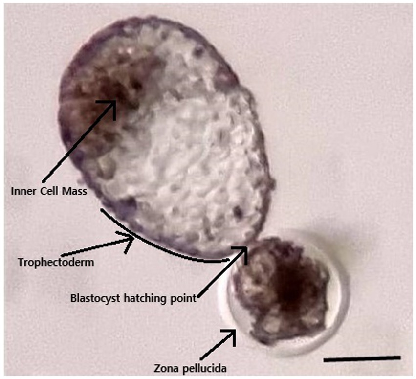
Figure 1. Photograph of the blastocyst transferred to the recipient. The blastocyst was 7 days old and started to hatch through the zona pellucida. The bar is 250 μm.
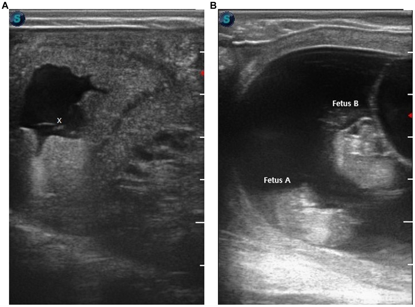
Figure 2. Ultrasonographic confirmation of pregnancy after transfer of cloned blastocyst. (A) The image shows a 17 mm cavity, 22 days after embryo transfer (x indicates fetus). (B) Ultrasonographic confirmation of twin pregnancy at 44 days after embryo transfer. The image shows a cavity having two fetuses; fetus A and fetus B.
The recipient spontaneously aborted twin fetuses on 278 days of pregnancy (Figure 3). The aborted fetuses were found on the ground in the morning. The recipient showed obvious signs of abortion. Hind legs were tinted with blood, and some portion of the placenta was hanging from the vagina. On autopsy, two full-grown fetuses were found. Their bodies weres separated. No congenital anomaly or malformation was seen in the fetuses. As the recipient aborted at late gestation, the brucellosis test was performed in fetal tissue and Dam’s blood, which was negative. Genomic DNA was extracted from aborted fetuses, recipient, and somatic cell donor using a Qiagen DNeasy Blood and Tissue Kit (Qiagen, Hilden, Germany) (8–10). Microsatellite analysis of 13 specific loci confirmed the fetuses as identical twins (Table 1).

Figure 3. Aborted twin fetuses at 278 days of gestation.
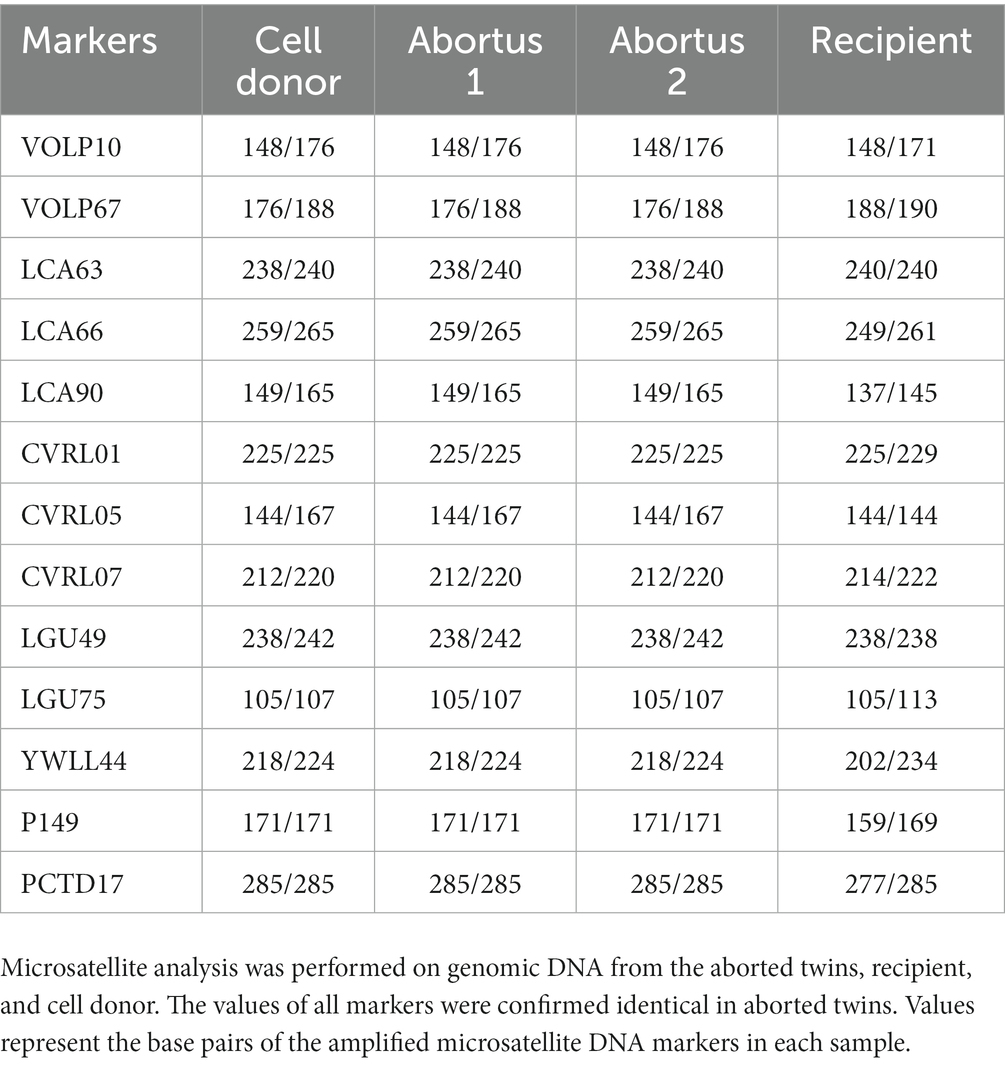
Table 1. Microsatellite analysis of aborted twins, recipient, and cell donor.
Discussion
Late-term pregnancy loss has severe economic consequences. Most of the pregnancy losses in camels occur in the first trimester of pregnancy; however, late-term pregnancy loss is also common (1). Here, we described a spontaneous abortion of twin fetuses at 278 days of gestation.
Camels have many fascinating reproductive phenomena, such as the induced ovulation by coitus, pregnancy only maintained in the left horn, higher rate of early pregnancy loss, especially in the case of twin pregnancies almost always one conceptus get reabsorbed within 60 days of pregnancy. Camels usually ovulate a single oocyte in each estrous cycle. However, around 14% of twin ovulation is reported; these twin ovulations normally result in twin pregnancies (4, 5). This kind of twin is known as a dizygotic twin, as two oocytes develop into two different embryos and is the most frequent form of twinning.
In the early zygotic stage, one embryo could be divided into two embryos and could develop into a monozygotic twin. In animals, there is no comprehensive study on the early development of monozygotic twins; however, different kinds of these twins were reported in humans, such as dichorionic-diamniotic twins, monochorinic-diamniotic twins, monoamniotic twins, or conjoined twins (11). The time of embryo division is the main determining factor for the type of twinning. In an elaborative study in humans, Hall (12) reported that most of the monozygotic twins develop within the first 7 days of embryonic life before blastocyst formation, and this early division gives a better survival chance to the twins. Monoamniotic twins develop when embryos divide after the blastocyst formation at around 8 to 13 days. Further delay in embryonic division exceeding 13 days develops conjoined twins in humans. We transferred a single 7 days-old blastocyst to the recipient, and we did not anticipate any twins. Therefore, the blastocyst divides after embryo transfer, and according to the reported chronology in human twins, this division seems to have occurred before 13 days as the twins were monoamniotic and not conjoined.
Artificial embryo splitting in the laboratory and the production of monozygotic twins have been reported in several livestock species, such as sheep (13), cattle (14), goats (15). Each of the splitted embryos implants separately, develops within its own sacs, and therefore increases fecundity. Spontaneous splitting of an embryo after blastocyst formation developed monochorionic and monoamniotic in nature. This kind of pregnancy has major discordant abnormalities, a higher rate of abortion, and perinatal mortality (7, 16).
The exact mechanism of spontaneous or natural embryo splitting and the development of monozygotic twins is not known. Illmen(see 17) explains that the collapse of a blastocyst may results in the splitting of its progenitor cells and develop into a monozygotic twin. We assume that mechanical pressure on the blastocyst during its loading or transfer may be responsible for blastocyst splitting as we transferred a fully expanded hatching blastocyst. The findings of this case report will support the growing embryo transfer practices in camel breeding. A fully grown hatching or hatched blastocyst is usually transferred to the recipient in multiple ovulation embryo transfer and cloning programs (18). Proper care and gentle handling of blastocyst could prevent the development of monoamniotic twins.
In conclusion, twin or multiple pregnancies are always associated with high maternal complications such as abortion, preterm delivery, preterm pre-labor rupture of fetal sacs, etc., and fetal complications such as malformations, fetal growth restrictions etc. In humans, fetal reduction during multifetal pregnancy decreases the possibilities of spontaneous pregnancy losses and dramatically increases the survival benefit of the remaining fetus (19, 20). We recommend that if spontaneous resorption of fetal sacs does not happen in camels in the first trimester of pregnancy, selective sac reduction using lesions from human medicine technics could benefit twin pregnancy management in camels.
Data availability statement
The original contributions presented in the study are included in the article/supplementary material, further inquiries can be directed to the corresponding author.
Ethics statement
The animal study was approved by Management of Scientific Centers and Presidential Camels. The study was conducted in accordance with the local legislation and institutional requirements.Written informed consent was obtained from the participant/patient(s) for the publication of this case report.
Author contributions
MH: Conceptualization, Writing – original draft. Y-BS: Conceptualization, Writing – original draft. YJ: Conceptualization, Writing – review & editing. MK: Conceptualization, Writing – review & editing. SL: Writing – review & editing. AT: Writing – review & editing, Conceptualization. WH: Conceptualization, Writing – review & editing, Supervision.
Funding
The author(s) declare that no financial support was received for the research, authorship, and/or publication of this article.
Acknowledgments
This project was made possible under the Patronage of H. H. Sheikh Mansour bin Zayed Al Nahyan, Vice President of the UAE. The authors acknowledge his support and inspiration in the initiation and mentoring of this project, without whom this project would not have been possible. The authors acknowledge the service of Camel Biotechnology Center (CBC), Presidential Camels and Camel Racing Affairs, Al Ain, for confirming the parentage of aborted twins by STR analysis.
Conflict of interest
The authors declare that the research was conducted in the absence of any commercial or financial relationships that could be construed as a potential conflict of interest.
Publisher’s note
All claims expressed in this article are solely those of the authors and do not necessarily represent those of their affiliated organizations, or those of the publisher, the editors and the reviewers. Any product that may be evaluated in this article, or claim that may be made by its manufacturer, is not guaranteed or endorsed by the publisher.
References
1. Karen, A, and Mansour, N. Factors affecting pregnancy rates and pregnancy losses after embryo transfer in dromedary camels. Anim Reprod Sci. (2020) 221:106580. doi: 10.1016/j.anireprosci.2020.106580
2. Ratto, M, Cervantes, M, Norambuena, C, Silva, M, Miragaya, M, and Huanca, W. Effect of location and stage of development of dominant follicle on ovulation and embryo survival rate in alpacas. Anim Reprod Sci. (2011) 127:100–5. doi: 10.1016/j.anireprosci.2011.07.003
3. Tibary, A, and Anouassi, A. Reproductive disorders of the female camelidae In: Theriogenology in Camelidae. Rabat: Institute Agronomique et Veterinaire Hassan II (1997). 317–74.
4. Tinson, AH, Kuhad, KS, Singh, K, Sambyal, R, Mugheiry, A, Rahman, A, et al. Twining in camels. Emir J Food Agric. (2001) 13:71–3. doi: 10.9755/ejfa.v13i1.5234
6. Rahbaran, M, Razeghian, E, Maashi, MS, Jalil, AT, Widjaja, G, Thangavelu, L, et al. Cloning and embryo splitting in mammalians: brief history, methods, and achievements. Stem Cells Int. (2021) 2021:2347506–11. doi: 10.1155/2021/2347506
7. Hack, KE, Derks, JB, Elias, SG, Franx, A, Roos, EJ, Voerman, SK, et al. Increased perinatal mortality and morbidity in monochorionic versus dichorionic twin pregnancies: clinical implications of a large Dutch cohort study. BJOG. (2008) 115:58–67. doi: 10.1111/j.1471-0528.2007.01556.x
8. Hossein, MS, Yu, X, Son, YB, Jeong, YI, Jeong, YW, Choi, EJ, et al. The resurrection of Mabrokan: production of multiple cloned offspring from decade-old vitrified tissue collected from a deceased champion show camel. Animals. (2021) 11:2691. doi: 10.3390/ani11092691
9. Son, YB, Jeong, YI, Jeong, YW, Yu, X, Olsson, PO, Cai, L, et al. Comparison of pregnancy outcomes following the transfer of early-developmental stage embryos and blastocysts produced by somatic cell nuclear transfer in Camelus dromedarius. Anim Reprod Sci. (2021) 233:106842. doi: 10.1016/j.anireprosci.2021.106842
10. Olsson, PO, Tinson, AH, Al Shamsi, N, Kuhad, KS, Singh, R, Son, YB, et al. Blastocyst formation, embryo transfer and breed comparison in the first reported large scale cloning of camels. Sci Rep. (2021) 11:14288. doi: 10.1038/s41598-021-92465-9
11. McNamara, HC, Kane, SC, Craig, JM, Short, RV, and Umstad, MP. A review of the mechanisms and evidence for typical and atypical twinning. Am J Obstet Gynecol. (2016) 214:172–91. doi: 10.1016/j.ajog.2015.10.930
13. Willadsen, SM . The viability of early cleavage stages containing half the normal number of blastomeres in the sheep. J Reprod Fertil. (1980) 59:357–62. doi: 10.1530/jrf.0.0590357
14. Johnson, WH, Loskutoff, NM, Plante, Y, and Betteridge, KJ. Production of four identical calves by the separation of blastomeres from an in vitro derived four-cell embryo. Vet Rec. (1995) 137:15–6. doi: 10.1136/vr.137.1.15
15. Tsunoda, Y, Tokunaga, T, Sugie, T, and Katsumata, M. Production of monozygotic twins following the transfer of bisected embryos in the goats. Theriogenology. (1985) 24:337–43. doi: 10.1016/0093-691x(85)90225-0
16. D’Antonio, F, Khalil, A, Dias, T, and Thilaganathan, B. Early fetal loss in monochorionic and dichorionic twin pregnancies: analysis of the Wouthwest Thames obstetric research collaborative (STORK) multiple pregnancy cohort. Ultrasound Obstet Gynecol. (2013) 41:632–6. doi: 10.1002/uog.12363
17. Illmensee, K, Levanduski, M, Vidali, A, Husami, N, and Goudas, VT. Human embryo twinning with applications in reproductive medicine. Fertil Steril. (2010) 93:423–7. doi: 10.1016/j.fertnstert.2008.12.098
18. Vettical, BS, Hong, SB, Umer, MA, and Wani, NA. Comparison of pregnancy rates with transfer of in vivo produced embryos derived using multiple ovulationand embryo transfer (MOET) with in vitro produced embryos by somatic cell nuclear transfer (SCNT) in the dromedary camel (Camelus dromedaries). Anim Reprod Sci. (2019) 209:106132. doi: 10.1016/j.anireprosci.2019.106132
19. Dadhwal, V, Sharma, AK, Deka, D, Chawla, L, and Agarwal, N. Selective fetal reduction in monochorionic twins: preliminary experience. J Turk Ger Gynecol Assoc. (2019) 20:79–83. doi: 10.4274/jtgga.galenos.2018.2018.0052
Keywords: monoamniotic twins, abortion, camels, late-term, pregnancy loss
Citation: Hossein MS, Son Y-B, Jeong YI, Kang M, Lee S, Tinson A and Hwang WS (2023) Case report: Spontaneous abortion of monoamniotic twins at the third trimester of pregnancy in Camelus dromedarius. Front. Vet. Sci. 10:1273791. doi: 10.3389/fvets.2023.1273791
Edited by:
Khalid El Allali, Agronomic and Veterinary Institute Hassan II, MoroccoReviewed by:
Muhammad Salman Waqas, Washington State University, United StatesHassan Ainani, Mohammed VI Polytechnic University, Morocco
Copyright © 2023 Hossein, Son, Jeong, Kang, Lee, Tinson and Hwang. This is an open-access article distributed under the terms of the Creative Commons Attribution License (CC BY). The use, distribution or reproduction in other forums is permitted, provided the original author(s) and the copyright owner(s) are credited and that the original publication in this journal is cited, in accordance with accepted academic practice. No use, distribution or reproduction is permitted which does not comply with these terms.
*Correspondence: Woo Suk Hwang, aHdhbmd3c0B1YWVicmMuYWU=
†These authors share first authorship