 Shimaa Ismail Farag1†
 David Cano-Terriza2,3†
 Moisés Gonzálvez2,4*Doaa Salman1
 Nasr-Eldin M. Aref5
 Murad A. Mubaraki6Débora Jiménez-Martín2
 Ignacio García-Bocanegra2,3
 Ehab Kotb Elmahallawy2,7*
- 1Department of Animal Medicine, Faculty of Veterinary Medicine, Sohag University, Sohag, Egypt
- 2Departamento de Sanidad Animal, Grupo de Investigación en Sanidad Animal y Zoonosis (GISAZ), UIC Zoonosis y Enfermedades Emergentes ENZOEM, Universidad de Córdoba, Córdoba, Spain
- 3CIBERINFEC, ISCIII CIBER de Enfermedades Infecciosas, Instituto de Salud Carlos III, Madrid, Spain
- 4Departamento de Sanidad Animal, Facultad de Veterinaria, Campus de Excelencia Internacional Regional “Campus Mare Nostrum”, Universidad de Murcia, Murcia, Spain
- 5Department of Animal Medicine, Faculty of Veterinary Medicine, Assiut University, Assiut, Egypt
- 6Clinical Laboratory Sciences Department, College of Applied Medical Sciences, King Saud University, Riyadh, Saudi Arabia
- 7Department of Zoonoses, Faculty of Veterinary Medicine, Sohag University, Sohag, Egypt
Toxoplasmosis, neosporosis, and Q fever are among the most important abortifacient diseases in ruminants worldwide. These diseases result in huge economic losses in livestock besides the fact that some of are of public health concern. The present study aimed to update the data about the current seroepidemiological situation of these diseases in Upper Egypt. A total of 411 blood samples were collected from small and large ruminants and serologically tested against the presence of T. gondii, N. caninum, and C. burnetii. Generalized estimating equation (GEE) models were performed to assess the potential risk factors associated with the exposure to these pathogens. The overall seroprevalence of T. gondii was 47.9% (197/411) with an individual seropositivity of 59.4% (63/106), 58.6% (17/29), 38.8% (54/139) and 46% (63/137) in cattle, buffalo, sheep and goats, respectively. Meanwhile, 9.7% (38/411) of the examined animals were tested positive for anti-N. caninum antibodies, with an individual seropositivity of 13.2% (12/106), 34.5% (10/29), 8.6% (12/139) and 2.9% (4/137) in cattle, buffalo, sheep and goats, respectively. Furthermore, the overall prevalence of antibodies against C. burnetii was 17.3% (63/411), and exposure to this pathogen was detected in 4.7% (5/106) of cattle, 19.3% (20/129) of sheep, 29.2% (38/130) of goats but none of the examined buffalo were found to be seropositive. A total of 12.1% (50/411) of the examined animals showed co-exposure to at least two of the tested pathogens. Regarding the potential risk factors, there were statistically significant differences among species in the frequency of exposure to the three tested pathogens. Age (> 6 months) was also shown to be a significant risk factor associated with T. gondii exposure. The results obtained provided updated information about the occurrence of three of the main reproductive pathogens in Upper Egypt. The high seropositivity values found for the tested zoonotic pathogens in most of the analyzed ruminant species suggest the necessity of performing additional in-depth studies to evaluate the epidemiology of these pathogens in the study area.
1. Introduction
Toxoplasmosis, neosporosis and Q fever are important abortifacient diseases associated with serious reproductive disorders in domestic ruminants and severe economic losses in livestock worldwide (1). They are caused by Toxoplasma gondii, Neospora caninum, (intracellular protozoan belongs phylum Apicomplexa) and Coxiella burnetii (obligate intracellular bacterium; family Coxiellaceae), respectively (2). Toxoplasma gondii and N. caninum have similar indirect life cycles, including a wide range of warm-blooded vertebrates as intermediate hosts and Felidae (T. gondii) or Canidae (N. caninum) as definitive hosts. These parasites are mainly transmitted through the ingestion of food and water contaminated with sporulated oocysts or congenitally (3–5). Although other infection routes have been reported for C. burnetti (e.g., vector and aerosol-borne transmission), contaminated food or water also play a key role in the epidemiology of this bacterium (6).
Nowadays, T. gondii is considered a major cause of abortion, stillbirth and weak lambs in sheep and cattle (7, 8), while N. caninum is the main cause of abortion and/or neonatal mortality in cattle worldwide (9). The hallmark of C. burnetii in domestic ruminants is late-term abortion, with rates as high as 80–90% (10, 11). In addition, other reproductive disorders of C. burnetii in cattle, goat and sheep include small, weak offspring, retained placenta and chronic metritis (8, 9, 12).
Even though most of the T. gondii and up to 60% of the C. burnetti infections are usually asymptomatic in humans (5, 13, 14), these two pathogens are of public health concern. Toxoplasmosis can lead to abortion, important neuromuscular diseases in immunocompromised people and even death (5). In addition, Q fever may be presented as acute febrile self-limited disease with headache, myalgia, pneumonia or hepatitis (15). Meanwhile, despite the considerable veterinary and economic importance of N. caninum, currently it is not considered to be relevant for human health (16).
Egypt, considered a developing country, has an estimated population of 16.3 million ruminants, which represents an important driver for the economy of rural areas (17). The traditional husbandry in Egypt is based on small holders who might own different animal species together, including cattle, buffalo and/or small ruminants, donkeys and camels usually reared nearby the other species too. Additionally, these smallholders commonly have one or more watchdogs in the herds, and stray or domestic cats usually roam freely around the farms. As a result, and taking into consideration the low socioeconomic conditions of the majority of Egyptian villages, hygienic and sanitary conditions are often inadequate in most of the farms.
Providing periodical update about the occurrence of the transmissible diseases is critical for implementation of effective control measures against infection. There are some reports describing the occurrence of T. gondii, N. caninum and C. burnetti in Northern part of Egypt (Lower Egypt) which are listed in Table 1 (18–48). However, there is a lack of information about the current epidemiological scenario at the Southern part of the country (Upper Egypt), particularly in Sohag governorate, which has obvious importance for livestock production besides its agricultural nature (49). Therefore, the aim of this study was to assess the seroprevalence and risk factors associated with domestic ruminants’ exposure to these reproductive pathogens in Sohag governorate, Egypt.
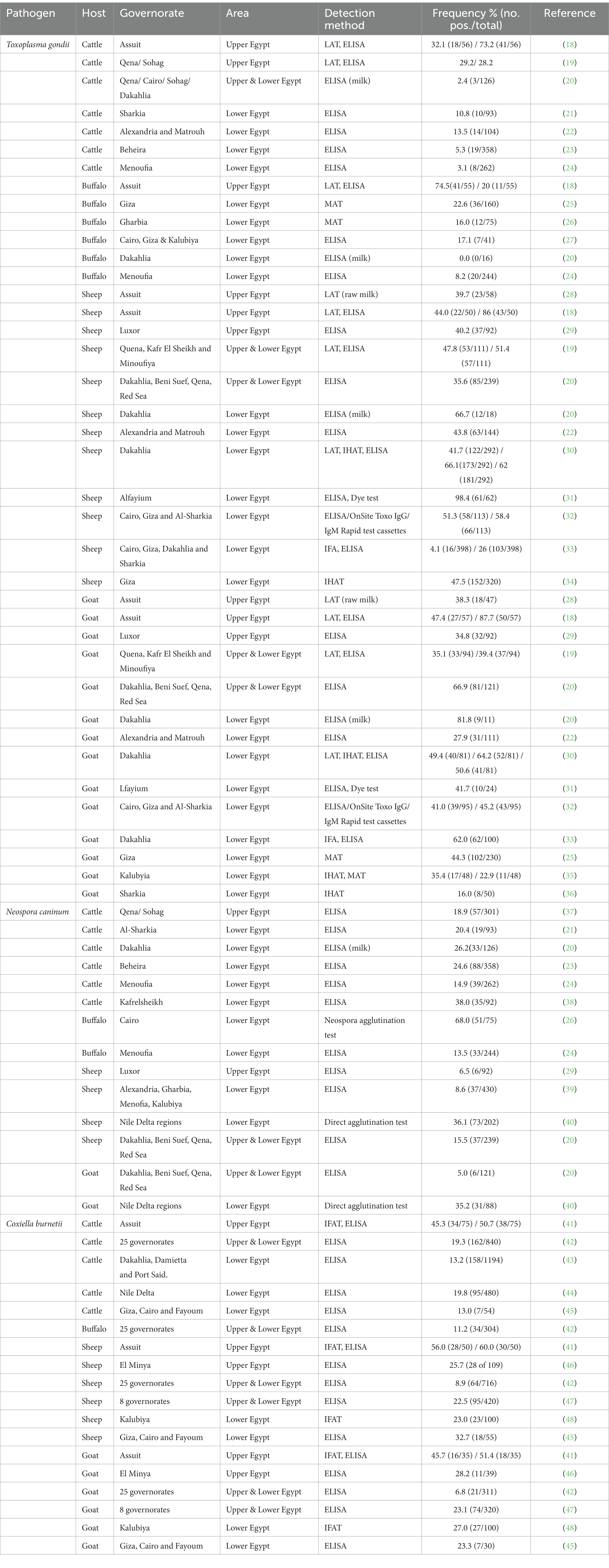
Table 1. Seroprevalence of studied pathogens in domestic ruminants in Egypt.
2. Materials and methods
2.1. Study design and sample collection
A cross-sectional study was carried out in domestic ruminant species in Sohag governorate (Upper Egypt) (26.56°N 31.7°E) between May and September 2021. The climate in the study area is characterized as desertic, with no rainfall during the year except little in winter, and a relative humidity ranging from 60% to less than 30% (50).
A total of 411 animals from small stakeholders were sampled, including 106 cattle, 29 buffalos, 139 sheep, and 137 goats in 13 different municipalities. The sample size was calculated using WinEpiscope 2.0.1 In consideration of the number of domestic ruminants in the study area (n > 10,000), an estimated prevalence of 50%, which provides the highest simple size in studies with unknown prevalence (51), the desired absolute precision was set at ±5% and confidence level at 95%, resulting in 385 animals to be sampled and a total of 411 animals from small stakeholders were finally included in the study. Blood samples were collected by jugular vein puncture using sterile tubes without anticoagulant (Vacutainer®, Becton-Dickinson, USA). Samples were transported to the laboratory (Department of Animal Medicine, Sohag University, Egypt) under refrigerated conditions (4–6°C) within 24–48 h following collection, then centrifugated at 400 g for 15 min to obtain serum, and preserved at −20°C until analysis. Information about each animal, including species, sex, age and some other general clinical information such a pregnancy, presence of ectoparasites, history of abortion, diarrohea and fever, were collected whenever possible (Table 2). None of surveyed animals was vaccinated against toxoplasmosis, neosporosis or Q fever.
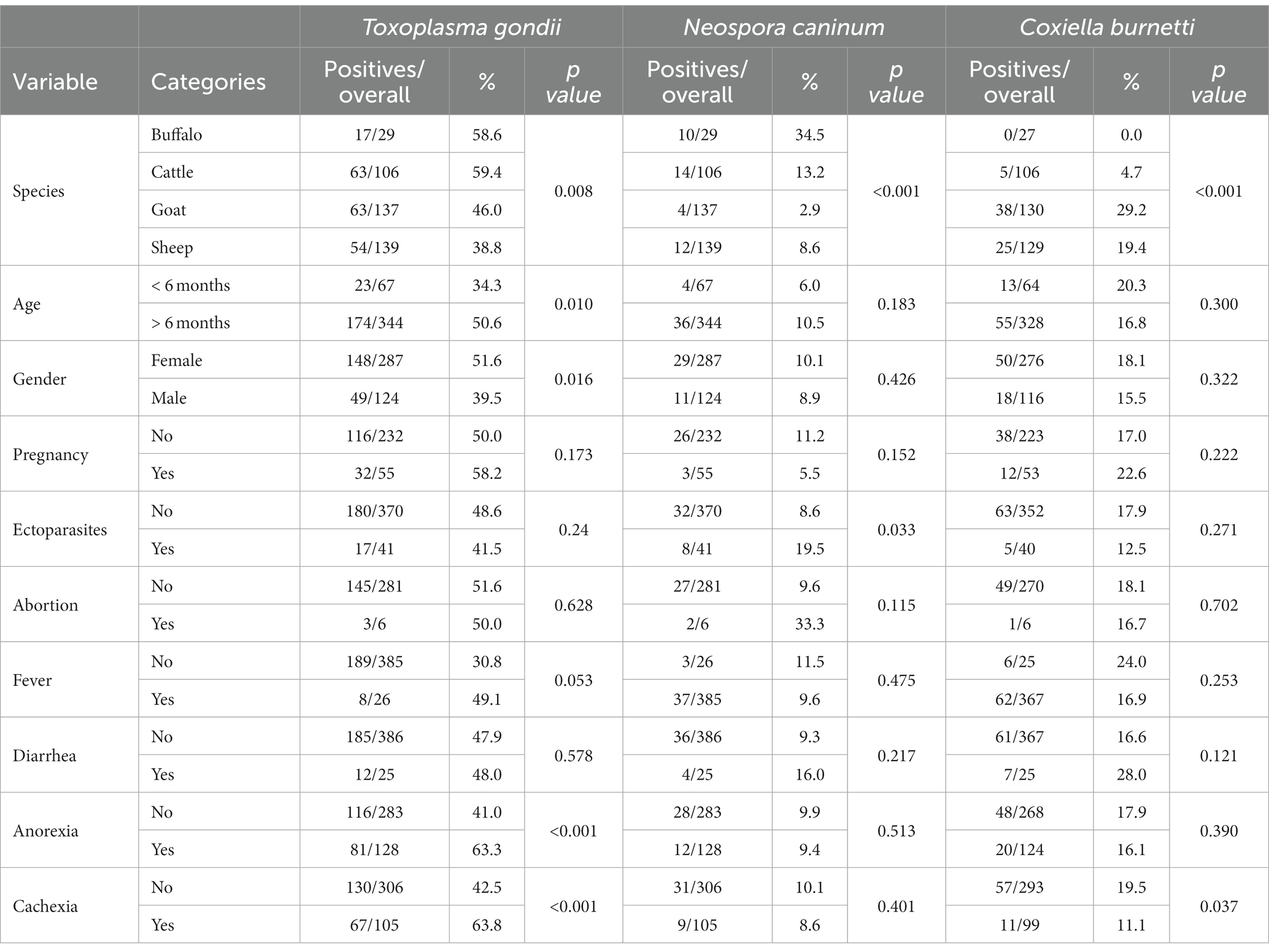
Table 2. Distribution of variables associated with seropositivity of studied pathogens in ruminants in Sohag governorate.
2.2. Serological analysis
The presence of T. gondii antibodies was detected using the modified agglutination test (MAT) as previously described (Dubey and Desmonts, 1987). Sera with titers ≥1:25 were considered positive. This technique has been employed broadly for the diagnosis of antibodies against T. gondii in both domestic and wildlife ruminants (Dubey, 2022). Sera were also analyzed to detect the presence of antibodies against N. caninum and C. burnetii using two commercial ELISA kits (ID Screen® Neospora caninum Competition and ID Screen® Q fever Indirect Multi-species, France) according to the manufacturer’s recommendation. The sensitivity and specificity values provided by the manufacturer for both ELISA were 100%. These ELISA kits have been used previously in different studies of domestic and wild ruminant species (52–55).
2.3. Statistical analysis
The individual seroprevalence against toxoplasmosis, neosporosis and Q fever was calculated from the ratio of seropositive samples to the total number of animals examined with a 95%CI. Associations between explanatory variables and serological results to the three pathogens analyzed (dependent variables) were performed using the Pearson’s chi-square or Fisher’s test, as required. Then, explanatory variables with value of p < 0.10 were selected for multivariate analysis. Collinearity between variables was also calculated using the Cramer’s V coefficients. Finally, generalized estimating equation (GEE) models were carried out for each tested pathogen. The number of seropositive animals was assumed to follow a binomial distribution and “municipality” was included as a random effect. Values with p < 0.05 were considered statistically significant. SPSS 25.0 software (IBM Corp., Armonk, NY, United States) was used to perform statistical analyses.
3. Results
Table 2 depicts the results of serosurvey for the studied pathogens besides pointing out some other general clinical information. As shown, T. gondii had overall seroprevalence of 47.9% (197/411; 95%CI: 43.1–52.8%). T. gondii antibodies against were detected in 59.4% (63/106) of cattle, 58.6% (17/29) buffaloes, 46.0% (63/137) goats and 38.8% (54/139) sheep. In addition, 9.7% (40/411; 95%CI: 6.9–12.6%) of the ruminants sampled showed anti-N. caninum antibodies. The highest seroprevalence was observed in buffaloes (34.5%; 10/29), followed by cattle (13.2%; 14/106), sheep (8.6%; 12/139) and goats (2.9%; 4/137). Finally, seropositivity of C. burnetii was detected in 17.3% (68/392; 95%CI: 13.6–21.1%) of the sampled ruminants, with a seropositivity of 29.2% (38/130) in goats, 19.4% (25/129) in sheep and 4.7% (5/106) in cattle. Anti-C. burnetii antibodies were no found in buffaloes (0/29). In relation to the co-exposure cases (Table 3), 15.3% (63/411) of the examined animals were found to be co-exposed to at least two of the tested pathogen; 29 (7.1%) animals had antibodies against both T. gondii and C. burnetii, 24 (5.8%) showed positive result to both T. gondii and N. caninum antibodies, 6 (1.5%) had antibodies against N. caninum and C. burnetii, and four (1.0%) individuals were found co-exposed by the three tested pathogens.
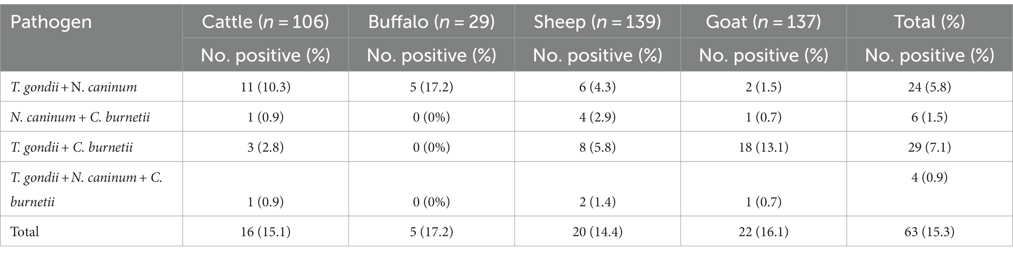
Table 3. Co-exposure of surveyed ruminants’ species with selected reproductive pathogens in Sohag governorate (Upper Egypt).
The independent variables selected in the bivariate analysis are summarized in Tables 2, 4. A total of six, two and two explanatory variables were selected (p < 0.10) for the multivariate analysis of T. gondii, N. caninum and C. burnetti, respectively. The final GEE model revealed two potential factors associated with T. gondii infection in ruminants: species (buffalo and cattle) and age (> 6 months). In addition, the multivariate analysis showed that species was also a risk factor related to N. caninum (buffalo, cattle and sheep) and C. burnetti (sheep and goat) exposure (Table 4).
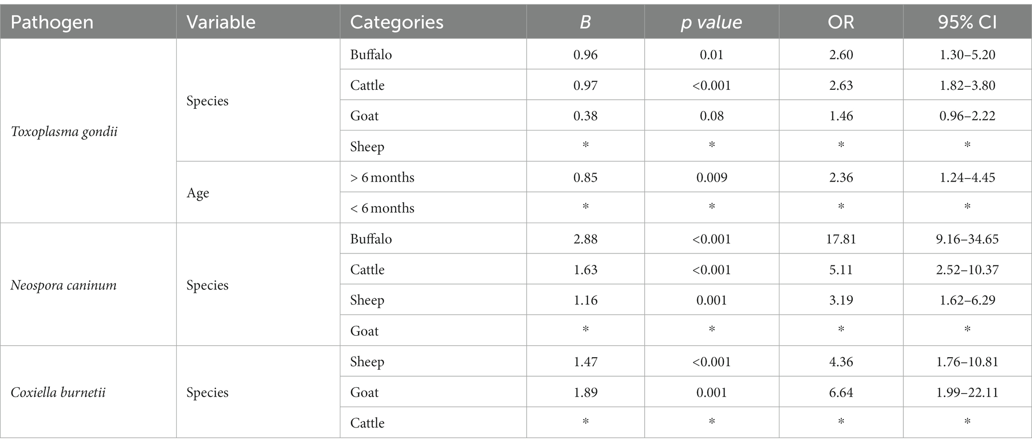
Table 4. Data of the generalized estimating equation (GEE) model of the potential risk factors associated with pathogens exposure in domestic ruminants in Sohag governorate (Upper Egypt).
4. Discussion
The present work revealed important baseline information about the seroprevalence of T. gondii, N. caninum and C. burnetti in Sohag governorate, Upper Egypt. Moreover, this work provides novel information about potential the co-exposure between these pathogens in the Upper part of the country. The study also provides updated information about the circulation of the three reproductive pathogens in domestic ruminants throughout the country (Table 1).
In relation to T. gondii, the high individual seroprevalence values obtained in cattle (59.4%), buffalo (58.6%), sheep (38.8%) and goats (46%) in our study indicate this parasite is widespread in Upper Egypt, which can be of animal and public health concern. The seroprevalence values falls within the previously reported range for this protozoa (20.0–87.7%) in small and large ruminants from Upper Egypt (Table 1). However, the seroprevalence of T. gondii in sheep in our study is slightly lower than those previously found in the study area (39.7–86%) (Table 1). Lower seroprevalence rate of T. gondii have also been previously reported in both small and large ruminants in Lower Egypt (0–22.6%) (Table 1). Moreover, the seroprevalence of T. gondii in goats falls within the reported range (16–81.8%) in this species in several governorates in Lower Egypt (Table 1).
We identified two risk factors (species and age) associated with T. gondii exposure. Seropositivity to T. gondii was significantly higher in large ruminants (buffalo and cattle) compared to small ruminants (goats and sheep). This may be attributed to the fact that cattle and buffaloes in this province are usually reared indoor and the tendency of the Egyptian owners to have cats at their homes which may have access to the animals or their feed (12). The risk factor analyses also showed the age as a risk factor associated with T. gondii exposure (Table 4). In this sense, the seroprevalence of T. gondii was higher in ruminants older than 6 months (50.6%) compared to young ones (34.3%). This finding is consistent with previous investigations (22, 56–59) reporting that a higher seroprevalence of T. gondii among older ages indicates that the contact with the pathogen and the persistence of antibodies increases with age (60, 61).
To author’s knowledge, epidemiological studies assessing N. caninum in Upper Egypt are limited. The seroprevalence rate obtained in the present work ranged between 2.9% in goat and 34.5% in buffalo (Table 3). The high differences between species, which was shown to be a risk factor for N. caninum exposure, are in line with those reported in the scientific literature not only in Egypt (Table 1) but also worldwide (8.6–68.0%) (9, 62). However, our study revealed the lowest seroprevalence rates of N. caninum obtained in small ruminant species in Upper and Lower Egypt so far (Table 1). This finding might be due to the variation in management and feeding practices of small ruminants which is usually based on pasture grazing besides the presence of a lot of stray dogs which in turn can contaminate feed and water and results in higher exposure to infection (9, 62).
Concerning C. burnetii, high difference between ruminant species were also found in the seropositivity to this zoonotic bacterium, being significantly higher in small ruminant species (29.2 and 19.3% in goat and sheep, respectively) than in large ruminants (0.0 and 4.7% in buffalo and cattle, respectively). Similarly, previous studies carried out in Upper Egypt revealed that the prevalence of antibodies against this pathogen was higher in small ruminants (25.7–60%) compared to cattle and buffalo (11.2–19.3%) (Table 1). In contrast, our survey reported lower seroprevalence values of C. burnetti in small and large ruminants than those reported in other studies in Upper and Lower Egypt (6.8 to 27%), respectively (Table 1). Regarding the risk factors analysis, the seropositivity of C. burnetii in small ruminants was significantly higher than in large ruminants, which came in stark contrast with some previous reports from Egypt (63). This finding could be explained by the nature of grazing of small ruminants or by differences in the systems of management in this area, where large ruminants are mostly kept indoor, and therefore small ruminants could be more exposed to this pathogen along their life (64).
Interestingly, the present study reports multiple cases of co-exposure by T. gondii, N. caninum and/or C. burnetti. To the best of our knowledge, this is the first seroepidemiological study evaluating jointly co-exposure of the three tested reproductive pathogens in different domestic ruminant species in Egypt. Metwally et al. (23) detected 1.9% (7/358) of T. gondii and N. caninum co-exposure in cattle in Beheira governorate in Lower Egypt. Similarly, Aboelwafa et al. (29) reported a co-exposure rate of 4.3% (4/92) of T. gondii and N. caninum in sheep in Luxor, Upper Egypt. Co-infections with T. gondii, N. caninum and C. burnetii is usually resulting in lower immunity, increased the risk of abortions, fetal losses and abnormalities which consequently leads to huge economic losses (65). Additional studies are warranted to assess the implications of co-infections by reproductive pathogens in livestock in the study region.
5. Conclusion
The present study provides updated seroepidemiological information about the circulation of T. gondii, N. caninum and C. burnetti in four domestic ruminant species to Upper Egypt. To author’s knowledge, the present work is considered the first seroepidemiological study documented the co-exposure of the three tested reproductive pathogens in different domestic ruminant species in Egypt. The circulation of the different selected pathogens was not homogeneous among the analyzed ruminant populations. The seroprevalence values of the tested zoonotic pathogens indicate a relevant epidemiological role of domestic ruminants in the maintenance of these pathogens. The present study point out the importance of improvement of the surveillance programs monitoring the circulation of reproductive pathogens at the domestic-human interface and the role of application of strict hygienic and biosecurity measures to control the infection in Upper Egypt. These measures should include control of access of dogs and cats to the farms, to ruminants rearing areas combined with application of proper vaccination programs to reduce the transmission of these pathogens at this area. Additional molecular and epidemiological surveys addressing the circulation of these reproductive pathogens at a large scale are needed to investigate both their economic and productive impact as well as the sanitary implications for animal and human health in Egypt. Further studies are also suggested to detect the mentioned pathogens on milk samples, meat juice with blood samples for explore the potential zoonotic link and the potential genetic relatedness of circulating strains.
Data availability statement
The original contributions presented in the study are included in the article/supplementary material, further inquiries can be directed to the corresponding authors.
Ethics statement
Ethical approval was not required for the study involving animals in accordance with the local legislation and institutional requirements because The collection of blood samples analysed in the present study was part of the official Animal Health Campaigns. Therefore, no ethical approval was necessary.
Author contributions
SF: Conceptualization, Data curation, Formal analysis, Investigation, Methodology, Validation, Visualization, Writing – original draft, Writing – review & editing. DC-T: Conceptualization, Data curation, Formal analysis, Investigation, Methodology, Software, Supervision, Validation, Visualization, Writing – original draft, Writing – review & editing. MG: Data curation, Formal analysis, Methodology, Software, Supervision, Validation, Visualization, Writing – original draft, Writing – review & editing. DS: Conceptualization, Data curation, Project administration, Supervision, Validation, Visualization, Writing – original draft. N-EA: Conceptualization, Supervision, Validation, Visualization, Writing – original draft. MM: Data curation, Formal analysis, Funding acquisition, Resources, Software, Writing – original draft. DJ-M: Data curation, Formal analysis, Investigation, Methodology, Software, Validation, Writing – original draft, Writing – review & editing. IG-B: Conceptualization, Data curation, Formal analysis, Funding acquisition, Investigation, Project administration, Resources, Supervision, Validation, Visualization, Writing – original draft, Writing – review & editing. EE: Conceptualization, Data curation, Formal analysis, Funding acquisition, Resources, Validation, Writing – original draft, Writing – review & editing.
Funding
The author(s) declare financial support was received for the research, authorship, and/or publication of this article. This work was partially financed by CIBER -Consorcio Centro de Investigación Biomédica en Red- (CB 2021), Instituto de Salud Carlos III, Ministerio de Ciencia e Innovación and Unión Europea – NextGenerationEU. MG was supported by a postdoctoral contract Margarita Salas (University of Murcia) from the Program of Requalification of the Spanish University System (Spanish Ministry of Universities) financed by the European Union-NextGenerationEU. DJ-M holds a PhD contract granted by Own Research Plan of the University of Córdoba. EE was supported by a postdoctoral contract María Zambrano (University of Córdoba) from the Program of Requalification of the Spanish University System (Spanish Ministry of Universities) financed by the European Union-NextGenerationEU. This study was supported by Researchers Supporting Project number (RSPD2023R655), King Saud University, Riyadh, Saudi Arabia.
Conflict of interest
The authors declare that the research was conducted in the absence of any commercial or financial relationships that could be construed as a potential conflict of interest.
The author(s) declared that they were an editorial board member of Frontiers, at the time of submission. This had no impact on the peer review process and the final decision.
Publisher’s note
All claims expressed in this article are solely those of the authors and do not necessarily represent those of their affiliated organizations, or those of the publisher, the editors and the reviewers. Any product that may be evaluated in this article, or claim that may be made by its manufacturer, is not guaranteed or endorsed by the publisher.
Footnotes
References
1. De Barros, LD , Garcia, JL , Bresciani, KDS , Cardim, ST , Storte, VS , and Headley, SA . A review of toxoplasmosis and neosporosis in water buffalo (Bubalus bubalis). Front Vet Sci. (2020) 7:455. doi: 10.3389/fvets.2020.00455
2. Tenter, AM , Heckeroth, AR , and Weiss, LM . Toxoplasma gondii: from animals to humans. Int J Parasitol. (2000) 30:1217–58. doi: 10.1016/S0020-7519(00)00124-7
3. Dubey, J , Carpenter, J , Speer, C , Topper, M , and Uggla, A . Newly recognized fatal protozoan disease of dogs. J Am Vet Med Assoc. (1988) 192:1269–85.
4. Trees, AJ , McAllister, M , Guy, C , McGarry, J , Smith, R , and Williams, DJ . Neospora caninum: oocyst challenge of pregnant cows. Vet Parasitol. (2002) 109:147–54. doi: 10.1016/S0304-4017(02)00234-0
5. Dubey, JP . Toxoplasmosis of animals and humans. 3rd ed Boca Raton, Florida, United States: CRC Press (2021).
6. Eldin, C , Mélenotte, C , Mediannikov, O , Ghigo, E , Million, M , Edouard, S, et al. From Q fever to Coxiella burnetii infection: a paradigm change. Clin Microbiol Rev. (2017) 30:115–90. doi: 10.1128/CMR.00045-16
7. Canada, N , Meireles, CS , Rocha, A , Correia da Costa, J , Erickson, M , and Dubey, J . Isolation of viable toxoplasma gondii from naturally infected aborted bovine fetuses. J Parasitol. (2002) 88:1247–8. doi: 10.1645/0022-3395(2002)088[1247:IOVTGF]2.0.CO;2
8. Innes, EA , Bartley, PM , Buxton, D , and Katzer, F . Ovine toxoplasmosis. Parasitology. (2009) 136:1887–94. doi: 10.1017/S0031182009991636
9. Dubey, J , Schares, G , and Ortega-Mora, L . Epidemiology and control of neosporosis and Neospora caninum. Clin Microbiol Rev. (2007) 20:323–67. doi: 10.1128/CMR.00031-06
10. Álvarez-Alonso, R , Basterretxea, M , Barandika, JF , Hurtado, A , Idiazabal, J , Jado, I, et al. A Q fever outbreak with a high rate of abortions at a dairy goat farm: Coxiella burnetii shedding, environmental contamination, and viability. Appl Environ Microbiol. (2018) 84:01650–18. doi: 10.1128/AEM.01650-18
11. Agerholm, JS . Coxiella burnetii associated reproductive disorders in domestic animals-a critical review. Acta Vet Scand. (2013) 55:1751–0147. doi: 10.1186/1751-0147-55-13
12. Stelzer, S , Basso, W , Benavides Silván, J , Ortega-Mora, LM , Maksimov, P , Gethmann, J, et al. Toxoplasma gondii infection and toxoplasmosis in farm animals: risk factors and economic impact. Food Waterborne Parasitol. (2019) 15:e00037. doi: 10.1016/j.fawpar.2019.e00037
13. Raoult, D , Marrie, T , and Mege, J . Natural history and pathophysiology of Q fever. Lancet Infect Dis. (2005) 5:219–26. doi: 10.1016/S1473-3099(05)70052-9
14. Voss, L , Huaman, J , Pacioni, C , Tolpinrud, A , Helbig, K , Carvalho, TG, et al. Seroprevalence of Coxiella burnetii antibodies in wild deer populations in eastern Australia. Aust Vet J. (2023) 101:106–14. doi: 10.1111/avj.13223
15. Morroy, G , van der Hoek, W , Albers, J , Coutinho, RA , Bleeker-Rovers, CP , and Schneeberger, PM . Population screening for chronic Q-fever seven years after a major outbreak. PLoS One. (2015) 10:e0131777. doi: 10.1371/journal.pone.0131777
16. Duarte, PO , Oshiro, LM , Zimmermann, NP , Csordas, BG , Dourado, DM , Barros, JC, et al. Serological and molecular detection of Neospora caninum and toxoplasma gondii in human umbilical cord blood and placental tissue samples. Sci Rep. (2020) 10:020–65991. doi: 10.1038/s41598-020-65991-1
17. USAID AaFSE . Livestock and Products Annual 2018. Egyptian Beef Prices Stable, Consumption and Imports to Rise in 2019. GAIN. (2019) 10:EG-18021
18. Kuraa, H , and Malek, S . Seroprevalence of toxoplasma gondii in ruminants by using latex agglutination test (LAT) and enzyme-linked immunosorbent assay (ELISA) in Assiut governorate. Trop Biomed. (2016) 33:711–25.
19. Fereig, RM , Mahmoud, HY , Mohamed, SG , AbouLaila, MR , Abdel-Wahab, A , Osman, SA, et al. Seroprevalence and epidemiology of toxoplasma gondii in farm animals in different regions of Egypt. Veterinary Parasitol: Regional Stud Reports. (2016) 3:1–6.
20. Fereig, RM , Abdelbaky, HH , Mazeed, AM , El-Alfy, E-S , Saleh, S , Omar, MA, et al. Prevalence of Neospora caninum and toxoplasma gondii antibodies and DNA in raw milk of various ruminants in Egypt. Pathogens. (2022) 11:1305. doi: 10.3390/pathogens11111305
21. Ibrahim, HM , Huang, P , Salem, TA , Talaat, RM , Nasr, MI , Xuan, X, et al. Prevalence of Neospora caninum and toxoplasma gondii antibodies in northern Egypt. The American journal of tropical medicine and hygiene. (2009) 80:263–7. doi: 10.4269/ajtmh.2009.80.263
22. Khattab, RA , Barghash, SM , Mostafa, OMS , Allam, SA , Taha, HA , and Ashour, AAE . Seroprevalence and molecular characterization of toxoplasma gondii infecting ruminants in the north-west of Egypt. Acta Trop. (2022) 225:106139. doi: 10.1016/j.actatropica.2021.106139
23. Metwally, S , Hamada, R , Sobhy, K , Frey, CF , and Fereig, RM . Seroprevalence and risk factors analysis of Neospora caninum and toxoplasma gondii in cattle of Beheira. Egypt Front Vet Sci. (2023) 10:1–9. doi: 10.3389/fvets.2023.1122092
24. Ibrahim, HM , Abdel-Rahman, AA , and Bishr, NM . Seroprevalence of Neospora caninum and toxoplasma gondii IgG and IgM antibodies among buffaloes and cattle from Menoufia Province. Egypt Journal of Parasitic Diseases. (2021) 45:952–8. doi: 10.1007/s12639-021-01386-x
25. Shaapan, R , Ma, H , and Khalil, F . Modified agglutination test for serologic survey of toxoplasma gndii infection in goats and water buffaloes in Egypt. Res J Parasitol. (2010) 5:13–7. doi: 10.3923/jp.2010.13.17
26. Dubey, JP , and Romand, S ., Hilali, M ., Kwok, O. C., & Thulliez, P . Seroprevalence of antibodies to Neospora caninum and toxoplasma gondii in water buffaloes (Bubalus bubalis) from Egypt. Int J Parasitol (1998) 28:527–529. doi: 10.1016/s0020-7519(97)00190-2
27. Elfadaly, H , Hassanain, N , Shaapan, R , Hassanain, M , Barakat, A , and Abdelrahman, K . Molecular detection and genotyping of toxoplasma gondii from Egyptian isolates. Asian J Epidemiol. (2017) 10:37–44. doi: 10.3923/aje.2017.37.44
28. Sadek, OA , ABDEL-HAMEED, M , and KURAA, M . Molecular detection of toxoplasma gondii DNA in raw goat and sheep milk with discussion of its public health importance in Assiut governorate. Assiut Vet Med J. (2015) 61:166–77. doi: 10.21608/avmj.2015.170200
29. Aboelwafa, S , Ali, A , Hamada, R , and Mahmoud, H . Seroprevalence of toxoplasma gondii and Neospora caninum in small ruminants in Luxor. Egypt Adv Anim Vet Sci. (2022) 10:412–20. doi: 10.17582/journal.aavs/2022/10.2.412.420
30. Younis, E , Abou-zeid, NZ , Zakaria, M , and Mhmoud, MR . Epidemiological studies on toxoplasmosis in small ruminants and equine in Dakahlia governorate. Egypt Assiut Vet Med J. (2015) 61:22–31. doi: 10.21608/avmj.2015.169756
31. Ghoneim, NH , Shalaby, SI , Hassanain, NA , Zeedan, GS , Soliman, YA , and Abdalhamed, AM . Comparative study between serological and molecular methods for diagnosis of toxoplasmosis in women and small ruminants in Egypt. Foodborne Pathog Dis. (2010) 7:17–22. doi: 10.1089/fpd.2008.0223
32. Abd El-Razik, KA , Barakat, AMA , Hussein, HA , Younes, AM , Elfadaly, HA , Eldebaky, HA, et al. Seroprevalence, isolation, molecular detection and genetic diversity of toxoplasma gondii from small ruminants in Egypt. J Parasit Dis. (2018) 42:527–36. doi: 10.1007/s12639-018-1029-4
33. Al-Kappany, YM , Abbas, IE , Devleesschauwer, B , Dorny, P , Jennes, M , and Cox, E . Seroprevalence of anti-toxoplasma gondii antibodies in Egyptian sheep and goats. BMC Vet Res. (2018) 14:1–5. doi: 10.1186/s12917-018-1440-1
34. Barakat, A , Elaziz, MA , and Fadaly, H . Comparative diagnosis of toxoplasmosis in Egyptian small ruminants by indirect hemagglutination assay and Elisa. Global Veterinaria. (2009) 3:9–14.
35. Ramadan, MY , Abdel-Mageed, AD , and Khater, HF . Seroprevalence and preliminary treatment of toxoplasmosis of pregnant goats in Kalubyia Gobernatore. Egypt Acta Scientiae Veterinariae. (2007) 35:295–301. doi: 10.22456/1679-9216.16119
37. Fereig, RM , AbouLaila, MR , Mohamed, SGA , Mahmoud, H , Ali, AO , Ali, AF, et al. Serological detection and epidemiology of Neospora caninum and Cryptosporidium parvum antibodies in cattle in southern Egypt. Acta Trop. (2016) 162:206–11. doi: 10.1016/j.actatropica.2016.06.032
38. Gaber, A , Hegazy, Y , Oreiby, A , and AL-GAABARY, M . Neosporosis: a neglected abortifacient disease in Egypt, seroprevalence and farmers’ knowledge, attitudes and practices. J Hellenic vet med Soci. (2021) 72:3109–16. doi: 10.12681/jhvms.28500
39. Selim, A , Khater, H , and Almohammed, HI . A recent update about seroprevalence of ovine neosporosis in northern Egypt and its associated risk factors. Sci Rep. (2021) 11:14041, 14043–6. doi: 10.1038/s41598-021-93596-9
40. El-Ghaysh, A , Khalil, F , Hilali, M , and Nassar, A . Serological diagnosis of Neospora caninum infection in some domestic animals from Egypt. Vet Med J-Giza. (2003) 51:355–62.
41. Abbass, H , Selim, SAK , Sobhy, MM , El-Mokhtar, MA , Elhariri, M , and Abd-Elhafeez, HH . High prevalence of Coxiella burnetii infection in humans and livestock in Assiut, Egypt: a serological and molecular survey. Vet World. (2020) 13:2578–86. doi: 10.14202/vetworld.2020.2578-2586
42. Klemmer, J , Njeru, J , Emam, A , El-Sayed, A , Moawad, AA , Henning, K, et al. Q fever in Egypt: epidemiological survey of Coxiella burnetii specific antibodies in cattle, buffaloes, sheep, goats and camels. PLoS One. (2018) 13:e0192188. doi: 10.1371/journal.pone.0192188
43. Gwida, M , El-Ashker, M , El-Diasty, M , Engelhardt, C , Khan, I , and Neubauer, H . Q fever in cattle in some Egyptian governorates: a preliminary study. BMC Res Notes. (2014) 7:1–5. doi: 10.1186/1756-0500-7-881
44. Selim, A , Marawan, MA , Abdelhady, A , and Wakid, MH . Seroprevalence and potential risk factors of toxoplasma gondii in dromedary camels. Agriculture. (2023) 13:129. doi: 10.3390/agriculture13010129
45. Nahed, HG , and Khaled, A . Seroprevalence of Coxiella burnetii antibodies among farm animals and human contacts in Egypt. J Am Sci. (2012) 8:619–21.
46. Abushahba, MFN , Abdelbaset, AE , Rawy, MS , and Ahmed, SO . Cross-sectional study for determining the prevalence of Q fever in small ruminants and humans at El Minya governorate. Egypt BMC Res Notes. (2017) 10:017–2868. doi: 10.1186/s13104-017-2868-2
47. Sobhy, M , Emm, I , and Kh, AA-G . Seroprevalence detection of antibodies of Coxiella burnetii in sheep, goats and human in some governorates in Egypt. Assiut Vet Med J. (2019) 65:68–73. doi: 10.21608/avmj.2019.169042
48. Khalifa, NO , Elhofy, FI , Fahmy, A , Sobhy, MM , and Agag, M . Seroprevalence and molecular detection of Coxiella burnetii infection in sheep, goats and human in Egypt. ISOI J Microbiol Biotechnol Food Sci. (2016) 2:1–7.
49. Galal, S , Elnahas, A , Mousa, E , Elshennawy, M , and Alsheikh, S . Factors affecting small ruminant holdings within the crop-livestock farming system in Sohag governorate. Egypt Egyptian Journal of Animal Production. (2011) 48:157–65. doi: 10.21608/ejap.2011.94068
50. Youssef, A , Abdel Moneim, A , and Abu El-Maged, S , Flood hazard assessment and its associated problems using geographic information systems, Sohag Governorate, Egypt. The Fourth International Conference on the Geology of Africa, Assiut, Egypt; (2005).
52. Cruz, R , Esteves, F , Vasconcelos-Nóbrega, C , Santos, C , Ferreira, AS , Mega, C, et al. A nationwide seroepidemiologic study on Q fever antibodies in sheep of Portugal. Vector-Borne and Zoonotic Diseases. (2018) 18:601–4. doi: 10.1089/vbz.2018.2294
53. Manca, R , Ciccarese, G , Scaltrito, D , and Chirizzi, D . Detection of anti-neospora caninum antibodies on dairy cattle farms in southern Italy. Vet Sci. (2022) 9:87. doi: 10.3390/vetsci9020087
54. Villari, S , Galluzzo, P , Arnone, M , Alfano, M , Geraci, F , and Chiarenza, G . Seroprevalence of Coxiella burnetii infection (Q fever) in sheep farms located in Sicily (southern Italy) and related risk factors. Small Rumin Res. (2018) 164:82–6. doi: 10.1016/j.smallrumres.2018.05.006
55. Bártová, E , Kobédová, K , Lamka, J , Kotrba, R , Vodička, R , and Sedlák, K . Seroprevalence of Neospora caninum and toxoplasma gondii in exotic ruminants and camelids in the Czech Republic. Parasitol Res. (2017) 116:1925–9. doi: 10.1007/s00436-017-5470-6
56. Alvarado-Esquivel, C , García-Machado, C , Vitela-Corrales, J , Villena, I , and Dubey, J . Seroprevalence of toxoplasma gondii infection in domestic goats in Durango state. Mexico Vet Parasitol. (2011) 183:43–6. doi: 10.1016/j.vetpar.2011.06.021
57. de Moura, AB , Ribeiro, A , de Souza, AP , da Silva, MO , Machado, G , Klauck, V, et al. Seroprevalence and risk factors for toxoplasma gondii infection in goats in southern Brazil. Acta Sci Vet. (2016) 44:1–7. doi: 10.22456/1679-9216.81073
58. Deng, H , Dam-Deisz, C , Luttikholt, S , Maas, M , Nielen, M , Swart, A, et al. Risk factors related to toxoplasma gondii seroprevalence in indoor-housed Dutch dairy goats. Prev Vet Med. (2016) 124:45–51. doi: 10.1016/j.prevetmed.2015.12.014
59. Garcia, G , Sotomaior, C , Nascimento, AJ , Navarro, IT , and Soccol, VT . Toxoplasma gondii in goats from Curitiba, Paraná, Brazil: risks factors and epidemiology. Rev Bras Parasitol Vet. (2012) 21:42–7. doi: 10.1590/S1984-29612012000100009
60. Dubey, JP , and Beattie, C . Toxoplasmosis of animals and man. Boca Raton, FL, USA: CRC Press, Inc. (1988).
61. Katzer, F , Brülisauer, F , Collantes-Fernández, E , Bartley, PM , Burrells, A , Gunn, G, et al. Increased toxoplasma gondii positivity relative to age in 125 Scottish sheep flocks; evidence of frequent acquired infection. Vet Res. (2011) 42:121. doi: 10.1186/1297-9716-42-121
62. Dubey, J, Review of N . Caninum and neosporosis in animals. Korean J Parasitol. (2003) 41:1–16. doi: 10.3347/kjp.2003.41.1.1
63. Selim, A , Marawan, MA , Abdelhady, A , Alshammari, FA , Alqhtani, AH , Ba-Awadh, HA, et al. Coxiella burnetii and its risk factors in cattle in Egypt: a seroepidemiological survey. BMC Vet Res. (2023) 19. doi: 10.1186/s12917-023-03577-5
64. Álvarez-Alonso, R , Zendoia, II , Barandika, JF , Jado, I , Hurtado, A , López, CM, et al. Monitoring Coxiella burnetii infection in naturally infected dairy sheep flocks throughout four lambing seasons and investigation of viable bacteria. Front Vet Sci. (2020) 7:352, 1–15. doi: 10.3389/fvets.2020.00352
Keywords: seroprevalence, Toxoplasma gondii , Neospora caninum , Coxiella burnetii , ruminants, Egypt
Citation: Farag SI, Cano-Terriza D, Gonzálvez M, Salman D, Aref N-EM, Mubaraki MA, Jiménez-Martín D, García-Bocanegra I and Elmahallawy EK (2023) Serosurvey of selected reproductive pathogens in domestic ruminants from Upper Egypt. Front. Vet. Sci. 10:1267640. doi: 10.3389/fvets.2023.1267640
Edited by:
Saber Esmaeili, Pasteur Institute of Iran (PII), IranReviewed by:
Eman Mohammed, South Valley University, EgyptAna Huertas López, Complutense University of Madrid, Spain
Copyright © 2023 Farag, Cano-Terriza, Gonzálvez, Salman, Aref, Mubaraki, Jiménez-Martín, García-Bocanegra and Elmahallawy. This is an open-access article distributed under the terms of the Creative Commons Attribution License (CC BY). The use, distribution or reproduction in other forums is permitted, provided the original author(s) and the copyright owner(s) are credited and that the original publication in this journal is cited, in accordance with accepted academic practice. No use, distribution or reproduction is permitted which does not comply with these terms.
*Correspondence: Moisés Gonzálvez, c2EyZ29qdW1AdWNvLmVz; Ehab Kotb Elmahallawy, c2EyZWxlbGVAdWNvLmVz
†These authors have contributed equally to this work