
95% of researchers rate our articles as excellent or good
Learn more about the work of our research integrity team to safeguard the quality of each article we publish.
Find out more
ORIGINAL RESEARCH article
Front. Vet. Sci. , 31 August 2023
Sec. Veterinary clinical, anatomical, and comparative pathology
Volume 10 - 2023 | https://doi.org/10.3389/fvets.2023.1234779
 Giulia D’Annunzio1,2*
 Fabio Ostanello2
 Luisa Vera Muscatello2
 Massimo Orioles3
 Niccolò Jacumin4Nicola Tommasini4Giorgio Leotti4Andrea Luppi1
 Luciana Mandrioli2
 Giuseppe Sarli2
Methods: This study aimed to examine the pathological impact of Porcine Circovirus type 2 (PCV2) and Porcine Reproductive and Respiratory Syndrome Virus (PRRSV) through histological and immunohistochemical analysis of 79 cases of Porcine Respiratory Disease Complex (PRDC) collected from 22 farms in Northern Italy. Lung tissue and several lymphoid organ samples were deployed to associate PCV2-positive stain with Circovirus-associated Diseases (PCVD).
Results: The most common lung lesion observed was interstitial pneumonia, alone or combined with bronchopneumonia. By immunohistochemistry, 44 lungs (55.7%) tested positive for PCV2, 34 (43.0%) for PRRSV, 16 (20.3%) for both viruses and in 17 cases (21.5%) neither virus was detected. Twenty-eight out of 44 (63.6%) PCV2-positive cases had lymphoid depletion or granulomatous inflammation in at least one of the lymphoid tissues examined; thus, they were classified as PCV2 Systemic Diseases (PCV2-SD). In the remaining 16 out of 44 cases (36.4%), PCV2-positive lung lesions were associated with hyperplastic or normal lymphoid tissues, which showed PCV2-positive centrofollicular dendritic cells in at least one of the lymphoid tissues examined. Therefore, these cases were classified as PRDC/PCV2-positive. In the PCV2-positive animals, 42.9% of the PCV2-SD cases (12/28) showed immunohistochemistry (IHC) positivity for PRRSV in the lung tissue, while 25.0% of PRDC/PCV2-positive cases (4/16) showed double positivity for PCV2 and PRRSV.
Discussion: In light of the caseload presented in this study, characterized by the high proportion of PCV2-SD cases alongside the overall respiratory symptomatology, it is imperative to emphasize the crucial role of a comprehensive sampling protocol. This is critical to avoid underestimating the harm caused by PCV2 in farms, particularly with respect to the systemic form of the disease. PCV2 and PRRSV remain the primary infections associated with PRDC in Italy that can significantly impact farm health and co-infections in the field can worsen the pathology, thus the selection of appropriate preventive measures is critical.
The term “Porcine Respiratory Disease Complex” (PRDC) refers to a multifactorial disease that arises from a combination of viral, bacterial and, less frequently, parasites, environmental, managerial, and genetic factors, leading to the development of pneumonia in pigs (1). The polymicrobic nature of PRDC presents a formidable diagnostic challenge as different infectious agents affecting the respiratory system often exhibit similar clinical signs (2). This can make it problematic to determine which of the many pathogens involved in PRDC are primarily responsible for respiratory disease. The complexity of interactions between pathogens and their ubiquity further complicate the study, prevention, and control of PRDC, as well as the identification of their direct involvement in the disease development and their correlation with lesions (1, 3, 4).
In suspected PRDC, a range of direct and indirect diagnostic tests can detect pathogens during an infection or monitor infections in a herd, respectively (5). Direct diagnosis of viral diseases is performed using molecular techniques such as Polymerase Chain Reaction (PCR), quantitative PCR (qPCR), or Multiplex PCR (3, 6–10). While these biomolecular methods are highly sensitive, they cannot determine the causal relationship between the pathogen and the related lesion; thus, histopathological investigations must be used to objectively determine the role of identified pathogens in the disease (11). Methods such as immunohistochemistry (IHC) and in situ hybridization (ISH) allow the co-localization of pathogens and associated lesions, thereby establishing their contribution to the disease (12).
One of the primary pathogens involved in PRDC is Porcine Circovirus type 2 (PCV2), which was first identified as the causative agent of Post-Weaning Multisystemic Wasting Syndrome (PMWS) in the 1990s also in Italy (13). PCV2 has since been linked to respiratory diseases, and histology and immunohistochemistry confirmed their association with histiocytic/granulomatous interstitial lung lesions and with proliferative and necrotizing pneumonia (PNP) often in co-infection with other viral agents (14–16). The respiratory form of PCV2 has been classified as PCV2-associated respiratory disease (17) or PCV2-lung disease (PCV2-LD) (18). However, it is essential to note that the systemic form of PCV2, also known as PCV2-SD, may induce respiratory signs, indicating some overlap between the two forms of PCV2 diseases (18). Despite its recognition as a significant pathogen involved in PRDC (19–22), PCV2-LD is frequently considered a negligible entity in a herd (23, 24). In their study, Ticó et al. (24) established a final diagnosis of PCV2-SD in many cases clinically identified as PCV2-associated PRDC, thanks to the detection of lymphoid depletion associated with PCV2-positive stain by immunohistochemistry in the lymphoid tissues.
It is widely acknowledged that co-infections caused by viral or bacterial pathogens significantly impact the development of Porcine Circovirus-associated Diseases (PCVD). Opriessnig and Halbur (25) and Ouyang et al. (26) have demonstrated that co-infections can increase the efficiency of disease reproduction and can be commonly observed in field settings linked to clinical signs and severe lesions.
In particular, co-infection of Porcine Circovirus type 2 (PCV2) and Porcine Reproductive and Respiratory Syndrome Virus (PRRSV) has been widely reported in respiratory clinical manifestations (27–31).
The combined impact of PCV2 and PRRSV infections leads to an immunosuppressive effect altering the host’s defence mechanism and increasing its susceptibility to secondary bacterial and viral infections (32–35). Considering the exacerbation of clinical signs resulting from the interaction of these two infections, it is imperative to promptly identify and implement effective management strategies for the two viruses.
In light of this, the present study aims to explore the impact of PCV2 and PRRSV in Porcine Respiratory Disease Complex (PRDC) cases in pigs from various farms in Northern Italy, area of the Country with the highest density of pig farms. The examination was carried out on lungs and lymphoid tissues to determine the extent to which the observed respiratory forms could be attributed to respiratory disease associated with PCV2 localized only in the lung or to PCV2 systemic disease. Furthermore, the study investigates the role played by PCV2 and PRRSV in co-infections in different forms of PRDC.
The study was conducted on 79 pigs of 5–8 weeks of age from wean-to-finishing and finishing sites of 22 different multi-site productions in Northern Italy. All the pigs died after suffering respiratory clinical signs and underwent necropsy for diagnostic purposes. Seven of these farms had a history of Porcine Reproductive and Respiratory Syndrome Virus (PRRSV) issues, and all farms had prophylactic vaccination strategies for Porcine Circovirus type 2 (PCV2). All pigs underwent diagnostic necropsy procedures, including tissue sampling (lung, mediastinal, tracheobronchial, and superficial inguinal lymph nodes, tonsils, and ileum) and formalin fixation for histopathological analysis.
Sections (3 μm) of the lung, tonsil, tracheobronchial and mediastinal lymph node, ileum, and superficial inguinal lymph node were stained with hematoxylin–eosin (H&E; Hematoxylin cat# 01HEMH2500; Eosin cat# 01EOY101000; Histo-Line Laboratories, Milan, Italy). Lung lesions were categorized into bronchopneumonia, interstitial pneumonia, proliferative and necrotizing pneumonia (PNP), and fibrinous pleuropneumonia according to the criteria established by Caswell and Williams (36). Lesions in lymphoid tissues were categorized as lymphoid depletion (37) or lymphoid hyperplasia (38). Immunohistochemistry (IHC) on three-micron-thick sections of the same tissues was performed to detect PCV2 antigen and only on lung sections to detect PRRSV antigen. Briefly, the sections were deparaffinized and rehydrated. The endogenous peroxidase was blocked in 3% peroxide for 30 min. Antigen retrieval was performed using protease type XIV at 0.5% in Tris-buffered saline (TBS) for 20 min at 37°C. After washing, blocking of unspecific bindings was carried out for 30 min at room temperature with bovine serum albumin–PBS Tween 20 solution (3%). Primary monoclonal antibodies for PCV2 (Ingenasa, 36A9) and PRRSV (Ingenasa, 1 AC7) were applied on the corresponding slide tissue and were incubated overnight at 4°C. At the end of the primary antibody incubation, the sections were rinsed three times in PBS, followed by a 30 min incubation in Scytek CRF Anti-Polyvalent HRP Polymer and by application to the sections of the final peroxidase substrate, prepared according to manufacturers’ instructions [CRF Anti-Polyvalent HRP Polymer (DAB) Stain Kit, ScyTek Laboratories, CPH080]. Slides were finally counterstained with Papanicolaou’s hematoxylin (Histo-Line Laboratories), and coverslips were applied using Bio Mount mounting medium (Bio-Optica, 05-BMHM100). The specificity of the immunohistochemical stain has been validated by adding, as the primary reagent, an aspecific antibody of the same isotype as the primary antibodies (IgG1). As positive control, samples positive by means of PCR to PCV2 or PRRSV were included.
The PCV2-positive cases of PRDC involved in this study were categorized based on the current diagnostic criteria for PCVD (23). The cases were considered indicative of PCV2 systemic disease (PCV2-SD) if the lungs were positive for PCV2 antigen and showed associated lymphocyte depletion in lymphoid tissues along with moderate to high amount of the virus. Cases with PCV2-positive lungs but without lymphoid tissue histopathological abnormalities were considered having PRDCs with a PCV2 role, and categorized as PRDC/PCV2-positive, considering that PCV2-LD should no longer be included among PCVDs (23).
Seventy-nine cases of pigs with respiratory signs from 22 farms located in Northern Italy were examined in the study. The most common lung lesion observed was interstitial pneumonia, alone or combined with bronchopneumonia (Figure 1).
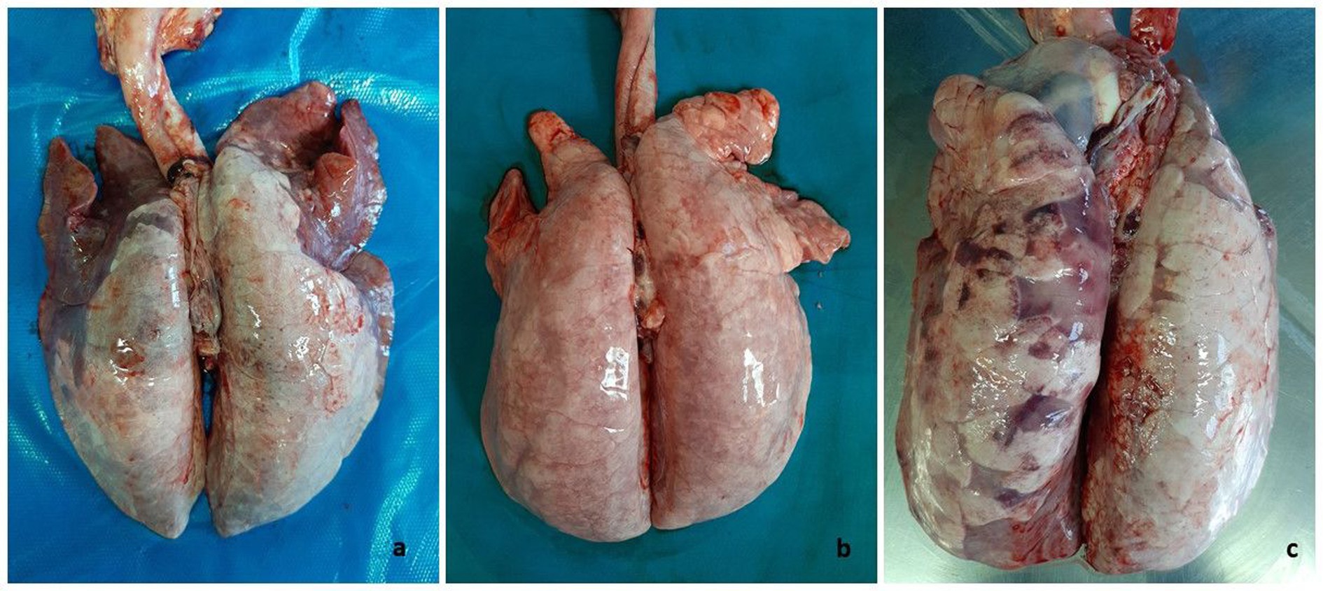
Figure 1. Swine. Lungs. Macroscopic pattern of pneumonia recognized in the caseload. (A) Cranioventral dark-red areas of consolidation (bronchopneumonia) associated to uncollapsed basal lobes showing discoloration and edema (interstitial pneumonia), this latter as main lesion appearing in (B). (C) Lung with dark areas of consolidation intermingled with interstitial pneumonia in basal lobes.
The following histologic associations were recorded: 44 cases (55.7%) exhibited pure interstitial pneumonia (illustrated in Figure 2A); 17 cases (21.5%) had bronchopneumonia associated with interstitial pneumonia (Figure 2B); 8 cases (10.1%) showed evidence of proliferative and necrotizing pneumonia (PNP) (Figures 2C,D); 2 cases (2.5%) had PNP in conjunction with interstitial pneumonia and exudative bronchopneumonia; 2 cases (2.5%) had fibrinous pleuropneumonia; 2 cases (2.5%) exhibited pure bronchopneumonia; 1 case (1.3%) had bronchopneumonia along with interstitial pneumonia and pleuropneumonia; 2 cases (2.5%) displayed only congestion and pulmonary hemorrhages; and 1 case (1.3%) had enzootic pneumonia (Figure 3).
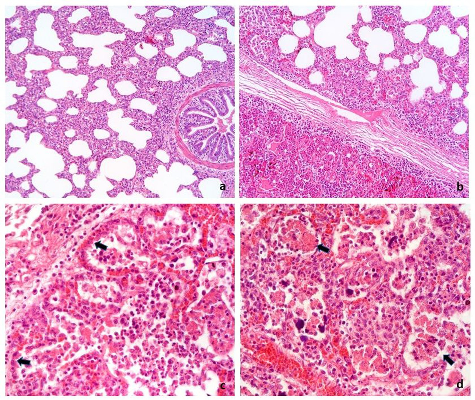
Figure 2. Swine, lung. Histologic features of the main pneumonia patterns recognized in 79 cases of PRDC. (A) Interstitial pneumonia characterized by irregular thickening of alveolar septa by inflammatory infiltrate. (B) Interstitial pneumonia (top of the figure) associated with bronchoalveolar exudate (bottom of the figure) of neutrophils (bronchopneumonia). (C,D) Proliferative and necrotizing pneumonia characterized by hyperplasia of type II pneumocytes (arrow in C) and necrosis of macrophages exudate in alveoli (arrow in D). H&E. A,B: 16×; C,D: 40 × .
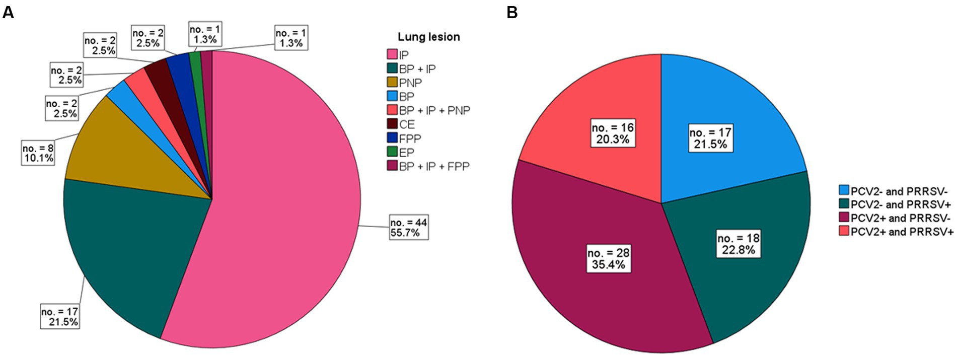
Figure 3. (A) Frequency of lung lesions in 79 cases of PRCD. Cases are grouped by single type of pneumonia or their associations. Interstitial pneumonia (IP) alone or associated with other pneumonias accounts for 82% of cases. (B) Frequency of PCV2 and/or PRRSV-positive or -negative stain in 79 cases of PRDC.
The results of the IHC staining on 79 cases of PRDC revealed that 44 (55.7%) tested PCV2 positive in the lung or in at least one out of the available lymphoid tissues, while 34 (43.0%) cases tested positive for PRRSV. In 16 (20.3%) of the total lung tested, both PCV2 and PRRSV were identified (Figure 4). In 17 cases (21.5%), neither virus was detected. Table 1 summarizes the IHC findings for each lung lesion in these cases.
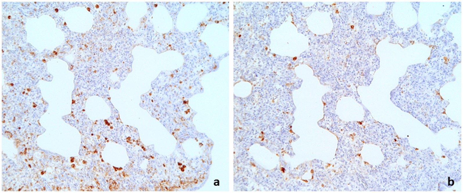
Figure 4. Swine lung. Interstitial pneumonia showing double positive immunostaining to PCV2 (A) and PRRSV (B). (A): IHC to PCV2, 16×; (B): IHC to PRRSV, 16×.
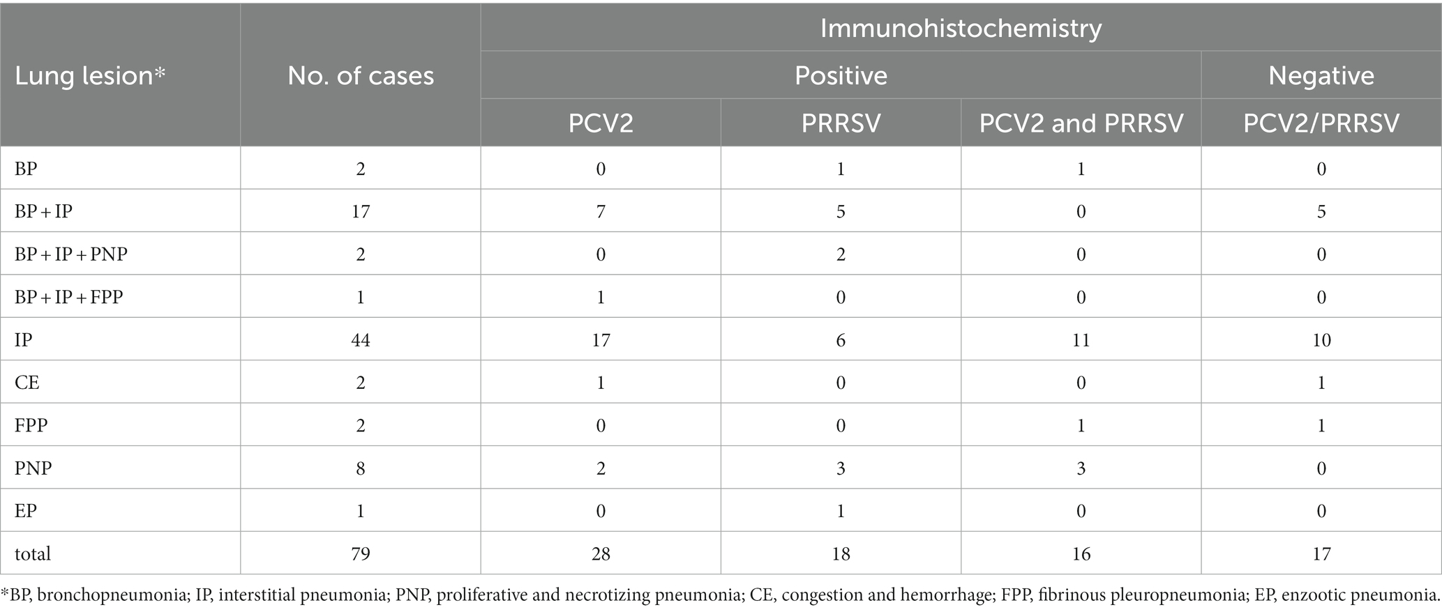
Table 1. Summary of results of PCV2 and PRRSV immunohistochemistry (IHC) associated with the pulmonary lesions.
Among the 79 cases, 10 (12.7%) showed lung lesions associated with PNP (alone or in conjunction with interstitial pneumonia and exudative bronchopneumonia), of which 5 (50.0%) were PCV2-negative and PRRSV-positive, 2 (20.0%) were PCV2-positive and PRRSV-negative, and 3 (30.0%) were double positive for both.
Among the 44 subjects with lymphoid tissues that tested PCV2-positive through IHC, 28 cases (63.6%) exhibited lesions compatible with lymphocytic depletion and showed IHC positivity for PCV2 in at least one of the lymphoid tissues examined. These cases were classified as PCV2-SD (Figure 5). In the remaining 16 out of 44 cases, in addition to PCV2-positive lung lesions, no lymphoid tissue lesions or only mild to moderate lymphoid hyperplasia were detected. In these cases, slight positivity to the PCV2 antigen was detected within the cytoplasm of centrofollicular dendritic cells in at least one of the lymphoid tissues examined. These cases were classified as PRDC/PCV2-positive (36.4%) (Figure 6).
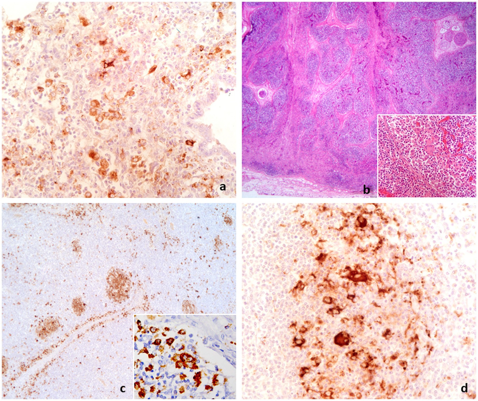
Figure 5. Diagnostic path conclusive for PCV2-SD. (A) PCV2-positive macrophages in lung with interstitial pneumonia associated with lymphoid depletion in lymphoid tissue as in lymph node (B). Inset in (B): collection of epithelioid and giant cells in lymph node. (C) Depleted lymph node showing PCV2 antigen in residual follicles (C,D) and in epithelioid and giant cells (inset in C). (A,D): IHC to PCV2, 40×; (B): H&E, 2.5×, inset 40×; (C): IHC to PCV2, 6.3×, inset 40×.
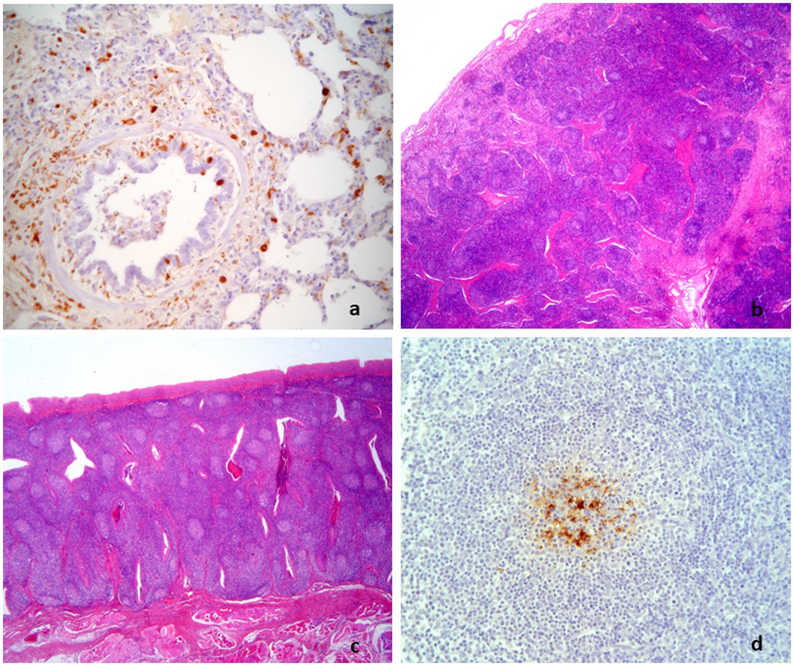
Figure 6. Diagnostic path conclusive for PRDC/PCV2-positive. Interstitial pneumonia with PCV2-positive macrophages (A) associated with hyperplastic lymphoid tissues [lymph node in (B) and tonsil in (C) showing PCV2-positive histiocytes in few normal and polarized follicles (D)]. (A,D): IHC to PCV2, 25×; (B,C): H&E, 2.5×.
The results of the classification and definition of individual cases in PCV2-SD and PRDC/PCV2-positive categories were subjected to further analysis by grouping them according to the farm of origin, as shown in Figure 7. A diagnosis of PCV2-SD was established in those farms where all subjects displayed systemic involvement (farms C, F, H, I, J, M, and N in Figure 7). Additionally, a diagnosis of PCVD-SD was made in farms B and L (Figure 7) when at least one pig in the group displayed such final diagnosis. However, a final diagnosis of PCVD-SD could not be reached in herds A, D, E, G, K, where only PRDC/PCV2-positive cases were identified.
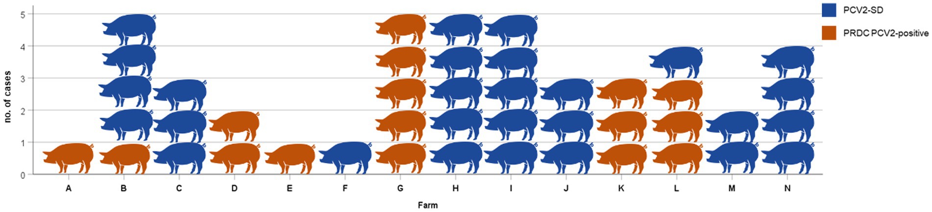
Figure 7. PCV2-SD or PRDC/PCV2-positive cases in each 14 PCV2-positive (A–N) farms. Considering the final diagnosis on the flock, in herds C, F, H, I, J, M, and N it is concluded PCV2-SD. A diagnosis of PCV2-SD is concluded also in farms B and L because at least one pig in the group displayed such final diagnosis. A final diagnosis of PCV2-SD was not reached in herds A, D, E, G, and K, where only PRDC/PCV2-positive was identified.
In the 44 PCV2-positive pigs, 42.9% of the PCV2-SD cases (12/28) showed IHC positivity for PRRSV in the lung tissue, while 25.0% of PRDC/PCV2-positive cases (4/16) showed double positivity for PCV2 and PRRSV (Figure 8).
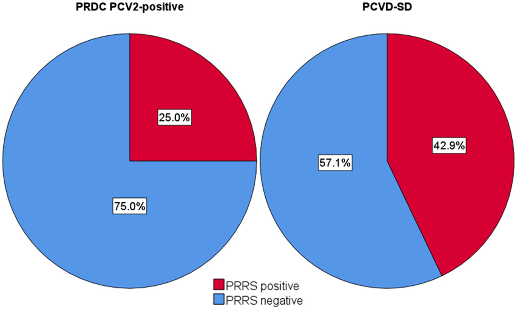
Figure 8. Frequency of PRRSV positive and negative pneumonias in PCV2-SD and PRDC/PCV2-positive cases.
Porcine Respiratory Disease Complex (PRDC) is a high-morbidity clinical condition affecting pigs over 4 weeks of age, exhibiting respiratory signs often unresponsive to antibiotics. This disease is frequently associated with a wide spectrum of lung lesions that are caused by the synergistic effect of both bacterial and viral microorganisms. In this study, interstitial pneumonia and mixed forms of both exudative bronchopneumonia and interstitial pneumonia were the most frequently (77.2%) observed microscopic lesions.
Viral infections, such as Porcine Circovirus type 2 (PCV2) and Porcine Reproductive and Respiratory Syndrome Virus (PRRSV), reaching the lung through the hematogenous route, are frequently associated with interstitial pneumonia in pigs. At the same time neutrophilic and macrophagic exudation in bronchiolo-alveolar spaces provides an indication of secondary bacterial infection further complicating the lung lesions (12). Microscopic pulmonary lesions, identified by the criteria used to diagnose PRDC, suggest that viral infections are the primary cause, exacerbated by secondary bacterial infections (1, 39).
In our study, the results of the immunohistochemistry (IHC) assay for PCV2 and PRRSV confirmed the link between the microscopic appearance of the lesions and their viral origin, as the antigens of the two viruses were identified in 78.5% of the overall cases and in 49 out of 64 cases (77%) of interstitial pneumonia included as single or combined diagnosis (Table 1).
PCV2 and PRRSV, although not strictly considered respiratory pathogens, are well-known viruses heavily involved in swine respiratory disease clinical manifestation (1). In 20.3% of the cases (16 out of 79) presented here, co-infection between PCV2 and PRRSV was identified through double-positive PCV2 and PRRSV results in lungs, as determined by IHC. PRRSV infection is known to act as a co-factor that exacerbates lesions and generates the clinical signs of PCVD (30, 40), and co-infection between PCV2 and PRRSV is a well-established phenomenon (27–31).
Consistent with previous research (28, 41), the majority of proliferative and necrotizing pneumonia (PNP) cases in this study were linked to PRRSV infection and the presence of viral antigen within lung lesions. Moreover, a significant proportion of PNP cases (3/10, 30%) are double-positive for PCV2 and PRRSV in the lung. PNP is a form of interstitial pneumonia characterized by hypertrophy and hyperplasia of type II pneumocytes associated with the presence of necrotic debris within alveolar spaces (42). PRRSV and PCV2 are the main viral agents involved in PNP, considering Aujezsky’s disease virus (ADV) and swine influenza A virus (IAV-S) playing a possible role in co-infections (43). Our study confirms previous findings by Morandi et al. (41), indicating that the severe form of interstitial pneumonia in Italy is primarily associated with PRRSV infection. Additionally, we observed here that co-infection with PCV2 resulted in more severe microscopical lesion.
The sampling method used in this study, which included a variety of lymphoid tissues in addition to the lung, allowed us to diagnose PCV2-SD in lung PCV2-positive cases by combining the results of lymphoid tissue lesions and IHC positivity. As a result, in 28 out of 44 PCV2-SD cases (63.6%), PCV2-positive stain was found in at least one lymphoid-depleted tissue, including the tonsil, tracheobronchial lymph node, superficial inguinal lymph node, spleen, and ileum, in addition to pulmonary lesions.
The diagnosis of systemic Porcine Circovirus type 2 disease has been well-established since the earliest descriptions of PCV2-related disease (18, 44) and continues to be used today (23). These criteria encompass the triad of clinical signs, lesions, and viral detection in the presence of recognizable lesions. The case series reviewed in this study showed that a higher proportion of cases had evidence of systemic infection despite presenting respiratory signs without severe emaciation and anemia, which are typical of PCV2-associated systemic disease (PCV2-SD). Thus, these cases were classified as PCV2-SD. This finding aligns with the study conducted by Ticó et al. (24), where on the 226 PCV2-positive animals suffering from PRDC, 184 cases had PCV2 systemic infection, suggesting that PCV2-LD is a negligible condition and possibly part of a systemic manifestation of infection.
The introduction of PCV2 vaccines has generated significant modifications in the epidemiology of the disease (45). As a result, the severity of disease presentation has decreased over time, while the prevalence of vague clinical signs has increased in affected animals (46). Nevertheless, although PCV2 vaccines are among the most effective used in swine herds, cases of PCV2-SD have been described in vaccinated farms (47, 48). The primary contributing factor to this phenomenon can be attributed to inappropriate vaccination protocols, whereby the timing of vaccine administration is either delayed or premature. Notably this is not driven by the circulation of new viral strains capable of evading vaccine-induced protection (49), but rather the disruption of maternal-derived immunity (23).
Additionally, it is noteworthy that PCVD may occur after a PCV2 infection triggered by co-infections with other viruses and bacteria (50). In this context, the Porcine Reproductive and Respiratory Syndrome Virus (PRRSV) has been identified as a key pathogen in co-infections in pigs (51, 52). In the current study, 7 out of the 22 farms had clinical PRRSV recrudescence reported in anamnesis, and 12 out of 28 pigs with a final diagnosis of PCV2-SD were also found to be PRRSV-positive in the lungs. Furthermore, experimental studies have demonstrated that co-infections with PCV2 and PRRSV can result in more severe clinical signs and lesions than infections with a single virus (53, 54). This synergism between the two viruses in regulating host innate and adaptive immunity (55–57) can lead to higher replication and pathogenicity of the viruses involved (29, 35, 58, 59). In the field, this also occurs in the case of vaccination against PRRSV in herds infected with PCV2; in fact, vaccination with live attenuated PRRSV vaccine has been shown to enhance PCV2 replication and increase viraemia with a mechanism identical to a PCV2/PRRSV coinfection (60). For the control of clinical forms, it is, therefore, pivotal to know the health status of the herd in order to choose the most suitable vaccination strategy (61).
The diagnosis of PCVD is a herd diagnosis and should be performed in a group of 3–5 animals (50). In our study among positive PCV2 farms (14/22), in 7 sites, all the examined pigs presented the criteria for categorization in PCV2-SD (Figure 7). In 5 herds, all forms observed were referable to PRDC/PCV2-positive, although the data may not be sufficient for a reliable herd diagnosis. Only one subject was conferred from two farms and only two from one, despite the 3-5 needed. On the other hand, in two herds, the results differed among the individual cases examined. However, the evidence of a systemic form of the disease in at least one animal tested supports the diagnostic conclusion of PCV2-SD in the group.
In this study, the individual cases classified PRDC/PCV2-positive (with specific lung lesions and immunohistochemical positive stain in the lung and in at least one of the examined lymphoid districts) were those in which the lymphocyte depletion in lymphoid tissues were not observed (16/44, 36.4%). The occurrence of microscopic lung lesions caused solely by PCV2 infection is rare in experimental conditions, as evidence by the findings of Hoogland et al. (62) and Opriessing et al. (63). However, in field conditions, the situation is considerably more complex, with numerous co-factors, infectious and non-infectious, potentially exacerbating lung damage. Notably, Grau-Roma et al. (64) reported a positive correlation between the PCV2 viral load in serum and tissues, implying a significant role of PCV2 in the onset of Porcine Respiratory Disease Complex (PRDC), even in subclinical cases and in the absence of lymphoid depletion.
Empirical evidence suggests that despite the absence of a definitive diagnosis of systemic Porcine Circovirus type 2 (PCV2) disease, the virus is prevalent in tissues and continues to circulate, invariably compromising the herd’s health.
Our case series reveals a strong association between PCV2 and a considerable percentage of respiratory signs, even when the systemic disease is absent. In such scenarios, where the likelihood of a bacterial origin for Porcine Respiratory Disease Complex (PRDC) is ruled out, detecting PCV2 infection should clearly indicate the need to strengthen control and preventive measures on the farm.
The results of this study highlight the significance of PCV2, particularly when its antigen is detected in the lung tissue in conjunction with microscopic interstitial pneumonia. The systemic form of PCVD (PCV2-SD) is diagnosed when the PCV2 antigen is present, given its strong association with both pulmonary issues but also lymphoid depletion. To avoid underestimating the potential harm caused by PCV2, it is imperative to employ appropriate sampling techniques to diagnose the systemic form of the disease, even in the absence of other clinical signs. Despite the outdated notion of PCV2-LD (23), PCV2, along with PRRSV, remains a prevalent infection linked to PRDC and has a negative impact on herd health. Co-infections can exacerbate the pathological condition, necessitating control measures based on pathological and analytical evidence.
The raw data supporting the conclusions of this article will be made available by the authors, without undue reservation.
No ethics approval is required. The study was conducted in accordance with the local legislation and institutional requirements.
GD’A and GS: conceptualization and writing—original draft preparation. GD’A, GS, AL, and MO: methodology. FO and LVM: validation. GD’A, GS, and LVM: investigation. GD’A, GS, and FO: data curation. MO and FO: writing—review and editing. NT, AL, and LuM: visualization. GS: supervision. NJ and GL: project administration. All authors contributed to the article and approved the submitted version.
NJ, NT, and GL were employed by Boehringer Ingelheim Animal Health Italia SpA.
The remaining authors declare that the research was conducted in the absence of any commercial or financial relationships that could be construed as a potential conflict of interest.
All claims expressed in this article are solely those of the authors and do not necessarily represent those of their affiliated organizations, or those of the publisher, the editors and the reviewers. Any product that may be evaluated in this article, or claim that may be made by its manufacturer, is not guaranteed or endorsed by the publisher.
1. Opriessnig, T, Giménez-Lirola, LG, and Halbur, PG. Polymicrobial respiratory disease in pigs. Anim Health Res Rev. (2011) 12:133–48. doi: 10.1017/S1466252311000120
2. Janke, BH. Diagnosis of viral respiratory disease in swine. J Swine Health Prod. (1995) 3:116–20.
3. Sunaga, F, Tsuchiaka, S, Kishimoto, M, Aoki, H, Kakinoki, M, Kure, K, et al. Development of a one-run real-time PCR detection system for pathogens associated with porcine respiratory diseases. J Vet Med Sci. (2020) 82:217–23. doi: 10.1292/jvms.19-0063
4. Yaeger, MJ, and Alstine, WG. Respiratory system In: J Zimmerman, L Karriker, A Ramirez, K Schwartz, G Stevenson, and J Zhang, editors. Diseases of Swine : Wiley (2019). 393–407.
5. Giles, TA, Belkhiri, A, Barrow, PA, and Foster, N. Molecular approaches to the diagnosis and monitoring of production diseases in pigs. Res Vet Sci. (2017) 114:266–72. doi: 10.1016/j.rvsc.2017.05.016
6. Christopher-Hennings, J, Erickson, GA, Hesse, RA, Nelson, EA, Rossow, S, Scaria, J, et al. Diagnostic tests, test performance, and considerations for interpretation In: J Zimmerman, L Karriker, A Ramirez, K Schwartz, G Stevenson, and J Zhang, editors. Diseases of Swine : Wiley (2019). 75–97.
7. Hernandez-Garcia, J, Robben, N, Magnée, D, Eley, T, Dennis, I, Kayes, SM, et al. The use of oral fluids to monitor key pathogens in porcine respiratory disease complex. Porc Health Manag. (2017) 3:7. doi: 10.1186/s40813-017-0055-4
8. Lung, O, Ohene-Adjei, S, Buchanan, C, Joseph, T, King, R, Erickson, A, et al. Multiplex PCR and microarray for detection of swine respiratory pathogens. Transbound Emerg Dis. (2017) 64:834–48. doi: 10.1111/tbed.12449
9. Trang, NT, Hirai, T, Yamamoto, T, Matsuda, M, Okumura, N, Giang, NTH, et al. Detection of porcine reproductive and respiratory syndrome virus in oral fluid from naturally infected pigs in a breeding herd. J Vet Sci. (2014) 15:361–7. doi: 10.4142/jvs.2014.15.3.361
10. Trang, N. T., Ngan, P. H., Hop, N.Van, and Hirai, T. (2018). Nasal swab – a new tool for the detection of porcine respiratory disease complex in natural infected pigs. Acta Sci Microbiol 1, 2–5.
11. Maes, RK, Langohr, IM, Wise, AG, Smedley, RC, Thaiwong, T, and Kiupel, M. Beyond H&E. Vet Pathol. (2014) 51:238–56. doi: 10.1177/0300985813505878
12. Sarli, G, D’Annunzio, G, Gobbo, F, Benazzi, C, and Ostanello, F. The role of pathology in the diagnosis of swine respiratory disease. Vet Sci. (2021) 8:256. doi: 10.3390/vetsci8110256
13. Marcato, PS, Sidoli, L, Mandrioli, L, Della Salda, L, Cerati, C, and Rolla, GL. Indagini clinico patologiche in un focolaio di PMWS (postweaning multisystemic wasting syndrome) in suini del Nord Italia. Large Animals Review. (1999) 5:47–62.
14. Harms, PA, Halbur, PG, and Sorden, SD. Three cases of porcine respiratory disease complex associated with porcine circovirus type 2 infection. J Swine Health Prod. (2002) 10:27–30.
15. Kim, J, Chung, HK, and Chae, C. Association of porcine circovirus 2 with porcine respiratory disease complex. Vet J. (2003) 166:251–6. doi: 10.1016/s1090-0233(02)00257-5
16. Segalés, J, Rosell, C, and Domingo, M. Pathological findings associated with naturally acquired porcine circovirus type 2 associated disease. Vet Microbiol. (2004) 98:137–49. doi: 10.1016/j.vetmic.2003
17. Opriessnig, T, Meng, XJ, and Halbur, PG. Porcine circovirus type 2 associated disease: update on current terminology, clinical manifestations, pathogenesis, diagnosis, and intervention strategies. J Vet Diagn Investig. (2007) 19:591–615. doi: 10.1177/104063870701900601
18. Segalés, J. Porcine circovirus type 2 (PCV2) infections: clinical signs, pathology and laboratory diagnosis. Virus Res. (2012) 164:10–9. doi: 10.1016/j.virusres.2011.10.007
19. Fablet, C, Marois-Créhan, C, Simon, G, Grasland, B, Jestin, A, Kobisch, M, et al. Infectious agents associated with respiratory diseases in 125 farrow-to-finish pig herds: a cross-sectional study. Vet Microbiol. (2012) 157:152–63. doi: 10.1016/j.vetmic.2011.12.015
20. Hansen, MS, Pors, SE, Jensen, HE, Bille-Hansen, V, Bisgaard, M, Flachs, EM, et al. An investigation of the pathology and pathogens associated with porcine respiratory disease complex in Denmark. J Comp Pathol. (2010) 143:120–31. doi: 10.1016/j.jcpa.2010.01.012
21. Paz-Sánchez, Y, Herráez, P, Quesada-Canales, Ó, Poveda, CG, Díaz-Delgado, J, Quintana-Montesdeoca, MDP, et al. Assessment of Lung disease in finishing pigs at slaughter: pulmonary lesions and implications on productivity parameters. Animals. (2021) 11:3604. doi: 10.3390/ani11123604
22. Wellenberg, GJ, Bouwkamp, FT, Wolf, PJ, Swart, WA, Mombarg, MJ, and de Gee, AL. A study on the severity and relevance of porcine circovirus type 2 infections in Dutch fattening pigs with respiratory diseases. Vet Microbiol. (2010) 142:217–24. doi: 10.1016/j.vetmic.2009.10.003
23. Segalés, J, and Sibila, M. Revisiting porcine circovirus disease diagnostic criteria in the current porcine circovirus 2 epidemiological context. Vet Sci. (2022) 9:110. doi: 10.3390/vetsci9030110
24. Ticó, G, Segalés, J, and Martínez, J. The blurred border between porcine circovirus type 2-systemic disease and porcine respiratory disease complex. Vet Microbiol. (2013) 163:242–7. doi: 10.1016/j.vetmic.2013.01.001
25. Opriessnig, T, and Halbur, PG. Concurrent infections are important for expression of porcine circovirus associated disease. Virus Res. (2012) 164:20–32. doi: 10.1016/j.virusres.2011.09.014
26. Ouyang, T, Zhang, X, Liu, X, and Ren, L. Co-infection of swine with porcine circovirus type 2 and other swine viruses. Viruses. (2019) 11:185. doi: 10.3390/v11020185
27. Burgara-Estrella, A, Montalvo-Corral, M, Bolaños, A, Ramírez-Mendoza, H, Valenzuela, O, and Hernández, J. Naturally co-infected boars with both porcine reproductive and respiratory syndrome virus and porcine circovirus type 2. Transbound Emerg Dis. (2012) 59:532–8. doi: 10.1111/j.1865-1682.2012.01313.x
28. Drolet, R, Larochelle, R, Morin, M, Delisle, B, and Magar, R. Detection rates of porcine reproductive and respiratory syndrome virus, porcine circovirus type 2, and swine influenza virus in porcine proliferative and necrotizing pneumonia. Vet Pathol. (2003) 40:143–8. doi: 10.1354/vp.40-2-143
29. Fan, P, Wei, Y, Guo, L, Wu, H, Huang, L, Liu, J, et al. Synergistic effects of sequential infection with highly pathogenic porcine reproductive and respiratory syndrome virus and porcine circovirus type 2. Virol J. (2013) 10:265. doi: 10.1186/1743-422X-10-265
30. Harms, PA, Sorden, SD, Halbur, PG, Bolin, SR, Lager, KM, Morozov, I, et al. Experimental reproduction of severe disease in CD/CD pigs concurrently infected with type 2 porcine circovirus and porcine reproductive and respiratory syndrome virus. Vet Pathol. (2001) 38:528–39. doi: 10.1354/vp.38-5-528
31. Zeng, Z, Liu, Z, Wang, W, Tang, D, Liang, H, and Liu, Z. Establishment and application of a multiplex PCR for rapid and simultaneous detection of six viruses in swine. J Virol Methods. (2014) 208:102–6. doi: 10.1016/j.jviromet.2014.08.001
32. Chase, C, and Lunney, JK. Immune System In: J Zimmerman, L Karriker, A Ramirez, K Schwartz, G Stevenson, and J Zhang, editors. Diseases of Swine : Wiley (2019). 264–91.
33. Loving, CL, Osorio, FA, Murtaugh, MP, and Zuckermann, FA. Innate and adaptive immunity against porcine reproductive and respiratory syndrome virus. Vet Immunol Immunopathol. (2015) 167:1–14. doi: 10.1016/j.vetimm.2015.07.003
34. Lunney, JK, Fang, Y, Ladinig, A, Chen, N, Li, Y, Rowland, B, et al. Porcine reproductive and respiratory syndrome virus (PRRSV): pathogenesis and interaction with the immune system. Annu Rev Anim Biosci. (2016) 4:129–54. doi: 10.1146/annurev-animal-022114-111025
35. Niederwerder, MC, Jaing, CJ, Thissen, JB, Cino-Ozuna, AG, McLoughlin, KS, and Rowland, RR. Microbiome associations in pigs with the best and worst clinical outcomes following coinfection with porcine reproductive and respiratory syndrome virus (PRRSV) and porcine circovirus type 2 (PCV2). Vet Microbiol. (2016) 188:1–11. doi: 10.1016/j.vetmic.2016.03.008
36. Caswell, JL, and Williams, KJ. Respiratory system In: Jubb, Kennedy & Palmer’s pathology of domestic animals, M. Grant Maxie. vol. 2: Elsevier (2015). 465–591.
37. Mandrioli, L, Sarli, G, Panarese, S, Baldoni, S, and Marcato, PS. Apoptosis and proliferative activity in lymph node reaction in postweaning multisystemic wasting syndrome (PMWS). Vet Immunol Immunopathol. (2004) 97:25–37. doi: 10.1016/j.vetimm.2003.08.017
38. Valli, VEO, Kiupel, M, and Bienzle, D (with Wood, R. D.),. Hematopoietic system In: Jubb, Kennedy & Palmer’s pathology of domestic animals, M. Grant Maxie. vol. 3: Elsevier (2015). 102–2068.
39. Loosli, CG. Synergism between respiratory viruses and bacteria. Yale J Biol Med. (1968) 40:522–40.
40. Park, C, Seo, HW, Park, SJ, Han, K, and Chae, C. Comparison of porcine circovirus type 2 (PCV2)-associated lesions produced by coinfection between two genotypes of PCV2 and two genotypes of porcine reproductive and respiratory syndrome virus. J Gen Virol. (2014) 95:2486–94. doi: 10.1099/vir.0.066290-0
41. Morandi, F, Ostanello, F, Fusaro, L, Bacci, B, Nigrelli, A, Alborali, L, et al. Immunohistochemical detection of aetiological agents of proliferative and necrotizing pneumonia in Italian pigs. J Comp Pathol. (2010) 142:74–8. doi: 10.1016/j.jcpa.2009.06.003
42. Morin, M, Girard, C, Elazhary, Y, Fajardo, R, Drolet, R, and Lagacé, A. Severe proliferative and necrotizing pneumonia in pigs: a newly recognized disease. Can Vet J. (1990) 31:837–9.
43. Grau-Roma, L, and Segalés, J. Detection of porcine reproductive and respiratory syndrome virus, porcine circovirus type 2, swine influenza virus and Aujeszky’s disease virus in cases of porcine proliferative and necrotizing pneumonia (PNP) in Spain. Vet Microbiol. (2007) 119:144–51. doi: 10.1016/j.vetmic.2006.09.009
44. Segalés, J, Allan, GM, and Domingo, M. Porcine circovirus diseases. Anim Health Res Rev. (2005) 6:119–42. doi: 10.1079/ahr2005106
45. Segalés, J. Best practice and future challenges for vaccination against porcine circovirus type 2. Expert Rev Vaccines. (2015) 14:473–87. doi: 10.1586/14760584.2015.983084
46. Kristensen, CS, Hjulsager, CK, and Larsen, LE. A two-year follow-up study of the PCV2 status of a Danish pig herd that was initially assumed to be PCV2-free. Porc Health Manag. (2015) 1:5. doi: 10.1186/2055-5660-1-5
47. Opriessnig, T, Xiao, CT, Gerber, PF, and Halbur, PG. Emergence of a novel mutant PCV2b variant associated with clinical PCVAD in two vaccinated pig farms in the U.S. concurrently infected with PPV2. Vet Microbiol. (2013) 163:177–83. doi: 10.1016/j.vetmic.2012.12.019
48. Seo, HW, Park, C, Kang, I, Choi, K, Jeong, J, Park, SJ, et al. Genetic and antigenic characterization of a newly emerging porcine circovirus type 2b mutant first isolated in cases of vaccine failure in Korea. Arch Virol. (2014) 159:3107–11. doi: 10.1007/s00705-014-2164-6
49. Sibila, M, Rocco, C, Franzo, G, Huerta, E, Domingo, M, Núñez, JI, et al. Genotyping of porcine circovirus 2 (PCV-2) in vaccinated pigs suffering from PCV-2-systemic disease between 2009 and 2020 in Spain. Pathogens. (2021) 10:1016. doi: 10.3390/pathogens10081016
50. Segalés, J, Allan, GM, and Domingo, M. Circoviruses In: J Zimmerman, L Karriker, A Ramirez, K Schwartz, G Stevenson, and J Zhang, editors. Diseases of Swine : Wiley (2019). 473–87.
51. Saade, G, Deblanc, C, Bougon, J, Marois-Créhan, C, Fablet, C, Auray, G, et al. Coinfections and their molecular consequences in the porcine respiratory tract. Vet Res. (2020) 51:80. doi: 10.1186/s13567-020-00807-8
52. Zhao, D, Yang, B, Yuan, X, Shen, C, Zhang, D, Shi, X, et al. Advanced research in porcine reproductive and respiratory syndrome virus coinfection with other pathogens in swine. Front Vet Sci. (2021) 8:699561. doi: 10.3389/fvets.2021.699561
53. Allan, GM, McNeilly, F, Ellis, J, Krakowka, S, Meehan, B, McNair, I, et al. Experimental infection of colostrum deprived piglets with porcine circovirus 2 (PCV2) and porcine reproductive and respiratory syndrome virus (PRRSV) potentiates PCV2 replication. Arch Virol. (2000) 145:2421–9. doi: 10.1007/s007050070031
54. Rovira, A, Balasch, M, Segalés, J, García, L, Plana-Durán, J, Rosell, C, et al. Experimental inoculation of conventional pigs with porcine reproductive and respiratory syndrome virus and porcine circovirus 2. J Virol. (2002) 76:3232–9. doi: 10.1128/jvi.76.7.3232-3239.2002
55. Dong, VH, Tu, PY, Tsai, PC, Lin, YH, Chang, HL, Kuo, TY, et al. Expression of toll-like receptor signalling-related genes in pigs co-infected with porcine reproductive and respiratory syndrome virus and porcine circovirus type 2. Res Vet Sci. (2015) 101:180–6. doi: 10.1016/j.rvsc.2015.05.006
56. Richmond, O, Cecere, TE, Erdogan, E, Meng, XJ, Piñeyro, P, Subramaniam, S, et al. The PD-L1/CD86 ratio is increased in dendritic cells co-infected with porcine circovirus type 2 and porcine reproductive and respiratory syndrome virus, and the PD-L1/PD-1 axis is associated with anergy, apoptosis, and the induction of regulatory T-cells in porcine lymphocytes. Vet Microbiol. (2015) 180:223–9. doi: 10.1016/j.vetmic.2015.09.014
57. Tu, PY, Tsai, PC, Lin, YH, Liu, PC, Chang, HL, Kuo, TY, et al. Expression profile of toll-like receptor mRNA in pigs co-infected with porcine reproductive and respiratory syndrome virus and porcine circovirus type 2. Res Vet Sci. (2015) 98:134–41. doi: 10.1016/j.rvsc.2014.12.003
58. Niederwerder, MC, Bawa, B, Serão, NV, Trible, BR, Kerrigan, MA, Lunney, JK, et al. Vaccination with a porcine reproductive and respiratory syndrome (PRRS) modified live virus vaccine followed by challenge with PRRS virus and porcine circovirus type 2 (PCV2) protects against PRRS but enhances PCV2 replication and pathogenesis compared to results for nonvaccinated Cochallenged controls. Clin Vaccine Immunol. (2015) 22:1244–54. doi: 10.1128/CVI.00434-15
59. Sinha, A, Shen, HG, Schalk, S, Beach, NM, Huang, YW, Meng, XJ, et al. Porcine reproductive and respiratory syndrome virus (PRRSV) influences infection dynamics of porcine circovirus type 2 (PCV2) subtypes PCV2a and PCV2b by prolonging PCV2 viremia and shedding. Vet Microbiol. (2011) 152:235–46. doi: 10.1016/j.vetmic.2011.05.005
60. Park, C, Oh, Y, Seo, HW, Han, K, and Chae, C. Comparative effects of vaccination against porcine circovirus type 2 (PCV2) and porcine reproductive and respiratory syndrome virus (PRRSV) in a PCV2-PRRSV challenge model. Clin Vaccine Immunol. (2013) 20:369–376. doi: 10.1128/CVI.00497-12
61. Chae, C. Porcine respiratory disease complex: Interaction of vaccination and porcine circovirus type 2, porcine reproductive and respiratory syndrome virus, and Mycoplasma hyopneumoniae. Vet J. (2016) 212:1–6. doi: 10.1016/j.tvjl.2015.10.030
62. Hoogland, MJ, Opriessnig, T, and Halbur, PG. Effects of adjuvants on porcine circovirus type 2-associated lesions. J Swine Health Prod. (2006) 14:133–9.
63. Opriessnig, T, Halbur, PG, Yu, S, Thacker, EL, Fenaux, M, and Meng, XJ. Effects of the timing of the administration of Mycoplasma hyopneumoniae bacterin on the development of lesions associated with porcine circovirus type 2. Vet Rec. (2006) 158:149–54. doi: 10.1136/vr.158.5.149
64. Grau-Roma, L, Hjulsager, CK, Sibila, M, Kristensen, CS, López-Soria, S, Enøe, C, et al. Infection, excretion and seroconversion dynamics of porcine circovirus type 2 (PCV2) in pigs from post-weaning multisystemic wasting syndrome (PMWS) affected farms in Spain and Denmark. Vet Microbiol. (2009) 135:272–82. doi: 10.1016/j.vetmic.2008.10.007
Keywords: histopathology, PCV2, PRRSV, porcine respiratory disease complex, PRDC, porcine health, Italy
Citation: D’Annunzio G, Ostanello F, Muscatello LV, Orioles M, Jacumin N, Tommasini N, Leotti G, Luppi A, Mandrioli L and Sarli G (2023) Porcine circovirus type 2 and porcine reproductive and respiratory syndrome virus alone or associated are frequent intralesional detected viruses in porcine respiratory disease complex cases in Northern Italy. Front. Vet. Sci. 10:1234779. doi: 10.3389/fvets.2023.1234779
Edited by:
Orlando Paciello, University of Naples Federico II, ItalyReviewed by:
Davide De Biase, University of Salerno, ItalyCopyright © 2023 D’Annunzio, Ostanello, Muscatello, Orioles, Jacumin, Tommasini, Leotti, Luppi, Mandrioli and Sarli. This is an open-access article distributed under the terms of the Creative Commons Attribution License (CC BY). The use, distribution or reproduction in other forums is permitted, provided the original author(s) and the copyright owner(s) are credited and that the original publication in this journal is cited, in accordance with accepted academic practice. No use, distribution or reproduction is permitted which does not comply with these terms.
*Correspondence: Giulia D’Annunzio, Z2l1bGlhLmRhbm51bnppb0BpenNsZXIuaXQ=
Disclaimer: All claims expressed in this article are solely those of the authors and do not necessarily represent those of their affiliated organizations, or those of the publisher, the editors and the reviewers. Any product that may be evaluated in this article or claim that may be made by its manufacturer is not guaranteed or endorsed by the publisher.
Research integrity at Frontiers
Learn more about the work of our research integrity team to safeguard the quality of each article we publish.