
95% of researchers rate our articles as excellent or good
Learn more about the work of our research integrity team to safeguard the quality of each article we publish.
Find out more
ORIGINAL RESEARCH article
Front. Vet. Sci. , 20 April 2023
Sec. Veterinary clinical, anatomical, and comparative pathology
Volume 10 - 2023 | https://doi.org/10.3389/fvets.2023.1139388
This article is part of the Research Topic Current Knowledge on Camelids Infectious and Parasitic Diseases View all 11 articles
 Ehab Kotb Elmahallawy1*
 Pamela C. Köster2
 Alejandro Dashti2
 Samia Qasem Alghamdi3
 Amira Saleh4
 Ahmed Gareh5
 Barakat M. Alrashdi6
 Carolina Hernández-Castro2,7
 Begoña Bailo2
 Maha S. Lokman8,9
 Eman A. A. Hassanen10
 David González-Barrio2
 David Carmena2,11*
Introduction: Few studies have investigated the occurrence of microeukaryotic gut parasites in dromedary camels in Egypt, and the majority of these investigations are based on microscopic analysis of fecal material.
Methods: Herein, we assessed the occurrence, molecular diversity, and zoonotic potential of protozoan (Cryptosporidium spp. and Giardia duodenalis) and microsporidian (Enterocytozoon bieneusi) pathogens in individual fecal samples (n = 102) of dromedary camels with (n = 26) and without (n = 76) diarrhea from Aswan Governorate, Upper Egypt. Other factors possibly associated with an increased risk of infection (geographical origin, sex, age, and physical condition) were also analyzed. The SSU rRNA or ITS genes were targeted by molecular (PCR and Sanger sequencing) techniques for pathogen detection and species identification.
Results and discussion: The most abundant species detected was G. duodenalis (3.9%, 4/102; 95% CI: 1.1–9.7), followed by Cryptosporidium spp. (2.9%, 3/102; 95% CI: 0.6–8.4). All samples tested negative for the presence of E. bieneusi. Sequence analysis data confirmed the presence of zoonotic C. parvum (66.7%, 2/3) and cattle-adapted C. bovis (33.3%, 1/3). These Cryptosporidium isolates, as well as the four Giardia-positive isolates, were unable to be amplified at adequate genotyping markers (Cryptosporidium: gp60; Giardia: gdh, bg, and tpi). Camels younger than 2 years old were significantly more likely to harbor Cryptosporidium infections. This connection was not statistically significant, although two of the three cryptosporidiosis cases were detected in camels with diarrhea. The spread of G. duodenalis infections was unaffected by any risk variables studied. This is the first report of C. parvum and C. bovis in Egyptian camels. The finding of zoonotic C. parvum has public health implications since camels may function as sources of oocyst pollution in the environment and potentially infect livestock and humans. Although preliminary, this study provides useful baseline data on the epidemiology of diarrhea-causing microeukaryotic parasites in Egypt. Further research is required to confirm and expand our findings in other animal populations and geographical regions of the country.
Globally, Cryptosporidium spp., Giardia duodenalis, and Enterocytozoon bieneusi are among the most prevalent diarrhea-causing enteric parasites in humans and livestock (1–5). These pathogens cause significant morbidity and, in the case of Cryptosporidium, mortality in children <5 years old and immunocompromised persons residing in low-resource settings with little or no access to safe drinking water and sanitation facilities (6, 7). They also pose a threat to public health in middle- and high-income nations (8). These pathogens are transmitted through the fecal–oral route or by direct contact with infected animals or humans. Adult livestock infected with Cryptosporidium spp., G. duodenalis, and E. bieneusi are usually asymptomatic carriers that release varied amounts of (oo)cysts/spores into the surrounding environment and remain a potential source of infection for other animals and humans (9, 10). However, infected neonatal animals may have diarrhea, loss of appetite, lethargy, dehydration, and in some cases, death can occur (11, 12). Importantly, infected neonatal animals can release substantial quantities of instantly infectious (oo)cysts/spores (13, 14), making them important contributors to the (oo)cysts/spore burden in the environment, including surface waters meant for human consumption (15).
Many clinical research facilities in low-income countries rely on microscopy analyses of fecal smears to diagnose enteric parasites (16). Although this method is cheap and easy to perform, it requires well-trained and experienced microscopists, takes time, and lacks diagnostic sensitivity (17). To overcome these limitations, several molecular biological methods for detecting and distinguishing microeukaryotic intestinal parasites have been developed. These include PCR-based genotyping, Sanger sequencing of PCR products, and fluorescence probe-based qPCR techniques (18–20). Molecular methods to improve epidemiological and epidemic studies by allowing researchers to monitor pathogen infection sites, transmission pathways, and virulent genetic variants. For this task, highly sensitive, multi-copy genes, including the small subunit ribosomal RNA (SSU rRNA) and the ribosomal internal transcribed spacer (ITS) markers, are widely used (21).
At least 44 Cryptosporidium species are considered taxonomically valid (22, 23). Nearly 15 species (C. andersoni, C. bovis, C. erinacei, C. felis, C. hominis, C. macropodum, C. muris, C. occultus, C. parvum, C. ryanae, C. scrofarum, C. suis, C. tyzzeri, C. ubiquitum, and C. xiaoi) have been reported in domestic ruminants globally, with C. parvum the most dominant species, particularly in cattle (3, 20, 24). Seven Cryptosporidium species (C. andersoni, C. bovis, C. hominis, C. muris, C. occultus, C. parvum, and C. ubiquitum), and two genotypes (rat IV and camel) have been identified circulating in camels to date (Table 1).
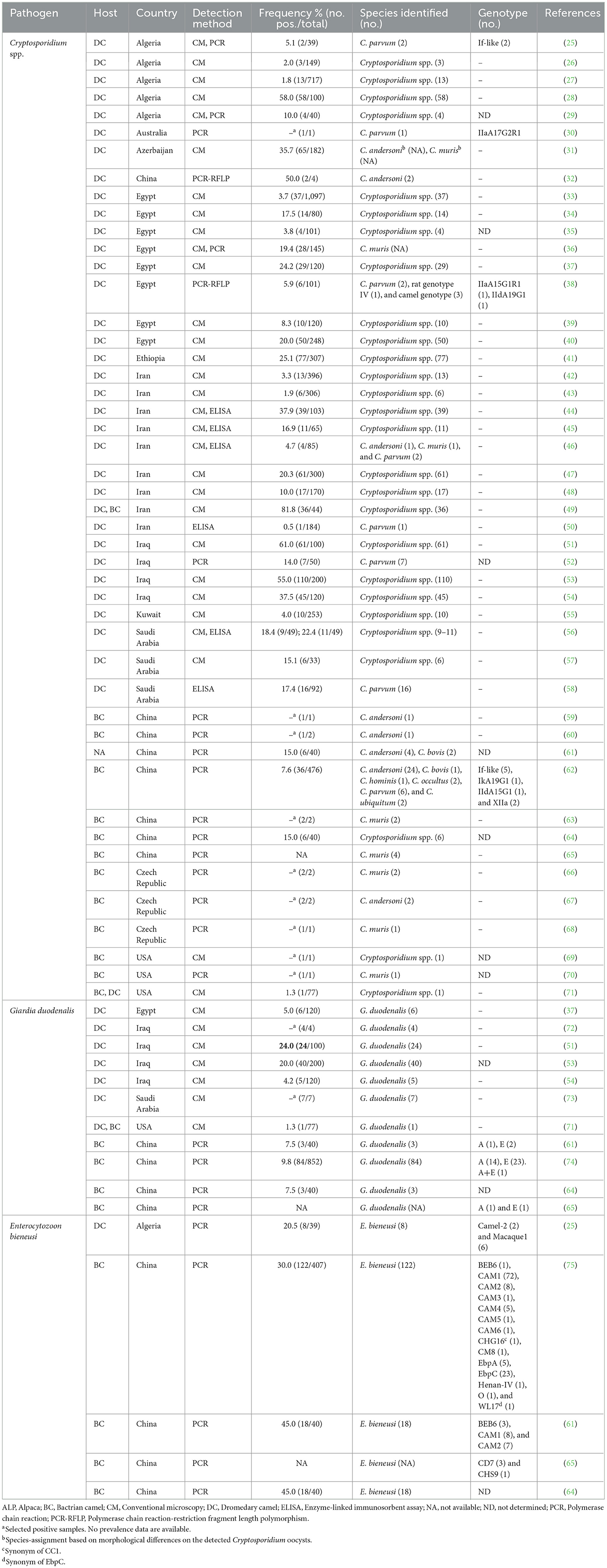
Table 1. Global occurrence and genetic diversity of Cryptosporidium spp., Giardia duodenalis, and Enterocytozoon bieneusi reported in camelids including Bactrian (Camelus bactrianus) and dromedary (Camelus dromedaries) camels.
Giardia duodenalis (syn. G. intestinalis and G. lamblia) is the only Giardia species able to infect domestic ruminants (22, 76). Giardia duodenalis is considered a complex cryptic species with eight distinct genetic variants (assemblages A to H), which differ in host distribution and specificity. Assemblages A and B are found in humans and in many other mammals, whereas C and D are found in canids, E in wild and domestic ungulates, F in felids, G in rodents, and H in marine pinnipeds (22, 76). Camels seem to be primarily infected by ungulate-adapted G. duodenalis assemblage E; however, zoonotic assemblage A infections have also been reported (Table 1). Remarkably, assemblage E is responsible for 8–100% of cases of human giardiasis documented in Egypt (77–79). More than 600 E. bieneusi genotypes have been identified and classified into 11 major phylogenetic groups, of which groups 1 and 2 contain most genotypes with zoonotic potential, and the remaining groups 3–11 include largely host-adapted genotypes associated with specific animal species (80, 81). Today, 15 E. bieneusi genotypes have been identified in camels globally, with CAM1 and EbpC accounting for nearly 70% of infections detected (Table 1).
Dromedary camels (Camelus dromedaries) have a significant economic, social, and ecological role in nomadic and/or pastoralist communities living in arid or semi-arid regions globally (79). They are natural hosts for a wide range of protists (Balantioides coli, Blastocystis sp., Cryptosporidium spp., Enterocytozoon bieneusi, Giardia spp., Toxoplasma gondii, and Trypanosoma spp.), helminth (Echinococcus granulosus, Fasciola hepatica, Schistosoma spp., and Trichinella spiralis), and arthropod (Linguatula serrata and Sarcoptes scabiei) zoonotic species, representing an often-unrecognized public health threat (27, 82, 83). In addition, infections by some of these pathogens result in significant economic loss due to decreased milk and meat output, diminished fertility, and mortality (84–86).
Several studies in Egypt have looked at the presence of parasite infections, such as Anaplasma, Babesia, Echinococcus, Sarcocystis, Sarcoptes, Theileria, and Trypanosoma in dromedary camels (87–90). However, evidence on the presence of Cryptosporidium spp., G. duodenalis, and E. bieneusi is even scarcer, with the drawback that most available data come from outdated microscopy-based studies (Table 1). Previous studies have suggested that camels infected with those microeukaryotic parasites might act as potential sources of human cryptosporidiosis, giardiasis, and microsporidiosis (25, 47). To bridge this knowledge gap, this study aims to assess the presence, genetic diversity, and zoonotic potential of Cryptosporidium spp., G. duodenalis, and E. bieneusi in dromedary camels with and without diarrhea in Aswan, the southernmost governorate in Upper Egypt.
A total of 102 individual fecal samples from dromedary camels were collected in three geographical areas (Abu Simbel, Edfu, and Kom Ombo) of the Aswan Governorate, Upper Egypt (Figure 1). The calculation of the sample size was performed as described elsewhere (38) based on a 95% confidence level. Fecal samples were collected during the period from August to December 2021. Local farmers were approached and encouraged to participate in the study after their agreement with the study's goals and procedures. Once permission was granted, fecal samples were directly collected from the rectum of the animals and placed into a sterile polystyrene plastic flask containing 70% ethanol as a preservation agent. Basic epidemiological information (geographical origin, sex, age, fecal consistency, and physical condition) was collected at the time of sampling. Animals were reared in an open system under conventional pasture grazing dependent on grazing food including hay and forages. In winter, camels were partly fed on natural grazing, but feeding was complemented by food crops gathered by breeders, and grains may have been added to the diet in certain episodes of production. Out of the 102 samples collected, 26 were diarrheic and 76 formed. Samples were delivered to the Department of Zoonoses, Faculty of Veterinary Medicine (Sohag University, Egypt) and stored at 4°C. Samples were subsequently transferred to the Parasitology Reference and Research Laboratory of the National Center for Microbiology (Majadahonda, Spain) for downstream molecular studies.
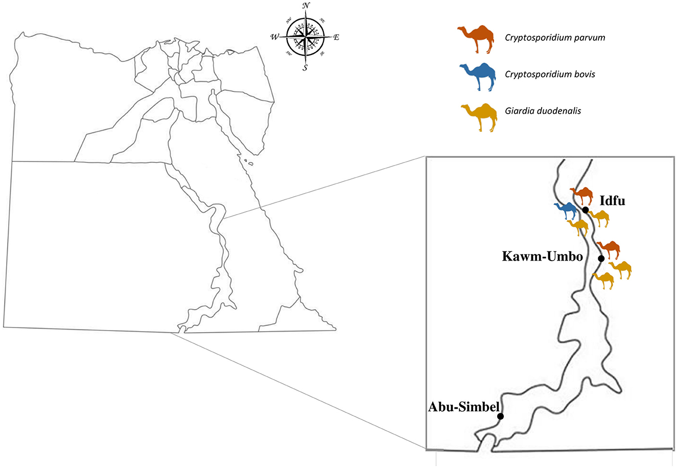
Figure 1. Map of Egypt showing the location of the sampling areas and the distribution of dromedary camels positive to Cryptosporidium spp. and G. duodenalis.
Genomic DNA was isolated from ~200 mg of each fecal sample using the QIAamp DNA Stool Mini Kit (Qiagen, Hilden, Germany) according to the manufacturer's instructions, with the exception that samples combined with InhibitEX buffer were incubated for 10 min at 95°C. DNA samples were extracted and purified before being eluted in 200 μl of PCR-grade water and stored at 4°C until further molecular analysis. A maximum of 18 weeks elapsed between sample collection and DNA extraction.
The presence of Cryptosporidium spp. was assessed using a nested-PCR protocol to amplify a 587-bp fragment of the SSU rRNA gene of the parasite (91). Approximately 3 μl of the DNA sample and 0.3 μM of the primer pairs CR-P1/CR-P2 in the primary reaction and CR-P3/CPB-DIAGR in the secondary reaction were used in the amplification procedures (50 μl) (Supplementary Table 1). Both PCR reactions were carried out as follows: one step of 94°C for 3 min, followed by 35 cycles of 94°C for 40 s, 50°C for 40 s, and 72°C for 1 min, concluding with a final extension of 72°C for 10 min.
Cryptosporidium parvum isolates were sub-typed by amplifying an 870-bp fragment of the gp60 locus using a nested PCR (92). Reaction mixtures (50 μl) contained 2–3 μl of template DNA and 0.3 μM of the primer pairs AL-3531/AL-3535 and AL-3532/AL-3534 in the primary and secondary reactions, respectively (Supplementary Table 1). The PCR protocol for the main reaction consisted of an initial step of 94°C for 5 min, followed by 35 cycles of 94°C for 45 s, 59°C for 45 s, and 72°C for 1 min, with a final extension of 72°C for 10 min. The secondary PCR settings were similar to the initial PCR except for the annealing temperature, which was 50°C.
Detection of G. duodenalis DNA was achieved using a real-time PCR (qPCR) method targeting a 62-bp region of the gene codifying the SSU rRNA of the parasite (93). Amplification reactions (25 μl) consisted of 3 μl of template DNA, 0.5 μM of each primer Gd-80F and Gd-127R, 0.4 μM of probe (Supplementary Table 1), and 12.5 μl TaqMan® Gene Expression Master Mix (Applied Biosystems, CA, USA). The parasite DNA was detected using a Corbett Rotor GeneTM 6000 real-time PCR system (QIAGEN, Hilden, Germany) with an amplification protocol consisting of an initial hold phase of 2 min at 55°C and 15 min at 95°C followed by 45 cycles of 15 s at 95°C and 1 min at 60°C. Samples with qPCR cycle threshold values <32 were re-analyzed at the glutamate dehydrogenase (gdh) (94), β-giardin (bg) (95), and triose phosphate isomerase (tpi) (96) markers using specific PCR protocols to attempt to identify their assemblages and sub-assemblages.
Detection of E. bieneusi was conducted by a nested PCR protocol to amplify the ITS region as well as portions of the flanking large and small subunits of the ribosomal RNA gene, as previously described (97). The outer EBITS3/EBTIS4 and inner EBITS1/EBITS2.4 primer sets (Supplementary Table 1) were used to generate PCR products of 435 and 390 bp, respectively. The main PCR was cycled at 94°C for 3 min, followed by 35 cycles of amplification (denaturation at 94°C for 30 s, annealing at 57°C for 30 s, and elongation at 72°C for 40 s), with a final extension at 72°C for 10 min. Conditions for the secondary PCR were identical to the primary PCR, except that only 30 cycles were performed at an annealing temperature of 55°C.
All of the aforementioned direct and nested PCR protocols were conducted on a 2720 Thermal Cycler (Applied Biosystems). Reaction mixes always included 2.5 units of MyTAQTM DNA polymerase (Bioline GmbH, Luckenwalde, Germany), and 5–10 μl of MyTAQTM Reaction Buffer with 5 mM dNTPs and 15 mM MgCl2. For each parasite species studied, laboratory-confirmed positive and negative DNA samples of human and animal origin were routinely used as controls and included in each round of PCR. PCR amplicons were visualized on 1.5% D5 agarose gels (Conda, Madrid, Spain) stained with Pronasafe (Conda) nucleic acid staining solutions. A 100-bp DNA ladder (Boehringer Mannheim GmbH, Baden-Wurttemberg, Germany) was used to size the obtained amplicons.
Positive-PCR products of the expected size were directly sequenced in both directions using appropriate internal primer sets (Supplementary Table 1). DNA sequencing was conducted by capillary electrophoresis using the BigDye® Terminator chemistry (Applied Biosystems) on an ABI PRISM 3130 automated DNA sequencer. Generated DNA consensus sequences were aligned to appropriate reference sequences using MEGA6 (98) for species confirmation and genotype identification. The sequences obtained in this study have been deposited in GenBank under accession numbers OP365100 (C. bovis) and OP365101–OP365102 (C. parvum).
Fisher's exact tests were used to assess the relationships between parasitic infections and the different independent factors addressed in the study (geographical origin, sex, age, fecal consistency, and physical condition). A P-value of < 0.05 was considered statistically significant. Analyses were conducted using the statistical package SPSS version 25 (IBM Corporation, Armonk, NY, USA).
Giardia duodenalis was the most prevalent species found (3.9%, 4/102; 95% CI: 1.1–9.7), followed by Cryptosporidium spp. (2.9%, 3/102; 95% CI: 0.6–8.4). In contrast, E. bieneusi DNA was not detected in the dromedary camel population under investigation. The distribution of the Cryptosporidium and G. duodenalis infections according to the variables considered in the study is shown in Table 2. Cryptosporidium infections were detected in male animals younger than 5 years age from Edfu and Kom Ombo localities. Two of the three infections were detected in animals that had diarrhea. One of the three cryptosporidiosis-infected animals had emaciation, weakness, and roughened skin. Giardia infections were also detected in male dromedary camels only from Edfu and Kom Ombo localities. In contrast to Cryptosporidium, all Giardia infections were found in animals older than 5 years of age, primarily without diarrhea and in good physical condition. None of the three intestinal protist species proved positive in the dromedary camels sampled at Abu Simbel.
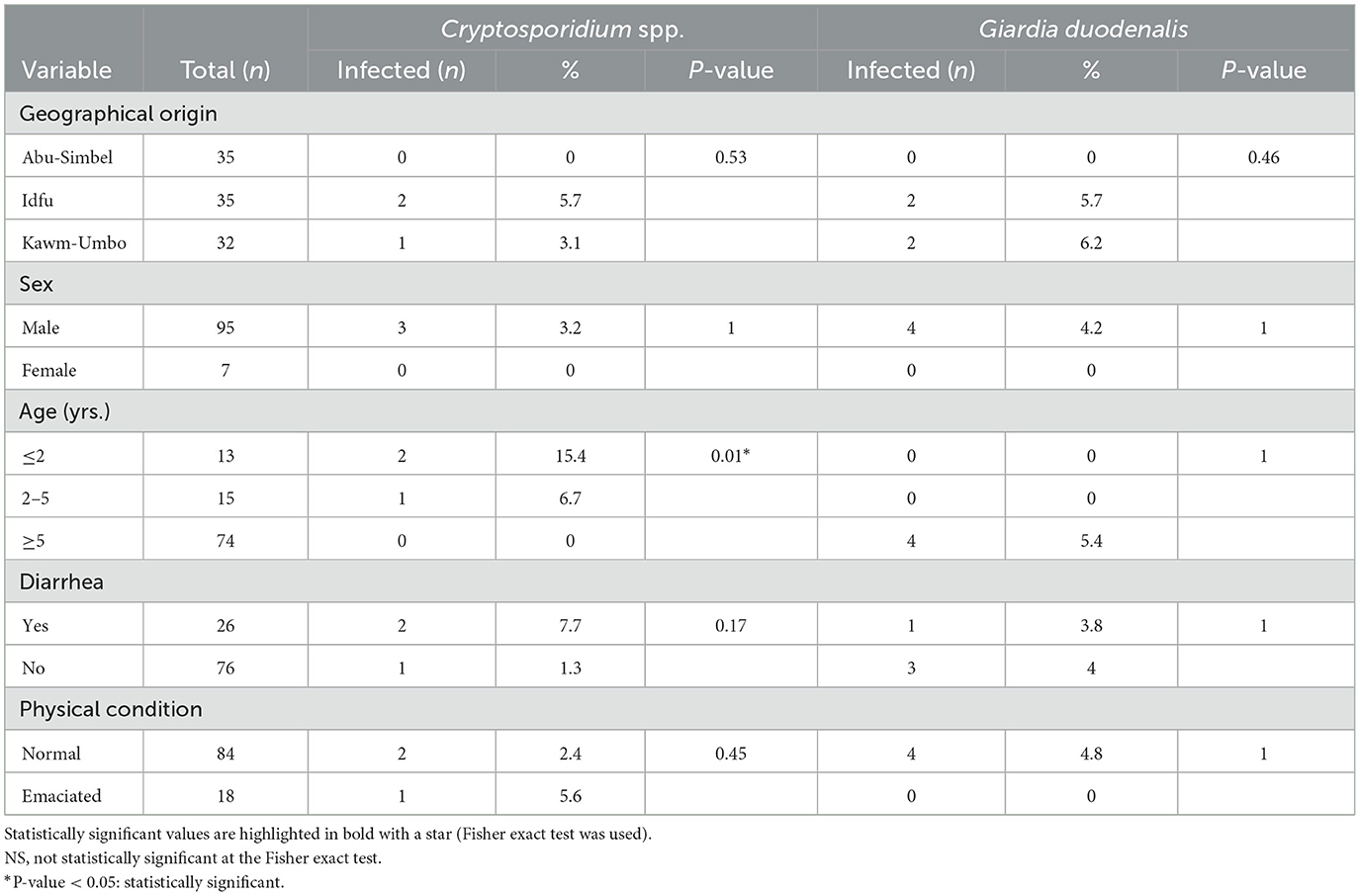
Table 2. Distribution of Cryptosporidium spp. and Giardia duodenalis infections according to geographical origin, sex, age, fecal consistency, and physical condition of examined camels (n = 102).
Dromedary camels younger than 2 years were significantly more likely to be infected by Cryptosporidium spp. than animals of older age (P < 0.05). None of the remaining variables were associated with an increased risk of infection by Cryptosporidium spp. or G. duodenalis.
The results of the Cryptosporidium sequencing analysis generated in the present study are summarized in Table 3. One of the three Cryptosporidium-positive samples was identified as cattle-adapted C. bovis, showing 100% identity with a stretch of 455 bp from position 315–770 of reference sequence AY741305. The remaining two samples were recognized as zoonotic C. parvum, and their sequences varied from reference sequence AF112571 by four to five single nucleotide polymorphisms (SNPs), including a TAAT deletion at positions 686–689 of AF112571. During the visual assessment of chromatograms, no ambiguous positions in the form of double peaks were found. Attempt to amplify the C. parvum isolates at the gp60 locus failed, so the subtype family of the parasite remained unknown.
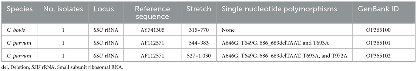
Table 3. Frequency and molecular diversity of Cryptosporidium spp. identified in camels in the present study.
All four G. duodenalis-positive isolates yielded CT values >32 (median: 35.9; range: 32.8–38.5) at qPCR, indicating a relatively low quantity of parasite DNA in the original samples. None of these samples could be amplified at the gdh, bg, and tpi loci.
This study adds to the body of knowledge about the occurrence and genetic diversity of the diarrhea-causing intestinal protists Cryptosporidium spp., G. duodenalis, and E. bieneusi in Egyptian dromedary camels. The main strength of the survey is the use of PCR and Sanger sequencing technologies, allowing for accurate detection, differentiation, and characterization of the investigated pathogens. The survey is also relevant because (i) it focuses on a host species (dromedary camel) for which parasite epidemiological data are particularly scarce in Egypt, (ii) it demonstrates that dromedary camels can act as the potential source of human cryptosporidiosis caused by C. parvum, and (iii) information gathered is useful for developing proper intervention and control strategies against oral–fecal transmitted diseases, including cryptosporidiosis and giardiasis (79, 99).
Cryptosporidium infections were detected in 3% of the investigated dromedary camels. Surprisingly, its incidence percentage was lower (4–24%) than those detected by conventional microscopy in other Egyptian camel populations (33–37, 39, 40). However, a slightly superior rate of 6% was reported in a similar study conducted using PCR-RFLP (38). These disparities between microscopy and PCR data might be attributed to fundamental epidemiological (infection pressure and geographical area) and host (age and immunological state) differences among the camel populations surveyed. However, unwanted false-positive results are prevalent during microscope investigation and might lead to overestimated prevalence rates (100). Similar highly variable Cryptosporidium prevalences have been observed by conventional microscopy or ELISA techniques in various Middle Eastern countries, including Iran (2–100%), Iraq (7–100%), and Saudi Arabia (15–22%; see Table 1). Our genotyping data revealed the presence of two Cryptosporidium species, including C. parvum (in two animals presenting with diarrhea) and C. bovis (in an asymptomatic animal). Cryptosporidium infections have been previously reported in diarrheic dromedary camels in Algeria (27) and Iran (48), whereas C. parvum has already been described in Egyptian dromedary camels (38); this is the first report of cattle-adapted C. bovis in this host species in the country and the third report globally after the description of the parasite in Bactrian camels in China (61, 62). In Egypt, previous research has revealed the occurrence of C. bovis in cattle and buffalo populations (101–105). These findings show that C. bovis cross-species transmission is likely in areas where different domestic ruminant species share habitat. Although the two dromedary camels infected with this Cryptosporidium species manifested diarrhea, we were unable to amplify the two C. parvum isolates at the gp60 locus. The lack of diagnostic data for viral or bacterial agents was an obstacle to unambiguously linking the occurrence of diarrhea with a given enteric pathogen. In this regard, light C. parvum infections associated with modest oocyst shedding might explain the amplification failure at the single copy gp60 gene, a marker known for its limited diagnostic sensitivity (21). Notably, C. parvum gp60 genotype families IIa and IId have been found in Egyptian dromedary camels (38). It should be stressed that C. parvum is regarded as a common zoonotic Cryptosporidium species with loose host specificity and worldwide distribution, whereas human cases of cryptosporidiosis caused by C. bovis are sporadically reported globally (22, 23). Therefore, our molecular data support the potential zoonotic spread of those Cryptosporidium species between infected dromedary camels and humans.
In the present study, G. duodenalis was the predominant (4%) protozoan parasite found among the examined camel population. Conventional microscopy revealed a fairly comparable G. duodenalis infection rate of 5% in the sole prior investigation undertaken on this host species in Egypt (37). Epidemiological information on camel populations in other Middle Eastern countries is also scarce and completely absent in African countries other than Egypt. Prevalence rates of 4–24% have been documented in Iraq (51, 53, 54). The parasite is also known to be circulating at an unknown infection rate in dromedary camels in Saudi Arabia (73). All the previously mentioned studies were based on conventional microscopy, so information on the G. duodenalis assemblages and sub-assemblages causing the infections is also lacking. It is noteworthy that G. duodenalis has been detected at occurrence rates of 7–10% in Chinese Bactrian camels by PCR (61, 64, 65, 74). All these infections were caused primarily by ungulate-adapted G. duodenalis assemblage E and, to a lesser extent, by zoonotic G. duodenalis assemblage A (see Table 1). In our study, the four G. duodenalis-positive samples (three in asymptomatic animals and one in a diarrheic animal) yielded high CT values (>32) at qPCR and impeded the completion of genotyping analyses at appropriate genetic markers, including the genes encoding for the glutamate dehydrogenase (gdh), beta-giardin (bg), and triosephosphate isomerase (tpi) proteins of the parasite. As in the case of the Cryptosporidium gp60 locus, the Giardia gdh, bg, and tpi loci are single-copy genes with limited diagnostic sensitivities, making them unsuitable for amplifying samples with a small amount of parasitic DNA. The high CT values obtained at qPCR are also indicative of light infections, compatible with the absence of gastrointestinal manifestations (diarrhea) in most Giardia-positive dromedary camels. The lack of genotyping data at the assemblage and sub-assemblage levels does not allow us to fully assess the zoonotic implications of our findings. More research should be conducted to ascertain the genetic diversity of G. duodenalis infections in camels and their role as potential sources of human giardiasis.
No DNA of the microsporidia E. bieneusi could be detected in any of the fecal DNA samples analyzed in the present study, suggesting that dromedary camels are not relevant hosts in the transmission of this pathogen in Egypt. Very few epidemiological studies have attempted to investigate the occurrence and genetic diversity of E. bieneusi in camels globally. In the only survey conducted in Africa to date, a PCR prevalence rate of 20% was estimated in Algerian dromedary camels (25). In that study, two E. bieneusi genotypes were detected, including Camel-2 and Macaque1. More information is available from Bactrian camels in China, where E. bieneusi seems to be a common finding with infection rates in the range of 30–45% (61, 64, 65, 75). Most of the infections detected were caused by camel-adapted E. bieneusi genotypes, including CAM1 to CAM6, but the presence of genotypes such as BEB6, EbpA, EbpC, and O (all four members of phylogenetic Groups 1 and 2, including zoonotic genetic variants of the parasite) indicate that Bactrian camels can serve as potential sources of E. bieneusi infections to humans (77, 78). It should be noted that, in Egypt, E. bieneusi has been previously detected in immunosuppressed patients with and without diarrhea (106, 107), in children attending day-care centers (108), and in domestic animals including cattle, buffaloes, rabbits, sheep, goats, cats, and dogs (109). These data highlight the need to investigate the role of other animal host species (including dromedary camels) as potential sources of human microsporidiosis by E. bieneusi in the country.
Regarding the analysis of variables potentially associated with an increased risk of infection by enteric protists, dromedary camels younger than 2 years of age were more likely to be infected by Cryptosporidium spp., this being the only statistically significant association found in the present study. This result is consistent with those obtained in a study that found greater Cryptosporidium infection rates in 1-year-old camel calves than in older animals in Iran (48). Discrepant results have been reported in other surveys. For instance, Cryptosporidium infections were more frequently identified in camels in the age groups of 1–4 years in Algeria (27) and 3–6 years in Iraq (51). A third study that was conducted in Iran revealed no significant associations between camel age and Cryptosporidium infection status (44). Although not statistically significant, all dromedary camels sampled at Abu Simbel tested negative for Cryptosporidium spp., G. duodenalis, and E. bieneusi, suggesting that environmental (e.g., geographical area of origin and local climatic conditions) and biological (e.g., host age and immunological status) conditions and management practices (e.g., contact with other livestock) might play a role in the occurrence and distribution of these pathogens. Taken into account, most of the studied animals were reared in resource-poor settings, including water and food sources, which, together with the management practices, affect the occurrence of the reported pathogens. A lack of access to safe drinking water and poor sanitation and hygiene practices were identified as potential factors linked with a higher risk of developing diarrhoeal illness (15). In relation to feeding habitat, several previous studies have revealed an obvious association between the occurrence of various parasites in camels and grazing performance, including bushes and grasses. In this respect, logging of shrubs, bushes, and trees for rain-fed production systems might enhance the probability of harvesting the ova and/or larvae from pasture (110). Given the above findings, our study pointed out that the application of strict control and hygienic measures represented by providing clean drinking water, improvement of sanitation and hygiene practices are mandatory preventive strategies to control these zoonotic pathogens. Furthermore, regular administration of antiparasitic drugs and treatment of infected camels in the studied area stand as major control measures for the infection and should be adopted, together with the strict quarantine of imported animals from neighboring regions.
Some design and methodological limitations might have biased the accuracy of the results obtained in the present study and should be considered when interpreting them. First, the smaller sample size may have led to underestimating true prevalence rates and lowered the power of the statistical analyses conducted. Second, the transversal nature of the study might not be adequate to capture potential temporal/seasonal variations in parasite occurrence. Third, the animal population under study was mainly composed of adult animals, which are less likely to be infected by the diseases studied. Fourth, suboptimal fecal sample storage and transportation conditions might have altered the quantity and quality of the DNA used for diagnostic and genotyping purposes. Fifth, the lack of genotyping data for some of the protist species investigated (e.g., G. duodenalis) made it difficult to fully analyze the epidemiological and zoonotic implications of our findings.
This is one of the very few molecular-based epidemiological studies aiming at investigating the presence and molecular diversity of diarrhea-causing enteric protist parasites in dromedary camels in African countries, including Egypt. Cryptosporidium spp. and G. duodenalis were identified at low (< 5%) infection rates. Sequence analyses revealed the presence of two Cryptosporidium species, including zoonotic C. parvum and cattle-adapted C. bovis. This is the first report of C. bovis in dromedary camels globally. The presence of C. parvum implies that dromedary camels play a role in the transmission of this Cryptosporidium species and can serve as potential sources of human cryptosporidiosis. Implementation of stricter hygienic measures and awareness raising are recommended to minimize the zoonotic hazard of camel pathogens to people in contact with these animals or their manure. Improving water and food resources in the studied area seems mandatory to reduce the transmission of infection by these zoonotic pathogens. Further research is warranted to corroborate and expand these preliminary findings in larger camel populations and other animal species in Upper Egypt.
The original contributions presented in the study are included in the article/Supplementary material, further inquiries can be directed to the corresponding authors.
The animal study was reviewed and approved by the Research Ethics Committee of the Faculty of Veterinary Medicine, Sohag University (Egypt) on 01.12.2019. Written informed consent was obtained from the owners for the participation of their animals in this study.
EE, SA, AS, AG, ML, BA, and EH collected the samples. EE, PK, AD, CH-C, and BB conducted laboratory experiments. PK and AD conducted sequence analyses. EE conducted statistical analyses. SA and ML secured the funding for conducting sampling and experimental work. EE, DG-B, and DC designed and supervised the experiments. EE and DC writing—original draft preparation. EE, SA, AG, AS, AD, DG-B, and DC writing—review and editing. The final version was read and approved by all authors.
This study was partially funded by the Health Institute Carlos III (ISCIII), Spanish Ministry of Economy and Competitiveness under project PI19CIII/00029. EE is the recipient of a postdoctoral fellowship funded by the Ministry of the Higher Education of the Arab Republic of Egypt. DG-B is the recipient of a Sara Borrell Research Contract (CD19CIII/00011) funded by the Spanish Ministry of Science, Innovation, and Universities. AD is the recipient of a PFIS contract (FI20CIII/00002) funded by the Spanish Ministry of Science and Innovation and Universities. CH-C is the recipient of a fellowship funded by the Fundación Carolina (Spain) and the University of Antioquia, Medellín (Colombia).
The authors declare that the research was conducted in the absence of any commercial or financial relationships that could be construed as a potential conflict of interest.
All claims expressed in this article are solely those of the authors and do not necessarily represent those of their affiliated organizations, or those of the publisher, the editors and the reviewers. Any product that may be evaluated in this article, or claim that may be made by its manufacturer, is not guaranteed or endorsed by the publisher.
The Supplementary Material for this article can be found online at: https://www.frontiersin.org/articles/10.3389/fvets.2023.1139388/full#supplementary-material
1. Squire SA, Ryan U. Cryptosporidium and Giardia in Africa: Current and future challenges. Parasit Vectors. (2017) 10:195. doi: 10.1186/s13071-017-2111-y
2. Santin M. Cryptosporidium and Giardia in ruminants. Vet Clin North Am Food Anim Pract. (2020) 36:223–38. doi: 10.1016/j.cvfa.2019.11.005
3. Hatam-Nahavandi K, Ahmadpour E, Carmena D, Spotin A, Bangoura B, Xiao L. Cryptosporidium infections in terrestrial ungulates with focus on livestock: A systematic review and meta-analysis. Parasit Vectors. (2019) 12:453. doi: 10.1186/s13071-019-3704-4
4. Taghipour A, Sharbatkhori M, Tohidi F, Ghanbari MR, Karanis P, Olfatifar M, et al. Global prevalence of Giardia duodenalis in cattle: A systematic review and meta-analysis. Prev Vet Med. (2022) 203:105632. doi: 10.1016/j.prevetmed.2022.105632
5. Li W, Xiao L. Ecological and public health significance of Enterocytozoon bieneusi. One Health. (2020) 12:100209. doi: 10.1016/j.onehlt.2020.100209
6. Messa A Jr, Köster PC, Garrine M, Gilchrist C, Bartelt LA, Nhampossa T, et al. Molecular diversity of Giardia duodenalis in children under 5 years from the Manhiça district, Southern Mozambique enrolled in a matched case-control study on the aetiology of diarrhoea. PLoS Negl Trop Dis. (2021) 15:e0008987. doi: 10.1371/journal.pntd.0008987
7. Kotloff KL, Nataro JP, Blackwelder WC, Nasrin D, Farag TH, Panchalingam S, et al. Burden and aetiology of diarrhoeal disease in infants and young children in developing countries (the Global Enteric Multicenter Study, GEMS): A prospective, case-control study. Lancet. (2013) 382:209–22. doi: 10.1016/S0140-6736(13)60844-2
8. Fletcher SM, Stark D, Harkness J, Ellis J. Enteric protozoa in the developed world: A public health perspective. Clin Microbiol Rev. (2012) 25:420–49. doi: 10.1128/CMR.05038-11
9. Fayer R, Trout JM, Graczyk TK, Lewis EJ. Prevalence of Cryptosporidium, Giardia and Eimeria infections in post-weaned and adult cattle on three Maryland farms. Vet Parasitol. (2000) 93:103–12. doi: 10.1016/S0304-4017(00)00356-3
10. Abarca N, Santín M, Ortega S, Maloney JG, George NS, Molokin A, et al. Molecular detection and characterization of Blastocystis sp. and Enterocytozoon bieneusi in cattle in Northern Spain. Vet Sci. (2021) 8:191. doi: 10.3390/vetsci8090191
11. Thomson S, Hamilton CA, Hope JC, Katzer F, Mabbott NA, Morrison LJ, et al. Bovine cryptosporidiosis: Impact, host-parasite interaction and control strategies. Vet Res. (2017) 48:42. doi: 10.1186/s13567-017-0447-0
12. Wang R, Li N, Jiang W, Guo Y, Wang X, Jin Y, et al. Infection patterns, clinical significance, and genetic characteristics of Enterocytozoon bieneusi and Giardia duodenalis in dairy cattle in Jiangsu, China. Parasitol Res. (2019) 118:3053–60. doi: 10.1007/s00436-019-06426-3
13. Nydam DV, Wade SE, Schaaf SL, Mohammed HO. Number of Cryptosporidium parvum oocysts or Giardia spp, cysts shed by dairy calves after natural infection. Am J Vet Res. (2001) 62:1612–5. doi: 10.2460/ajvr.2001.62.1612
14. Castro-Hermida JA, Almeida A, González-Warleta M, Correia da Costa JM, Rumbo-Lorenzo C, Mezo M. Occurrence of Cryptosporidium parvum and Giardia duodenalis in healthy adult domestic ruminants. Parasitol Res. (2007) 101:1443–8. doi: 10.1007/s00436-007-0624-6
15. Carmena D. Waterborne transmission of Cryptosporidium and Giardia: detection, surveillance and implications for public health. In: Current Research, Technology and Education Topics in Applied Microbiology and Microbial Biotechnology, ed A. Méndez-Vilas (Badajoz: Formatex) (2010). p. 3–14.
16. Elmahallawy EK, Sadek HA, Aboelsoued D, Aloraini MA, Alkhaldi AAM, Abdel-Rahman SM, et al. Parasitological, molecular, and epidemiological investigation of Cryptosporidium infection among cattle and buffalo calves from Assiut Governorate, Upper Egypt: Current status and zoonotic implications. Front Vet Sci. (2022) 9:899854. doi: 10.3389/fvets.2022.899854
17. Adeyemo FE, Singh G, Reddy P, Stenström TA. Methods for the detection of Cryptosporidium and Giardia: From microscopy to nucleic acid based tools in clinical and environmental regimes. Acta Trop. (2018) 184:15–28. doi: 10.1016/j.actatropica.2018.01.011
18. Khan A, Shaik JS, Grigg ME. Genomics and molecular epidemiology of Cryptosporidium species. Acta Trop. (2018) 184:1–14. doi: 10.1016/j.actatropica.2017.10.023
19. Verweij JJ, Stensvold CR. Molecular testing for clinical diagnosis and epidemiological investigations of intestinal parasitic infections. Clin Microbiol Rev. (2014) 27:371–418. doi: 10.1128/CMR.00122-13
20. Hijjawi N, Zahedi A, Al-Falah M, Ryan U. A review of the molecular epidemiology of Cryptosporidium spp. and Giardia duodenalis in the Middle East and North Africa (MENA) region. Infect Genet Evol. (2022) 98:105212. doi: 10.1016/j.meegid.2022.105212
21. Dacal E, Köster PC, Carmena D. Diagnóstico molecular de parasitosis intestinales. Enferm Infecc Microbiol Clin. (2020) 38:24–31. doi: 10.1016/j.eimc.2020.02.005
22. Ryan UM, Feng Y, Fayer R, Xiao L. Taxonomy and molecular epidemiology of Cryptosporidium and Giardia—A 50 year perspective (1971-2021). Int J Parasitol. (2021) 51:1099–119. doi: 10.1016/j.ijpara.2021.08.007
23. Feng Y, Ryan UM, Xiao L. Genetic diversity and population structure of Cryptosporidium. Trends Parasitol. (2018) 34:997–1011. doi: 10.1016/j.pt.2018.07.009
24. Cardona GA, de Lucio A, Bailo B, Cano L, de Fuentes I, Carmena D. Unexpected finding of feline-specific Giardia duodenalis assemblage F and Cryptosporidium felis in asymptomatic adult cattle in Northern Spain. Vet Parasitol. (2015) 209:258–63. doi: 10.1016/j.vetpar.2015.02.028
25. Baroudi D, Zhang H, Amer S, Khelef D, Roellig DM, Wang Y, et al. Divergent Cryptosporidium parvum subtype and Enterocytozoon bieneusi genotypes in dromedary camels in Algeria. Parasitol Res. (2018) 117:905–10. doi: 10.1007/s00436-017-5734-1
26. Laatamna AK, Belkessa S, Khalil A, Afidi A, Benmahdjouba K, Belalmi R, et al. Prevalence of Cryptosporidium spp. in farmed animals from steppe and high plateau regions in Algeria. Trop Biomed. (2018) 35:724–35.
27. Bouragba M, Laatamna A, Cheddad FE, Baroudi D, Houali K, Hakem A. Gastrointestinal parasites of dromedary camel (Camelus dromedarius) in Algeria. Vet World. (2020) 13:1635–40. doi: 10.14202/vetworld.2020.1635-1640
28. Saidi R, Mimoune N, Chaibi R, Abdelouahed K, Khelef D, Kaidi R, et al. Camel gastrointestinal parasites in southern Algeria. Veterinarska Stanica. (2022) 53:283–94. doi: 10.46419/vs.53.3.7
29. Ouchene N, Khelifi-Touham NA. Prevalence of Cryptosporidium spp. in humans and dromedaries (Camelus dromedarius) in Algeria. Veterinarska Stanica. (2023) 54:2023. doi: 10.46419/vs.54.2.6
30. Zahedi A, Lee GKC, Greay TL, Walsh AL, Blignaut DJC, Ryan UM. First report of Cryptosporidium parvum in a dromedary camel calf from Western Australia. Acta Parasitol. (2018) 63:422–7. doi: 10.1515/ap-2018-0049
31. Gaibova H, Iskenderova N, Hajieva N. Cryptosporidia of the Bactrian camel in Azerbaijan. Proc Azerbaijan Instit Zool. (2011) 2011:347–51.
32. Gu Y, Wang X, Zhou C, Li P, Xu Q, Zhao C, et al. Investigation on Cryptosporidium infections in wild animals in a zoo in Anhui Province. J Zoo Wildl Med. (2016) 47:846–54. doi: 10.1638/2015-0301.1
33. Saleh M, Mahran O. A preliminary study on cryptosporidiosis in dromedary camels at Shalatin Area, Egypt. Assiut Vet Med J. (2007) 53:1–14. doi: 10.21608/avmj.2007.175974
34. El Kelesh EA, Abdel-Maogood SZ, Abdel-Wahab AM. Comparative studies on some Cryptosporidium species infecting different animals. Egypt Vet Med Soc Parasitol J. (2009) 5:19–36.
35. Wahba A, Radwan IGH. Some studies on protozoal parasites of camels in Egypt. Egypt J Comp Path Clinic Path. (2009) 22:41–53.
36. Abdel-Wahab A, Abdel-Maogood S. Identification of Cryptosporidium species infecting camels (Camelus dromedarius) in Egypt. J Am Sci. (2011) 7:609–12.
37. Khedr EA, El-Shanat SK, Fadly RS, Alsokkary MY, Otify YZ. Studies on blood and enteric protozoans infecting camels at Behera Province, Egypt. Egypt Vet Med Soc Parasitol J. (2015) 11:123–30. doi: 10.21608/evmspj.2015.38001
38. El-Alfy ES, Abu-Elwafa S, Abbas I, Al-Araby M, Al-Kappany Y, Umeda K, et al. Molecular screening approach to identify protozoan and trichostrongylid parasites infecting one-humped camels (Camelus dromedarius). Acta Trop. (2019) 197:105060. doi: 10.1016/j.actatropica.2019.105060
39. El-Khabaz KA, Abdel-Hakeem SS, Arfa MI. Protozoan and helminthes parasites endorsed by imported camels (Camel dromedaries) to Egypt. J Parasit Dis. (2019) 43:607–15. doi: 10.1007/s12639-019-01138-y
40. Elshahawy I, AbouElenien F. Seroprevalence of Cryptosporidium and risks of cryptosporidiosis in residents of Sothern Egypt: A cross-sectional study. Asian Pac J Trop Med. (2019) 12:232. doi: 10.4103/1995-7645.259244
41. Abraha A, Urge B, Kemal J, Siyoum T, Tadele M, Muktar Y. Major microbial and parasitic pathogens causing calf diarrhea of dromedary camel in selected areas of eastern Ethiopia. Livestock Res Results. (2020) 1:7834026. doi: 10.1155/2020/7834026
42. Nouri M, Razmyar J, Keyhani P. A Cryptosporidium muris like parasite in large ruminants in various parts of Iran. J Fac Vet Med Univ Tehran. (1995) 50:1–5.
43. Borj H, Razmi G, Movassaghi A, Naghibi A, Maleki M. Prevalence of Cryptosporidium and Eimeria infections in dromedary (Camelus dromedaries) in abattoir of Mashhad, Iran. J Camel Pract Res. (2009) 16:167–70.
44. Razawi SM, Oryan A, Bahrami S, Mohammadalipour A, Gowhari M. Prevalence of Cryptosporidium infection in camels (Camelus dromedarius) in a slaughterhouse in Iran. Trop Biomed. (2009) 26:267–73.
45. Nazifi S, Behzadi MA, Haddadi SH, Raayat Jahromi A, Mehrshad S, Tamadon A. Prevalence of Cryptosporidium isolated from dromedary camels (Camelus dromedarius) in Qeshm Island, Southern Iran. Comp Clin Path. (2010) 19:311–4. doi: 10.1007/s00580-009-0862-3
46. Radfar MH, Aminzadeh Gowhari M. Common gastrointestinal parasites of indigenous camels (Camelus dromedarius) with traditional husbandry management (free-ranging system) in central deserts of Iran. J Parasit Dis. (2013) 37:225–30. doi: 10.1007/s12639-012-0170-8
47. Sazmand A, Rasooli A, Nouri M, Hamidinejat H, Hekmatimoghaddam S. Prevalence of Cryptosporidium spp. in camels and involved people in Yazd Province, Iran. Iran J Parasitol. (2012) 7:80–4.
48. Yakhchali M, Moradi T. Prevalence of Cryptosporidium-like infection in one-humped camels (Camelus dromedarius) of northwestern Iran. Parasite. (2012) 19:71–5. doi: 10.1051/parasite/2012191071
49. Asghari A, Pirestani M, Ebrahimzadeh H, Sadraei J, Mirza-Ghavami M. Detection of Cryptosporidium spp. in one-hump and two-hump (dromedary and Bactrian) camels of Ardabil province, northwest Iran: Implication for zoonosis transmission (2015). In: 9th International and 14th National Congress on Quality Improvement in Clinical Laboratories, Tehran, April 19–22 2016. Tehran.
50. Shahraki FRM. Prevalence of Cryptosporidium parvum in the Cities of Sistan Region by ELISA and Evaluation of Risk Factors of Season, Age and Sex. (2016). Available online at: https://ganj.irandoc.ac.ir (accessed September 10, 2022).
51. Hussin A, Khalaf J, Ali H. Detection of intestinal protozoa in camels and their breeders in Najef, Iraq. Res J Vet Pract. (2015) 3:53–7. doi: 10.14737/journal.rjvp/2015/3.3.53.57
52. Ahmed HS, Hassan A, Mohammed NQ. Detection of Cryptosporidium parvum from feces samples of human and camels by using direct Polymerase Chain Reaction assay technique. Al-Qadisiyah J Vet Med Sci. (2016) 15:59–62.
53. Jawad HH, Jasim GA. Molecular study of Cryptosporidium spp. and Giardia lamblia which cause diarrhea in camels (Camelus dromedarius) in Al-Diwaniyah and Al-Najaf provinces/Iraq Al-Qadisiyah J Vet Med Sci. (2016) 15:70–5.
54. Hasan M, Alani A, Aghwan S. Investigations on gastrointestinal parasites in camels rearing in Nineveh Governorate. Egyptian J Vet Sci. (2021) 52:131–8. doi: 10.21608/ejvs.2020.44519.1192
55. Abd-Al-Aal Z, El-Kabbany A, Tahrani L. Comparison between two diagnostic methods for detection of Cryptosporidium spp. infecting farm animals in Kuwait. Bull Fac Sci Zagazig Univ. (2016) 38:31075. doi: 10.21608/bfszu.2016.31075
56. Al-Megrin WA. Comparison of ELISA and microscopy for detection of Cryptosporidium oocysts in animals. Pak J Biol Sci. (2015) 18:341–5. doi: 10.3923/pjbs.2015.341.345
57. El Wathig M, Faye B. Camel calf diarrhoea in Riyadh Region, Saudi Arabia. J Camel Pract Res. (2016) 23:283–5. doi: 10.5958/2277-8934.2016.00047.3
58. El Hassan E-AM, Al-Jabr OA, El-Bahr SM. Prevalence of Cryptosporidium parvum in diarrheic camel-calves (Camelus dromedarius) in Al-Ahsa, Saudi Arabia. Alex J Vet Sci. (2020) 66:104663. doi: 10.5455/ajvs.104663
59. Wang R, Zhang L, Ning C, Feng Y, Jian F, Xiao L, et al. Multilocus phylogenetic analysis of Cryptosporidium andersoni (Apicomplexa) isolated from a bactrian camel (Camelus bactrianus) in China. Parasitol Res. (2008) 102:915–20. doi: 10.1007/s00436-007-0851-x
60. Liu X, Zhou X, Zhong Z, Deng J, Chen W, Cao S, et al. Multilocus genotype and subtype analysis of Cryptosporidium andersoni derived from a Bactrian camel (Camelus bactrianus) in China. Parasitol Res. (2014) 113:2129–36. doi: 10.1007/s00436-014-3863-3
61. Zhang Q, Zhang Z, Ai S, Wang X, Zhang R, Duan Z. Cryptosporidium spp, Enterocytozoon bieneusi, and Giardia duodenalis from animal sources in the Qinghai-Tibetan Plateau Area (QTPA) in China. Comp Immunol Microbiol Infect Dis. (2019) 67:101346. doi: 10.1016/j.cimid.2019.101346
62. Cao Y, Cui Z, Zhou Q, Jing B, Xu C, Wang T, et al. Genetic diversity of Cryptosporidium in Bactrian camels (Camelus bactrianus) in Xinjiang, Northwestern China. Pathogens. (2020) 9:946. doi: 10.3390/pathogens9110946
63. Wang L, Cao L, Zheng S, Chang Y, Zhang K, Zhang S, et al. Molecular identification and biological characterization of Cryptosporidium muris from camels (Camelus bactrianus) in China. Parasit Vectors. (2021) 14:365. doi: 10.1186/s13071-021-04862-8
64. Wang X, Zhang Z, Yin W, Zhang Q, Wang R, Duan Z. Interactions between Cryptosporidium, Enterocytozoon, Giardia and intestinal microbiota in Bactrian camels on Qinghai-Tibet Plateau, China. Appl Sci. (2021) 11:3595. doi: 10.3390/app11083595
65. Zhang K, Zheng S, Wang Y, Wang K, Wang Y, Gazizova A, et al. Occurrence and molecular characterization of Cryptosporidium spp, Giardia duodenalis, Enterocytozoon bieneusi, and Blastocystis sp. in captive wild animals in zoos in Henan, China. BMC Vet Res. (2021) 17:332. doi: 10.1186/s12917-021-03035-0
66. Morgan UM, Xiao L, Monis P, Sulaiman I, Pavlasek I, Blagburn B, et al. Molecular and phylogenetic analysis of Cryptosporidium muris from various hosts. Parasitology. (2000) 120:457–64. doi: 10.1017/S0031182099005703
67. Ryan U, Xiao L, Read C, Zhou L, Lal AA, Pavlasek I. Identification of novel Cryptosporidium genotypes from the Czech Republic. Appl Environ Microbiol. (2003) 69:4302–7. doi: 10.1128/AEM.69.7.4302-4307.2003
68. Kvac M, Sak B, Kvetonova D, Ditrich O, Hofmannova L, Modry D, et al. Infectivity, pathogenicity, and genetic characteristics of mammalian gastric Cryptosporidium spp. domestic ruminants. Vet Parasitol. (2008) 153:363–7. doi: 10.1016/j.vetpar.2008.01.033
69. Fayer R, Phillips L, Anderson BC, Bush M. Chronic cryptosporidiosis in a Bactrian camel (Camelus bactrianus). J Zoo Wildl Med. (1991) 22:228–32.
70. Xiao L, Escalante L, Yang C, Sulaiman I, Escalante AA, Montali RJ, et al. Phylogenetic analysis of Cryptosporidium parasites based on the small-subunit rRNA gene locus. Appl Environ Microbiol. (1999) 65:1578–83. doi: 10.1128/AEM.65.4.1578-1583.1999
71. Locklear TR, Videla R, Breuer RM, Mulon PY, Passmore M, Mochel JP, et al. Presentation, clinical pathology abnormalities, and identification of gastrointestinal parasites in camels (Camelus bactrianus and Camelus dromedarius) presenting to two North American veterinary teaching hospitals. A retrospective study: 1980-2020. Front Vet Sci. (2021) 8:651672. doi: 10.3389/fvets.2021.651672
72. Radhy AM, Khalaf JM, Faraj A. Some gastro-intestinal protozoa of zoonotic importance observed in captive animals of al-Zawraa zoo in Baghdad. Int J Sci Nat. (2013) 4:567–70.
73. Al-Jabr OA, Mohammed GE, Al-Hamdan BA. Giardiosis in camels (Camelus dromedarius). Vet Rec. (2005) 157:350–2. doi: 10.1136/vr.157.12.350
74. Zhao SS, Li YH, Zhang Y, Zhou Q, Jing B, Xu CY, et al. Multilocus genotyping of Giardia duodenalis in Bactrian camels (Camelus bactrianus) in China. Parasitol Res. (2020) 119:3873–80. doi: 10.1007/s00436-020-06905-y
75. Qi M, Li J, Zhao A, Cui Z, Wei Z, Jing B, et al. Host specificity of Enterocytozoon bieneusi genotypes in Bactrian camels (Camelus bactrianus) in China. Parasit Vectors. (2018) 11:219. doi: 10.1186/s13071-018-2793-9
76. Feng Y, Xiao L. Zoonotic potential and molecular epidemiology of Giardia species and giardiasis. Clin Microbiol Rev. (2011) 24:110–40. doi: 10.1128/CMR.00033-10
77. Foronda P, Bargues MD, Abreu-Acosta N, Periago MV, Valero MA, Valladares B, et al. Identification of genotypes of Giardia intestinalis of human isolates in Egypt. Parasitol Res. (2008) 103:1177–81. doi: 10.1007/s00436-008-1113-2
78. Helmy YA, Klotz C, Wilking H, Krücken J, Nöckler K, Von Samson-Himmelstjerna G, et al. Epidemiology of Giardia duodenalis infection in ruminant livestock and children in the Ismailia province of Egypt: Insights by genetic characterization. Parasit Vectors. (2014) 7:321. doi: 10.1186/1756-3305-7-321
79. Abdel-Moein KA, Saeed H. The zoonotic potential of Giardia intestinalis assemblage E in rural settings. Parasitol Res. (2016) 115:3197–202. doi: 10.1007/s00436-016-5081-7
80. Li W, Feng Y, Santin M. Host specificity of Enterocytozoon bieneusi and public health implications. Trends Parasitol. (2019) 35:436–51. doi: 10.1016/j.pt.2019.04.004
81. Zhang Y, Koehler AV, Wang T, Gasser RB. Enterocytozoon bieneusi of animals-With an ‘Australian twist'. Adv Parasitol. (2021) 111:1–73. doi: 10.1016/bs.apar.2020.10.001
82. Sazmand A, Joachim A, Otranto D. Zoonotic parasites of dromedary camels: So important, so ignored. Parasit Vectors. (2019) 12:610. doi: 10.1186/s13071-019-3863-3
83. Joachim A. Parasitic diseases of camels in Iran (1931-2017)—A literature review. Parasite. (2017) 24:21. doi: 10.1051/parasite/2017024
84. Mochabo MO, Kitala PM, Gathura PB, Ogara WO, Eregae EM, Kaitho TD, et al. The socio-economic impact of important camel diseases as perceived by a pastoralist community in Kenya. Onderstepoort J Vet Res. (2006) 73:269–74.
85. Debela E, Abdulahi B, Megersa B, Kumsa B, Abunna F, Sheferaw D, et al. Hydatidosis of camel (Camelus dromedarius) at Jijiga municipal abattoir, Eastern Ethiopia: prevalence, associated risk factors and financial implication. J Parasit Dis. (2015) 39:730–5. doi: 10.1007/s12639-014-0430-x
86. Salah A, Robertson I, Mohamed AS. Modelling the potential benefits of different strategies to control infection with Trypanosoma evansi in camels in Somaliland. Trop Anim Health Prod. (2016) 48:199–205. doi: 10.1007/s11250-015-0942-0
87. Gareh A, Soliman M, Saleh AA, El-Gohary FA, El-Sherbiny HMM, Mohamed RH, et al. Epidemiological and histopathological investigation of Sarcocystis spp. in slaughtered dromedary camels (Camelus dromedarius) in Egypt. Vet Sci. (2020) 7:162. doi: 10.3390/vetsci7040162
88. Gareh A, Saleh AA, Moustafa SM, Tahoun A, Baty RS, Khalifa RMA, et al. Epidemiological, morphometric, and molecular investigation of cystic echinococcosis in camel and cattle from Upper Egypt: Current status and zoonotic implications. Front Vet Sci. (2021) 8:750640. doi: 10.3389/fvets.2021.750640
89. Ahmed MA, Elmahallawy EK, Gareh A, Abdelbaset AE, El-Gohary FA, Elhawary NM, et al. Epidemiological and histopathological investigation of sarcoptic mange in camels in Egypt. Animals. (2020) 10:1485. doi: 10.3390/ani10091485
90. Salman D, Sivakumar T, Otgonsuren D, Mahmoud ME, Elmahallawy EK, Khalphallah A, et al. Molecular survey of Babesia, Theileria, Trypanosoma, and Anaplasma infections in camels (Camelus dromedaries) in Egypt. Parasitol Int. (2022) 90:102618. doi: 10.1016/j.parint.2022.102618
91. Tiangtip R, Jongwutiwes S. Molecular analysis of Cryptosporidium species isolated from HIV-infected patients in Thailand. Trop Med Int Health. (2002) 7:357–64. doi: 10.1046/j.1365-3156.2002.00855.x
92. Feltus DC, Giddings CW, Schneck BL, Monson T, Warshauer D, McEvoy JM. Evidence supporting zoonotic transmission of Cryptosporidium spp. in Wisconsin. J Clin Microbiol. (2006) 44:4303–8. doi: 10.1128/JCM.01067-06
93. Verweij JJ, Schinkel J, Laeijendecker D, van Rooyen MA, van Lieshout L, Polderman AM. Real-time PCR for the detection of Giardia lamblia. Mol Cell Probes. (2003) 17:223–5. doi: 10.1016/S0890-8508(03)00057-4
94. Read CM, Monis PT, Thompson RC. Discrimination of all genotypes of Giardia duodenalis at the glutamate dehydrogenase locus using PCR-RFLP. Infect Genet Evol. (2004) 4:125–30. doi: 10.1016/j.meegid.2004.02.001
95. Lalle M, Pozio E, Capelli G, Bruschi F, Crotti D, Cacciò SM. Genetic heterogeneity at the beta-giardin locus among human and animal isolates of Giardiaduodenalis and identification of potentially zoonotic subgenotypes. Int J Parasitol. (2005) 35:207–13. doi: 10.1016/j.ijpara.2004.10.022
96. Sulaiman IM, Fayer R, Bern C, Gilman RH, Trout JM, Schantz PM, et al. Triosephosphate isomerase gene characterization and potential zoonotic transmission of Giardia duodenalis. Emerg Infect Dis. (2003) 9:1444–52. doi: 10.3201/eid0911.030084
97. Buckholt MA, Lee JH, Tzipori S. Prevalence of Enterocytozoon bieneusi in swine: An 18-month survey at a slaughterhouse in Massachusetts. Appl Environ Microbiol. (2002) 68:2595–9. doi: 10.1128/AEM.68.5.2595-2599.2002
98. Tamura K, Stecher G, Peterson D, Filipski A, Kumar S. MEGA6: Molecular Evolutionary Genetics Analysis version 60. Mol Biol Evol. (2013) 30:2725–9. doi: 10.1093/molbev/mst197
99. Webster JP, Gower CM, Knowles SC, Molyneux DH, Fenton A. One health—An ecological and evolutionary framework for tackling Neglected Zoonotic Diseases. Evol Appl. (2016) 9:313–33. doi: 10.1111/eva.12341
100. Khurana S, Chaudhary P. Laboratory diagnosis of cryptosporidiosis. Trop Parasitol. (2018) 8:2–7. doi: 10.4103/tp.TP_34_17
101. Helmy YA, Krücken J, Nöckler K, von Samson-Himmelstjerna G, Zessin KH. Molecular epidemiology of Cryptosporidium in livestock animals and humans in the Ismailia province of Egypt. Vet Parasitol. (2013) 193:15–24. doi: 10.1016/j.vetpar.2012.12.015
102. Amer S, Zidan S, Adamu H, Ye J, Roellig D, Xiao L, et al. Prevalence and characterization of Cryptosporidium spp. in dairy cattle in Nile River delta provinces, Egypt. Exp Parasitol. (2013) 135:518–23. doi: 10.1016/j.exppara.2013.09.002
103. Mahfouz ME, Mira N, Amer S. Prevalence and genotyping of Cryptosporidium spp. in farm animals in Egypt. J Vet Med Sci. (2014) 76:1569–75. doi: 10.1292/jvms.14-0272
104. Naguib D, El-Gohary AH, Mohamed AA, Roellig DM, Arafat N, Xiao L. Age patterns of Cryptosporidium species and Giardia duodenalis in dairy calves in Egypt. Parasitol Int. (2018) 67:736–41. doi: 10.1016/j.parint.2018.07.012
105. Abdelaziz AR, Tahoun A, El-Sharkawy H, Abd El-Salam MM, Alorabi M, El-Shehawi AM, et al. Overview on Cryptosporidium bovis and its effect on calves in some governorates in Egypt. J Trop Med. (2022) 2022:4271063. doi: 10.1155/2022/4271063
106. Hammouda NA, Sadaka HA, El-Gebaly WM, El-Nassery SM. Opportunistic intestinal protozoa in chronic diarrhoeic immunosuppressed patients. J Egypt Soc Parasitol. (1996) 26:143–53.
107. Shehab AY, Moneer EA, Allam AF, Khalil SS, Tolba MM. Intestinal Microsporidia infection in leukemic children: Microscopic and molecular detection. Acta Parasitol. (2021) 66:346–53. doi: 10.1007/s11686-020-00283-2
108. Naguib D, Roellig DM, Arafat N, Xiao L. Prevalence and genetic characterization of Enterocytozoon bieneusi in children in Northeast Egypt. Parasitol Res. (2022) 121:2087–92. doi: 10.1007/s00436-022-07546-z
109. Al-Herrawy AZ, Gad MA. Microsporidial spores in fecal samples of some domesticated animals living in Giza, Egypt. Iran J Parasitol. (2016) 11:195–203.
Keywords: epidemiology, genotyping, protists, microsporidia, Zoonoses, transmission
Citation: Elmahallawy EK, Köster PC, Dashti A, Alghamdi SQ, Saleh A, Gareh A, Alrashdi BM, Hernández-Castro C, Bailo B, Lokman MS, Hassanen EAA, González-Barrio D and Carmena D (2023) Molecular detection and characterization of Cryptosporidium spp., Giardia duodenalis, and Enterocytozoon bieneusi infections in dromedary camels (Camelus dromedaries) in Egypt. Front. Vet. Sci. 10:1139388. doi: 10.3389/fvets.2023.1139388
Received: 06 January 2023; Accepted: 24 March 2023;
Published: 20 April 2023.
Edited by:
Alireza Sazmand, Bu-Ali Sina University, IranReviewed by:
Reza Nabavi, Bu-Ali Sina University, IranCopyright © 2023 Elmahallawy, Köster, Dashti, Alghamdi, Saleh, Gareh, Alrashdi, Hernández-Castro, Bailo, Lokman, Hassanen, González-Barrio and Carmena. This is an open-access article distributed under the terms of the Creative Commons Attribution License (CC BY). The use, distribution or reproduction in other forums is permitted, provided the original author(s) and the copyright owner(s) are credited and that the original publication in this journal is cited, in accordance with accepted academic practice. No use, distribution or reproduction is permitted which does not comply with these terms.
*Correspondence: Ehab Kotb Elmahallawy, ZWVoYWFAdW5pbGVvbi5lcw==; David Carmena, ZGFjYXJtZW5hQGlzY2lpaS5lcw==
Disclaimer: All claims expressed in this article are solely those of the authors and do not necessarily represent those of their affiliated organizations, or those of the publisher, the editors and the reviewers. Any product that may be evaluated in this article or claim that may be made by its manufacturer is not guaranteed or endorsed by the publisher.
Research integrity at Frontiers
Learn more about the work of our research integrity team to safeguard the quality of each article we publish.