 Agustín Rebollada-Merino1,2
 Marta Pérez-Sancho1,3*
 Antonio Rodríguez-Bertos1,2
 Nerea García1,3
 Irene Martínez1
 Alejandro Navarro1
 Lucas Domínguez1,3
 Teresa García-Seco1
- 1VISAVET Health Surveillance Centre, Complutense University of Madrid, Madrid, Spain
- 2Department of Internal Medicine and Animal Surgery, Faculty of Veterinary Medicine, Complutense University of Madrid, Madrid, Spain
- 3Department of Animal Health, Faculty of Veterinary Medicine, Complutense University of Madrid, Madrid, Spain
Porcine brucellosis, caused by Brucella suis (B. suis), is a notifiable disease causing significant economic losses in production systems. Most infected pigs may act as carriers and shed B. suis even if asymptomatic. This can contribute to environmental persistence, thus hindering control efforts. Here, the environment and the offspring were investigated during and after a B. suis outbreak at a sow breeding farm. The diagnosis of B. suis in sows (n = 1,140) was performed by culture and polymerase chain reaction (PCR) from vaginal swabs, indirect enzyme-linked immunosorbent assay (I-ELISA) from sera, and brucellin skin test (BST). B. suis diagnosis in post-weaning pigs (n = 899) was performed by I-ELISA in sera and BST. The environmental surveillance programme was implemented by placing gauze sponges (n = 175) pre-hydrated in a surfactant and inactivating liquid for Brucella DNA detection by PCR in different farm areas. Our results showed that the offspring of infected sows reacted to in vivo techniques for B. suis. Furthermore, the offspring born during the outbreak displayed higher seropositivity (I-ELISA) and reactivity (BST) than those pigs born after. Brucella DNA was detected in pregnant sow areas, boxes, boots, and post-weaning pig areas. In addition, Brucella DNA environmental detection was higher during the B. suis outbreak than the post B. suis outbreak. The environmental approach has proven to be a simple, practical, valuable, and safe method to detect and monitor B. suis. These results suggest a role of the environment and the offspring that should be considered in porcine brucellosis surveillance and control programmes.
Introduction
Brucellosis [Brucella abortus (B. abortus), B. melitensis, B. suis] is a notifiable disease according to the World Organization for Animal Health (OIE) (1). Porcine brucellosis is a worldwide-distributed, re-emerging disease caused by B. suis biovars 1, 2, and 3, of which biovar 2 is the most prevalent in domestic swine in Europe (2). B. suis surveillance is mandatory in insemination centers and during exports–imports in the European Union (EU) [Commission Delegated Regulation (EU) 2020/688 of 17 December 2019].
Definitive diagnosis is achieved by bacterial culture and isolation followed by polymerase chain reaction (PCR) confirmation (1), which is a time-consuming approach, limited by laboratory resources, and with a variable sensitivity (3). In vivo diagnosis in domestic swine relies on humoral-based and/or cellular-based techniques. Serological assays comprise the buffered Brucella antigen tests, the complement fixation test, the indirect enzyme-linked immunosorbent assay (I-ELISA), the fluorescence polarization assay, and the competitive enzyme-linked immunosorbent assay. The brucellin skin test (BST) is a cellular-based assay founded on the delayed type IV hypersensitivity reaction to cytosolic and periplasmatic protein extracts inoculated in the skin (4, 5). The complementary use of I-ELISA and BST increases diagnostic accuracy (3, 6).
Brucella suis infection leads to significant economic losses (7). B. suis can cause infertility and reproductive failure at any moment during pregnancy, mainly in the last third (8, 9). Shedding occurs via semen, uterine/vaginal discharges, placenta, and tissues from abortions/dead piglets, as well as in urine and milk, and it is transmitted via direct contact with mucous membranes (mating, perinatal, and throughout ingestion of milk by piglets or aborted remains by sows) (10, 11). However, the pathogenesis and epidemiology of brucellosis in swine are not widely characterized. Most infected pigs may act as asymptomatic carriers and shedders, contributing to the maintenance and spread of the disease in the herd due to the ability of B. suis to survive in the environment (11, 12). Despite this, the role of the environment in the epidemiology of porcine brucellosis is yet to be ascertained.
Porcine brucellosis pre-movement surveillance programme in the EU lays down that the pigs must come from a farm with no cases of brucellosis during the 42 days prior to departure, and where for at least 12 months prior to departure the pigs have been subjected to surveillance for brucellosis using immunological assays demonstrating the absence of brucellosis at a target prevalence of 10% (EU Commission Delegated Regulation 2020/688 of 17 December 2019). Despite the fact that the use of single or multiple diagnostic techniques in sows and boars has been widely studied, there is a lack of research published on the use of diagnostic techniques in young pigs. Thus, the humoral- and cellular-based immune responses in offspring born from B. suis-infected sows are still unknown.
Herein, we present the results of research focused on the environment and offspring during and after a B. suis outbreak at a sow breeding farm, which, to the best of the authors' knowledge, has not been previously evaluated in the context of porcine brucellosis. We sought to research the environmental bacterial DNA distribution and persistence as the control measures take place using B. suis-inactivating surfactant-hydrated sponges that allowed DNA detection. By monitoring post-weaning pigs, we aimed to assess the reactivity to commonly-employed diagnostic techniques and the correlation with the diagnostic results obtained in sows.
Materials and Methods
The diagnostic procedures were carried out at a sow breeding farm (n = 500) with an open production system for a two-year period. Part of the post-weaned pigs was raised for up to 2 months (n = 3,000). The replacement rate was 60%, and the percentage of abortions historically did not exceed 2%. From January 2016 onwards, a gradual rise in reproductive failures, especially in late-term abortions up to 6%, raised suspicions of a reproductive problem compatible with B. suis infection.
In vivo Assays for B. suis Diagnosis in Sows
Brucella suis diagnosis in sows was performed by culture, isolation, and PCR confirmation in vaginal swabs; I-ELISA and BST, in accordance with the Manual of Diagnostic Tests and Vaccines for Terrestrial Animals (1). The diagnosis was grouped in rounds of 3 months to screen the highest number of sows. Therefore, 35% of the farm census (n = 84) was screened during the period the abortions lasted (3 months—first round), and 100% of the farm census was screened twice after the last B. suis-abortion (n = 468, from months 1–5 after the last abortions—second round, and n = 588, from months 6–10 after abortions—third round).
Samples of vaginal swabs (n = 1,140) of sows displaying reproductive failures (including abortions), and also vaginal swabs from sows without reproductive failures (1-week post-delivery), were collected in Amies transport medium (Deltalab, Barcelona, Spain). The vaginal swabs were cultured, and DNA was extracted using a commercial extraction kit (MagMAX CORE Nucleic Acid Purification Kit, Applied Biosystems, Foster City, CA) and an automated extraction robot (KingFisher Flex, Thermo Fisher Scientific). Brucella detection was performed using a previously described PCR protocol (13). A commercial multiplex conventional PCR was used for the identification of B. suis biovars 1 to 5 (INgene Bruce-ladder Suis, Ingenasa, Madrid; Spain). There were negative results by specific PCR techniques after direct extraction of DNA/RNA for swine abortive agents such as porcine reproductive and respiratory syndrome virus (14), porcine herpesvirus types 1 and 2 (15), porcine circovirus type 2 (16), porcine parvovirus type 1 (17), Leptospira interrogans (18), Chlamydia suis (19), and Toxoplasma gondii (20).
To determine the presence of antibodies against Brucella, blood samples were collected. Sera were tested with a commercial I-ELISA kit that detects IgG against Brucella lipopolysaccharide (LPS) (Ingezim Brucella Porcina, Ingenasa, Madrid, Spain). Results were interpreted according to the manufacturer's instructions.
To assess the cellular immune response to Brucella, 0.1 ml of a commercial antigen (Brucellergene OCB, Zoetis, Parsippany-Troy Hills, NJ) was inoculated intradermically in the base of the tail, as described previously (3, 4). A reaction in the inoculation site 48 h post-inoculation, associated with a delayed-type IV hypersensitivity response, was considered positive if inflammation or hemorrhage was present.
In vivo Assays for B. suis Diagnosis in Post-Weaning Pigs
In 2-month-old post-weaned pigs, I-ELISA and BST were performed monthly at seven moments in time: four consecutive samplings in weaned pigs born during the time the abortions lasted and three consecutive samplings in pigs born after the last B. suis-abortion.
To determine the presence of antibodies against Brucella, blood samples were collected. Sera were tested with a commercial I-ELISA kit that detects IgG against Brucella LPS (Ingezim Brucella Porcina, Ingenasa, Madrid, Spain). Results were interpreted according to the manufacturer's instructions.
To assess the cellular immune response to Brucella, 0.1 ml of a commercial antigen (Brucellergene OCB, Zoetis, Parsippany-Troy Hills, NJ) was inoculated intradermically in the base of the tail, as described previously (3, 4). A reaction in the inoculation site 48 h post-inoculation, associated with a delayed-type IV hypersensitivity response, was considered positive if inflammation or hemorrhage was present.
In vitro Assays for the Validation of B. suis Inactivation and Conservation of Bacterial DNA in a New Surfactant Isotonic Liquid for Environmental Samplings
The new surfactant liquid designed for environmental sampling (Spanish patent, number P2115ES00) was obtained by mixing equal parts of solution 1 (isopropyl alcohol 99.8%, ethanol 99.8%, methanol 99.9%, and glycerol) and solution 2 (disodium phosphate, sodium dodecyl sulfate 0.1%, and nuclease-free water). This surfactant liquid has proven to inactivate microorganisms of animal and public health importance as well as to preserve their genetic material for molecular detection tests (21–23).
First, a test was carried out to confirm the surfactant isotonic liquid to inactivate B. suis. Briefly, B. suis colony growth in purity was suspended in a 0.5 McFarland 0.85% sterile saline solution (SS). Afterward, 1 ml of the suspension was dispensed into a tube with 9 ml of sterile 0.85% SS (tube A, viability and purity control, 107 CFU/ml B. suis expected concentration) and two tubes with 9 ml of the surfactant isotonic liquid (tubes B and C). The suspensions were homogenized by vortexing, and the incubation was performed at three moments in time: 10 min, 1, and 24 h. After each of the times, 100 μl of the mass suspension in solid medium Agar Columbia was seeded in each tube in order to evaluate B. suis inactivation. The seeding of each tube at each time was performed in duplicate. In addition, in the case of viability and purity control tube, serial dilutions in phosphate-buffered saline (PBS) were performed on a 1:10 basis to estimate the concentration of the inoculum. Incubation of the plates was carried out in aerobiosis at 37°C for 24 h. After incubation, the plates were read to check whether or not there was bacterial growth. In order to evaluate B. suis DNA preservation in the sampling liquid, samples from tubes A, B, and C after 24 h of incubation were subjected to DNA extraction, purification, and B. suis DNA detection by PCR in accordance with the protocols described in “DNA extraction and real time polymerase chain reaction (PCR)” section.
The performance of the surfactant liquid was compared with buffered peptone water (BPW). BPW is a culture and transport medium commonly used in surface and carcass sampling for isolation and detection of bacterial species such as Salmonella and Listeria. By doing so, once the B. suis outbreak was confirmed, a preliminary environmental sampling was performed on the farm using the surfactant liquid and BPW in parallel.
B. suis DNA Detection in Environmental Samples
To monitor the environmental presence of Brucella, Dry Sponges 3 M (3 M Dry-Sponge; 3 M, Madrid, Spain) were pre-hydrated in the previously cited surfactant and pathogen-inactivating isotonic liquid (15 ml/sponge).
The sponges were randomly placed around the farm facilities in different locations: boxes, pregnant sow areas, post-weaning pig areas, and boots (Table 1). Seven samplings were performed: three consecutive samplings during the time the abortions lasted, and four samplings after the last B. suis-abortion. After sampling, the sponges were preserved at room temperature in a plastic bag ensuring bio-safety.

Table 1. Results for the polymerase chain reaction (PCR) evaluation of environmental samples at different moments in time (months) and in different locations throughout the study.
Environmental samples (from in vitro inactivation assays, from the preliminary assay comparing the surfactant liquid and BPW, and from the environmental sponges embedded in the surfactant liquid) were extracted using a commercial extraction kit (MagMAX CORE Nucleic Acid Purification Kit, Applied Biosystems, Foster City, CA) and an automated extraction robot (KingFisher Flex, Thermo Fisher Scientific). Brucella detection was performed using a previously described PCR protocol (13).
Statistical Analysis
All statistical analyses were performed using IBM SPSS Statistics Software v. 25 (IBM; Armonk, NY, USA). Multiple comparisons of proportions were estimated using Z-test with a Bonferroni adjustment. Comparisons of two proportions were estimated by using Fisher's test. The level of significance was set at p < 0.05. Concordance between diagnostic techniques was evaluated by Cohen's kappa coefficient, according to the following interpretation: 0.0–0.2 insignificant, 0.2–0.4 low, 0.4–0.6 moderate, 0.6–0.8 good, and 0.8–1.0 very good.
Results
Brucella suis biovar 2 was diagnosed as the cause of abortions. Consequently, the farm's veterinary staff implemented a control programme over a 2-year period based on a test culling strategy in sows and post-weaning pigs.
B. suis Diagnosis in Sows
Most vaginal swabs recovered from aborted sows were positive for Brucella (69/84, 82.1%) during the first round (B. suis-abortions) of PCR testing. Vaginal swabs taken during the second and third rounds (after the last B. suis-abortion) were negative for B. suis.
Reproductive failures associated with B. suis occurred mainly during the last third of gestation (37/84, 44.0%), while the first (23/84, 27.4%) and second (24/84, 28.6%) thirds were equally represented. The distribution of the abortions per productive cycle showed an increase in the cases in primiparous sows (25/84, 29.8%) and sows in their second (20/84, 23.8%) and third (14/84, 16.7%) cycles. Abortions were minimal in sows of the fourth (6/84, 7.1%), fifth (7/84, 8.3%), sixth (9/84, 10.7%), and seventh cycles (3/84, 3.6%).
The percentage of I-ELISA-positive sows showed a statistically significant decrease in each round with regard to the previous one: 92.3% (180/195) in the first, 79.7% (373/468) in the second, and 30.4% (179/588) in the third rounds (p < 0.05).
For BST, positive reactions were found in 51.1% (285/558) in the first, 78.6% (368/468) in the second, and 34.3% (202/588) in the third rounds, with statistically significant differences between phases (p < 0.05).
B. suis Diagnosis in Post-Weaning Pigs
The results of I-ELISA showed that pigs born during B. suis abortions displayed higher seropositivity rates (4–30%) than after (0–1%) (Figure 1).
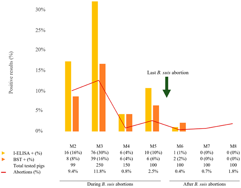
Figure 1. Histogram representing the number of post-weaning pigs, positive or negative to indirect enzyme-linked immunosorbent assay (I-ELISA, yellow) and brucellin skin test (BST, orange), related to total abortions registered in the farm (red line). Results are grouped in pigs born during and after B. suis abortions.
The BST results showed increased rates of positive reactors during (4–16%) than after B. suis abortions (0–2%) (Figure 1). The positive reactor proportion was lower than seropositive pigs throughout the study.
The concordance between both diagnostic techniques was moderate (Cohen's kappa coefficient = 0.522).
B. suis Inactivation and DNA Preservation Using Environmental Sponges
The total inactivation of B. suis after 10 min of contact with the surfactant isotonic liquid was demonstrated, according to the total absence of colonies observed when samples after 10 min, 1 h, and 24 h of contact with the surfactant liquid were cultured in agar Columbia medium, contrasting with a growth corresponding with 107 UFC/ml of control B. suis inoculum re-suspended in 0.85% SS.
Furthermore, PCR results obtained after the duplicate extraction of both tubes inoculated with B. suis in the surfactant liquid and the control tube (B. suis in 0.85% of SS) were equivalent, amplifying at cycles 26–27 in all cases.
The preliminary assay comparing the surfactant liquid and BPW revealed a total of 20 PCR-positive samples (20/26, 76.9%): 14 positives with both methods, 4 positives with the surfactant liquid alone, and 2 positives with BPW alone.
Brucella DNA Detection in the Environment
Brucella DNA was detected in boxes (18/64, 28.1%), pregnant sow areas (22/64, 34.4%), post-weaning pig areas (4/39, 10.3%), and boots (2/8, 25.0%) (Table 1).
Furthermore, Brucella DNA environmental detection was higher during (47.2–68.8%) than after B. suis abortions (0–18.5%) (Table 1).
Discussion
The presence of B. suis in the environment may contribute to the transmission of the disease on farms (8, 12). However, there are few baseline data demonstrating that the environment may represent a component contributing to transmission. Here, the presence of environmental B. suis DNA was determined during and after the outbreak. To the authors' knowledge, this is the first study describing environmental surveillance and B. suis DNA presence at porcine farm facilities.
Brucella culture is challenging, and it is difficult to isolate from environmental samples (24). To solve this, we pre-hydrated gauze sponges in the isotonic surfactant and pathogen-inactivating liquid described to simplify application and enhance bio-safety during transport and sample handling while preserving the DNA. The detection of B. suis DNA indicates the usefulness of this sampling method in brucellosis environmental surveillance. Despite the fact that DNA detection does not allow viable bacteria to be distinguished (25), environmental DNA detection provides valuable information in the context of surveillance programmes in swine (26). In this context, environmental sampling could be a useful tool for assessing B. suis distribution and persistence.
The detection of bacterial or viral nucleic acids using the same pre-hydrated sponge approach has been successfully applied in the environmental detection of such notifiable pathogens as Mycobacterium tuberculosis complex (23), Mycobacterium avium complex (unpublished data), SARS-CoV-2 (21), and African swine fever virus (22). Environmental sampling using methods other than sponges has already proven useful for the detection of slow cultured bacteria such as Mycobacterium tuberculosis complex (27) and Mycoplasma hyopneumoniae (26), and abortive agents such as Chlamydia suis (28) and Coxiella burnetii (29, 30). Environmental detection of Brucella microti-like during an outbreak at a frog farm has also been described (31).
The initial results observed here indicate that B. suis could be detected in the environment, where it may be shed by infected pigs and may be associated with the high prevalence observed. It also constitutes a potential focus of indirect infection to naïve pigs if cleaning and disinfection measures are not properly emphasized. Thus, positive environmental samples were detected months after the last confirmed B. suis abortion, during periods without detection of PCR-positive vaginal swabs, even in locations subject to cleaning and disinfection procedures. Environmental sampling could be a complementary (or even alternative) and non-invasive and safe technique to animal testing in routine screenings in closed loop productions (32). Moreover, unlike vaginal swabs, which are only useful during or shortly after parturition, environmental samples can be used throughout the production cycle, especially in farm zones areas where non-pregnant pigs are raised (25). In fact, B. suis DNA was detected in the post-weaning areas, suggesting that post-weaning pigs may excrete into the environment and may thus contribute to porcine brucellosis perpetuation and the risk of their spread to other farms.
Environmental presence of Brucella DNA progresivelly decreased up to a complete absence 2 years after the outbreak. These results suggested that B. suis was permanently shed until control measures were implemented, and/or that the cleaning and disinfection procedures were not effective in totally removing environmental B. suis. In fact, positive environmental samples continued to be observed long after the last Brucella-associated abortion was detected, so it can be assumed that even if outbreaks are controlled, the pigs may remain at risk of infection for a long period of time if all potential shedders are not diagnosed and sent to slaughter.
We assume that the environmental reduction of DNA Brucella was provided by a reduction in the potential shedders and by an improvement in the cleaning and disinfection protocols and bio-safety. The results obtained in the present study support the need to implement new control measures for porcine brucellosis on farms. Specifically, the use of environmental sampling has proven to be a simple, practical, valuable, and safe method to detect and monitor B. suis DNA persistence at farm facilities, and a suitable system to implement in order to evaluate the efficacy of B. suis control programmes on farms. Our results also highlight the need to carry out successive sampling after a negative result to ensure negative environmental presence, as this may be subject to variations due to different factors (such as shedders and non-sampled areas). Further, controlled experimental studies are necessary to ascertain the role of the environment on B. suis transmission by means of placing naïve pigs into a Brucella-contaminated environment.
Herein, we also aimed to compare how weaned pigs, born in the context of a clinical brucellosis outbreak, reacted to diagnostic techniques in periods with and without abortions caused by B. suis, which, to our knowledge, has not been previously reported. This may be useful to understand the epidemiology of B. suis during outbreaks and the specific roles of post-weaning pigs in porcine brucellosis aside from the immunological response. Our results show that pigs born during the period of time that abortions lasted display higher seropositivity and reactivity than those born after the last B. suis-confirmed abortion (30 vs. 1% positivity for I-ELISA and 16 vs. 2% for BST), suggesting that the immune response of post-weaned pigs correlates with the epidemiological scenario on the farm. The progressive decrease in on-farm infection pressure due to the measures implemented may have prevented both the vertical and horizontal infection of piglets from a certain point in time onwards.
Also, we have observed a reduced percentage of seropositive post-weaning pigs compared with sows. This coincides with previous studies that observed a lower number of seropositive in the progeny compared with females in buffalo (33, 34), bison (35), and domestic cattle (36) infected with Brucella. We found that post-weaning pigs displayed a cellular response to Brucella antigens by means of BST, which to our knowledge has not been evaluated before in the offspring of females infected by any Brucella. The use of BST in post-weaning pigs may help to truly distinguish infections from “false” seropositive piglets due to colostrum intake within the first month of life (37).
The combination of humoral-based and cellular-based diagnostic assays in the context of porcine brucellosis allows us to avoid correlation errors as each technique is biologically independent (2). Here, the moderate concordance (Cohen's kappa coefficient = 0.522) between both tests in post-weaning pigs shows the usefulness of employing both techniques in parallel in order to increase the detection of positive pigs. Concordance is widely variable depending on the epidemiological context, as previously described (38). This study suggests that a representative sampling of post-weaning pigs could serve as an indirect indicator of B. suis infection in sows. This approach could be useful in the national and international commercial trade of young pigs, as the early detection of infected pigs that may act as potential shedders reduces the risk of B. suis dissemination (8, 11).
Data Availability Statement
The original contributions presented in the study are included in the article/supplementary material, further inquiries can be directed to the corresponding author/s.
Ethics Statement
Ethical review and approval was not required for the animal study because the samples were obtained from routinary diagnostic procedures in a porcine farm.
Author Contributions
MP-S, LD, and TG-S: conceptualization. AR-M, AR-B, NG, IM, and AN: methodology. AR-M and TG-S: software and formal analysis. MP-S, AR-B, NG, IM, LD, and TG-S: validation. AR-M, MP-S, AR-B, NG, IM, AN, and TG-S: investigation. LD: resources and visualization. AR-M: writing—original draft preparation. MP-S and TG-S: writing—review and editing. AR-B, LD, and TG-S: supervision. AR-B and LD: funding acquisition. All authors contributed to the article and approved the submitted version.
Funding
This research was partially funded by the Spanish Ministry of Science and Innovation and the Spanish Ministry of Universities (RTI-2018/098658-B-C22) as AR-M was a recipient of a Spanish government-funded Ph.D. contract for research staff training (FPI).
Conflict of Interest
The authors declare that the research was conducted in the absence of any commercial or financial relationships that could be construed as a potential conflict of interest.
Publisher's Note
All claims expressed in this article are solely those of the authors and do not necessarily represent those of their affiliated organizations, or those of the publisher, the editors and the reviewers. Any product that may be evaluated in this article, or claim that may be made by its manufacturer, is not guaranteed or endorsed by the publisher.
Acknowledgments
The authors thank the veterinarians and farm staff for collaborating during the study and English language services provided by Gabinete Lingüístico del Centro de Idiomas (Complutense University of Madrid).
References
1. World Organization for Animal Health. Manual of Diagnostic Tests and Vaccines for Terrestrial Animals. 8th edition. Paris: World Organization for Animal Health (2018).
2. More S, Bøtner A, Butterworth A, Calistri P, Depner K, Edwards S, et al. Assessment of listing and categorisation of animal diseases within the framework of the animal health law [regulation (EU) No 2016/429]: infection with Brucella abortus, B. melitensis and B. suis. EFSA J. (2017) 15:4889. doi: 10.2903/j.efsa.2017.4889
3. Dieste-Pérez L, Blasco JM, De Miguel MJ, Marín CM, Barberán M, Conde-Álvarez R, et al. Performance of skin tests with allergens from B. melitensis B115 and rough B. abortus mutants for diagnosing swine brucellosis. Vet Microbiol. (2014) 168:161–8. doi: 10.1016/j.vetmic.2013.10.024
4. Riber U, Jungersen G. Cell-mediated immune responses differentiate infections with Brucella suis from Yersinia enterocolitica serotype O:9 in pigs. Vet Immunol Immunopathol. (2017) 116:13–25. doi: 10.1016/j.vetimm.2006.12.006
5. Dieste-Pérez L, Barberán M, Muñoz P M, Moriyón I, Blasco JM. Clinical and histological features of brucellin skin test responses in Brucella suis biovar 2 infected pigs. Vet Immunol Immunopathol. (2015) 163:77–85. doi: 10.1016/j.vetimm.2014.11.009
6. Dieste-Pérez L, Blasco JM, de Miguel MJ, Moriyón I, Muñoz PM. Diagnostic performance of serological tests for swine brucellosis in the presence of false positive serological reactions. J Microbiol Meth. (2015) 111:57–63. doi: 10.1016/j.mimet.2015.02.001
7. Risco D, García A, Serrano E, Fernández-Llario P, Benítez JM, Martínez R, et al. High-density dependence but low impact on selected reproduction parameters of Brucella suis Biovar 2 in wild boar hunting estates from south-western Spain. Transbound Emerg Dis. (2013) 61:555–62. doi: 10.1111/tbed.12060
8. Pilo C, Tedde MT, Orrù G, Addis G, Liciardi M. Brucella suis infection in domestic pigs in Sardinia (Italy). Epid Infect. (2014) 143:2170–7. doi: 10.1017/S0950268814003513
9. Zriba S, Garcia-Gonzalez DG, Khalaf OH, Wheeler L, Chaki SP, Rice-Ficht A, et al. Vaccine safety studies of Brucella abortus S19 and S19ΔvjbR in pregnant swine. Vaccine. (2019) 3:100041. doi: 10.1016/j.jvacx.2019.100041
10. Di Sabatino D, Garofolo G, Di Provvido A, Zilli K, Foschi G, Di Giannatale E, et al. Brucella suis biovar 2 multi locus sequence type ST16 in wild boars (Sus scrofa) from Abruzzi region, Italy. Introduction from Central-Eastern Europe? Infect Gen Evol. (2017) 55:63–67. doi: 10.1016/j.meegid.2017.08.031
11. Shome R, Kalleshamurthy T, Natesan K, Jayaprakash K R, Byrareddy K, Mohandoss N, et al. Serological and molecular analysis for brucellosis in selected swine herds from Southern India. J Infect Publ Health. (2018) 12:247–51. doi: 10.1016/j.jiph.2018.10.013
12. Muñoz PM, Mick V, Sacchini L, Janowicz A, de Miguel MJ, Cherfa MA, et al. Phylogeography and epidemiology of Brucella suis biovar 2 in wildlife and domestic swine. Vet Microbiol. (2019) 233:68–77. doi: 10.1016/j.vetmic.2019.04.025
13. Bounaadja L, Albert D, Chénais B, Hénault S, Zygmunt MS, Poliak S, et al. Real-time PCR for identification of Brucella spp.: a comparative study of IS711, bcsp31 and per target genes. Vet Microbiol. (2009) 137:156–64. doi: 10.1016/j.vetmic.2008.12.023
14. Kono Y, Kanno T, Shimizu M, Yamada S, Ohashi S, Nakamine M, et al. Nested PCR for detection and typing of porcine reproductive and respiratory syndrome (PRRS) virus in pigs. J Vet Med Sci. (1996) 58:941–6. doi: 10.1292/jvms.58.10_941
15. Liu XQ, Xu K, Zhang ZY. A touchdown multiplex PCR for porcine circovirus type 2 and pseudorabies virus. Acta Virol. (2012) 56:163–5. doi: 10.4149/av_2012_02_163
16. Hamel AL, Lin LL, Sachvie C, Grudeski E, Nayar GP. PCR detection and characterization of type-2 porcine circovirus. Can J Vet Res. (2000) 64:44–52.
17. Wilhelm S, Zimmermann P, Selbitz HJ, Truyen U. Real-time PCR protocol for the detection of porcine parvovirus in field samples. J Virol Methods. (2006) 134:257–60. doi: 10.1016/j.jviromet.2006.01.004
18. Plata-Luis J, Foronda P, Martín-Alonso A, Feliu C, Alves J, Gil H, et al. Leptospira interrogans in rodents from cape verde. Vector Borne Zoonotic Dis. (2016) 16:731–3. doi: 10.1089/vbz.2016.1989
19. De Puysseleyr K, De Puysseleyr L, Geldhof J, Cox E, Vanrompay D. Development and validation of a real-time PCR for Chlamydia suis diagnosis in swine and humans. PLoS ONE. (2014) 9:e96704. doi: 10.1371/journal.pone.0096704
20. Wang H, Zhang L, Ren Q, Yu F, Yang Y. Diagnosis of swine toxoplasmosis by PCR and genotyping of Toxoplasma gondii from pigs in Henan, Central China. BMC Vet Res. (2017) 13:152. doi: 10.1186/s12917-017-1079-3
21. Fernández-de-Mera IG, Rodríguez del-Río FJ, de la Fuente J, Pérez-Sancho M, Hervás D, Moreno I, et al. Detection of environmental SARS-CoV-2 RNA in a high prevalence setting in Spain. Transb Emerg Dis. (2020) 68:1487–1492. doi: 10.1111/tbed.13817
22. Kososwka A, Barasona JA, Barroso-Arévalo S, Rivera B, Domínguez L, Sánchez-Vizcaíno JM. A new method for sampling African swine fever virus genome and its inactivation in environmental samples. Sci Rep. (2021) 11:21560. doi: 10.1038/s41598-021-00552-8
23. Martínez-Guijosa J, Romero B, Infantes-Lorenzo JA, Díez E, Boadella M, Balseiro A, et al. Environmental DNA: a promising factor for tuberculosis risk assessment in multi-host settings. PLoS ONE. (2020) 15:e0233837. doi: 10.1371/journal.pone.0233837
24. Abril C, Thomann A, Brodard I, Wu N, Ryser-Degiorgis MP, Frey J, et al. A novel isolation method of Brucella species and molecular tracking of Brucella suis biovar 2 in domestic and wild animals. Vet Microbiol. (2011) 150:405–410. doi: 10.1016/j.vetmic.2011.02.056
25. Carrié P, Barry S, Rousset E, de Crémoux R, Sala C, Calavas D, et al. Swab cloths as a tool for revealing environmental contamination by Q fever in ruminant farms. Transbound Emerg Dis. (2019) 66:1202–9. doi: 10.1111/tbed.13137
26. Garza-Moreno L, Vilalta C, Pieters M. Environmental detection of Mycoplasma hyopneumoniae in breed-to-wean farms. Res Vet Sci. (2022) 145:188–92. doi: 10.1016/j.rvsc.2022.02.009
27. Barasona JA, Vicente J, Díez-Delgado I, Aznar J, Gortázar C, Torres MJ. Environmental presence of Mycobacterium tuberculosis complex in aggregation points at the wildlife/livestock interface. Transbound Emerg Dis. (2016) 64:1148–58. doi: 10.1111/tbed.12480
28. De Puysseleyr K, De Puysseleyr L, Dhondt H, Geens T, Braeckman L, Morré SA, et al. Evaluation of the presence and zoonotic transmission of Chlamydia suis in a pig slaughterhouse. BMC Infect Dis. (2014) 14:560. doi: 10.1186/s12879-014-0560-x
29. De Bruin A, Janse I, Koning M, de Heer L, van der Plaats RQJ, van Leuken JPG, et al. Detection of Coxiella burnetii DNA in the environment during and after a large Q fever epidemic in the Netherlands. J Appl Microbiol. (2013) 114:1395–404. doi: 10.1111/jam.12163
30. Kersh GJ, Fitzpatrick KA, Self JS, Priestley RA, Kelly AJ, Lash RR, et al. Presence and persistence of Coxiella burnetii in the environments of goat farms associated with a Q fever outbreak. Appl Envir Microbiol. (2013) 79:1697–703. doi: 10.1128/AEM.03472-12
31. Jaý M, Freddi L, Mick V, Durand B, Girault G, Perrot L, et al. Brucella microti- like prevalence in French farms producing frogs. Transbound Emerg Dis. (2019) 67:617–625. doi: 10.1111/tbed.13377
32. Vilalta C, Sanhueza J, Garrido J, Murray D, Morrison R, Corzo CA, et al. Indirect assessment of porcine reproductive and respiratory syndrome virus status in pigs prior to weaning by sampling sows and the environment. Vet Microbiol. (2019) 237:108406. doi: 10.1016/j.vetmic.2019.108406
33. Wilesmith JW. The persistence of Brucella abortus infection in calves: a retrospective study of heavily infected herds. Vet Res. (1978) 103:149–53. doi: 10.1136/vr.103.8.149
34. Akhtar S, Mirza MA. Rates of seroconversion in the progeny of Brucella abortus seropositive and seronegative cattle and buffalo. Rev Sci Tech. (1995) 14:711–8. doi: 10.20506/rst.14.3.861
35. Rhyan JC, Aune K, Roffe T, Ewalt D, Hennager S, Gidlewski T, et al. Pathogenesis and epidemiology of brucellosis in yellowstone bison: serologic and culture results from adult females and their progeny. J Wildl Dis. (2009) 45:729–39. doi: 10.7589/0090-3558-45.3.729
36. Del Campo MR, Tamayo R, Del Campo CH. A serologic study of the progeny of a seropositive cow. Theriogenol. (1989) 31:1249–51. doi: 10.1016/0093-691X(89)90095-2
37. Rooke J, Bland I. The acquisition of passive immunity in the new-born piglet. Livestock Prod Sci. (2002) 78:13–23. doi: 10.1016/S0301-6226(02)00182-3
Keywords: Brucella suis, brucellin skin test, environment, post-weaning pigs, surveillance
Citation: Rebollada-Merino A, Pérez-Sancho M, Rodríguez-Bertos A, García N, Martínez I, Navarro A, Domínguez L and García-Seco T (2022) Environment and Offspring Surveillance in Porcine Brucellosis. Front. Vet. Sci. 9:915692. doi: 10.3389/fvets.2022.915692
Received: 08 April 2022; Accepted: 11 May 2022;
Published: 21 June 2022.
Edited by:
Peck Toung Ooi, Putra Malaysia University, MalaysiaReviewed by:
Maryam Dadar, Razi Vaccine and Serum Research Institute, IranAman Ullah Khan, University of Veterinary and Animal Sciences, Pakistan
Copyright © 2022 Rebollada-Merino, Pérez-Sancho, Rodríguez-Bertos, García, Martínez, Navarro, Domínguez and García-Seco. This is an open-access article distributed under the terms of the Creative Commons Attribution License (CC BY). The use, distribution or reproduction in other forums is permitted, provided the original author(s) and the copyright owner(s) are credited and that the original publication in this journal is cited, in accordance with accepted academic practice. No use, distribution or reproduction is permitted which does not comply with these terms.
*Correspondence: Marta Pérez-Sancho, bWFwZXJlenNAdWNtLmVz