- 1College of Veterinary Medicine, Northwest A&F University, Yangling, China
- 2Key Laboratory of Animal Biotechnology, Ministry of Agriculture and Rural Affairs, Northwest A&F University, Yangling, China
- 3Yangling Nongfu Agriculture and Animal Husbandry Technology Co., Ltd., Yangling, China
- 4Animal Husbandry Industry Test and Demonstration Center of Shaanxi Province, Xianyang, China
Further optimization of reproduction management programs in dairy cows is a contemporary research topic. In this context, our study aimed to compare a hormone program, named “uterus-ovary monitoring and classified use of hormone program” (M+C), with the Pre-OvSynch program. The M+C was based on regular application of B-mode ultrasonography during a voluntary waiting period to monitor the uterus and ovaries, while using various treatments under different conditions. Results of the 30–33-day and 60-day pregnancy/artificial insemination after the first AI of M+C were significantly better than the Pre-OvSynch (p < 0.05). The pregnancy rates within 180 days in milk after M+C was significantly higher than that after Pre-OvSynch (p < 0.05). The total number of inseminations used for M+C was significantly lower than that for Pre-OvSynch (p < 0.01). The number of open days was fewer after M+C than after the Pre–OvSynch throughout the experimental period with highly significant differences (p < 0.01). In summary, the use of M+C enhances reproductive benefits and reduces the need for hormone drugs among cows.
Introduction
Fertility in dairy cows is a major factor affecting the stable growth of core cattle herds and a sustained increase in milk production. It also has direct effects on the economic benefits of dairy farms (1). Improving cow fertility remains a highly researched issue worldwide as fertility is closely related to many factors, including nutrition (2), genetics (3), environment (4), disease (5), and stress (6), as well as to the implementation and optimization of the relevant reproduction programs.
Since its development, the synchronization of ovulation (OvSynch) and timed artificial insemination (TAI) program (OvSynch+TAI) has significantly reduced the time of the waiting period and the workload of reproductive workers. The Pre-OvSynch, Double-OvSynch, and ReSynch programs (7), all of which are based on the OvSynch program, have been optimized for large-scale batch production management. Nevertheless, some OvSynch+TAI programs have demonstrated practical problems such as long handling time and the number of handlings, usage of hormones, high drug costs, and lack of screening for uterine and ovarian subclinical diseases before beginning the OvSynch protocol.
Endometritis, mainly due to bacterial infection, and ovarian dysfunction, caused by endocrine disorders, is the main reason for low postpartum reproductive function and delayed recovery (7, 8). These conditions severely affect the outcome of OvSynch+TAI and reduce reproductive capability. In addition to synchronization programs, hormone drugs can also be used to treat reproductive disorders (9); for example, gonadotropin-releasing hormone (GnRH) is used to treat ovarian dysfunction caused by ovarian cysts and postpartum anestrus (10). Moreover, prostaglandin F2α (PGF2α), which initiates luteolysis, can be used to treat corpus luteum cysts, and promotes tissue debris excretion and inflammatory secretion from the uterus, thus preventing and resolving endometritis (11). Nevertheless, a recent meta-analysis on the effect of PGF2α in cows undergoing endometritis treatment reported that PGF2α cannot be recommended to treat cows to improve their reproductive performance (12).
Hormone drugs should be used with caution in clinical practice. The causes of reproductive disorders are complicated, and hormone treatment alone does not show optimal curative results. Moreover, the wide use of hormones also results in economic waste. Although the side effects of hormone drugs have long been taken seriously in clinical medicine (13), relevant research in cattle remains insufficient. In dairy farms, the use of routine monitoring and disease detection has been widely applied for effective disease detection and early intervention. Ultrasonography provides unique advantages for examining organ status. With further development, ultrasonography could be accurately and conveniently used to monitor the status of both the uterus (14) and the ovaries (15), making it a practical and effective method for diagnosing endometritis and ovarian diseases (16, 17). Thus, the development and promotion of ultrasonography for disease monitoring demonstrate high potential (18).
Therefore, in this study, in order to effectively diagnose and treat endometritis, reduce the use of hormones, improve the reaction of dairy cows to the OvSynch program before AI, and optimize reproductive efficiency comprehensively, we develop and evaluate a precise hormone program, named the “uterus-ovary monitoring and classified use of hormone program” (M+C), based on regular application of B-mode ultrasonography during a voluntary waiting period to monitor the uterus and ovaries.
Materials and Methods
Cows and Dairy Farm
This experiment was conducted at a commercial dairy farm in Northwest of China, from June to August 2020. At the time of the experiment, this farm had 3,826 Holstein cows, of which 1,708 were lactating; their average parity number was 2.65, and average annual milk yield was 9,134 kg. All cows were fed a TMR three times a day and managed using the herd management system “Aladdin” (Beijing HemuXingbang Network Technology Co., LTD., China). The hardware facilities, diet formulation, staff, and herd structure remained consistent during the experimental period. Within 60 days after delivery in this farm during the whole experiment period, incidence of lameness, ketosis, placental retention, metritis, and milk fever were 5.1, 12.3, 1.9, 2.4, and 4.2%, respectively. After calving, postpartum cows with different parity were randomly divided into two groups, M+C group and Pre-OvSynch group, respectively, and the voluntary waiting period was 60 days for both. Finally, the cows that received the complete M+C (n = 262) or Pre-OvSynch (n = 264) program were included in subsequent statistical analysis. All animal handling procedures were approved by the Committee for the Ethics on Animal Care and Experiments at Northwest A&F University.
M+C and Pre-OvSynch Program
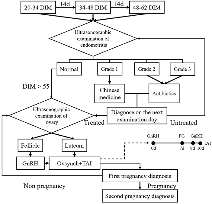
In the M+C program, each cow underwent three regular reproductive organ inspections using B-mode ultrasonography (British BCF, model Easi-scan3) by an experienced technician before artificial insemination (Figure 1). The three examinations were performed at 20–34, 34–48, and 48–62 days in milk (DIM). Subsequently, different treatment protocols were adopted for cows depending on the ovarian cycle phase as examined by B-mode ultrasonography. If there was a mature corpus luteum in the ovary, a OvSynch+TAI protocol was chosen; GnRH (100 μg/stem; Ningbo Sansheng Pharmaceutical, China) was injected first (Day 0), followed by an injection of PGF2α (25 mg/pc, Ningbo Sansheng Pharmaceutical, China) on Day 7, and another GnRH injection on Day 9. If there was a mature follicle in the ovary (follicular phase), a pre-injection of GnRH was given, followed after 7 days by a complete OvSynch+TAI protocol as above. The typical ultrasound imaging findings of the mature corpus luteum were given as uniform gray image of regular boundary with 2–3 cm long and 1.5–2.5 cm wide. Mature follicles range from 1.5 to 2.0 cm in diameter (long axis).

Figure 1. Schematic diagram of the M+C program. Each cow underwent three regular reproductive organ inspections before insemination. The first inspection was performed at 20–34 days in milk (DIM). B-mode ultrasonography was used to examine the uterus and ovary transrectally and to take appropriate measurements under different conditions. After 14 and 28 days, the same examination and treatment were performed again. At 20–34, 34–48, and 48–62 DIM, the images for cow uterus size and abnormal fluid in uterus cavity were classified into four grades: normal and endometritis level I to III; Chinese medicine was used to treat cows with endometritis, and antibiotics were used to treat cows with endometritis level II or III. If a cow was diagnosed with endometritis during examination and treated, we waited 14 days until the next examination for diagnosis. In all cows with endometritis, TAI was performed only after endometritis was resolved. Then, if DIM was >55 at this time, different treatment protocols were adopted for cows without abnormalities depending on their ovary phase. If the corpus luteum was mature with a still developing ovarian follicle in the cow ovary (i.e., the luteal phase), then we applied OvSynch+TAI; if the cow ovary had a mature ovarian follicle (i.e., the follicular phase), GnRH was used first, followed by OvSynch+TAI after 7 days.
In the Pre-OvSynch program, no ultrasonography took place but cows were treated with 25 mg of PGF2α at 14–21 DIM, followed by two 25-mg treatments of PGF2α at 14-day intervals, followed after 11 days by a complete OvSynch+TAI protocol.
Cows were scheduled to receive TAI ~16 h after the last GnRH treatment of the M+C and Pre-OvSynch protocols. The artificial insemination personnel were randomly assigned to three artificial insemination technicians on the dairy farm. The first pregnancy diagnosis through B-mode ultrasonography was performed by experienced veterinarians 30–33 days after TAI to observe signs of pregnancy. The second pregnancy diagnosis was performed 60 days after TAI to determine pregnancy loss. Cows that were not pregnant repeatedly underwent ultrasonographic examination of the ovary in M+C (Figure 1), or perform the OvSynch+TAI program in the Pre-OvSynch group.
B-Mode Ultrasonography for Diagnosis of Endometritis Followed by Classification and Treatment
In this study, uterus size and abnormal uterine fluids at 20–34, 34–48, and 48–62 DIM were examined using B-mode ultrasonography (19, 20), and then classified into four grades based on endometritis occurrence and severity:
1. No endometritis (Normal, Grade 0): No abnormal findings were noted in the uterus (no fluid or echo-free); the cervical diameter was <7.5 cm, and no abnormalities were noted on clinical examination.
2. Endometritis grade I: A small amount of abnormal fluid (discontinuous hyperechogenic, thin <1 mm) was detected in the uterus.
3. Endometritis grade II: A considerable amount of abnormal fluid (continuous hyperechogenic, thin ≥1 mm) was detected in the uterus.
4. Endometritis grade III: A considerable amount of abnormal fluid was detected in the uterus, accompanied by purulent fluid accumulation with some fluctuation (large amount of storage material that looked like a snowstorm).
Clinical examination of the cow tail, perineum, and vulva for purulent secretion in the uterus was also performed. If uterine purulent secretion was detected, the cows were directly classified with endometritis grade III.
For cows with endometritis grade I, Gongyankang (commercial Chinese medicine pills, with active ingredients including Epimedium, Actinolite, Angelica sinensis, Cyperus rotundus, Leonurus japonicus, and Sophora flavescens; 10 capsules/bottle; Jilida, Jilin, China) were administered by uterine infusion. Cows with endometritis grade II or III were given antibiotics (21) (10% oxytetracycline uterine injectant; Eastern Along Pharmaceutical Co., LTD; China). If cows were diagnosed with endometritis, they would receive a course of treatment, and then receive B-mode ultrasonography diagnose on the next examination (14 days later) to determine therapeutic effect. Endometritis was considered completely cured when B-mode ultrasonography and clinical examination were at normal grades. For all cows with endometritis, TAI was only performed when endometritis was cured. The number of cows diagnosed as returning to grade 0 before AI expressed as a percentage of the total number of cows diagnosed with endometritis.
Statistical Analysis
All data are presented as the means ± standard error of the mean (SEM) and analyzed using SPSS (version 24.0). Logistic regression was used to analyze the pregnancy rates at 30–33 and 60 days after the first AI. Survival analysis with right censoring was used to analyze pregnancy rates within 180 DIM. The chi-square test was used to analyze the incidence and rate of endometritis. One-way analysis of variance was used to analyze the number of frozen semen straws, the cost of drugs, hormones, and frozen semen. Differences were considered significant when p < 0.05.
Results
Effects of M+C and Pre–OvSynch Program on P/AI After First AI of Post-partum Cows
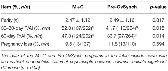
As shown in Table 1, no significant difference in parity was observed between the two programs (p > 0.05). The 30–33 and 60-day P/AI of M+C were significantly higher than those of Pre-OvSynch (p < 0.05). In addition, the pregnancy loss was not significantly different between the two programs (p > 0.05). The above results indicated that M+C might significantly improve P/AI after the first AI of postpartum cows.

Table 1. The parity, 30–33-day P/AI, 60-day P/AI after first AI, and pregnancy loss of postpartum cows treated with the M+C and Pre-OvSynch program.
Pregnancy Rates Within 180 DIM, Number of Frozen Semen, and Cow Open Days With M+C and Pre–OvSynch Program
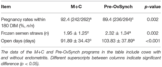
The pregnancy rates within 180 DIM after M+C was significantly higher than that after the Pre–OvSynch program (Table 2, p < 0.05). The total number of frozen semen used for M+C was significantly lower than that of Pre–OvSynch (Table 2, p < 0.05). Meanwhile, the number of open days was significantly lower in M+C than in Pre–OvSynch (Table 2, p < 0.05). These results suggest that the application of M+C could significantly reduce the number of frozen semen straws and the number of open days.

Table 2. The pregnancy rates within 180 DIM, number of frozen semen, and cow open days of the M+C and Pre–OvSynch program.
Ultrasonography-Based Diagnosis of Endometritis and Ovaries in M+C Program
Ultrasonography for the diagnosis of endometritis in M+C was performed at 20–34, 34–48, and 48–62 days, respectively (Table 3). Approximately 15% of cows were diagnosed with endometritis of grade I at 20–34 days postpartum, whereas the diagnostic rates gradually declined at 34–48 (about 7%) and 48–62 days (about 3%), respectively. Meanwhile, the total diagnostic rates of endometritis in grades II and III at 20–34 days postpartum were 6.5% and 6.1%, respectively, and the diagnostic rates also gradually declined at 34–48 and 48–62 days.
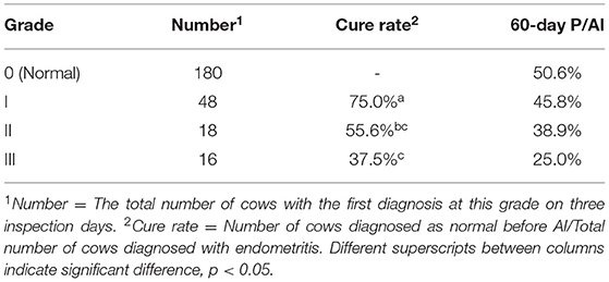
To evaluate the effect of treatment of cases with different grades of endometritis, the cure rate and 60-day P/AI after the first AI of cows were analyzed (Table 4). As the endometritis grade increased, the cure rate and 60-day P/AI after the first AI decreased; the cure rate for endometritis grade I was significantly higher than that for endometritis grade III (p < 0.05). However, no significant differences were detected in 60-day P/AI after the first AI among the three endometritis grades (p > 0.05).
The percentages of corpus luteum and ovarian follicles in the ovary at the first implementation of the OvSynch program in M+C were 82.4 and 17.6%, respectively, with those of 60-day P/AI after the first AI being 44.9 and 58.7%, respectively (Table 5, p > 0.05).

Table 5. Ultrasonography-based diagnosis of ovaries at the first implementation of the OvSynch program and 60-day P/AI after the first AI in M+C program.
Cost of Drugs, Hormone, and Frozen Semen in M+C vs. Pre–OvSynch in 180 Days
As shown in Table 6, in addition to the extra drug cost in M+C, the average cost of hormone and frozen semen was significantly lower in M+C than in Pre-OvSynch (p < 0.05).
Discussion
Increased pregnancy rate, reduced number of frozen semen straws used, and shortened number of open days are ideal attributes for dairy farms. At the same time, reducing the workload of reproductive workers in farms is also essential. Accordingly, the development and application of OvSynch+TAI has achieved remarkable results (22). Further optimization of synchronization programs has been a dynamic topic of research, mainly focusing on increasing hormonal management of reproduction (23), evaluating pregnancy diagnosis time (24), using novel drugs (25), enhancing estrus and hormone monitoring (26), and monitoring the ovary cycle (27). It is well-known that the OvSynch program provides the best results when it is started at day 5–9 of the estrous cycle, and the Pre-OvSynch program was developed for this purpose, but increasing hormone use. Nevertheless, increased hormone dosage and frequency may reduce personnel working enthusiasm and potentially lead to negative effects. For example, Pre-OvSynch may increase the proportion of male calves (28).
Funakura et al. (29) reported that using a portable ultrasound device to objectively evaluate the presence or absence of a functional corpus luteum could achieve a higher pregnancy rate than the conventional AI method based on estrus detection after administration of PGF alone in beef herds. Silva et al. (30) also concluded that assessing the corpus luteum functionality of Holstein cows using ultrasound at the first GnRH injection of Pre-OvSynch protocol could resolve the anovular condition before TAI and thus improve reproductive performance. In addition, using the synchronization program based on functionality of the corpus luteum as determined by ultrasonography may be a viable alternative to OvSynch, with an additional benefit of having lower costs (31). Similarly, in the current study, before performing the synchronization programs, we first examined the ovaries using B-mode ultrasonography. If a corpus luteum with a developing dominant follicle was noted on days 5–12 of the estrus cycle, the cow was considered to have high sensitivity to GnRH, and the OvSynch+TAI procedure was performed directly. If mature follicles were present, GnRH was used first to promote follicular expulsion and formation of a new corpus luteum, followed by the OvSynch program 7 days later to induce a new follicular cycle and ovulation. The results showed that compared with the Pre–OvSynch, M+C significantly increased the P/AI of cows, reduced the number of frozen semen straws required, shortened the number of open days, and reduced the usage of hormone drugs.
The incidence of endometritis, which affects the internal environment of the uterus and destroys normal ovary function, resulting in reduced postpartum fertility in cows, can reach up to 40% (32). Endometritis can be divided into clinical and subclinical types, with the former demonstrating typical clinical signs (33), while the latter usually requires endometrial cytology, biopsy, and leukocyte esterase testing to confirm the diagnosis (34), all of which are difficult to implement in dairy farms. Currently, endometritis is usually diagnosed after 21 DIM once in farms (16, 20); however, because ultrasonography inspection can be susceptible to the probe position and orientation and the uterine state of cows (35), it cannot be effectively used for diagnosing subclinical cases where no secretions are observed in the uterus (20). Hence, selecting multiple time points for checking can help improve sensitivity. PGF2α and intrauterine infusion of antibiotics are commonly used for endometritis treatment, but their effectiveness remains controversial (12, 36); however, the use of some herbal extracts has been investigated in the treatment of endometritis because they have antibacterial and anti-inflammatory characteristics, cause less irritability, and do not require milk to be discarded (12, 37). Based on the above reasons, in the current study, the M+C program referred and optimized the diagnosis method of endometritis used by Lenz et al. (19) and Kasimanickam et al. (17). The results showed a high incidence of endometritis at all grades in this trial within 20–34 days postpartum, with an overall diagnostic rate of 26.3%. The cure rate for endometritis grade I was significantly higher than that for endometritis grade II and III, and may be due to L. japonicus and S. flavescens as the main ingredients of Gongyankang have been reported to have a beneficial anti-inflammatory effect on endometritis (38, 39). However, our outcome for endometritis II and III treatment is still not ideal, and follow-up studies focusing on improving the treatment of severe endometritis are warranted. The M+C exhibits two advantages: first, endometritis cases were detected in a timely and effective manner through three regular examinations of the uterus before insemination. The effect of the treatment could be assessed on the next examination day, ensuring that the internal environment of the uterus is improved as much as possible before ovulation synchronization is performed. Second, cows categorized as having either mild or severe endometritis could be treated more specifically using different drugs, thus avoiding large-scale, blind use of hormone drugs, reducing drug costs, and achieving satisfactory results.
The measurement of economic benefits is an important part of evaluating the effectiveness of any program. In the evaluation of the optimized OvSynch+TAI program, the measurement of economic benefits usually requires a large amount of data and mathematical modeling, and based on preliminary analyses, compared with the Pre–OvSynch, M+C could reduce the amount of hormone drugs and frozen semen used; however, the costs of treatment drugs and B-mode ultrasonography were increased. In the future, we aim to continually improve and promote M+C and collect data to completely evaluate the cost of the different echographic examinations and the required time to make such examinations.
Conclusions
The advantages of using M+C include effective endometritis detection, classification, and treatment and precise hormone administration according to the ovary cycle. These enhance reproductive benefits and reduce the need for hormone drugs in cows. Thus, M+C is convenient and feasible with value for promotion and application.
Data Availability Statement
The original contributions presented in the study are included in the article/supplementary material, further inquiries can be directed to the corresponding author/s.
Ethics Statement
The animal study was reviewed and approved by Committee for the Ethics on Animal Care and Experiments in Northwest A&F University (Approval ID: 2011ZX08008-002). Written informed consent was obtained from the owners for the participation of their animals in this study.
Author Contributions
BW, YJin, and PL: conceptualization and resources. BW: methodology. JX and YM: software. CG, YJia, and HL: validation. JX: formal analysis. BW and JX: investigation. JX and PL: writing—original draft preparation. BW, JX, YM, CG, HL, YJia, YJin, and PL: writing—review and editing. YJin and PL: supervision. All authors contributed to the article and approved the submitted version.
Funding
This research was funded by the Key Research and Development (R&D) program in Ningxia Hui Autonomous Region (2021BBF02037; 2018BBF33001) and the Science and Technology Innovation Project of Shaanxi Province.
Conflict of Interest
BW was employed by Yangling Nongfu Agriculture and Animal Husbandry Technology Co., Ltd.
The remaining authors declare that the research was conducted in the absence of any commercial or financial relationships that could be construed as a potential conflict of interest.
Publisher's Note
All claims expressed in this article are solely those of the authors and do not necessarily represent those of their affiliated organizations, or those of the publisher, the editors and the reviewers. Any product that may be evaluated in this article, or claim that may be made by its manufacturer, is not guaranteed or endorsed by the publisher.
References
1. Rutten CJ, Steeneveld W, Inchaisri C, Hogeveen H. An ex ante analysis on the use of activity meters for automated estrus detection: to invest or not to invest? J Dairy Sci. (2014) 11:6869–87. doi: 10.3168/jds.2014-7948
2. Rodney RM, Celi P, Scott W, Breinhild K, Santos J, Lean IJ. Effects of nutrition on the fertility of lactating dairy cattle. J Dairy Sci. (2018) 6:5115–33. doi: 10.3168/jds.2017-14064
3. Lucy MC. Symposium review: selection for fertility in the modern dairy cow-current status and future direction for genetic selection. J Dairy Sci. (2019) 4:3706–21. doi: 10.3168/jds.2018-15544
4. Craig H, Stachowicz K, Black M, Parry M, Burke CR, Meier S, et al. Genotype by environment interactions in fertility traits in New Zealand dairy cows. J Dairy Sci. (2018) 12:10991–1003. doi: 10.3168/jds.2017-14195
5. Chaters G, Rushton J, Dulu TD, Lyons NA. Impact of foot-and-mouth disease on fertility performance in a large dairy herd in Kenya. Prev Vet Med. (2018) 2018:57–64. doi: 10.1016/j.prevetmed.2018.08.006
6. Gernand E, Konig S, Kipp C. Influence of on-farm measurements for heat stress indicators on dairy cow productivity, female fertility, and health. J Dairy Sci. (2019) 7:6660–71. doi: 10.3168/jds.2018-16011
7. Nyabinwa P, Kashongwe OB, Hirwa CD, Bebe BO. Effects of endometritis on reproductive performance of zero-grazed dairy cows on smallholder farms in Rwanda. Anim Reprod Sci. (2020) 2020:106584. doi: 10.1016/j.anireprosci.2020.106584
8. Dahiya S, Kumari S, Rani P, Onteru SK, Singh D. Postpartum uterine infection & ovarian dysfunction. Indian J Med Res. (2018) 148:S64–70. doi: 10.4103/ijmr.IJMR_961_18
9. Gundling N, Feldmann M, Hoedemaker M. [Hormonal treatments for fertility disorders in cattle]. Tierarztl Prax Ausg G Grosstiere Nutztiere. (2012) 4:255–63. doi: 10.1055/s-0038-1623121
10. Sterry RA, Silva E, Kolb D, Fricke PM. Strategic treatment of anovular dairy cows with GNRH. Theriogenology. (2009) 3:534–42. doi: 10.1016/j.theriogenology.2008.08.020
11. Yu GM, Bai JH, Liu Y, Maeda T, Zeng SM. A weekly postpartum pgf2alpha protocol enhances uterine health in dairy cows. Reprod Biol. (2016) 4:295–9. doi: 10.1016/j.repbio.2016.10.006
12. Haimerl P, Heuwieser W, Arlt S. Short communication: meta-analysis on therapy of bovine endometritis with prostaglandin f2alpha-an update. J Dairy Sci. (2018) 11:10557–64. doi: 10.3168/jds.2018-14933
13. Darendeliler F. Growth and growth hormone: recent papers on efficacy and adverse effects of growth hormone and world health organization growth standards. J Pediatr Endocrinol Metab. (2018) 1:1–3. doi: 10.1515/jpem-2017-0531
14. Rizzo A, Gazza C, Silvestre A, Maresca L, Sciorsci RL. Scopolamine for uterine involution of dairy cows. Theriogenology. (2018) 2018:35–40. doi: 10.1016/j.theriogenology.2018.08.025
15. Hanzen C, Pieterse M, Scenczi O, Drost M. Relative accuracy of the identification of ovarian structures in the cow by ultrasonography and palpation per rectum. Vet J. (2000) 2:161–70. doi: 10.1053/tvjl.1999.0398
16. Meira EJ, Henriques LC, Sa LR, Gregory L. Comparison of ultrasonography and histopathology for the diagnosis of endometritis in holstein-friesian cows. J Dairy Sci. (2012) 12:6969–73. doi: 10.3168/jds.2011-4950
17. Kasimanickam R, Duffield TF, Foster RA, Gartley CJ, Leslie KE, Walton JS, et al. Endometrial cytology and ultrasonography for the detection of subclinical endometritis in postpartum dairy cows. Theriogenology. (2004) 1–2:9–23. doi: 10.1016/j.theriogenology.2003.03.001
18. Smith RF, Oultram J, Dobson H. Herd monitoring to optimise fertility in the dairy cow: making the most of herd records, metabolic profiling and ultrasonography (research into practice). Animal. (2014) 2014:185–98. doi: 10.1017/S1751731114000597
19. Lenz M, Drillich M, Heuwieser W. [Evaluation of the diagnosis of subclinical endometritis in dairy cattle using ultrasound]. Berl Munch Tierarztl Wochenschr. (2007) 5–6:237–44.
20. Koyama T, Omori R, Koyama K, Matsui Y, Sugimoto M. Optimization of diagnostic methods and criteria of endometritis for various postpartum days to evaluate infertility in dairy cows. Theriogenology. (2018) 2018:225–32. doi: 10.1016/j.theriogenology.2018.07.002
21. Sheldon IM, Noakes DE. Comparison of three treatments for bovine endometritis. Vet Rec. (1998) 21:575–9. doi: 10.1136/vr.142.21.575
22. Pursley JR, Mee MO, Wiltbank MC. Synchronization of ovulation in dairy cows using pgf2alpha and GNRH. Theriogenology. (1995) 7:915–23. doi: 10.1016/0093-691X(95)00279-H
23. Karakaya-Bilen E, Ribeiro ES, Bisinotto RS, Gumen A, Santos J. Effect of presynchronization with prostaglandin f2alpha before the 5-d timed AI protocol on ovarian responses and pregnancy in dairy heifers. Theriogenology. (2019) 2019:138–43. doi: 10.1016/j.theriogenology.2019.03.019
24. Pereira RV, Caixeta LS, Giordano JO, Guard CL, Bicalho RC. Reproductive performance of dairy cows resynchronized after pregnancy diagnosis at 31 (+/-3 days) after artificial insemination (AI) compared with resynchronization at 31 (+/-3 days) after AI with pregnancy diagnosis at 38 (+/-3 days) after AI. J Dairy Sci. (2013) 12:7630–9. doi: 10.3168/jds.2013-6723
25. de Lima RS, Martins T, Lemes KM, Binelli M, Madureira EH. Effect of a puberty induction protocol based on injectable long acting progesterone on pregnancy success of beef heifers serviced by TAI. Theriogenology. (2020) 154:128–34. doi: 10.1016/j.theriogenology.2020.05.036
26. Madureira A, Polsky LB, Burnett TA, Silper BF, Soriano S, Sica AF, et al. Intensity of estrus following an estradiol-progesterone-based ovulation synchronization protocol influences fertility outcomes. J Dairy Sci. (2019) 4:3598–608. doi: 10.3168/jds.2018-15129
27. Sauls-Hiesterman JA, Voelz BE, Stevenson JS. A shortened resynchronization treatment for dairy cows after a non-pregnancy diagnosis. Theriogenology. (2020) 2020:105–12. doi: 10.1016/j.theriogenology.2019.09.013
28. Youssefi R, Vojgani M, Gharagozlou F, Akbarinejad V. More male calves born after presynch-ovsynch protocol with 24-hour timed AI in dairy cows. Theriogenology. (2013) 5:890–4. doi: 10.1016/j.theriogenology.2013.01.007
29. Funakura H, Shiki A, Tsubakishita Y, Mido S, Katamoto H, Kitahara G, et al. Validation of a novel timed artificial insemination protocol in beef cows with a functional corpus luteum detected by ultrasonography. J Reprod Dev. (2018) 2:109–15. doi: 10.1262/jrd.2017-135
30. Silva E, Sterry RA, Fricke PM. Assessment of a practical method for identifying anovular dairy cows synchronized for first postpartum timed artificial insemination. J Dairy Sci. (2007) 7:3255–62. doi: 10.3168/jds.2006-779
31. McArt JA, Caixeta LS, Machado VS, Guard CL, Galvao KN, Sa FO, et al. Ovsynch versus ultrasynch: reproductive efficacy of a dairy cattle synchronization protocol incorporating corpus luteum function. J Dairy Sci. (2010) 6:2525–32. doi: 10.3168/jds.2009-2930
32. Sheldon IM, Molinari P, Ormsby T, Bromfield JJ. Preventing postpartum uterine disease in dairy cattle depends on avoiding, tolerating and resisting pathogenic bacteria. Theriogenology. (2020) 150:158–65. doi: 10.1016/j.theriogenology.2020.01.017
33. LeBlanc SJ, Duffield TF, Leslie KE, Bateman KG, Keefe GP, Walton JS, et al. Defining and diagnosing postpartum clinical endometritis and its impact on reproductive performance in dairy cows. J Dairy Sci. (2002) 9:2223–36. doi: 10.3168/jds.S0022-0302(02)74302-6
34. Wagener K, Gabler C, Drillich M. A review of the ongoing discussion about definition, diagnosis and pathomechanism of subclinical endometritis in dairy cows. Theriogenology. (2017) 94:21–30. doi: 10.1016/j.theriogenology.2017.02.005
35. Barlund CS, Carruthers TD, Waldner CL, Palmer CW. A comparison of diagnostic techniques for postpartum endometritis in dairy cattle. Theriogenology. (2008) 6:714–23. doi: 10.1016/j.theriogenology.2007.12.005
36. Kasimanickam R, Cornwell JM, Nebel RL. Effect of presence of clinical and subclinical endometritis at the initiation of presynch-ovsynch program on the first service pregnancy in dairy cows. Anim Reprod Sci. (2006) 3–4:214–23. doi: 10.1016/j.anireprosci.2005.10.007
37. Heuwieser W, Tenhagen BA, Tischer M, Luhr J, Blum H. Effect of three programmes for the treatment of endometritis on the reproductive performance of a dairy herd. Vet Rec. (2000) 12:338–41. doi: 10.1136/vr.146.12.338
38. Wu H, Dai A, Chen X, Yang X, Li X, Huang C, et al. Leonurine ameliorates the inflammatory responses in lipopolysaccharide-induced endometritis. Int Immunopharmacol. (2018) 61:156–61. doi: 10.1016/j.intimp.2018.06.002
Keywords: reproduction program, ultrasonography, endometritis, hormone, dairy cow
Citation: Wang B, Xiao J, Ma Y, Gao C, Li H, Jia Y, Jin Y and Lin P (2022) Comparison of the Evaluation of Combination of Ultrasonography of the Reproductive Tract With Hormone Administration on Dairy Cow Fertility. Front. Vet. Sci. 9:840724. doi: 10.3389/fvets.2022.840724
Received: 21 December 2021; Accepted: 31 January 2022;
Published: 15 March 2022.
Edited by:
Kangfeng Jiang, Yunnan Agricultural University, ChinaReviewed by:
Christian Hanzen, University of Liège, BelgiumAsghar Mogheiseh, Shiraz University, Iran
Copyright © 2022 Wang, Xiao, Ma, Gao, Li, Jia, Jin and Lin. This is an open-access article distributed under the terms of the Creative Commons Attribution License (CC BY). The use, distribution or reproduction in other forums is permitted, provided the original author(s) and the copyright owner(s) are credited and that the original publication in this journal is cited, in accordance with accepted academic practice. No use, distribution or reproduction is permitted which does not comply with these terms.
*Correspondence: Pengfei Lin, bGlucGVuZ2ZlaSYjeDAwMDQwO253c3VhZi5lZHUuY24=; Yaping Jin, eWFwaW5namluJiN4MDAwNDA7MTYzLmNvbQ==
†These authors have contributed equally to this work
 Bingke Wang1,2,3†
 Jinbang Xiao
 Yaping Jin
 Pengfei Lin

