 Sandra Valéria Inácio1
 Jancarlo Ferreira Gomes2,3
 Alexandre Xavier Falcão3Bianca Martins dos Santos2Felipe Augusto Soares2Saulo Hudson Nery Loiola2Stefani Laryssa Rosa2Celso Tetsuo Nagase Suzuki3
 Katia Denise Saraiva Bresciani1*
- 1São Paulo State University (Unesp), School of Veterinary Medicine, Araçatuba, Brazil
- 2School of Medical Sciences, University of Campinas (UNICAMP), Campinas, Brazil
- 3Institute of Computing (IC), University of Campinas (UNICAMP), Campinas, Brazil
The increasingly close proximity between people and animals is of great concern for public health, given the risk of exposure to infectious diseases transmitted through animals, which are carriers of more than 60 zoonotic agents. These diseases, which are included in the list of Neglected Tropical Diseases, cause losses in countries with tropical and subtropical climates, and in regions with temperate climates. Indeed, they affect more than a billion people around the world, a large proportion of which are infected by one or more parasitic helminths, causing annual losses of billions of dollars. Several studies are being conducted in search for differentiated, more sensitive diagnostics with fewer errors. These studies, which involve the automated examination of intestinal parasites, still face challenges that must be overcome in order to ensure the proper identification of parasites. This includes a protocol that allows for elimination of most of the debris in samples, satisfactory staining of parasite structures, and a robust image database. Our objective here is therefore to offer a critical description of the techniques currently in use for the automated diagnosis of intestinal parasites in fecal samples, as well as advances in these techniques.
Introduction
Parasitic infectious diseases pose an important public health problem, particularly in developing countries, where basic sanitation services are often poor, and diseases are aggravated by environmental factors such as temperature, type of soil, seasonal precipitation and overall climate in each geographic region (1).
The increasingly close proximity between people and their pet animals, which are kept for companionship, entertainment and emotional support, also increases the risk of exposure to infectious diseases, since animals are carriers of more than 60 zoonotic agents (2). Despite advances in tools for the management and control of parasitic diseases, veterinarians and other health professionals still consider the occurrence of intestinal parasites in pet animals very important (3–6).
These diseases are included in the list of “Neglected Tropical Diseases,” causing losses in countries with tropical and subtropical climates and in regions with temperate climates and affecting more than a billion people, one sixth of the world population, a large proportion of which are infected by one or more helminths, causing billions of dollars in losses every year (7).
The agents responsible for amebiasis, ascariasis, hookworms and trichuriasis are among the ten most prevalent infectious parasites in the human population worldwide. However, although their mortality rate is low, complications are common and many cases require hospitalization (8).
Malabsorption, diarrhea, hemorrhage, impaired work capacity, reduced growth rate and impaired cognitive skills are serious health and social problems linked to intestinal parasitic infections that cause serious economic burdens on populations (8).
Gastrointestinal parasites are common in dogs and cats and can cause major damage to the gastrointestinal tract, although some animals may be asymptomatic (9–13). The most common endoparasites of dogs and cats, which can be a source of transmission to humans and are considered zoonotic and of concern for public health (10), are Giardia spp., Toxocara spp., and Ancylostoma spp. (4, 9, 10).
Giardiasis can be asymptomatic, but it can also cause acute or chronic diarrhea, in addition to delayed growth in humans and animals, as well as decreased cognitive functions and chronic fatigue. It can also lead to post-infectious functional gastrointestinal disorders, such as irritable bowel syndrome and functional dyspepsia (14).
Parasites of the genus Toxocara cause infections that are often asymptomatic, but when their larvae migrate from the small intestine into the bloodstream, they reach the tissues and cause the syndrome called Visceral Larva Migrans (VLM), which can migrate to the eyes and result in Ocular Larva Migrans (OLM) syndrome. Other pathologies associated with this parasite are neurotoxocariasis and covert toxocariasis (15, 16). Commonly affected organs are liver, lungs, heart, brain and eyes, causing an intense inflammatory response, eosinophilia, and high levels of total IgE (15, 17–20).
Furthermore, parasites of the species Ancylostoma braziliense and Ancylostoma caninum can cause Cutaneous Larva Migrans (21), with infective larvae penetrating the skin and moving to the dermis, causing inflammation with severe pruritus (13, 22, 23).
Helminth larvae in dogs are present in the intestine, where they produce thousands of eggs that are excreted in feces and contaminate the environment. Transmission occurs through contaminated water, ingestion of poorly washed or cooked greens and vegetables, and through ingestion by children who play on contaminated soil and touch their mouths with dirty hands. Thus, helminth eggs, cysts and oocysts of protozoa are excreted in the feces of infected animals, contaminating the environment, which is the main source of infections in animals and humans (8, 24).
To detect the presence of parasites in the stool, it is necessary to make use of parasitological laboratory techniques. The techniques most frequently used are Flotation in Saturated Sodium Chloride Solution (25), Centrifugal Flotation in Saturated Zinc Sulfate Solution (26) and Spontaneous Sedimentation (27). These techniques are used mainly due to their low cost and because they are practical and direct (13, 28–32).
Notably, the literature reports that the diagnostic sensitivity of the above-mentioned analytical techniques may be low to moderate. This limitation may be attributed to differences these in techniques, from sample collection to laboratory processing. The interpretation of laboratory analyses may be impaired if performed by a professional with little experience in identifying the wide variety of existing parasites (30, 33–35). These challenges must be overcome so that a more precise technique with specific results, involving a wide variety of parasites, can be developed (36).
Such good results can be achieved by using a new technique known as the TF-Test (Three Fecal Test), which has performed well, showing good sensitivity in studies with fecal samples from humans, cattle, sheep and dogs. This can be accomplished by means of triple sampling, suitable preservatives in sample collection tubes, transport, homogenization and an appropriate protocol (29, 30, 33–35, 37–39).
An extensive scientific and technological study for automated diagnostics is under development, aiming to reduce the types of errors described above. The system consists of a parasitology protocol, personal computer, and a microscope coupled to a high resolution digital camera equipped with an appropriate optical tube and platinum motorized dome (40, 41). This new system is called “Automated Diagnosis of Intestinal Parasites” [DAPI] (Figure 1) (37, 40, 41).
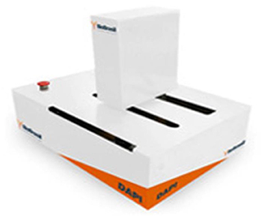
Figure 1. Image courtesy of Laboratory of Image Data Science (LIDS)- Unicamp. Techniques commonly used in routine laboratory procedures and the tendency toward automated diagnostics.
In the field of veterinary medicine, this new protocol has shown good performance in the diagnosis of intestinal parasites in dogs. This justifies the continuing development of automated diagnostics, which requires a protocol to obtain a cleaner slides, free of impurities and debris, enabling the computer system to more accurately identify parasite structures (38).
Immunological and molecular techniques, which are widely used in epidemiological surveys, scientific research and for the description of parasite species, are expensive, thus restricting their use in laboratory routines (33, 35).
Therefore, we emphasize the importance of an initial protocol, i.e., one that enables parasite structures to be more clearly visible in order to assist the automatic identification system. Traditionally accepted techniques widely published in the literature leave behind a lot of debris and impurities that end up hindering this identification. Hence, a technique that includes an initial protocol aimed at reducing these problems is ideal for this identification through good software programs. Our objective is therefore to critically describe the techniques currently in use for the automated diagnosis of intestinal parasites in fecal samples, as well as advances in these techniques, thus demonstrating the necessary requirements for their automation (Figure 2).
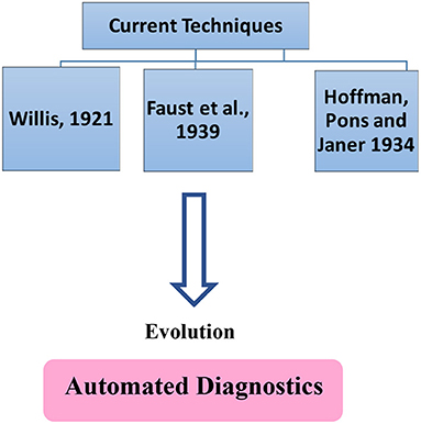
Figure 2. Techniques commonly used in routine laboratory procedures and the tendency towards automated diagnostics.
Article Retrieval Method
The articles were retrieved from the SciELO, ScienceDirect, and Google Scholar databases, and were selected after reading their contents.
Development
Advances and Limits of Computational Diagnostic Techniques for the Identification of Intestinal Parasites
Several studies are being conducted to devise differentiated, more sensitive diagnostics with fewer errors. These studies, which involve the automated examination of intestinal parasites (Table 1), still face challenges that must be overcome in order to ensure the proper identification of parasites. This includes a protocol for the preparation of microscope slides containing less debris, good staining of parasite structures, as well as a robust image database (38).
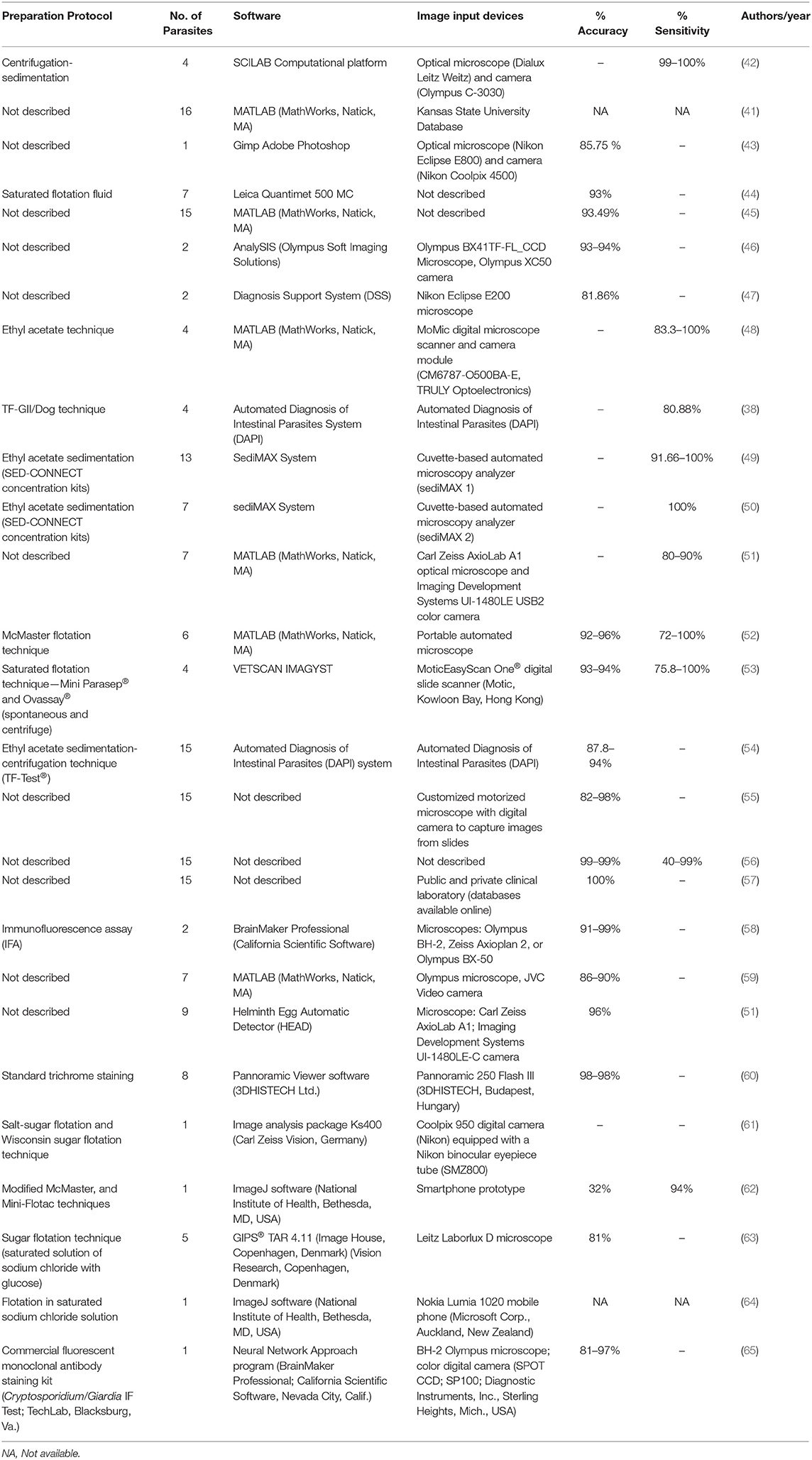
Table 1. Articles pertaining to the automatic identification of gastrointestinal parasites according to the computational techniques employed.
Over the last decade, researchers have been working on digital image processing models and pattern techniques for the automatic recognition of parasite eggs in microscopic images. The main objective is to reduce human errors that occur in the diagnosis of fecal parasites, and to produce faster and highly accurate results. These research efforts clearly illustrate the crucial importance of the automated identification of intestinal parasites in producing efficient and reliable results (43).
Intestinal parasites are considered of great interest in the implementation of algorithms for automated identification based on diagnostic imaging, because these organisms have stages of development with well-defined and reasonably homogeneous morphology (44).
Notably, the use of automated diagnostic techniques to identify and count eggs, cysts, and oocysts of helminths and protozoa is also very important. Moreover, with regard to the volume of samples processed in a short time, automated diagnostics offer high precision in the identification of host positivity and parasite load, allied to less fatigue and less time spent counting eggs on a computer screen when compared to the traditional microscope process (45).
Given the increasing use of computational technologies, the production and storage of visual and textual databases is essential. This means that an effective and efficient tool is needed to obtain such information to satisfy automation requirements (46, 47). Thus, image annotation methods have been based on several types of supervised classifiers (48), Bayesian classifier (49–51), Support Vector Machines (SVM) (52, 53), Artificial Neural Networks (ANNs) (66, 67), k-nearest neighbor (k-NN) (68, 69), Decision Tree (DT) (51, 52, 60), and Optimum-Path Forest (OPF) (61, 62).
ANNs implement algorithms to reproduce the processing functions of neural networks, in which neurons are arranged in layers (each neuron being connected to all the other neurons in the preceding layer) and process the information. The information is applied to an activation function and passes on signals to other neurons within the system. Using this structure of interconnected neurons, neural networks undergo a training procedure in which they “learn” how to discern patterns in data (63, 64). Backpropagation, a common learning algorithm employed by ANN, involves two steps. The first step is direct processing of input data through the neurons, which produce a predicted solution, while the second step involves the correction of weights within the layers of neurons to minimize the errors of the predicted solution relative to the true solution (63).
Initial studies on automated diagnostics involved the analysis of fecal samples from cattle (56, 65, 70) and pigs (71, 72). These studies of fecal samples from livestock differentiated the parasite eggs based on their characteristics (56, 65, 70–72). The use of the parameter of texture, which is the variation of the gray level in digital image processing, has also been investigated, and improves the classification result when used together with the characteristics of shape and size (70).
In a preliminary study to detect helminths using digital image processing techniques (73) and an artificial neural network (ANN) system (74), 82 images of seven human parasites were acquired. This study achieved an execution and identification rate of 84% and 83% respectively, and proved the applicability of the developed algorithms to the fully automatic examination system (75). This same ANN system was used in immunofluorescence staining (76) and 4′,6-diamidino-2-phenylindole [DAPI] (77) to identify the protozoan Cryptosporidium spp. in order to solve errors in mechanical identification resulting from factors such as technician fatigue and inexperience (77, 78).
The study generated from automated diagnostics has also evolved to the counting of bovine parasitic nematode eggs. The work in question involved a comparison of two techniques, Salt-Sugar Flotation (SSF) and Wisconsin Sugar Flotation (WSF). The former technique proved to be significantly better than the latter, since it allows a larger number of samples to be processed and provides a high degree of precision. Although these techniques use solutions that are close to saturation, they were diluted in the research in question (45).
Another study involved working on images of the protozoa Cryptosporidium spp. and Giardia spp. taken from slides stained with fluorescein-labeled monoclonal antibodies. The images were taken with a color digital camera, and the color information was discarded through dither filtering. These two protozoans were detected using Artificial Neural Networks (ANN), which correctly identified 91.8 of the images of the Cryptosporidium oocyst and 99.6% the Giardia cyst, respectively, indicating that it can be extremely useful in automated diagnostics (66). The above-mentioned techniques have several limitations, such as the difficulty of quantifying morphological characteristics, allied to the high complexity of the algorithms (44).
The purpose of image analysis is to classify and recognize objects of interest in digital images. This can be done in several ways, e.g., by identifying the colors, textures, shapes, movements and position of the objects in the images (44). Thus, different species of Eimeria found in farmyard chickens were included in the study of automated identification. This involved working with three groups of features, namely, characterization of the curvature, size, and symmetry of the internal structure, and quantification, i.e., its morphology studied over the multiscale curvature, geometry and texture (44). The image identification process carried out in this study included three components, which are image pre-processing, feature extraction and pattern recognition. The features were extracted automatically and used to compose a 3-dimensional feature vector for each oocyst image. Various problems were identified during this study, such as out of focus images, improperly positioned oocysts, compromised morphological structures, and the presence of debris and bacteria, which can make these variables difficult to use in the segmentation of the objects in question, impairing the identification of parasites (44).
Eimeria species are difficult to differentiate because of their highly similar morphology. However, a study using the Bayesian classifier was found to produce more accurate results than the SVM (Support Vector Machines). In fact, the Bayesian classifier presented 99.21% correct identification of Eimeria maxima, although Eimeria necatrix was incorrectly identified as Eimeria acervulina (6.10%) and as Eimeria tenella (9.94%). Moreover, 12.53% of E. necatrix was also mistaken for E. acervulina, 10.94% was mistaken for Eimeria praecox, and 12.22% for E. tenella. These results indicate that E. necatrix and E. praecox are the species most frequently identified, because their morphological similarity makes it difficult to differentiate them from each other and from other species. Nevertheless, in general, 85.75% of the parasites identified belonged to the genus Eimeria (44).
Researchers made a study based on Moment Invariants (MI) and a classifier of the Adaptive Neuro-Fuzzy Inference System (ANFIS) for the identification of 16 helminth parasite eggs in humans. This MI-ANFIS system consists of four stages: pre-processing, feature extraction, classification and testing. The ANFIS method, a more elaborate model of ANN, is considered a hybrid fuzzy logic algorithm and ANN. Therefore, the ANFIS classifier has all the advantages of these systems. The feature extraction stage uses Hu's seven moment invariants. The MI-ANFIS system showed a 93.49% correctness rate, and the main reason for incorrect classifications was the similarity in the shape of eggs, indicating the need for future advances in the technique (67).
The Multi-Class Support Vector Machine (MCSVM) classifier was tested in a study of 16 human parasites, which resulted in a 97.70% success rate. However, the same problem occurred as in the preceding study, i.e., the incorrect identification of similar shaped eggs. The pre-processing stage was considered the most important part of this proposal (68).
To improve the efficiency of existing conventional automated methods, a study was conducted using the MATLAB image processing toolbox (46). This study proposed a technique able to detect the presence of Ascaris lumbricoides and Trichuris trichiura parasites in a few seconds per image; however, this research was carried out only on these two parasite species (69).
In a routine laboratory parasitology diagnosis, debris, fecal impurities (41) and similarities in parasite structure (67, 68) pose real challenges for automated image analysis. Research has focused on the automatic segmentation and classification of microscopy images containing fecal impurities, and has detected the 15 most common species of protozoa, eggs and helminth larvae in Brazil. These species comprise A. lumbricoides, Enterobius vermicularis, Ancylostomatidae, T. trichiura, Hymenolepis diminuta, Hymenolepis nana, Taenia spp., Schistosoma mansoni, Strongyloides stercoralis, Entamoeba histolytica and Entamoeba dispar, Giardia duodenalis, Entamoeba coli, Endolimax nana, Iodamoeba butschlii, and Blastocystis hominis. Comparisons have been made of the performance of the OPF, ANN-MLP and SVM classifiers, with and without Bagging and AdaBoost, which are methods for building classifier committees. This evaluation demonstrated that the OPF classifier was the most suitable for the species in question, achieving 90.38% sensitivity, 98.32% specificity and 98.19% efficiency, with κ equal to 0.79 (41). For this study, a fecal sample processing technique was employed, called the TF-Test, which facilitates the concentration of parasite structures and help eliminate fecal impurities (41). In a later study, Suzuki et al. (41) proposed a complete solution for the diagnosis of intestinal parasites, with automated image acquisition from microscope slides and faster algorithms to reduce image processing time, and processed fecal samples using the TF-Test Modified technique (79). This study used an image base with 6,068 impurities and 1,791 parasites, and attained an average sensitivity of 93.00%, average specificity of 99.17% and average κ of 0.84 (80).
Based on a software program developed using morphometric analysis, area, perimeter and circularity, information on morphological specificity and characteristics of the parasites, 81.86% of the parasites were correctly identified. In that study, 85 images of A. lumbricoides and 54 images of T. trichiura were used. However, one of the main limitations of the automated technique is linked to debris and impurities from fecal samples left on microscope slides. This may explain the fact that the percentage of parasites not identified by automated means was 18.13%, which was attributed to the large amount of such impurities found in the evaluated samples (81).
Pattern classifiers are usually trained using a parasite image database annotated by a specialist. The automated reading of microscope slides can generate a large number of images to be annotated, rendering the process of manual annotation time-consuming and subject to errors. To facilitate this process, an active learning technique was developed whereby a specialist checks a small set of images, enabling the resulting classifier to automatically annotate the rest of the database. The proposed technique, called RDS (Root Distance-Based Sampling), organizes the dataset only once, as a pre-processing procedure, and adequately balances the diversity of classes as well as the sample uncertainty for the selection of useful samples during the learning process of a classifier, requiring verification of only a small part of the dataset (42).
A study of intestinal parasites was conducted using a cuvette-based automated microscopy analyzer, registered under the name of sediMAX 1®, which was developed for urine analysis. This equipment consists of a microscope, camera and high quality image processing software that can detect and classify particles in urine (82, 83). In this study, the equipment was used to automatically capture images from fecal samples, although the detection of parasites was performed by visual inspection. This device provides a practical way to store images for educational purposes, including the training of technicians in the detection of intestinal parasites (84).
In a study involving wastewater, a system was developed to identify and quantify up to seven species of helminth eggs. Images were captured manually using a microscope and color digital camera, and the system analyzed each image in <60 s. As in other studies of fecal samples, this study also came up against problems with debris and impurities that hindered the identification of eggs. Therefore, it is advisable to dilute concentrated sediment in tap water in a proportion of 1/1 or 1/2 (v/v). The system showed a detection specificity of 99% and its sensitivity varied from 80 to 90% (85).
The use of technology in the identification of intestinal parasites continues to expand rapidly and even involves mobile phones applications, as was the case with a study focusing on A. lumbricoides. In the study by Sowerby et al. (86), samples were processed by the flotation technique in saline solution and then identified using a mobile phone. Much still remains to be studied in the future, particularly the expansion of diagnostics for other parasites, the problem of impurities, and other challenges (86).
Using simple multivariate logistic regression, an algorithm was developed in the open-source program SCILAB to identify Taenia spp., Fasciola hepatica, T. trichiura, and Diphyllobothrium latum in stool smear images. The algorithm achieved sensitivity and specificity rates of 99.10 and 98.29% for Taenia spp., 99.15 and 98.18% for Fasciola hepatica, 100 and 98.38% for T. trichiura, and 100 and 98.13% for D. latum. A total of 200 samples were processed using rapid sedimentation (centrifugation) (87).
Aiming to provide automated treatment of gastrointestinal parasites in rural areas, a study was developed using a total of 30 microscope slides stained with iodine and containing eggs of A. lumbricoides, T. trichiura and hookworm species, in addition to four slides containing Schistosoma haematobium. These slides were digitized using a reference slide scanner and a mobile microscope. This new method aims to perform the diagnosis in less time but with high quality and accuracy, and at low cost. This proof-of-concept study demonstrates that the image of an inexpensive digital microscope suffices for a reliable diagnosis of the four helminth species that were worked on. Nevertheless, further studies are needed for improvement in order to overcome the challenges mentioned earlier herein (88).
Another analysis using the sediMAX® 2 was performed to compare the improvement achieved in the detection of protozoa in stool samples when compared with the sediMAX® 1. In this study, improvements were found in total reading time, which decreased from the original 10 min to about 5 min. SediMAX® 2 also allows amoebae to be differentiated by species, according to the number of nuclei present in the cells, as its focus is adjusted by hand. However, advances are still needed for an adequate diagnosis, such as the development of software for automatic image analysis, and to capture more than 15 images per sample (89).
Smartphones were also used in research to identify parasites, which are studied and analyzed for diagnostics in veterinary medicine. This research was based on Strongylus eggs found in horses, and an initial analysis indicated that the technique showed limitations in the identification of two eggs positioned close to each other or overlapping, and eggs covered by debris, making their identification difficult. This method was compared with the MiniFLOTAC and McMaster techniques, and was considered more sensitive than specific, generating false positive results; hence, further studies are still needed to improve this technique (90). A smartphone was also used in a study of three helminths that infect humans (A. lumbricoides, T. trichiura and hookworms), using Kankanet, an artificial neural network-based object detection smartphone application. The authors of the study reported sensitivity and specificity rates of 66.7 and 85.7% for T. trichiura, of 100 and 87.5% for A. lumbricoides, and of 100 and 100% for hookworms (91).
The use of automated diagnostics in the detection of intestinal parasites in various host species, such as sheep, canines, primates, and others was also developed for use in places where there are few resources. This is a portable method involving the McMaster flotation technique, which makes it low cost, fast and without requiring a trained professional to identify parasites. This technique attained good results, presenting an overall accuracy of 92 or 96% for Eimeria in the counting of one or four grids, respectively, and 100% for nematodes using one or four grids. In this study, debris was not a limitation, since the software was trained to recognize it (92).
Two classifiers were used in a study to identify eggs of the parasite Ascaris spp. in pigs, namely, the Multiclass Support Vector Machine (MC-SVM) and Artificial Neural Network (ANN). In this study, the parasite eggs were counted automatically. The accuracy rate of Ascaris spp. identification using the MC-SVM and ANN classifiers was ~95 and 93%, respectively (43).
In another study based on edge detection, image segmentation and recognition patterns, the detection and extraction of parasites in microscope images were fully automated, using the image pixel as a descriptor. This research made great strides in the detection of 15 human intestinal parasites, achieving an identification rate of 100% (93).
Staining also makes a significant difference in automated analysis for the more accurate identification of parasites. Thus, a convolutional neural network (CNN or ConvNet) model was developed to detect intestinal protozoa in human fecal samples stained with trichrome. However, this study showed limitations, such as the scantiness of some species. Even so, data from this study revealed that image capture using a slide scanner and Artificial Intelligence (AI) software allows for a 98.88% positive agreement [95% Confidence Interval from 93.76 to 99.98%], and 98.11% negative agreement [95% Confidence Interval from 93.35 to 99.77%] when compared to the correctness rates achieved with manual microscopy (94).
The performance of the VETSCAN IMAGYST system for the detection of parasites in dogs and cats was evaluated in another study, in which 100 fecal samples were analyzed, 84 from dogs and 16 from cats. The study revealed several limitations of the system, such as the lack of examination of the edges or outside the cover slip due to the reading area, and the study involved a low number of fecal samples, especially samples of Trichuris spp., Toxocara spp. and Taeniidae, which also limited the assessment of the system's diagnostic sensitivity and specificity (95).
In order to improve the diagnostic accuracy of the DAPI system (41) without compromising its efficiency and cost, a hybrid approach was proposed that combines two decision-making systems for the classification of images obtained from microscope slides. The study combines a simple system based on rapid extraction of characteristics from the images and SVM classifier, and a more complex system based on a deep neural network. The proposed system reached an average Kappa of 94.9, 87.8, and 92.5% in helminth eggs, helminth larvae and protozoan cysts, respectively (96).
A protocol designed to create a cleaner slide free of impurities contributed to a significant advance in the automated diagnosis of gastrointestinal parasites in the field of veterinary medicine. This technique was tested in a study involving four genera of canine intestinal parasites of high prevalence in an endemic region of the state of São Paulo, Brazil. Fecal samples from 104 dogs were collected to test this new protocol, which reached a Kappa index of 0.7636. It was therefore concluded that the new Prototic Coproparasitological Test for Dogs (PC-Test Dog) allows for a clearer view of parasite structures and presented a favorable result for the automated diagnosis of intestinal parasites in dogs (38).
More recently, Cringoli et al. (97) developed a portable versatile low-cost Kubic FLOTAC microscope (KFM) for students of veterinary medicine. The authors of the study prepared slides of bovine feces using the Mini-FLOTAC or FLOTAC method. Moreover, they stated that the KFM can be used to quantify parasite structures, and that the results were highly successful (97).
Final Remarks
Fecal samples in which parasite structures are easily detectable make microscope slides easy to analyze (43). That is why most researchers use microscope images of fecal samples and use digital processing of technical images to eliminate fecal impurities and detect the presence of parasite structures (43). However, debris, impurities and parasite load are major limitations in the development of automated diagnostics.
Most of the studies described in this paper do not include an adequate protocol for the preparation of slides for use in automated diagnostics, which is a crucial aspect in the identification of intestinal parasites (96). To ensure the successful advance of automated parasitological diagnosis, a holistic view of the entire procedure must be adopted, from sample collection to identification on computers. In other words, samples must be collected and stored carefully, a processing technique should be used that reduces impurities and concentrates the parasites, as well as a suitable dye and a proper software program. These steps will undoubtedly be helpful in the advancements of automated diagnostics in both human and veterinary medicine.
Author Contributions
SI, JG, and AF: conceptualization. SI: writing—original draft preparation. SI, BM, SR, CN, and KB: writing—review and editing. FS and SN: creation of the table. All the authors have read and agreed to the published version of the manuscript.
Funding
This work was supported by Fundação de Amparo à Pesquisa do Estado de São Paulo (FAPESP) - Process No. 2017/14189-9 – Thematic Project Process No. 2014/12236-1 and National Council for Scientific and Technological Development (CNPQ) No. 303808/2018-7.
Conflict of Interest
The authors declare that the research was conducted in the absence of any commercial or financial relationships that could be construed as a potential conflict of interest.
Publisher's Note
All claims expressed in this article are solely those of the authors and do not necessarily represent those of their affiliated organizations, or those of the publisher, the editors and the reviewers. Any product that may be evaluated in this article, or claim that may be made by its manufacturer, is not guaranteed or endorsed by the publisher.
References
1. Punsawad C, Phasuk N, Thongtup K, Nagavirochana S, Viriyavejakul P. Prevalence of parasitic contamination of raw vegetables in Nakhon Si Thammarat province, southern Thailand 11 Medical and Health Sciences 1117 Public Health and Health Services. BMC Public Health. (2019) 19:34. doi: 10.1186/s12889-018-6358-9
2. Macpherson CNL. Human behaviour and the epidemiology of parasitic zoonoses. Int J Parasitol. (2005) 35:1319–31. doi: 10.1016/j.ijpara.2005.06.004
3. Alho AM, Lima C, Colella V, Madeira De Carvalho L, Otranto D, Cardoso L. Awareness of zoonotic diseases and parasite control practices: a survey of dog and cat owners in Qatar. Parasites Vectors. (2018) 11:133. doi: 10.1186/s13071-018-2720-0
4. Itoh N, Kanai K, Kimura Y, Chikazawa S, Hori Y, Hoshi F. Prevalence of intestinal parasites in breeding kennel dogs in Japan. Parasitol Res. (2015) 114:1221–224. doi: 10.1007/s00436-015-4322-5
5. Monteiro MFM, Ramos RAN, Calado AMC, Lima VFS, Ramos ICDN, Tenório RFL, et al. Parasitas gastrointestinais em gatos no Brasil: frequência e risco zoonótico. Rev Brasil Parasitol Vet. (2016) 25:254–7. doi: 10.1590/S1984-29612016019
6. Pereira A, Martins Â, Brancal H, Vilhena H, Silva P, Pimenta P, et al. Parasitic zoonoses associated with dogs and cats: a survey of Portuguese pet owners' awareness and deworming practices. Parasites Vectors. (2016) 9:245. doi: 10.1186/s13071-016-1533-2
7. CDC. CDC - Parasites - About Parasites. Centers for Disease Control and Prevention (2020). Available online at: https://www.cdc.gov/parasites/about.html (accessed October 30, 2020).
8. WHO. Prevention and Control of Intestinal Parasitic Infections. WHO Technical Report Series N° 749. WHO (2016).
9. Beugnet F, Bourdeau P, Chalvet-Monfray K, Cozma V, Farkas R, Guillot J, et al. Parasites of domestic owned cats in Europe: co-infestations and risk factors. Parasites Vectors. (2014) 7:291. doi: 10.1186/1756-3305-7-291
10. Kostopoulou D, Claerebout E, Arvanitis D, Ligda P, Voutzourakis N, Casaert S, et al. Abundance, zoonotic potential and risk factors of intestinal parasitism amongst dog and cat populations: the scenario of Crete, Greece. Parasites Vectors. (2017) 10:43. doi: 10.1186/s13071-017-1989-8
11. Ortuño A, Scorza V, Castellà J, Lappin M. Prevalence of intestinal parasites in shelter and hunting dogs in Catalonia, Northeastern Spain. Vet J. (2014) 199:465–7. doi: 10.1016/j.tvjl.2013.11.022
12. Robertson ID, Thompson RC. Enteric parasitic zoonoses of domesticated dogs and cats. Microbes Infect. (2002) 4:867–73. doi: 10.1016/S1286-4579(02)01607-6
13. Taylor MA, Coop RL, Wall R. Parasites of dogs and cats. In: Parasitology, Veterinary, 3rd ed. Carlton, VIC: Blackwell Publishing Asia Pty Ltd (2007), 356–458.
14. Halliez MCM, Buret AG. Extra-intestinal and long term consequences of Giardia duodenalis infections. World J Gastroenterol. (2013) 19:8974–85. doi: 10.3748/wjg.v19.i47.8974
15. Phasuk N, Punsawad C. Seroprevalence of Toxocara canis infection and associated risk factors among primary schoolchildren in rural Southern Thailand. Trop Med Health. (2020) 48:23. doi: 10.1186/s41182-020-00211-0
16. Smith H, Holland C, Taylor M, Magnaval JF, Schantz P, Maizels R. How common is human toxocariasis? Towards standardizing our knowledge. Trends Parasitol. (2009) 25:182–8. doi: 10.1016/j.pt.2009.01.006
17. Boldiš V, Ondriska F, Špitalská E, Reiterová K. Immunodiagnostic approaches for the detection of human toxocarosis. Exp Parasitol. (2015) 159:252–8. doi: 10.1016/j.exppara.2015.10.006
18. Dattoli VCC, Freire SM, Mendonça LR, Santos PC, Meyer R, Alcantara-Neves NM. Toxocara canis infection is associated with eosinophilia and total IgE in blood donors from a large Brazilian centre. Trop Med Int Health. (2011) 16:514–7. doi: 10.1111/j.1365-3156.2010.02719.x
19. Magnaval JF, Glickman LT, Dorchies P, Morassin B. Highlights of human toxocariasis. Korean J Parasitol. (2001) 39:1–11. doi: 10.3347/kjp.2001.39.1.1
20. Mirdha BR. Ocular toxocariasis in a North Indian population. J Trop Pediatr. (2002) 48:328–30. doi: 10.1093/tropej/48.6.328
21. Bowman DD, Montgomery SP, Zajac AM, Eberhard ML, Kazacos KR. Hookworms of dogs and cats as agents of cutaneous larva migrans. Trends Parasitol. (2010) 26:162–7. doi: 10.1016/j.pt.2010.01.005
22. George S, Levecke B, Kattula D, Velusamy V, Roy S, Geldhof P, et al. Molecular identification of hookworm isolates in humans, dogs and soil in a tribal area in Tamil Nadu, India. PLOS Negl Trop Dis. (2016) 10:e0004891. doi: 10.1371/journal.pntd.0004891
23. Nazzaro G, Angileri L, Parducci BA, Veraldi S. Hookworm-related cutaneous larva migrans: Our 201st patient. J Infect Dev Countries. (2017) 11:437–9. doi: 10.3855/jidc.8930
24. Simonato G, Cassini R, Morelli S, Di Cesare A, La Torre F, Marcer F, et al. Contamination of Italian parks with canine helminth eggs and health risk perception of the public. Prev Vet Med. (2019) 172:104788. doi: 10.1016/j.prevetmed.2019.104788
25. Willis HH. A simple levitation method for the detection of Hookworm Ova. Med J Australia. (1921) 2:375–6. doi: 10.5694/j.1326-5377.1921.tb60654.x
26. Faust EC, Sawitz W, Tobie J, Odom V, Peres C, Lincicome DR. Comparative efficiency of various technics for the diagnosis of protozoa and helminths in feces. J Parasitol. (1939) 25:241–62. doi: 10.2307/3272508
27. Hoffman WA, Pons JA, Janer JL. The sedimentation-concentration method in Schistosomiasis Mansoni. PR J Public Health Trop Med. (1934) 9:288–91.
28. Barbecho JM, Bowman DD, Liotta JL. Comparative performance of reference laboratory tests and in-clinic tests for Giardia in canine feces. Parasites Vectors. (2018) 11:444. doi: 10.1186/s13071-018-2990-6
29. Coelho WMD, Gomes JF, do Amarante AFT, Bresciani KDS, Lumina G, Koshino-Shimizu S, et al. A new laboratorial method for the diagnosis of gastrointestinal parasites in dogs. Rev Brasil Parasitol Vet. (2013) 22:1–5. doi: 10.1590/S1984-29612013000100002
30. Coelho WMD, Gomes JF, Falcão AX, Dos Santos BM, Soares FA, Nagase Suzuki CT, et al. Estudo comparativo de cinco técnicas para o diagnóstico de parasitos gastrointestinais caninos. Rev Brasil Parasitol Vet. (2015) 24:223–26.
31. de Santana BB, da Silva TLB, Ramos RAN, Alves LC, de Carvalho GA. Evaluation of different parasitological techniques for diagnosing intestinal parasites in dogs. Open J Vet Med. (2015) 05:19–24. doi: 10.4236/ojvm.2015.52003
32. Katagiri S, Oliveira-Sequeira TCG. Comparison of three concentration methods for the recovery of canine intestinal parasites from stool samples. Exp Parasitol. (2010) 126:214–6. doi: 10.1016/j.exppara.2010.04.027
33. Gomes JF, Hoshino-Shimizu S S, Dias LC, Araujo AJSA, Castilho VLP, Neves FAMA. Evaluation of a novel kit (TF-Test) for the diagnosis of intestinal parasitic infections. J Clin Lab Anal. (2004) 18:132–8. doi: 10.1002/jcla.20011
34. de Carvalho GLX, Moreira LE, Pena JL, Marinho CC, Bahia MT, Machado-Coelho GLL. A comparative study of the TF-test®, Kato-Katz, Hoffman-Pons-Janer, Willis and Baermann-Moraes coprologic methods for the detection of human parasitosis. Memorias Inst Oswaldo Cruz. (2012) 107:80–4. doi: 10.1590/S0074-02762012000100011
35. Carvalho JB de, Santos BM dos, Gomes JF, Suzuki CTN, Hoshino Shimizu S, Falcão AX, et al. TF-Test modified: new diagnostic tool for human enteroparasitosis. J Clin Lab Anal. (2016) 30:293–300. doi: 10.1002/jcla.21854
36. Soares FA, Benitez ADN, Dos Santos BM, Loiola SHN, Rosa SL, Nagata WB, et al. A historical review of the techniques of recovery of parasites for their detection in human stools. Rev Soc Brasil Med Trop. (2020) 53:1–9. doi: 10.1590/0037-8682-0535-2019
37. Inácio SV, Ferreira Gomes J, Xavier Falcão A, Nagase Suzuki CT, Bertequini Nagata W, Nery Loiola SH, et al. Automated diagnosis of canine gastrointestinal parasites using image analysis. Pathogens. (2020) 9:139. doi: 10.3390/pathogens9020139
38. Inácio SV, Gomes JF, Oliveira BCM, Falcão AX, Suzuki CTN, dos Santos BM, et al. Validation of a new technique to detect Cryptosporidium spp. oocysts in bovine feces. Prev Vet Med. (2016) 134:1–5. doi: 10.1016/j.prevetmed.2016.09.020
39. Lumina G, Bricarello PA, Ferreira Gomes J, Talamini AF. Avaliação do kit “TF-Test” para o diagnóstico das infecções por parasitas gastrintestinais em ovinos. Braz J Vet Res Anim Sci. (2006) 43:496–501.
40. Falcão AX, Gomes JF, Suzuki CTN, Hoshino-Shimizu S, Dias LCS, et al. Sistema para Diagnóstico de Parasitos Intestinais por Análise Computadorizada de Imagens e Uso de Referido Sistema (Concessão de Patente—INPI: PI0802292-5). Campinas: INPI (2008).
41. Suzuki CTN, Gomes JF, Falcão AX, Papa JP, Hoshino-Shimizu S. Automatic segmentation and classification of human intestinal parasites from microscopy images. IEEE Transac Biomed Eng. (2013) 60:803–12. doi: 10.1109/TBME.2012.2187204
42. Saito PTM, Suzuki CTN, Gomes JF, De Rezende PJ, Falcão AX. Robust active learning for the diagnosis of parasites. Pattern Recogn. (2015) 48:3572–83. doi: 10.1016/j.patcog.2015.05.020
43. Das AK, Nayak J, Naik B, Pati SK, Pelusi D. Computational Intelligence in Pattern Recognition: Proceedings of CIPR 2019, 1st ed. Springer Nature Singapore Pte Ltd (2019) 1046. doi: 10.1007/978-981-13-9042-5
44. Castañón CAB, Fraga JS, Fernandez S, Gruber A, da Costa FL. Biological shape characterization for automatic image recognition and diagnosis of protozoan parasites of the genus Eimeria. Pattern Recogn. (2007) 40:1899–910. doi: 10.1016/j.patcog.2006.12.006
45. Mes THM, Ploeger HW, Terlou M, Kooyman FNJ, Van Der Ploeg MPJ, Eysker M. A novel method for the isolation of gastro-intestinal nematode eggs that allows automated analysis of digital images of egg preparations and high throughput screening. Parasitology. (2001) 123:309–14. doi: 10.1017/S0031182001008496
46. Tamura H, Yokoya N. Image database systems: a survey. Pattern Recogn. (1984) 17:29–43. doi: 10.1016/0031-3203(84)90033-5
47. Chang SK, Hsu A. Image information systems: where do we go from here? IEEE Transac Knowl Data Eng. (1992) 4:431–42. doi: 10.1109/69.166986
48. Zhang D, Islam MM, Lu G. A review on automatic image annotation techniques. Pattern Recogn. (2012) 45:346–62. doi: 10.1016/j.patcog.2011.05.013
49. Jeon J, Lavrenko V, Manmatha R. Automatic Image Annotation and Retrieval Using Cross-Media Relevance Models. Association for Computing Machinery (ACM), 119.
50. Carneiro G, Chan AB, Moreno PJ, Vasconcelos N. Supervised learning of semantic classes for image annotation and retrieval. IEEE Transac Pattern Anal Mach Intellig. (2007) 29:394–410. doi: 10.1109/TPAMI.2007.61
51. Liu Y, Zhang D, Lu G. Region-based image retrieval with high-level semantics using decision tree learning. Pattern Recogn. (2008) 41:2554–70. doi: 10.1016/j.patcog.2007.12.003
52. Goh KS, Chang EY, Li B. Using one-class and two-class SVMs for multiclass image annotation. IEEE Transac Knowl Data Eng. (2005) 17:1333–46. doi: 10.1109/TKDE.2005.170
53. Qi X, Han Y. Incorporating multiple SVMs for automatic image annotation. Pattern Recogn. (2007) 40:728–41. doi: 10.1016/j.patcog.2006.04.042
54. Park SB, Lee JW, Kim SK. Content-based image classification using a neural network. Pattern Recogn Lett. (2004) 25:287–300. doi: 10.1016/j.patrec.2003.10.015
55. Del Frate F, Pacifici F, Schiavon G, Solimini C. Use of neural networks for automatic classification from high-resolution images. IEEE Transac Geosci Remote Sens. (2007) 45:800–9. doi: 10.1109/TGRS.2007.892009
56. Sommer C. Quantitative characterization, classification and reconstruction of oocyst shapes of Eimeria species from cattle. Parasitology. (1998) 116:21–8. doi: 10.1017/S003118209700187X
57. Jain P, Kapoor A. Active Learning for Large Multi-Class Problems. Institute of Electrical and Electronics Engineers (IEEE). p. 762–9.
58. Tang J, Hong R, Yan S, Chua TS, Qi GJ, Jain R. Image annotation by k nn-sparse graph-based label propagation over noisily tagged web images. ACM Transac Intell Syst Technol. (2011) 2:1–15. doi: 10.1145/1899412.1899418
59. Vens C, Struyf J, Schietgat L, DŽeroski S, Blockeel H. Decision trees for hierarchical multi-label classification. Mach Learn. (2008) 73:185–214. doi: 10.1007/s10994-008-5077-3
60. Wong RCF, Leung CHC. Automatic semantic annotation of real-world web images. IEEE Transac Pattern Anal Mach Intellig. (2008) 30:1933–44. doi: 10.1109/TPAMI.2008.125
61. Da Silva AT, Falcão AX, Magalhães LP. Active learning paradigms for CBIR systems based on optimum-path forest classification. Pattern Recogn. (2011) 44:2971–8. doi: 10.1016/j.patcog.2011.04.026
62. Saito PTM, De Rezende PJ, Falcão AX, Suzuki CTN, Gomes JF. A data reduction and organization approach for efficient image annotation. In: Proceedings of the ACM Symposium on Applied Computing. New York, NY: ACM Press (2013). p. 53–5.
63. Widrow B, Lehr MA. 30 Years of adaptive neural networks: perceptron, madaline, and backpropagation. Proc IEEE. (1990) 78:1415–42. doi: 10.1109/5.58323
64. Basheer IA, Hajmeer M. Artificial neural networks: fundamentals, computing, design, and application. J Microbiol Methods. (2000) 43:3–31. doi: 10.1016/S0167-7012(00)00201-3
65. Sommer C. Digital image analysis and identification of eggs from bovine parasitic nematodes. J Helminthol. (1996) 70:143–51. doi: 10.1017/S0022149X00015303
66. Widmer KW, Srikumar D, Pillai SD. Use of artificial neural networks to accurately identify Cryptosporidium oocyst and Giardia cyst images. Appl Environ Microbiol. (2005) 71:80–4. doi: 10.1128/AEM.71.1.80-84.2005
67. Dogantekin E, Yilmaz M, Dogantekin A, Avci E, Sengur A. A robust technique based on invariant moments - ANFIS for recognition of human parasite eggs in microscopic images. Expert Syst Appl. (2008) 35:728–38. doi: 10.1016/j.eswa.2007.07.020
68. Avci D, Varol A. An expert diagnosis system for classification of human parasite eggs based on multi-class SVM. Expert Syst Appl. (2009) 36:43–8. doi: 10.1016/j.eswa.2007.09.012
69. Ghazali KH, Hadi RS, Mohamed Z. Automated system for diagnosis intestinal parasites by computerized image analysis. Modern Appl Sci. (2013) 7:98–114. doi: 10.5539/mas.v7n5p98
70. Sommer C. Quantitative characterization of texture used for identification of eggs of bovine parasitic nematodes. J Helminthol. (1998) 72:179–82. doi: 10.1017/S0022149X00016370
71. Joachim A, Dülmer N, Daugschies A. Differentiation of two Oesophagostomum spp. from pigs, O. dentatum and O. quadrispinulatum, by computer-assisted image analysis of fourth-stage larvae. Parasitol Int. (1999) 48:63–71. doi: 10.1016/S1383-5769(99)00003-3
72. Daugschies A, Imarom S, Bollwahn W. Differentiation of porcine Eimeria spp. by morphologic algorithms. Vet Parasitol. (1999) 81:201–10. doi: 10.1016/S0304-4017(98)00246-5
73. Gonzalez R, Woods R. Digital Image Process, 3rd ed. Upper Saddle River, NJ: Prentice-Hall, Inc. (1992).
74. Freeman JA, Skapura DM. Neural Networks: Algorithms, Applications, and Programming Techniques. Reading, MA: Addison-Wesley (1991).
75. Yoon Seok Yang, Duck Kun Park, Hee Chan Kim, Choi MH, Chai JY. Automatic identification of human helminth eggs on microscopic fecal specimens using digital image processing and an artificial neural network. IEEE Transac Biomed Eng. (2001) 48:718–30. doi: 10.1109/10.923789
76. Widmer KW, Oshima KH, Pillai SD. Identification of Cryptosporidium parvum oocysts by an artificial neural network approach. Appl Environ Microbiol. (2002) 68:1115–21. doi: 10.1128/AEM.68.3.1115-1121.2002
77. Widmer KW. Development of artificial neural networks capable of identifying cryptosporidium parvum oocysts stained with 4', 6 diamidino-2-phenylindole. J Rapid Methods Automat Microbiol. (2003) 11:97–110. doi: 10.1111/j.1745-4581.2003.tb00033.x
78. Clancy JL, Gollnitz WD, Tabib Z. Commercial labs: how accurate are they? J Am Water Works Assoc. (1994) 86:89–97. doi: 10.1002/j.1551-8833.1994.tb06198.x
79. Falcão AX, Gomes JF, Shimizu SH, Suzuki CTN. Method for Preparing a Faecal Copro-Parasitological Specimen, and Clarifying Composition (2010 PTC: Prot. 018100037856) (2010). Available online at: http://www.wipo.int/pctdb.
80. Suzuki CTN, Gomes JF, Falcao AX, Shimizu SH, Papa JP. Automated diagnosis of human intestinal parasites using optical microscopy images. In: Proceedings - International Symposium on Biomedical Imaging. San Francisco, CA (2013). p. 460–3.
81. Gomes AP, Noguerol L, Bez MR, Tavares RG. Performance analysis of software for identification of intestinal parasites. J Brasil Patol Med Lab. (2015) 51:218–23. doi: 10.5935/1676-2444.20150036
82. Zaman Z, Fogazzi GB, Garigali G, Croci MD, Bayer G, Kránicz T. Urine sediment analysis: analytical and diagnostic performance of sediMAX® - a new automated microscopy image-based urine sediment analyser. Clin Chim Acta. (2010) 411:147–54. doi: 10.1016/j.cca.2009.10.018
83. Falbo R, Sala MR, Signorelli S, Venturi N, Signorini S, Brambilla P. Bacteriuria screening by automated whole-field-image-based microscopy reduces the number of necessary urine cultures. J Clin Microbiol. (2012) 50:1427–9. doi: 10.1128/JCM.06003-11
84. Intra J, Taverna E, Sala MR, Falbo R, Cappellini F, Brambilla P. Detection of intestinal parasites by use of the cuvette-based automated microscopy analyser sediMAX®. Clin Microbiol Infect. (2016) 22:279–84. doi: 10.1016/j.cmi.2015.11.014
85. Jiménez B, Maya C, Velásquez G, Torner F, Arambula F, Barrios JA, et al. Identification and quantification of pathogenic helminth eggs using a digital image system. Exp Parasitol. (2016) 166:164–72. doi: 10.1016/j.exppara.2016.04.016
86. Sowerby SJ, Crump JA, Johnstone MC, Krause KL, Hill PC. Smartphone microscopy of parasite eggs accumulated into a single field of view. Am J Trop Med Hyg. (2016) 94:227–30. doi: 10.4269/ajtmh.15-0427
87. Alva A, Cangalaya C, Quiliano M, Krebs C, Gilman RH, Sheen P, et al. Mathematical algorithm for the automatic recognition of intestinal parasites. PLOS ONE. (2017) 12:1–13. doi: 10.1371/journal.pone.0175646
88. Holmström O, Linder N, Ngasala B, Mårtensson A, Linder E, Lundin M, et al. Point-of-care mobile digital microscopy and deep learning for the detection of soil-transmitted helminths and Schistosoma haematobium. Global Health Action. (2017) 10:1337325. doi: 10.1080/16549716.2017.1337325
89. Intra J, Sala MR, Falbo R, Cappellini F, Brambilla P. Improvement in the detection of enteric protozoa from clinical stool samples using the automated urine sediment analyzer sediMAX® 2 compared to sediMAX® 1. Eur J Clin Microbiol Infect Dis. (2017) 36:147–51. doi: 10.1007/s10096-016-2788-4
90. Scare JA, Slusarewicz P, Noel ML, Wielgus KM, Nielsen MK. Evaluation of accuracy and precision of a smartphone based automated parasite egg counting system in comparison to the McMaster and Mini-FLOTAC methods. Vet Parasitol. (2017) 247:85–92. doi: 10.1016/j.vetpar.2017.10.005
91. Yang A, Bakhtari N, Langdon-Embry L, Redwood E, Lapierre SG, Rakotomanga P, et al. Kankanet: an artificial neural network-based object detection smartphone application and mobile microscope as a point-of-care diagnostic aid for soil-transmitted helminthiases. PLOS Neglect Trop Diseases. (2019) 3:e0007577. doi: 10.1371/journal.pntd.0007577
92. Li Y, Zheng R, Wu Y, Chu K, Xu Q, Sun M, et al. A low-cost, automated parasite diagnostic system via a portable, robotic microscope and deep learning. J Biophot. (2019) 12:e201800410. doi: 10.1002/jbio.201800410
93. Tchinda BS, Noubom M, Tchiotsop D, Louis-Dorr V, Wolf D. Towards an automated medical diagnosis system for intestinal parasitosis. Inform Med Unlocked. (2019) 13:101–111. doi: 10.1016/j.imu.2018.09.004
94. Mathison BA, Kohan JL, Walker JF, Smith RB, Ardon O, Ardon O, et al. Detection of intestinal protozoa in trichrome-stained stool specimens by use of a deep convolutional neural network. J Clin Microbiol. (2020) 58:e02053–19. doi: 10.1128/JCM.02053-19
95. Nagamori Y, Hall Sedlak R, Derosa A, Pullins A, Cree T, Loenser M, et al. Evaluation of the VETSCAN IMAGYST: an in-clinic canine and feline fecal parasite detection system integrated with a deep learning algorithm. Parasites Vectors. (2020) 13:346. doi: 10.1186/s13071-020-04215-x
96. Osaku D, Cuba CF, Suzuki CTN, Gomes JF, Falcão AX. Automated diagnosis of intestinal parasites: a new hybrid approach and its benefits. Comput Biol Med. (2020) 123:103917. doi: 10.1016/j.compbiomed.2020.103917
Keywords: automated, parasite, protozoan, animal, human, gastrointestinal, technological progress, helminths
Citation: Inácio SV, Gomes JF, Falcão AX, Martins dos Santos B, Soares FA, Nery Loiola SH, Rosa SL, Nagase Suzuki CT and Bresciani KDS (2021) Automated Diagnostics: Advances in the Diagnosis of Intestinal Parasitic Infections in Humans and Animals. Front. Vet. Sci. 8:715406. doi: 10.3389/fvets.2021.715406
Received: 26 May 2021; Accepted: 19 October 2021;
Published: 23 November 2021.
Edited by:
Alessia Libera Gazzonis, University of Milan, ItalyReviewed by:
Daniel A. Abugri, Alabama State University, United StatesLaura Rinaldi, University of Naples Federico II, Italy
Copyright © 2021 Inácio, Gomes, Falcão, Martins dos Santos, Soares, Nery Loiola, Rosa, Nagase Suzuki and Bresciani. This is an open-access article distributed under the terms of the Creative Commons Attribution License (CC BY). The use, distribution or reproduction in other forums is permitted, provided the original author(s) and the copyright owner(s) are credited and that the original publication in this journal is cited, in accordance with accepted academic practice. No use, distribution or reproduction is permitted which does not comply with these terms.
*Correspondence: Katia Denise Saraiva Bresciani, a2F0aWEuYnJlc2NpYW5pQHVuZXNwLmJy