- 1State Key Laboratory of Agricultural Microbiology, College of Veterinary Medicine, Huazhong Agricultural University, Wuhan, China
- 2MOST International Research Center for Animal Disease, Cooperative Innovation Center for Sustainable Pig Production, Huazhong Agricultural University, Wuhan, China
- 3Diagnostic Center of Animal Diseases, Wuhan Keqian Biology Co., Ltd, Wuhan, China
- 4MARA Key Laboratory of Prevention and Control Agents for Animal Bacteriosis, Institute of Animal Husbandry and Veterinary, Hubei Academy of Agricultural Sciences, Wuhan, China
Bordetella bronchiseptica is a leading cause of respiratory diseases in pigs. However, epidemiological data of B. bronchiseptica in pigs particularly in China, the largest pig rearing country in the world is still limited. We isolated 181 B. bronchiseptica strains from 4259 lung samples of dead pigs with respiratory diseases in 14 provinces in China from 2018 to 2020. The average isolation rate of this 3-year period was 4.25% (181/4259). Antimicrobial susceptibility testing performed by disc diffusion method revealed that most of the B. bronchiseptica isolates in this study were resistant to ampicillin (83.98%), while a proportion of isolates were resistant to cefotaxime (30.39%%), chloramphenicol (12.71%), gentamicin (11.60%), florfenicol (11.60%), tetracycline (8.84%), amoxicillin (8.29%), tobramycin (6.63%), ceftriaxone (4.97%), and cefepime (0.55%). There were no isolates with resistant phenotypes to imipenem, meropenem, polymyxin B, ciprofloxacin, enrofloxacin, and amikacin. In addition, ~13.18% of the isolates showed phenotypes of multidrug resistance. Detection of antimicrobial resistance genes (ARGs) by PCR showed that 16.57% of the B. bronchiseptica isolates in this study was positive to aac(3)-IV, while 3.87%, 2.21%, 1.10%, 0.55%, 0.55%, and 0.55% of the isolates were positive to aac6'-Ib, rmtA, blaTEM, blaSHV, oqxB, and tetA, respectively. Detection of virulence factors encoding genes (VFGs) by conventional PCR showed that over 90% of the pig B. bronchiseptica isolates in this study were positive to the five VFGs examined (fhaB, 97.24%; prn, 91.16%; cyaA, 98.34%; dnt, 98.34%; betA, 92.82%). These results demonstrate B. bronchiseptica as an important pathogen associated with pig respiratory disorders in China. The present work contributes to the current understanding of the prevalence, antimicrobial resistance and virulence genes of B. bronchiseptica in pigs.
Introduction
Bordetella bronchiseptica is an aerobic, motile, gram-negative rod, or coccobacillus belonging to genus Bordetella. It is an important pathogenic bacterium in agriculture and in veterinary medicine (1). In veterinary medicine, B. bronchiseptica is a leading cause of many respiratory infections including rhinitis, tracheitis, bronchitis, and pneumonia in a wide spectrum of animals (2). It can also enhance respiratory colonization of Streptococcus suis and Haemophilus parasuis, promote disease caused by S. suis, and interact with porcine reproductive and respiratory syndrome virus (PRRSV) and swine influenza virus (SIV) to increase severity of respiratory disease (3). While rarely to be reported, B. bronchiseptica is also potentially involved in infections in humans, and human cases are frequently associated with direct contact with infected animals such as swine, dog, rabbit and/or cat (4–6). Similar to the other members belonging to genus Bordetella, many B. bronchiseptica produces several important virulence factors, including filamentous hemagglutinin, and protein toxins, adenylate cyclase toxin, pertussis toxin, dermonecrotic toxin as well as type III secretion system (T3SS) and effector proteins, contributing to its pathogenesis (7, 8).
In swine, B. bronchiseptica is proposed as a main causative agent of porcine respiratory disease complex (PRDC) and atrophic rhinitis; both of which are economically-important diseases in pig industry (9, 10). Continuously monitoring the prevalence, antimicrobial resistance (AMR) and virulence profiles of B. bronchiseptica in pigs are beneficial for the prevention and control of swine bordetellosis. However, the relevant data are still limited. China is the largest pig-farming and pork consuming country in the world. Although the outbreak of African Swine Fever in August 2018 caused a huge loss of pigs in China, there are still more than 406 million pigs rearing in China in 2020 (11). To understand the current epidemiological and microbiological characteristics such as the antimicrobial resistance profiles of B. bronchiseptica isolates from pigs in China, we performed bacterial isolation of B. bronchiseptica strains from lung samples of dead pigs with a history of respiratory disorders in China from 2018 to 2020 in this study. These isolates were characterized by testing the antimicrobial susceptibility and detecting the antimicrobial resistance genes (ARGs) as well as virulence encoding genes (VFGs).
Materials and Methods
Study Design, Sample Collection, and Ethic Statement
Study design was shown in Figure 1A. From 2018 to 2020, a total of 4259 lung samples (3022 samples in 2018, 841 samples in 2019, 396 samples in 2020) from 14 provinces (Guangdong, Henan, Hubei, Shandong, Fujian, Hebei, Zhejiang, Hunan, Anhui, Sichuan, Shanxi, Inner Mongolia, Xinjiang, Guizhou) in China were used for B. bronchiseptica isolation and identification (Figure 1B). All of the clinical samples used in this study were submitted by veterinarians/or the farm owners to the Veterinary Diagnostic Laboratory of Huazhong Agricultural University (Wuhan, China) for routine testing.

Figure 1. Study design and isolation of B. bronchiseptica from swine lung samples in different regions in China from 2018 to 2020. (A) Shows study design of this work; (B) shows the geographic sites of sample collection. Numbers of B. bronchiseptica from the samples as well as numbers of samples collected from each of the provinces are shown; (C) shows the isolation rates of B. bronchiseptica from swine lung samples in different regions in China from 2018 to 2020.
Bacterial Isolation and Identification
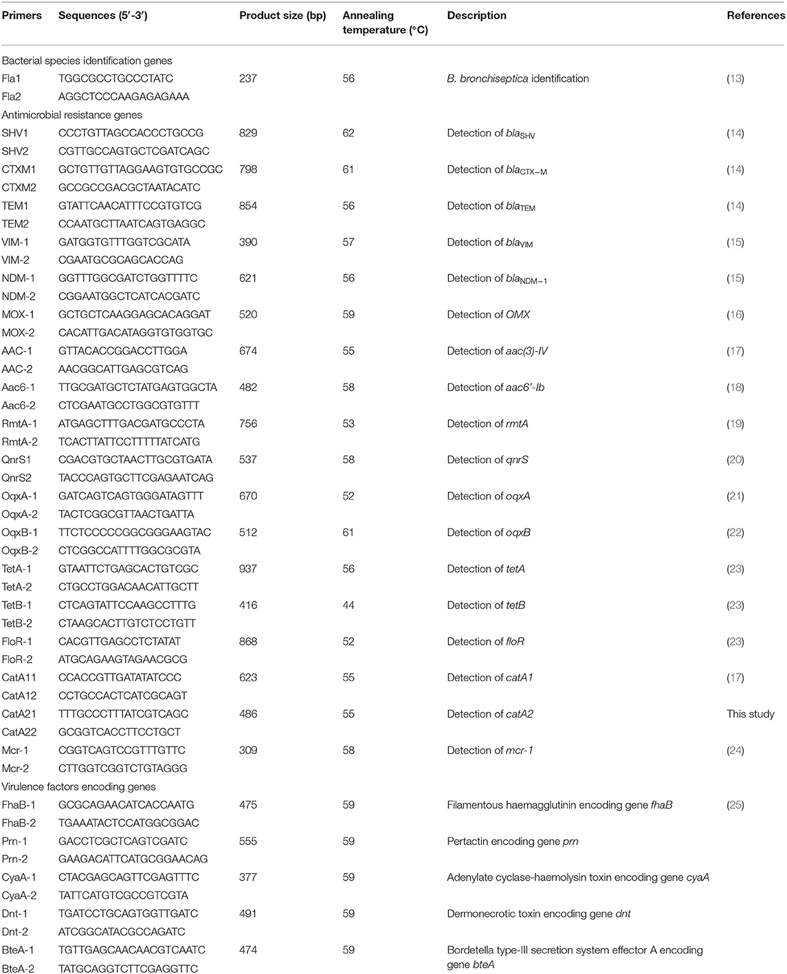
Collected samples (~10 grams per sample) were cut into pieces and lysed in sterile 0.9% normal saline by using a TissueLyser II (QIAGEN, Venlo, Netherlands). Thereafter, tissue homogenates of each sample were streak-plated onto one tryptic soy agar (TSA; Becton, Dickinson and Company, MD, USA) containing 10 μg/ml nicotinamide adenine dinucleotide (NAD; Sigma, St. Louis, MO) and 10% new-born bovine serum. The agar plates were incubated at 37°C for 24~48 h. Isolates growing on the plates were then purified and cultured following the standard methods used for bacterial identification (12). On each of the agar plates, five colonies with similar morphological characteristics to B. bronchiseptica [small circular glistening or rough colonies with 0.5 to 1.0 mm in diameter after 48 h of incubation in air at 37°C (4)] were selected for biochemical test. Presumptive isolates of B. bronchiseptica were finally confirmed using polymerase chain reaction (PCR) assay amplifying the species-specific gene fla with the primers listed in Table 1 (26). Considering B. bronchiseptica possesses only one serotype (27), we therefore chose one colony confirmed by both PCR and biochemical tests (positive for fla and displaying similar biochemical characteristics to B. bronchiseptica) to represent B. bronchiseptica strain recovered for its corresponding sample.
Antimicrobial Susceptibility Testing
Antimicrobial susceptibility of the B. bronchiseptica isolates was tested by using Disk diffusion method following the Clinical and Laboratory Standards Institute (CLSI) antimicrobial susceptibility testing standards (28). Briefly, purified overnight-cultured colonies of B. bronchiseptica were picked up from TSA plates and resuspended in sterile 0.9% normal saline to 0.5 McFarland standard. The suspension was then prepared by swabbing on Mueller-Hinton (MH) agar (Sigma-Aldrich, 102 St. Louis, MO) using sterile swabs. After dry for ~5 min, disks containing specific antibiotics (Hangzhou Microbial Reagent, Hangzhou, China) were dispensed onto the plates. All plates were finally incubated overnight at an incubation temperature of 37°C. A total of 16 types of antibiotics including amikacin [AMK; 30 μg], gentamicin [GEN; 10 μg], tobramycin [TOB; 10 μg], ceftriaxone [CRO; 30 μg], cefotaxime [CTX; 30 μg], cefepime [CPM; 30 μg], imipenem [IPM; 10 μg], meropenem [MRP; 10 μg], enrofloxacin [ENR; 10 μg], ciprofloxacin [CIP; 5 μg], chloramphenicol [CHL; 30 μg], florfenicol [FLO; 30 μg], amoxicillin [AMX; 20 μg], ampicillin [AMP; 10 μg], tetracycline [TET; 30 μg], and polymyxin B [PMB; 300 IU] were tested. The zone diameter values were measured and the results were interpreted according to CLSI document (28). As clinic breakpoints specific to B. bronchiseptica are limited available (2), we thereby used breakpoints to Enterobacteriaceae published in CLSI document M100 for result-interpretation in this study. Breakpoints used are listed in Table 2. Escherichia coli ATCC®* 25922 was used as quality control.
Detection of Antimicrobial Resistance Genes
PCR assays were performed to detect the presence of putative genes conferring resistance to aminoglycosides [aac(3)-IV, aac6'-Ib, rmtA], β-lactams (blaVIM, blaNDM−1, blaTEM, blaSHV, blaCTX−M, MOX), quinolones (qnrS, oqxA, oqxB), phenicols (floR, catA1, catB1), tetracyclines (tetA, tetB), and polymyxins (mcr-1) in each of the B. bronchiseptica isolates with the primers listed in Table 1. PCR assays were performed in a 20-μL reaction mixture comprised of 2-μL bacterial DNA, each of the forward and reverse primers 1-μL, 2 × Taq Master Mix (Dye Plus) 10-μL, DMSO 2-μL, and ddH2O 4-μL. The cycling conditions were 94°C for 5 min, followed by 35 cycles consisting of denaturation for 30 s at 94°C, annealing for 30 s at 52~63°C, and extension for 30 s at 72°C, and a final extension at 72°C for 5 min. PCR products were analyzed by electrophoresis on a 1% agarose gel. Genomic DNAs extracted from our previously sequenced multidrug resistant E. coli strain RXD033 (GenBank accession no. SQQZ00000000) (29) and drug-sensitive bacterium Pasteurella multocida strain HND05 (GenBank accession no. PPWG00000000) (30) were used as positive and negative controls, respectively.
Detection of Virulence Factors Encoding Genes
The presence of five well-characterized VFGs, including the filamentous haemagglutinin encoding gene fhaB, the pertactin encoding gene prn, the adenylate cyclase-haemolysin toxin encoding gene cyaA, the dermonecrotic toxin encoding gene dnt, and the Bordetella type-III secretion system effector A encoding gene bteA in each of the isolates were examined by PCR with primers listed in Table 1, as described previously (25). PCR assays were performed in a 20-μL reaction mixture comprised of 2-μL bacterial DNA, each of the forward and reverse primers 1-μL, 2 × Taq Master Mix (Dye Plus) 10-μL, DMSO 2-μL, and ddH2O 4-μL. The cycling conditions were 94°C for 5 min, followed by 35 cycles of 94°C for 30 s, 59°C for 30 s and 72°C for 30 s, and a final extension at 72°C for 5 min. Our laboratory stored B. bronchiseptica strain HH0809 (31) and the sterile ddH2O were included as the positive and negative controls, respectively. PCR products were analyzed by electrophoresis on a 1% agarose gel.
Statistical Analysis
We used SAS version 9.0 (SAS Institute Inc.) software to perform statistical analyses in this study, as described previously (26). Univariate association between variables and isolation rates of B. bronchiseptica was determined by using univariate ordinary logistic regression analysis. P <0.05 was considered to be significant.
Results
B. bronchiseptica Isolation and Identification
From 2018 to 2020, we isolated a total of 181 B. bronchiseptica strains (4.25%) from 4259 lung samples of dead pigs with respiratory diseases. The isolation rates of B. bronchiseptica over the 3 years were 3.51, 5.47, and 7.32%, respectively. Rates of isolation across different provinces in China ranged from 2.49 to 29.17% (Figures 1B,C). Biochemical tests revealed that B. bronchiseptica isolates could not ferment fructose, glucose, mannitol, maltose, rhamnose, and lactose; the methyl red (MR), voges-proskauer (VP), and indole reactions were negative. It is positive testes for oxidase and catalase.
Antimicrobial Susceptibility Testing
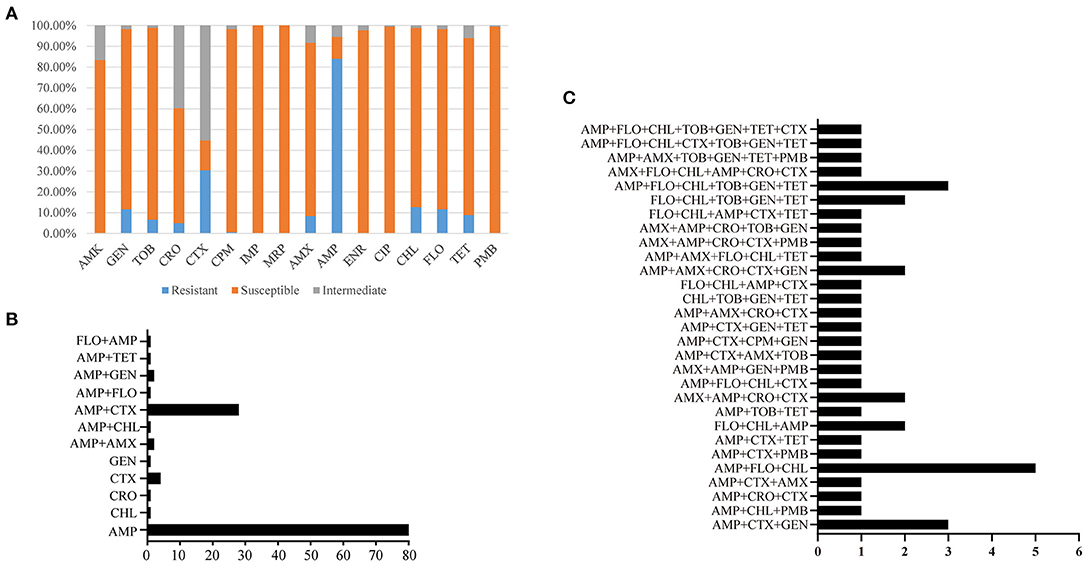
Antimicrobial susceptibility testing (AST) revealed that 9.39% (n = 17) of the B. bronchiseptica isolates recovered in this study were susceptible to all of the 16 types of the antibiotics tested while the remaining 90.61% (n = 164) of the isolates were resistant to at least one type of the antibiotics. All of the B. bronchiseptica isolates recovered in this study were susceptible to imipenem (100%, n = 181), meropenem (100%, n = 181), and polymyxin B (100%, n = 181); more than 80% of the B. bronchiseptica isolates were susceptible to ciprofloxacin (99.45%, n = 180), cefepime (97.79%, n = 177), enrofloxacin (97.79%, n = 177), tobramycin (92.27%, n = 167), gentamicin (86.74%, n = 157), florfenicol (86.74%, n = 157), chloramphenicol (86.19%, n = 156), tetracycline (85.08%, n = 154), amikacin (83.43%, n = 151), and amoxicillin (83.43%, n = 151) (Figure 2A). Approximately 55.25% (n = 100) of the B. bronchiseptica isolates were susceptible to ceftriaxone, while only 14.36% (n = 26) and 10.50% (n = 19) of the B. bronchiseptica isolates were susceptible to cefotaxime and ampicillin, respectively (Figure 2A). Among the 164-drug resistant B. bronchiseptica isolates, resistance rates to 1 type, 2 types, 3 types, 4 types, 5 types, 6 types, and 7 types of drugs were 53.05% (n = 87), 23.17% (n = 38), 7.32% (n = 12), 6.10% (n = 10), 4.88% (n = 8), 3.66% (n = 6), and 1.22% (n = 2), respectively (Figure 2B). Approximately 50.00% (n = 82), 26.83% (n = 44), 17.07% (n = 28), 9.76% (n = 16), and 4.88% (n = 8) of the isolates were resistant to at least 2 types, 3 types, 4 types, 5 types, and 6 types of the antibiotics tested, respectively (Figures 2B,C).

Figure 2. Resistance phenotypes of B. bronchiseptica from pigs in China. (A) Shows percent isolates susceptible or resistant to the 16 kinds of antibiotics tested; (B,C) display the number of isolates with different resistance patterns. In (B,C), X axes show the number of B. bronchiseptica strains while Y axes indicate different resistance patterns. AMK, amikacin; GEN, gentamicin; TOB, tobramycin; CRO, ceftriaxone; CTX, cefotaxime; CPM, cefepime; IPM, imipenem; MRP, meropenem; ENR, enrofloxacin; CIP, ciprofloxacin; CHL, chloramphenicol; FLO, florfenicol; AMX, amoxicillin; AMP, ampicillin; TET, tetracycline; PMB, polymyxin B.
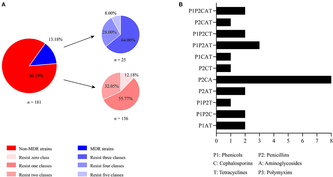
The tested antibiotics in the present study could be divided into eight classes: aminoglycosides (AMK, GEN, TOB), broad-spectrum-cephalosporins (CRO, CTX, CPM), carbapenems (IPM, MRP), fluoroquinolones (ENR, CIP), phenicols (CHL, FLO), penicillins (AMX, AMP), tetracyclines (TET), and polymyxins (PMB). Most of the B. bronchiseptica isolates (86.19%, n = 156) in this study were resistant to less than three classes of the antibiotics. Among these isolates, 55.77% (n = 87) and 32.05% (n = 50) of them were resistant to one and two classes of drugs, respectively (Figure 3A). Approximately 13.18% (n = 25) of the isolates were resistant to more than three classes of the antibiotics. According to the international expert proposal for interim standard definitions for acquired resistance (32), these 25 B. bronchiseptica isolates could be defined as multidrug resistant (MDR) strains. Among these MDR strains, proportions of isolates resistance to three-, four-, and five-classes of drugs were 64.00% (n = 20), 28.00% (n = 7), and 8.00% (n = 2), respectively (Figure 3A). Most MDR-strains possessed a phenotype of co-resistance to aminoglycosides, broad-spectrum-cephalosporins, and penicillins (37.93%, n = 11) (Figure 3B).

Figure 3. Distribution of multidrug resistant (MDR) strains and non-MDR strains of B. bronchiseptica from pigs in China. (A) Shows the percentages of MDR and non-MDR strains as well as percent strains resisting 0, 1, 2, 3, 4, and 5 classes of drugs; (B) displays the number of isolates resistance to different groups of drug classes. In (B), X axis shows the number of B. bronchiseptica strains while Y axis indicates different resistance patterns.
Detection of Antimicrobial Resistance Genes
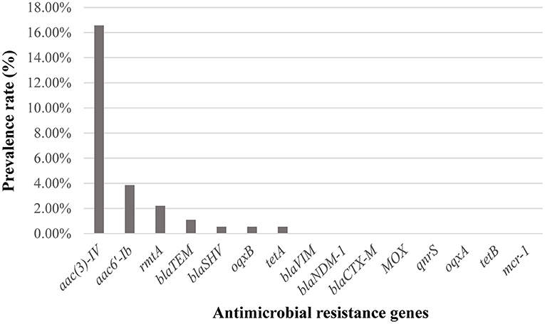
Detection of ARGs showed that 16.57% (n = 30) of the B. bronchiseptica isolates in this study was positive to aac(3)-IV, while 3.87% (n = 7), 2.21% (n = 4), 1.10% (n = 2), 0.55% (n = 1), 0.55% (n = 1), and 0.55% (n = 1) of the isolates were positive to aac6'-Ib, rmtA, blaTEM, blaSHV, oqxB, and tetA, respectively (Figure 4). All isolates were negative to the other ARGs detected (blaVIM, blaNDM−1, blaCTX−M, MOX, qnrS, oqxA, tetB, and mcr-1).

Figure 4. Distribution of antimicrobial resistance genes (ARGs) among B. bronchiseptica isolates in this study.
Detection of Virulence Factors Encoding Genes
Screening of VFGs revealed that 98.90% (n = 179) of the B. bronchiseptica isolates in this study was positive to at least one of the five VFGs detected while the remaining 1.10% (n = 2) ones were negative to all VFGs. The detection rates of fhaB, prn, cyaA, dnt, and betA were 97.24% (n = 176), 91.16% (n = 165), 98.34% (n = 178), 98.34% (n = 178), and 92.82% (n = 168), respectively (Figures 5A,B). Among the VFG-positive isolates, 84.36% (n = 151) of the isolates contained fhaB, prn, cyaA, dnt, and betA, simultaneously (Figure 5C). The remaining isolates harbored “fhaB+prn+cyaA+dnt” (6.15%, n = 11), “fhaB+cyaA+dnt+betA” (7.26%, n = 13), “prn+cyaA+dnt+betA” (1.68%, n = 3), and “fhaB+dnt+betA” (0.56%, n = 1), respectively (Figure 5C).

Figure 5. PCR detection of virulence factors encoding genes (VFGs) among B. bronchiseptica isolates in this study. (A) Shows agarose gel analysis on the PCR products on the five VFGs cyaA (band 1, 377 bp), betA (band 2, 474 bp), fhaB (band 3, 475 bp), dnt (band 4, 491 bp), and prn (band 5, 555 bp); (B) shows the detection rates of the five VFGs while (C) shows the number of strains containing different groups of VFGs. In (C), X axis shows the number of B. bronchiseptica strains while Y axis indicates different groups of VFGs.
Discussion
Although B. bronchiseptica is a well-known leading cause of pig respiratory disorders and an important causative agent of PRDC, there is not too much report on the epidemiology of B. bronchiseptica in pigs round the world, particularly in China, the largest pig rearing and production country. In this study, we described the isolation and characterization of B. bronchiseptica in pigs in China from 2018 to 2020. The average isolation rate of this 3-year period was 4.25% (181/4259), which is much lower than that reported in pigs with clinical respiratory disease in China from 2003 to 2008 (4.25 vs. 18.6%, P <0.05) (26). The average isolation rates of B. bronchiseptica in pigs in different regions from 2018 to 2020 were also much lower than those reported in the same regions from 2003 to 2008 (Hubei: 3.48 vs. 18.0%, P <0.05; Henan: 3.42 vs. 19.6%, P <0.05; Fujian: 4.14 vs. 18.4%, P <0.05; Hunan: 5.96 vs. 19.2%, P <0.05; Anhui: 7.32 vs. 18.0%, P <0.05; Shandong: 3.98 vs. 20.7%, P <0.05) (26). The significant decreasing average isolation rate of B. bronchiseptica from 2018 to 2020 compared to that from 2003 to 2008 might be owing to China's continuously efforts to promote transformation and upgrading of pig industry as well as improve the level of disease prevention and control in pig farms. In addition, the outbreak of African Swine Fever in 2018 and its continuous circulation in pigs in China also accelerates the improvement and enhancement of biosecurity on pig farms in recent years (33), which may also be beneficial for the control of B. bronchiseptica and the other pathogens.
Administration of antimicrobials is still one of the most effective way to control B. bronchiseptica and the other bacteria, but the emergence of drug-resistant bacteria may lead to the failure of using antibiotics in clinic (34–36). Therefore, monitoring the drug resistance profile of clinical microbiology is an important aspect in many epidemiological studies (25, 37, 38). In this study, we characterized the resistance phenotypes of B. bronchiseptica from pigs in China from 2018 to 2020. The results revealed that all isolates were susceptible to imipenem (100%), meropenem (100%), and polymyxin B (100%). All of these three types of antibiotics are proposed to be the last-resort antibiotics for the treatment of infections caused by gram-negative bacteria (29), and they are not approved to be used in veterinary medicine in China. In addition, the majority of the isolates were sensitive to ciprofloxacin (99.45%), cefepime (97.79%), enrofloxacin (97.79%), tobramycin (92.27%), gentamicin (86.74%), florfenicol (86.74%), chloramphenicol (86.19%), tetracycline (85.08%), amikacin (83.43%), and amoxicillin (83.43%). These results are in agreement with the results of previous studies in China (25, 39), as well as in other countries such as Germany and Korea (2, 40–42), suggesting these antibiotics might be suitable candidates for treating B. bronchiseptica infections when necessary. A high level of resistance was found for ampicillin (83.98%), followed by resistance for cefotaxime (30.39%). These findings are also in agreement with those from the other articles (2, 25, 39), and in particular, B. bronchiseptica is documented to be commonly resistant to ampicillin (2). Therefore, these drugs are not recommended to be used in clinic settings. It should be also reminded that several B. bronchiseptica isolates from pigs in China displayed a level of multidrug resistance, particularly co-resistance to aminoglycosides, broad-spectrum-cephalosporins, and penicillins. Continues studies should be taken to monitor the prevalence and change-trend of these MDR-isolates in clinic, as some antibiotics belonging to aminoglycosides, broad-spectrum-cephalosporins, and penicillins are commonly used for treating B. bronchiseptica infections in veterinary medicine (2, 35).
Virulence factors (VFs) play an important role in the pathogenesis of bacteria (43). For B. bronchiseptica, important VFs include filamentous haemagglutinin (FHA), pertactin (PRN), adenylate cyclase-haemolysin toxin, dermonecrotic toxin (DNT), and types III secretion system (44–48), and the expression of these VFs facilitates the invasion of B. bronchiseptica in hosts (49). In the present study, we examined five genes encoding these VFs, including fhaB which encodes filamentous haemagglutinin; prn which encodes pertactin; cyaA which encodes adenylate cyclase-haemolysin toxin; dnt which encodes DNT; and bteA which encodes the T3SS effector A. Surprisingly, over 90% of the pig B. bronchiseptica isolates in this study were positive to these five VFGs examined (fhaB, 97.24%; prn, 91.16%; cyaA, 98.34%; dnt, 98.34%; betA, 92.82%). Importantly, approximately 84.36% of the isolates contained these five kinds of VFGs simultaneously. These results are also in agreement with those reported in B. bronchiseptica isolates from rabbits in China (25), suggesting carrying of these VFGs are broad characteristics of B. bronchiseptica. Laboratory studies have shown that FHA, and PRN expressed in E. coli and Salmonella enterica, as well as adenylate cyclase-haemolysin toxin expressed in B. bronchiseptica provide protection against fatal infections with B. bronchiseptica in mouse models (5, 50, 51).
Despite the findings, this work has several limitations that should be noted. First, all samples used for bacterial isolation were submitted by pig farms from different provinces in China. This way of sample collection may have some influences on the isolation rate. However, the outbreak of African Swine Fever since 2018 and its continuous circulation in pigs in China, and more recently, the worldwide pandemic of the novel coronavirus disease since the late 2019 (COVID-19) made it very difficult for us to collect samples initiatively. Second, the results of antimicrobial susceptibility testing in this study were interpreted by using breakpoints to Enterobacteriaceae published in CLSI document M100, and this is because clinic breakpoints specific to B. bronchiseptica are limited available (2). Third, a very few published epidemiological studies of swine B. bronchiseptica in China are available to date [On March 18, 2021, we searched PubMed with key words “(((Bordetella bronchiseptica) AND (Prevalence)) AND (Pigs)) AND (China)” for reports published, with no language restrictions. Our search identified two articles (26, 39) of relevance to this study. All of them were published by our group in 2011], therefore, we only compared the results we obtained from this study to those reported in our previously published two studies in 2011 (26, 39). However, the results from this work could still help understand the current epidemiological and microbiological characteristics of B. bronchiseptica in pigs in China.
In summary, we reported the isolation, antimicrobial resistance phenotypes, the detection of ARGs and VFGs of B. bronchiseptica from pigs in China from 2018 to 2020 in this study. Our results showed that B. bronchiseptica remains an important pathogen associated with pig respiratory disorders in China. While most of the isolates were still susceptible to ciprofloxacin, cefepime, enrofloxacin, tobramycin, gentamicin, florfenicol, chloramphenicol, tetracycline, amikacin, and amoxicillin, MDR-isolates were still determined. These isolates should receive more attentions and further studies are necessary to monitor the prevalence of drug-resistant B. bronchiseptica. In addition, our results also revealed that several VFGs, including fhaB, prn, cyaA, dnt, and betA displayed a high level of detection rate.
Data Availability Statement
The original contributions presented in the study are included in the article/supplementary material, further inquiries can be directed to the corresponding authors.
Author Contributions
YZ, ZP, and BW delineated the study conception and design. ZP and BW supervised the study. YZ, HY, LG, MZ, FW, WS, LH, LW, WL, and XT collected the bacterial isolates and performed laboratory tests as well as analyzed the data. ZP and YZ wrote the manuscript and approved the final version for publication. ZP, BW, and WL participated in the manuscript discussion and revision. All authors have read and approved the final version of the manuscript.
Funding
This work was supported in part by the Agricultural Science and Technology Innovation Program of Hubei Province (Grant Number: 2018skjcx05). ZP acknowledges the financial support from China Postdoctoral Science Foundation (Grant Numbers: 2020T130232 and 2018M640719). The funders have no role in the study design, data collection and interpretation, or the decision to submit the work for publication.
Conflict of Interest
LG and XT were employed by the company Wuhan Keqian Biology Co., Ltd.
The remaining authors declare that the research was conducted in the absence of any commercial or financial relationships that could be construed as a potential conflict of interest.
References
1. Mattoo S, Cherry JD. Molecular pathogenesis, epidemiology, and clinical manifestations of respiratory infections due to Bordetella pertussis and other Bordetella subspecies. Clin Microbiol Rev. (2005) 18:326–82. doi: 10.1128/CMR.18.2.326-382.2005
2. Kadlec K, Schwarz S. Antimicrobial resistance in Bordetella bronchiseptica. Microbiol Spectr. (2018) 6:ARBA-0024-2017. doi: 10.1128/microbiolspec.ARBA-0024-2017
3. Brockmeier SL, Register KB, Nicholson TL, Loving CL. Bordetellosis. In: Zimmerman JJ, Karriker LA, Ramirez A, Schwartz KJ, Stevenson GW, Zhang J, editors. Diseases of Swine, Eleventh Edition. New York, NY: John Wiley & Sons, Inc. (2019). p. 767–77. doi: 10.1002/9781119350927.ch49
4. Woolfrey BF, Moody JA. Human infections associated with Bordetella bronchiseptica. Clin Microbiol Rev. (1991) 4:243–55. doi: 10.1128/CMR.4.3.243
5. Gueirard P, Weber C, Le Coustumier A, Guiso N. Human Bordetella bronchiseptica infection related to contact with infected animals: persistence of bacteria in host. J Clin Microbiol. (1995) 33:2002–6. doi: 10.1128/JCM.33.8.2002-2006.1995
6. Register KB, Sukumar N, Palavecino EL, Rubin BK, Deora R. Bordetella bronchiseptica in a paediatric cystic fibrosis patient: possible transmission from a household cat. Zoonoses Public Health. (2012) 59:246–50. doi: 10.1111/j.1863-2378.2011.01446.x
7. Linz B, Ivanov YV, Preston A, Brinkac L, Parkhill J, Kim M, et al. Acquisition and loss of virulence-associated factors during genome evolution and speciation in three clades of Bordetella species. BMC Genomics. (2016) 17:767. doi: 10.1186/s12864-016-3112-5
8. Kamanova J. Bordetella type III secretion injectosome and effector proteins. Front Cell Infect Microbiol. (2020) 10:466. doi: 10.3389/fcimb.2020.00466
9. Brockmeier SL, Halbur PG, Thacker EL. Chapter 13: Porcine respiratory disease complex. In: Brogden KA, Guthmiller JM, editors. Polymicrobial Diseases. Washington, DC: ASM Press (2002) p. 231–58.
10. Horiguchi Y. Swine atrophic rhinitis caused by Pasteurella multocida toxin and Bordetella dermonecrotic toxin. Curr Top Microbiol Immunol. (2012) 361:113–29. doi: 10.1007/82_2012_206
11. National Bureau of Statistics. Statistical Bulletin of the National Economic and Social Development of the People's Republic of China. (2020). Available online at: http://www.stats.gov.cn/tjsj/zxfb/202102/t20210227_1814154.html (accessed February 28, 2021).
12. Jorgensen JH, Pfaller MA, Carroll KC, Funke G, Landry ML, Richter SS, et al. Manual of Clinical Microbiology, Eleventh Edition. Washington, DC: ASM Press (2015). doi: 10.1128/9781555817381
13. Hozbor D, Fouque F, Guiso N. Detection of Bordetella bronchiseptica by the polymerase chain reaction. Res Microbiol. (1999) 150:333–41. doi: 10.1016/S0923-2508(99)80059-X
14. González-Sanz R, Herrera-León S, de la Fuente M, Arroyo M, Echeita MA. Emergence of extended-spectrum beta-lactamases and AmpC-type beta-lactamases in human Salmonella isolated in Spain from 2001 to 2005. J Antimicrob Chemother. (2009) 64:1181–6. doi: 10.1093/jac/dkp361
15. Poirel L, Walsh TR, Cuvillier V, Nordmann P. Multiplex PCR for detection of acquired carbapenemase genes. Diagn Microbiol Infect Dis. (2011) 70:119–23. doi: 10.1016/j.diagmicrobio.2010.12.002
16. Pérez-Pérez FJ, Hanson ND. Detection of plasmid-mediated AmpC beta-lactamase genes in clinical isolates by using multiplex PCR. J Clin Microbiol. (2002) 40:2153–62. doi: 10.1128/JCM.40.6.2153-2162.2002
17. Guerra B, Junker E, Miko A, Helmuth R, Mendoza MC. Characterization and localization of drug resistance determinants in multidrug-resistant, integron-carrying Salmonella enterica serotype Typhimurium strains. Microb Drug Resist. (2004) 10:83–91. doi: 10.1089/1076629041310136
18. Guerra B, Helmuth R, Thomas K, Beutlich J, Jahn S, Schroeter A. Plasmid-mediated quinolone resistance determinants in Salmonella spp. isolates from reptiles in Germany. J Antimicrob Chemother. (2010) 65:2043–5. doi: 10.1093/jac/dkq242
19. Granier SA, Hidalgo L, San Millan A, Escudero JA, Gutierrez B, Brisabois A, et al. ArmA methyltransferase in a monophasic Salmonella enterica isolate from food. Antimicrob Agents Chemother. (2011) 55:5262–6. doi: 10.1128/AAC.00308-11
20. Cavaco LM, Frimodt-Møller N, Hasman H, Guardabassi L, Nielsen L, Aarestrup FM. Prevalence of quinolone resistance mechanisms and associations to minimum inhibitory concentrations in quinolone-resistant Escherichia coli isolated from humans and swine in Denmark. Microb Drug Resist. (2008) 14:163–9. doi: 10.1089/mdr.2008.0821
21. Hansen LH, Sørensen SJ, Jørgensen HS, Jensen LB. The prevalence of the OqxAB multidrug efflux pump amongst olaquindox-resistant Escherichia coli in pigs. Microb Drug Resist. (2005) 11:378–82. doi: 10.1089/mdr.2005.11.378
22. Kim HB, Wang M, Park CH, Kim EC, Jacoby GA, Hooper DC. oqxAB encoding a multidrug efflux pump in human clinical isolates of Enterobacteriaceae. Antimicrob Agents Chemother. (2009) 53:3582–4. doi: 10.1128/AAC.01574-08
23. Sáenz Y, Briñas L, Domínguez E, Ruiz J, Zarazaga M, Vila J, et al. Mechanisms of resistance in multiple-antibiotic-resistant Escherichia coli strains of human, animal, and food origins. Antimicrob Agents Chemother. (2004) 48:3996–4001. doi: 10.1128/AAC.48.10.3996-4001.2004
24. Liu YY, Wang Y, Walsh TR, Yi LX, Zhang R, Spencer J, et al. Emergence of plasmid-mediated colistin resistance mechanism MCR-1 in animals and human beings in China: a microbiological and molecular biological study. Lancet Infect Dis. (2016) 16:161–8. doi: 10.1016/S1473-3099(15)00424-7
25. Wang J, Sun S, Chen Y, Chen D, Sang L, Xie X. Characterisation of Bordetella bronchiseptica isolated from rabbits in Fujian, China. Epidemiol Infect. (2020) 148:e237. doi: 10.1017/S0950268820001879
26. Zhao Z, Wang C, Xue Y, Tang X, Wu B, Cheng X, et al. The occurrence of Bordetella bronchiseptica in pigs with clinical respiratory disease. Vet J. (2011) 188:337–40. doi: 10.1016/j.tvjl.2010.05.022
27. Buboltz AM, Nicholson TL, Karanikas AT, Preston A, Harvill ET. Evidence for horizontal gene transfer of two antigenically distinct O antigens in Bordetella bronchiseptica. Infect Immun. (2009) 77:3249–57. doi: 10.1128/IAI.01448-08
29. Peng Z, Li X, Hu Z, Li Z, Lv Y, Lei M, et al. Characteristics of carbapenem-resistant and colistin-resistant Escherichia coli co-producing NDM-1 and MCR-1 from pig farms in China. Microorganisms. (2019) 7:482. doi: 10.3390/microorganisms7110482
30. Peng Z, Liang W, Wang F, Xu Z, Xie Z, Lian Z, et al. Genetic and phylogenetic characteristics of Pasteurella multocida isolates from different host species. Front Microbiol. (2018) 9:1408. doi: 10.3389/fmicb.2018.01408
31. Ai W, Peng Z, Wang F, Zhang Y, Xie S, Liang W, et al. A marker-free Bordetella bronchiseptica aroA/bscN double deleted mutant confers protection against lethal challenge. Vaccines. (2019) 7:176. doi: 10.3390/vaccines7040176
32. Magiorakos AP, Srinivasan A, Carey RB, Carmeli Y, Falagas ME, Giske CG, et al. Multidrug-resistant, extensively drug-resistant and pandrug-resistant bacteria: an international expert proposal for interim standard definitions for acquired resistance. Clin Microbiol Infect. (2012) 18:268–81. doi: 10.1111/j.1469-0691.2011.03570.x
33. Xie S, Liang W, Wang X, Chen H, Fan J, Song W, et al. Epidemiological and genetic characteristics of porcine reproduction and respiratory syndrome virus 2 in mainland China, 2017-2018. Arch Virol. (2020) 165:1621–32. doi: 10.1007/s00705-020-04661-z
34. Ventola CL. The antibiotic resistance crisis: part 1: causes and threats. P T. (2015) 40:277–83.
35. Lappin MR, Blondeau J, Boothe D, Breitschwerdt EB, Guardabassi L, Lloyd DH, et al. Antimicrobial use guidelines for treatment of respiratory tract disease in dogs and cats: antimicrobial guidelines working group of the International Society for Companion Animal Infectious Diseases. J Vet Intern Med. (2017) 31:279–94. doi: 10.1111/jvim.14627
36. Kadlec K, Kehrenberg C, Wallmann J, Schwarz S. Antimicrobial susceptibility of Bordetella bronchiseptica isolates from porcine respiratory tract infections. Antimicrob Agents Chemother. (2004) 48:4903–6. doi: 10.1128/AAC.48.12.4903-4906.2004
37. Russo TP, Pace A, Varriale L, Borrelli L, Gargiulo A, Pompameo M, et al. Prevalence and antimicrobial resistance of enteropathogenic bacteria in yellow-legged gulls (Larus michahellis) in Southern Italy. Animals. (2021) 11:275. doi: 10.3390/ani11020275
38. Kasumba IN, Pulford CV, Perez-Sepulveda BM, Sen S, Sayed N, Permala-Booth J, et al. Characteristics of Salmonella recovered from stools of children enrolled in the Global Enteric Multicenter Study. Clin Infect Dis. (2021). ciab051. doi: 10.1093/cid/ciab051. [Epub ahead of print].
39. Zhao Z, Xue Y, Wang C, Ding K, Wu B, He Q, et al. Antimicrobial susceptibility of Bordetella bronchiseptica isolates from pigs with respiratory diseases on farms in China. J Vet Med Sci. (2011) 73:103–6. doi: 10.1292/jvms.10-0184
40. Prüller S, Rensch U, Meemken D, Kaspar H, Kopp PA, Klein G, et al. Antimicrobial susceptibility of Bordetella bronchiseptica isolates from Swine and Companion Animals and Detection of Resistance Genes. PLoS ONE. (2015) 10:e0135703. doi: 10.1371/journal.pone.0135703
41. El Garch F, de Jong A, Simjee S, Moyaert H, Klein U, Ludwig C, et al. Monitoring of antimicrobial susceptibility of respiratory tract pathogens isolated from diseased cattle and pigs across Europe, 2009-2012: VetPath results. Vet Microbiol. (2016) 194:11–22. doi: 10.1016/j.vetmic.2016.04.009
42. Shin SJ, Kang SG, Nabin R, Kang ML, Yoo HS. Evaluation of the antimicrobial activity of florfenicol against bacteria isolated from bovine and porcine respiratory disease. Vet Microbiol. (2005) 106:73–7. doi: 10.1016/j.vetmic.2004.11.015
43. Sharma AK, Dhasmana N, Dubey N, Kumar N, Gangwal A, Gupta M, et al. Bacterial virulence factors: secreted for survival. Indian J Microbiol. (2017) 57:1–10. doi: 10.1007/s12088-016-0625-1
44. Cotter PA, Yuk MH, Mattoo S, Akerley BJ, Boschwitz J, Relman DA, et al. Filamentous hemagglutinin of Bordetella bronchiseptica is required for efficient establishment of tracheal colonization. Infect Immun. (1998) 66:5921–9. doi: 10.1128/IAI.66.12.5921-5929.1998
45. Inatsuka CS, Xu Q, Vujkovic-Cvijin I, Wong S, Stibitz S, Miller JF, et al. Pertactin is required for Bordetella species to resist neutrophil-mediated clearance. Infect Immun. (2010) 78:2901–9. doi: 10.1128/IAI.00188-10
46. Guiso N. Bordetella adenylate cyclase-hemolysin toxins. Toxins. (2017) 9:277. doi: 10.3390/toxins9090277
47. Brockmeier SL, Register KB, Magyar T, Lax AJ, Pullinger GD, Kunkle RA. Role of the dermonecrotic toxin of Bordetella bronchiseptica in the pathogenesis of respiratory disease in swine. Infect Immun. (2002) 70:481–90. doi: 10.1128/IAI.70.2.481-490.2002
48. Kuwae A, Matsuzawa T, Ishikawa N, Abe H, Nonaka T, Fukuda H, et al. BopC is a novel type III effector secreted by Bordetella bronchiseptica and has a critical role in type III-dependent necrotic cell death. J Biol Chem. (2006) 281:6589–600. doi: 10.1074/jbc.M512711200
49. Fingermann M, Hozbor D. Acid tolerance response of Bordetella bronchiseptica in avirulent phase. Microbiol Res. (2015) 181:52–60. doi: 10.1016/j.micres.2015.09.001
50. Zhao Z, Xue Y, Wu B, Tang X, Hu R, Xu Y, et al. Subcutaneous vaccination with attenuated Salmonella enterica serovar Choleraesuis C500 expressing recombinant filamentous hemagglutinin and pertactin antigens protects mice against fatal infections with both S. enterica serovar Choleraesuis and Bordetella bronchiseptica. Infect Immun. (2008) 76:2157–63. doi: 10.1128/IAI.01495-07
Keywords: Bordetella bronchiseptica, isolation, antimicrobial resistance, virulence factors encoding genes, pigs
Citation: Zhang Y, Yang H, Guo L, Zhao M, Wang F, Song W, Hua L, Wang L, Liang W, Tang X, Peng Z and Wu B (2021) Isolation, Antimicrobial Resistance Phenotypes, and Virulence Genes of Bordetella bronchiseptica From Pigs in China, 2018–2020. Front. Vet. Sci. 8:672716. doi: 10.3389/fvets.2021.672716
Received: 26 February 2021; Accepted: 18 May 2021;
Published: 08 June 2021.
Edited by:
Marina Spinu, University of Agricultural Sciences and Veterinary Medicine of Cluj-Napoca, RomaniaReviewed by:
Faham Khamesipour, Shahid Beheshti University of Medical Sciences, IranAbdelaziz Ed-Dra, Zhejiang University, China
Copyright © 2021 Zhang, Yang, Guo, Zhao, Wang, Song, Hua, Wang, Liang, Tang, Peng and Wu. This is an open-access article distributed under the terms of the Creative Commons Attribution License (CC BY). The use, distribution or reproduction in other forums is permitted, provided the original author(s) and the copyright owner(s) are credited and that the original publication in this journal is cited, in accordance with accepted academic practice. No use, distribution or reproduction is permitted which does not comply with these terms.
*Correspondence: Zhong Peng, cGVuZ3pob25nQG1haWwuaHphdS5lZHUuY24= orcid.org/0000-0001-5249-328X; Bin Wu, d3ViQG1haWwuaHphdS5lZHUuY24=
 Yue Zhang1,2
 Mengfei Zhao
 Wan Liang
 Zhong Peng
 Bin Wu
