 Irina Amorim
 Fatima Faria1
 Marian Taulescu
 Fatima Gärtner
- 1Department of Pathology and Molecular Immunology of the Institute of Biomedical Sciences Abel Salazar, University of Porto, Porto, Portugal
- 2Instituto de Investigação e Inovação em Saúde, Universidade do Porto, Porto, Portugal
- 3Institute of Molecular Pathology and Immunology of the University of Porto, Porto, Portugal
- 4Department of Pathology, Faculty of Veterinary Medicine, University of Agricultural Sciences and Veterinary Medicine Cluj-Napoca, Cluj-Napoca, Romania
- 5Synevovet Laboratory, Bucharest, Romania
- 6Visionvet Veterinary Ophthalmology Clinic, Bologna, Italy
This report provides a clinical, histological, and immunohistochemical description of an unusual hibernoma (pale cell variant) in the subepidermal area of the nipple of a six-year-old bitch. Furthermore, an extensive literature review of hibernomas in animals was made. Physical examination revealed a nodular lesion in the subepidermal area of the third nipple of the left mammary chain. The histopathological findings included lobules of round to oval cells with abundant pale to eosinophilic cytoplasm, containing one or multiple optically empty vacuoles, consistent with nipple hibernoma. Immunohistochemically, the neoplastic cells were negative for cytokeratin AE1/AE3 and p53 but showed strong immunoreaction for vimentin and uncoupling protein-1, thus confirming the brown adipose tissue origin. Local recurrence was not detected after 18 months of follow-up. Hibernomas are rare and benign neoplastic lesions, originating from brown adipose tissue. Due to their histological and molecular resemblance with liposarcoma, a correct diagnosis of these neoplasms is required. In addition, the literature review suggests that hibernomas may present different features, according to species.
Introduction
Hibernomas are rare soft tissue benign neoplasms that originate in brown fat (brown adipose tissue, BAT) (1). These tumors may originate from BAT remnants, ectopic growth or migration of brown adipose cells or aberrant differentiation of mesenchymal cells (2). BAT is a specialized form of adipose tissue specifically designed to generate heat in response to cold (nonshivering thermogenesis) and food ingestion (diet-induced thermogenesis). BAT heat production is activated whenever the organism requires extra heat, like in the postnatal period, during hibernation or when entering into a febrile state (3). The brown aspect of BAT is attributed to the large numbers of cellular mitochondria and abundant blood capillaries (4). In the human fetus, BAT has been identified in the interscapular area, posterior abdominal wall, suprailiac, and peripancreatic adipose tissue, and near autonomic ganglia. Thus, it is not surprising that hibernomas have often been reported in these locations. However, only a few hibernomas were reported in sites where brown fat is usually absent, such as breast or thigh, so far (5). The distribution of brown fat is not as well-characterized in dogs (6).
Currently, there are several reported cases of hibernomas in veterinary medicine, mostly in the orbital region of dogs (7). In the present study, we describe the clinical, pathological, and immunohistochemical features of a hibernoma located in the subepidermal area of the nipple in a bitch. Furthermore, an extensive literature review of hibernomas in animals was conducted.
Case Description
A six-year-old intact female mixed breed dog was presented with acute pruritus of ears, muzzle, and abdomen. The dog has been previously vaccinated and treated for both internal and external parasites. The physical examination revealed a decreased diameter of the auditory canal of the left ear with inflammation and brown granular content with a strong rancid odor, compatible with the overgrowth of Malassezia spp. that was further confirmed by cytology. Erythema and papular skin lesions were present on the abdomen. An additional nodular lesion was detected in the subepidermal area of the third nipple of the left mammary chain. The owner was aware of this lesion for at least 1 year before the dog was presented to the hospital, without causing any pain or discomfort to the animal. The dog was treated with corticosteroids (prednisolone administered orally at 0.5 mg/kg BID for 3 days, followed by the same dosage SID for 3 days, and then QID for other 3 days) along with cleansing and topical application of Canaural® ear drops for the treatment of otitis externa. Excision of the mammary mass was advised and performed ~2 months later, after complete recovery of the dermatological signs.
The lesion was surgically removed, fixed in 10% neutral buffered formalin and submitted for histological examination.
For histology, the samples were cut and processed routinely, and sections were stained with hematoxylin and eosin (HE) and periodic acid-Schiff (PAS). Immunohistochemistry was performed using the Novolink™ Max-Polymer detection system (Novocastra, Newcastle, UK), employing the following monoclonal antisera: pan-cytokeratin (clone AE1/AE3, Zymed) diluted 1:50; vimentin (clone V9, Dako) diluted 1:100; uncoupling protein-1 (UCP1) (clone ab23841, Abcam) diluted 1:20 and p53 (clone BP53.12, Zymed) diluted 1:400. Positive controls consisted of sections of mouse perirenal adipose tissue (Supplementary Figure 1) and canine sebaceous gland adenocarcinoma (Supplementary Figure 2) known to express UCP1 and p53, respectively. A section of subcutaneous adipose tissue of a dog was used as negative control for UCP1 (Supplementary Figure 3).
Grossly, the tissue fragment was represented by a nipple, with an exophytic nodular and dense mass, measuring 12 mm × 6 mm × 4 mm. In cross-section, the lesion was well-delimited, partially encapsulated, with a homogenous appearance and white-yellow color.
Histological examination revealed a well-defined, partially encapsulated, multinodular and highly cellular neoplastic mass, effacing and expanding the subepidermal area of the nipple (Figure 1A). The tumor was composed of round to polygonal cells arranged in nests and delimited by thin fibrovascular stroma. The neoplastic cells presented variably distinct cellular borders, clear or pale eosinophilic cytoplasm, containing one or multiple optically empty vacuoles, mainly resembling lipocytes (Figure 1B). The vacuolar content was PAS-negative and was consistent with, although not proven to be, lipid. The nuclear:cytoplasm (N:C) ratio was low, and the nuclei were round to oval, central or occasionally eccentrically positioned, with fine granular chromatin and 1–2 small, basophilic nucleoli. Anisocytosis and anisokaryosis were mild, and the number of mitotic figures varied from 0 to 2 per high power field (0.237 mm2) (Figure 1C). Scattered mononuclear inflammatory cells, represented by small lymphocytes, plasma cells, and some macrophages were observed within the lesion. There were no evidence for vascular invasion of neoplastic cells.
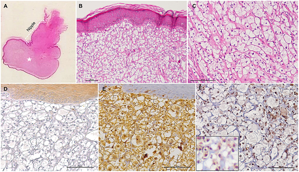
Figure 1. Histological and immunohistochemical features of a hibernoma (pale cell variant) in the subepidermal area of the nipple in a bitch. (A) The subepidermal area of the nipple is distended by an expansile, benign tumor (white asterisk), HE stain. (B,C) The tumor is composed of round to polygonal neoplastic brown fat cells arranged into sheets and poorly defined lobules, separated by a fine fibrovascular stroma. The neoplastic cells show abundant vacuolated cytoplasm, and mild anisocytosis and anisokaryosis, HE stain. (D) The neoplastic cells are negative for cytokeratin AE1/AE3, but positive for Vimentin (E) and UCP1 [(F) and higher magnification in the inset]. IHC.
The neoplastic cell population did not exhibit any immunoreactivity for cytokeratin AE1/AE3 (Figure 1D) and p53 (Supplementary Figure 4) but showed strong positive immunostaining for vimentin (Figure 1E) and UCP1 (Figure 1F).
Based on morphological, histochemical, and immunohistochemical findings, a diagnosis of a subepidermal nipple hibernoma was made.
Outcome
No local recurrence was detected after 18 months of follow-up.
Discussion
A hibernoma is a rare and benign soft tissue neoplasm of the brown adipose tissue (BAT). It was originally described and named “pseudolipoma” by Merkel, in 1906. In 1914, Gery Louis (8) derived the name hibernoma from the tumor's histological similarity to brown fat in hibernating animals.
BAT is present in both human and animal fetuses, being gradually replaced with white adipose tissue (WAT) with aging. However, it may persist in variable degrees throughout adult life, namely in the cervical and interscapular areas, axilla, mediastinum and periaortic and perirenal zones (3). In WAT, fatty acids are metabolized to produce energy in the form of adenosine triphosphate (ATP). In BAT instead, these are metabolized in the form of heat, due to the presence of abundant mitochondria and thermogenin/UCP1 (3). UCP1 is a specialized protein located in the inner mitochondrial membrane which uncouples mitochondrial fatty acid oxidation from ATP synthesis and dissipates energy of substrate oxidation as heat (9). Thus, in both humans and animals, immunohistochemistry for UCP1 has been considered a useful marker to differentiate between BAT and WAT and for diagnosing hibernomas (7, 10, 11). Despite being a valuable marker, UCP1 is not specific and exclusive for hibernomas, since a recent investigation concluded that it was expressed in 23 out of 25 canine liposarcomas, even though neoplastic cells showed reduced immunoreactivity intensity when compared with the positive control (canine abdominal brown adipose tissue) (12). In the present study, and in accordance with the findings reported by Ravi et al. (7), at the subcellular level UCP1 was immunoexpressed in both cytoplasm and nuclei of the neoplastic cells. Cinti et al. (13) demonstrated that UCP1 mRNA expression in rodent brown adipocytes had a positivity pattern very similar to that of the protein IHC localization. Furthermore, immunopositive cells were identified close to negative ones, and some cells showed nuclear staining, with or without cytoplasmic immunoreaction.
In veterinary medicine there are <20 reports of hibernomas in few animal species and in different anatomical sites (Table 1). In dogs, there seems to be a slight predilection in males older than 8 years, while in rats, females are mainly affected (Table 1). In addition, the periorbital region seems to be the most frequent site in dogs, whereas in rats, hibernomas are most often encountered intrathoracically, into the mediastinum (Table 1). The latter location may give rise to new inferences about the histogenesis of these tumors, at least in these two species. In rodents, BAT depots are described in this precise location (3) further suggesting that these lesions may originate from BAT remnants. Contrariwise, to the best of our knowledge, to date there are no reports on the presence of BAT in the periorbital region of animals. Thus, the description of hibernomas in sites that are assumed to be devoid of BAT seems to reinforce the hypothesis that abnormal mesenchymal differentiation may be related with the histogenesis of this neoplastic lesion. Previously, Champigny et al. (25) showed that the adrenergic stimulation of WAT taken from five different locations of adult dogs resulted in reactivation of WAT UCP, which is a classical feature of BAT. These authors postulated that tumors may originate from “dormant” BAT deposits that histologically present WAT-compatible morphology but are in fact constituted by brown preadipocyte cells. Nevertheless, in order to prove this hypothesis, the complete identification and distribution of BAT in dogs needs to be fully characterized.
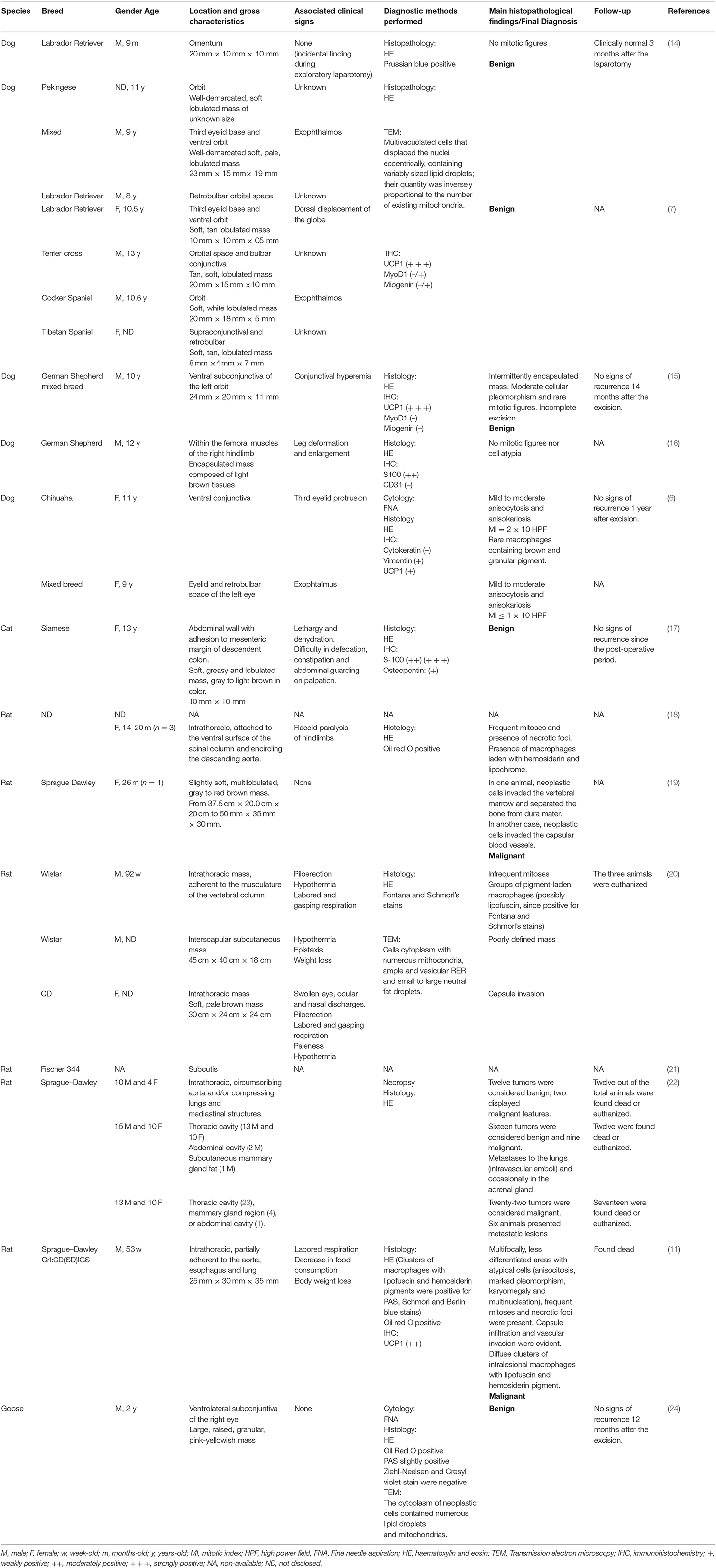
Table 1. Literature available data concerning spontaneously occurring hibernomas in animals.
Herein, the tumor was peculiar not only for the associated anatomical site (nipple), but also for its superficial and subepidermal location. An identical lesion was described in 9-year old boy (26).
In the majority of cases, hibernomas present as asymptomatic, painless, and slowly growing neoplasms, that are usually detected either as a palpable mass or as incidental imaging or necropsy findings, similarly to those occurring in humans (27). According to the literature, when present, the clinical signs are often related to the mass expansion and subsequent adjacent tissue compression, which may further dictate the clinical course (Table 1).
In human medicine, six histological variants of hibernoma are described according to WHO classification: granular or eosinophilic, pale cell, lipoma-like, myxoid, spindle cell, and hybrid (28). Herein, we described a neoplastic lesion that was classified as a pale cell hibernoma. However, details regarding the histological subtype of hibernomas in animals are usually not disclosed in the literature, impairing further comparisons.
Although complete surgical excision is advised to avoid recurrence, most authors claim that these neoplasms do not have the propensity for local recurrence or aggressive behavior. Once again, the existing bibliography seems to be consensual: all cases reported in dogs had benign features, regardless of the anatomical site or whether the surgical excision was complete; irregular and poorly defined boundaries, partial encapsulation, presence of cytological atypia and frequent mitosis, foci of necrosis and invasion of adjacent tissues and vessels are characteristics of malignancy mainly seen in rats. In specific cases, these features eventually led to potential paraneoplastic systemic manifestations, such as marked weight loss (29). Gadea et al. (30) also described the effect of a hibernoma resection on human body composition and metabolism. One year after the resection, the patient gained 15 kg of body weight associated with visceral fat gain, exposing the patient to high risks of metabolic disorder.
Differential diagnosis that should be considered: residual or normal brown fat, which usually does not form a distinct mass; xanthogranulomatous (lipogranulomatous) inflammation, which normally presents with abundant foamy macrophages admixed with mature adipocytes and inflammatory and multinucleated giant cells, both foreign body and Touton types; classical lipoma, although adipocytes are not multivacuolated cells; lipoblastoma, normally composed of immature cells that resemble fetal adipose tissue in different development stages; and liposarcoma, which usually presents with infiltrative behavior, high cellular atypia and high mitotic activity.
P53 overexpression was observed in two human lesions, leading to the hypothesis that functional inactivation of the protein product of this gene may be important in the development of these tumors (23). Furthermore, in hibernomas of transgenic mice, a simian virus 40 (SV40)-transforming gene linked to specific regulatory regions of adipocytes was identified, and the binding of SV40 large T antigen to tumor suppressor genes, including p53 was reported (23). Nevertheless, in the current case, no p53 protein expression was identified amongst the neoplastic cells. Interestingly, in a 2-year carcinogenicity study with tofacitinib (an oral Janus kinase inhibitor for the treatment of rheumatoid arthritis), an increased incidence of hibernoma was noted in female rats. In this research, it was hypothesized that Janus kinase/Signal Transducer and Activator of Transcription inhibition in BAT along with sympathetic stimulation might contribute to the genesis of hibernomas (31). Both investigations call into question the simplicity of these neoplasms and highlight the possible metabolic complexity of hibernomas.
Despite the great abundance and long-life maintenance of the brown adipose tissue in animals, its neoplastic transformation appears to be a rarer event than in humans given the scarcity of cases reported in veterinary medicine. Additionally, following analysis of the existing literature, it was found that these neoplasms may present different features or behavior, according to species. Thus, a correct diagnosis as well as proper monitorization and documentation of hibernomas is needed in order to get insights into their putative metabolic impact, and to avoid unreasonable, radical therapeutic approaches and overtreatments.
Data Availability Statement
The raw data supporting the conclusions of this article will be made available by the authors, without undue reservation.
Ethics Statement
Ethical review and approval was not required for the animal study because it was not the case.
Author Contributions
IA and FF contributed to the sample collection and performed the pathological studies and interpretation of the results. IA and CT analyzed the data and wrote the paper. MT and FF participated in immunohistochemical interpretation. FG contributed to the design, supervised the study, and helped to draft the manuscript. All authors read and approved the final manuscript.
Conflict of Interest
The authors declare that the research was conducted in the absence of any commercial or financial relationships that could be construed as a potential conflict of interest.
Acknowledgments
IPATIMUP integrates the i3S Research Unit, which is partially supported by FCT, the Portuguese Foundation for Science and Technology.
Supplementary Material
The Supplementary Material for this article can be found online at: https://www.frontiersin.org/articles/10.3389/fvets.2021.627288/full#supplementary-material
References
1. Merkel H. Uber ein pseudolipom der mamma (Eigenartigr Fettzellentumor). Beitr Z Pathol Anat. (1906) 39, 152–7.
2. Udwadia ZF, Kumar N, Bhaduri AS. Mediastinal hibernoma. Eur J Cardiothorac Surg. (1999) 15, 533–5. doi: 10.1016/S1010-7940(99)00051-2
3. Vosselman MJ, van Marken Lichtenbelt WD, Schrauwen P. Energy dissipation in brown adipose tissue: from mice to men. Mol Cell Endocrinol. (2013) 379, 43–50. doi: 10.1016/j.mce.2013.04017
4. Oelkrug R, Polymeropoulos ET, Jastroch M. Brown adipose tissue: physiological function and evolutionary significance. J Comp Physiol B. (2015) 185:587–606. doi: 10.1007/s00360-015-0907-7
5. Furlong MA, Fanburg-Smith JC, Miettinen M. The morphologic spectrum of hibernoma: a clinicopathologic study of 170 cases. Am J Surg Pathol. (2001) 25:809–14. doi: 10.1097/00000478-200106000-00014
6. Piccione J, Dial SM. Cytologic appearance of hibernoma in two dogs. Vet Clin Pathol. (2020) 49:125–9. doi: 10.1111/vcp12836
7. Ravi M, Schobert CS, Kiupel M, Dubielzig RR. Clinical, morphologic, and immunohistochemical features of canine orbital hibernomas. Vet Pathol. (2014) 51, 563–568. doi: 10.1177/0300985813493913
8. Louis G. Discussion of tumeur du creux de l' aisselle by F Bonnel. Bull Soc Anat Paris. (1914) 89:110–2.
9. Klingenberg M, Echtay KS. Uncoupling proteins: the issues from a biochemist's point of view. Biochim Biophys Acta. (2001) 1504, 128–43. doi: 10.1016/S0005-2728(00)00242-5
10. Zancanaro C, Pelosi G, Accordini C, Balercia G, Sbabo L, Cinti S. Immunohistochemical identification of the uncoupling protein in human hibernoma. Biol Cell. (1994) 80:75–8. doi: 10.1016/0248-4900(94)90021-3
11. Anagawa A, Okazaki Y, Murakami Y, Tsubota K, Ono M, Matsumoto M, et al. A case of spontaneous malignant hibernoma in a Crl:CD(SD)IGS Rat. Toxicol Pathol. (2009) 22:205–8. doi: 10.1293/tox.22205
12. LaDouceur EEB, Stevens SE, Wood J, Reilly CM. Immunoreactivity of canine liposarcoma to muscle and brown adipose antigens. Vet Pathol. (2017) 54:885–91. doi: 10.1177/0300985817723691
13. Cinti S, Cancello R, Zingaretti MC, Ceresi E, De Matteis R, Giordano A, et al. CL316,243 and cold stress induce heterogeneous expression of UCP1 mRNA and protein in rodent brown adipocytes. J Histochem Cytochem. (2002) 50:21–31. doi: 10.1177/002215540205000103
15. Stuckey JA, Rankin AJ, Romkes G, Slack J, Kiupel M, Dubielzig RR. Subconjunctival hibernoma in a dog. Vet Ophthalmol. (2015) 18:78–82. doi: 10.1111/vop12124
16. Dzimira S, Kapusniak V, Madej JA. Immunohistochemical diagnostic of hibernoma in dog. Pol J Vet Sci. (2015) 18:233–6. doi: 10.1515/pjvs-2015-0029
17. Ozturk-Gurgen H, Egeden E, Calp-Egedem O, Gurel A. Hibernoma in a cat: a case report. Kafkas Univ Vet Fak Derg. (2020) 26:449–52.
18. Carter RL. Tumours of the soft tissues. In: Turusov VS, editor. Pathology of Tumours in Laboratory Animals (Lyon: IARC) (1973). pp. 151–68.
19. Coleman GL. Four intrathroracic hibernomas in rats. Vet Pathol. (1980) 17:634–7. doi: 10.1177/030098588001700514
20. Al Zubaidy AJ, Finn JP. Brown fat tumours (hibernomas) in rats: histopathological and ultrastructural study. Lab Anim. (1983) 17:13–7. doi: 10.1258/002367783781070939
21. Stefanski SA, Elwell MR, Yoshitomi K. Malignant hibernoma in a Fischer 344 rat. Lab Anim Sci. (1987) 37:347–50.
22. Bruner RH, Novilla MN, Picut CA, Kirkpatrick JB, O'Neill TP, Scully KL, et al. Spontaneous hibernomas in Sprague-Dawley rats. Toxicol Pathol. (2009) 37:547–52. doi: 10.1177/0192623309335061
23. Lele SM, Chundru S, Chaljub G, Adegboyega P, Haque AK. Hibernoma: a report of 2 unusual cases with a review of the literature. Arch Pathol Lab Med. (2002) 126:975–8. doi: 10.5858/2002-126-0975-H
24. Murphy CJ, Bellhorn RW, Buyukmihci NC. Subconjunctival hibernoma in a goose. J Am Vet Med Assoc. (1986) 189:1109–10.
25. Champigny O, Ricquier D, Blondel O, Mayers RM, Briscoe MG, Holloway BR. Beta 3-adrenergic receptor stimulation restores message and expression of brown-fat mitochondrial uncoupling protein in adult dogs. Proc Natl Acad Sci U S A. (1991) 88:10774–7. doi: 10.1073/pnas.88.2310774
27. Riley MP, Karamchandani DM. Mammary hibernoma: a rare entity. Arch Pathol Lab Med. (2015) 139:1565–67. doi: 10.5858/arpa2014-0318-RS
28. Miettinen MM, Fanbourg JC, Mandl N. Hibernoma. In: Fletcher CDM, Unni KK, Mertens F, editors. Pathology and Genetics of Tumours of Soft Tissue and Bone (Lyon: IARC Press) (2002). pp. 33–5.
29. Essadel A, Bensaid Alaoui S, Mssrouri R, Mohammadine E, Benamr S, Taghy A, et al. Hibernoma: a rare case of massive weight loss. Ann Chir. (2002) 127:215–7. doi: 10.1016/S0003-3944(01)00715-5
30. Gadea E, Thivat E, Paulon R, Mishellany F, Gimbergues P, Capel F, et al. Hibernoma: a clinical model for exploring the role of brown adipose tissue in the regulation of body weight? J Clin Endocrinol Metab. (2014) 99:1–6. doi: 10.1210/jc.2013-2829
Keywords: brown adipose tissue, hibernoma, UCP1, cancer, canine
Citation: Amorim I, Faria F, Taulescu M, Taulescu C and Gärtner F (2021) Nipple Hibernoma in a Dog: A Case Report With Literature Review. Front. Vet. Sci. 8:627288. doi: 10.3389/fvets.2021.627288
Received: 08 November 2020; Accepted: 13 April 2021;
Published: 12 May 2021.
Edited by:
Tracy Stokol, Cornell University, United StatesReviewed by:
Sharon Dial, University of Arizona, United StatesLaura Pena, Complutense University of Madrid, Spain
Copyright © 2021 Amorim, Faria, Taulescu, Taulescu and Gärtner. This is an open-access article distributed under the terms of the Creative Commons Attribution License (CC BY). The use, distribution or reproduction in other forums is permitted, provided the original author(s) and the copyright owner(s) are credited and that the original publication in this journal is cited, in accordance with accepted academic practice. No use, distribution or reproduction is permitted which does not comply with these terms.
*Correspondence: Marian Taulescu, bWFyaWFuLnRhdWxlc2N1QHVzYW12Y2x1ai5ybw==