 Fábio Ranyeri Nunes Rodrigues1
 Jeniffer Mendes da Silva Freire2Luana de Aguiar Paes Fidelis2Alexandra Ariadne Bittencourt Gonçalves Pereira1
 Davi Emanuel Ribeiro de Sousa1Tais Meziara Wilson1
 Benito Soto-Blanco3*
 Márcio Botelho de Castro1
- 1Veterinary Pathology Laboratory, Campus Darcy Ribeiro, University of Brasília, Brasília, Brazil
- 2Veterinary Teaching Hospital, Campus Darcy Ribeiro, University of Brasília, Brasília, Brazil
- 3Department of Veterinary Clinics and Surgery, Veterinary College, Universidade Federal de Minas Gerais, Belo Horizonte, Brazil
Over the last 20 years, substantial knowledge has been developed in Veterinary oncology, and tumors previously reported only in humans have been identified in animals. Primary paragangliomas of the tongue are extremely rare tumors in human beings and have never been reported in animals. A Chow Chow dog showed an ulcerated nodule at the lingual body, deeply infiltrated, which extended to the base of the tongue. A full clinical and pathological investigation was conducted, and a post-surgical follow-up of 6 months did not detect recurrence. Cytological, histological, and immunohistochemical features are presented and support the diagnosis of lingual paraganglioma. The paraganglioma of the tongue reported in this Chow Chow dog shares many similarities with the human counterpart.
Background
Over the last 20 years, substantial knowledge has been developed in Veterinary oncology. Tumors previously reported only in humans have been identified in animals, and there is a considerable improvement in the diagnosis and treatment of neoplasms and an increase of survival. Neoplasms of the tongue accounted for 54% of lingual lesions, and 64% are malignant tumors, with a particularly high incidence in Chow Chows and Chinese Shar-Peis (1).
Primary paragangliomas of the tongue are extremely rare tumors in human beings and have never been reported in animals. Paragangliomas of the head and neck usually have a parasympathetic origin and generally are non-secretory (2, 3). Extra-adrenal paragangliomas (EPs), also known as chemodectomas, are neuroendocrine tumors derived from paraganglia of the autonomic nervous system (3, 4). In dogs, the most common locations of EPs are mediastinum (aortic body), head and neck (carotid and jugular bodies), and also occur in other infrequent sites such as orbit and abdomen (4–8). EPs have also been reported in orbit (9) and abdomen of horses (10), and in the retroperitoneal and renal region of cats (11, 12).
We describe the occurrence of paraganglioma in the tongue of a Chow Chow dog that shares similarities with the human counterpart.
Case Description
An 11-year-old female Chow Chow dog was referred for clinical care with sialorrhea, oral bleeding during eating, fetid breath (halitosis), and dysphagia for 20 days. The inspection of the oral cavity showed a nodule with an irregular surface, exophytic, and ulcerated at the right side of the lingual body, deeply infiltrated, which extended to the base of the tongue (Figure 1A). Regional lymph nodes did not present changes at clinical evaluation, and no other tumoral masses were detected in the oral cavity. Thoracic x-ray, abdominal ultrasonography, and computed tomography (CT) of the head, complete blood cell count, and biochemical assay, pre-surgical fine needle biopsy aspiration of the neoplasm, and a partial glossectomy was indicated.
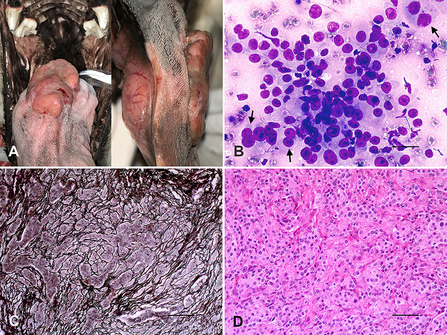
Figure 1. Tongue, paraganglioma. (A) Ulcerated exophytic nodule at the right side of the lingual body, which extended to the base of the tongue. (B) Round to polygonal cells, moderate anisocytosis and anisokaryosis, nuclei with finely granular chromatin, evident nucleoli, and some naked nuclei and binucleated cells (arrows). Romanovsky stain. Bar = 25 μm. (C) Neoplastic cells arranged in rounded nests surrounded by thin fibrovascular trabeculae (“Zellballen” appearance). Reticulin stain. Bar = 100 μm. (D) Tumoral cells with rounded nuclei, heterogeneous chromatin, and eosinophilic cytoplasm. H&E. Bar = 50 μm.
Samples of tissues were fixed in 10% phosphate-buffered formalin (pH 7.0), embedded in paraffin, and sections of 4 μm stained with hematoxylin and eosin (H&E), periodic acid-Schiff (PAS) and reticulin silver stains. Tumoral samples were also submitted to immunohistochemistry using the biotin-peroxidase-streptavidin method (ImmunoDetector DAB, HRP, BioSB Inc.) with primary antibodies incubated overnight (Table 1). The primary antibodies were omitted on the tissue sections and used as negative controls.
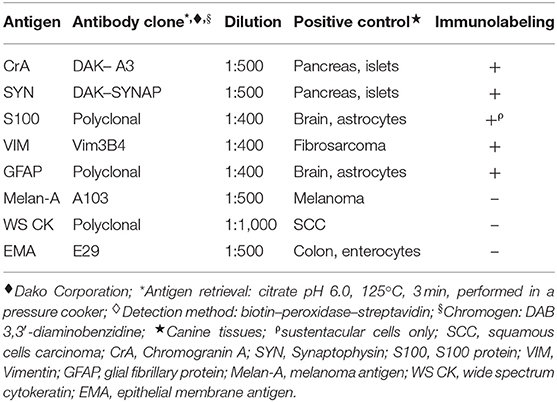
Table 1. Antigen, origin of the primary antibodies♦, dilutions used in the immunostaining protocols, and immunolabeling of the paraganglioma of tongue in the Chow Chow dog.
Additionally, we made a review of manuscripts published on tongue paraganglioma in animals and humans. A PubMed query was conducted with the following keywords: paraganglioma, tongue, pharynx, dog, canine, animals, veterinary, head, and neck. Articles were selected based on the location of paragangliomas and scientific relevance and were used to compare features between the dog and the human counterpart.
CT revealed a poorly delimited neoformation in the body of the tongue, isodense, heterogeneous at the proper lingual, styloglossus, hyoglossus, and genioglossus muscles. Thoracic x-ray and abdominal ultrasonography did not detect tumoral masses or other abnormalities. The pre-surgical evaluation showed an increase in serum activity of creatine phosphokinase (CK 435.7 UI/L, reference range: 69-214 UI/L). The CBC and the serum levels of alanine aminotransferase, aspartate aminotransferase, alkaline phosphatase, γ-glutamyl transferase, creatinine, urea nitrogen, total protein, albumin, cholesterol, triglycerides, calcium, and phosphorus were within the reference ranges (13).
Fine needle biopsy aspiration of the tongue mass revealed round to polygonal cells arranged in groups or isolated, moderate anisocytosis and anisokaryosis, nuclei with finely granular chromatin, some naked nuclei, and the presence of one or two nucleoli. The tumor cells had a slightly basophilic cytoplasm and occasional acinar-like structures, and binucleated cells were also observed (Figure 1B). The partial glossectomy was successful with the complete resection of a solid and firm white-brown nodule of 6.5 × 5.0 × 4.0 cm.
Histologically, the lingual mass was densely cellular and composed of round to polygonal cells arranged in distinctly rounded nests surrounded by a thin fibrovascular stroma (“Zellballen” appearance) evidenced by the reticulin stain (Figure 1C). Neoplastic cells showed rounded nuclei, heterogeneous chromatin, moderate anisocytosis, and anisokaryosis, and occasional evident single nucleoli (Figure 1D). Tumoral mass also showed two mitotic figures per 10 high-power fields (2.37 mm2). The cytoplasm was eosinophilic and slightly granular, and PAS-negative stained. There were multifocal tumoral infiltrations to the muscular tissues and scarce surgical margins. Immunohistochemistry of tumor samples (Table 1) showed strong immunolabeling for the neuroendocrine markers Chromogranin A (Figure 2A) and Synaptophysin (Figure 2B), and vimentin, moderate GFAP positivity, and remarkable S100 protein immunostaining of sustentacular cells (Figure 2C).
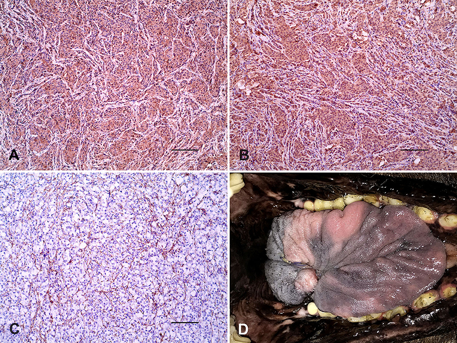
Figure 2. Tongue, paraganglioma. (A) Strong positivity of neoplastic cells for Chromogranin A. (B) Immunolabeling of tumoral cells for Synaptophysin. (C) S100 protein immunostaining of sustentacular cells. Immunoperoxidase. Bar = 100 μm. (D) Proper healing of the tongue 1 month after partial glossectomy.
The dog returned 1 month after partial glossectomy for clinical follow-up, and there was an improvement in food intake and weight gain. The tongue showed proper healing (Figure 2D), and halitosis disappeared. Post-surgical follow-up occurred for 6 months, and there was no recurrence of the lingual tumor. Morphological features and immunohistochemistry assay supported the diagnosis of primary paraganglioma of the tongue. The query performed in PubMed failed to demonstrate paragangliomas of the tongue in the Veterinary literature and a few cases in human beings. Table 2 summarizes the main features of paragangliomas of the tongue in the Chow Chow dog and human patients.
Table 2. General features of paragangliomas of the tongue in the Chow Chow dog and human cases.
Discussion
The paraganglioma of the tongue in the Chow Chow dog shares many similarities with the human counterpart. Difficulty swallowing associated with a partial tongue dysfunction owing to tumor infiltration and traumatic injuries enabled sialorrhea, difficulty feeding, bleeding, and halitosis in the dog. Hemorrhage during eating was also related to traumatic injuries of a tumoral mass on the base of the tongue in the first report of primary lingual paraganglioma in humans (14). Bleeding has not been detected in other cases of paragangliomas in humans with small neoplasms on the tongue (15–17), which possibly reduced the risk of traumatic injuries and hemorrhage.
Pharyngeal paresthesia (14), pain and swelling of the tongue (15), throat irritation (16), choke (17), and sleep snoring (18) are other clinical signs detected in human patients with paragangliomas of the tongue. Anatomical location of the tumor on the tongue possibly did not have an effect on pharyngeal areas, reducing the variability of clinical signs in the Chow Chow dog. It is also important to consider the difficulty in evaluating some clinical signs and sensations in dogs concerning paraganglioma of the tongue in comparison with their counterparts in humans.
The large ulcerated tumoral mass in the dog showed deep infiltration of the lingual muscles from the body of the tongue to the base. The high serum activity of creatine phosphokinase possibly demonstrates muscular damage related to tumor growth and infiltration. The location of paragangliomas in the tongue of humans is similar to that observed in the dog, and ulceration and serum biochemistry abnormalities have not been reported (14–17). The absence of other hematological or biochemical changes indicates the general good physical condition of the dog despite feeding difficulties and weight loss.
Cytological features of the ulcerated lingual lesion in the Chow Chow dog, such as acinar-like configuration or loose clusters of cells with round to oval nuclei within stippled chromatin, moderate anisocytosis and anisokaryosis, and some naked nuclei (19–21), provided the pre-surgical suspicion of a neuroendocrine origin tumor. Similar cytological findings were also reported in a cat with renal paraganglioma (12). The moderate cellular pleomorphism suggested a malignant neoplasm; however, the follow-up of 6 months did not detect recurrence or metastasis. An orbital paraganglioma in a dog with mild pleomorphism of neoplastic cells showed no post-surgical recurrence for 25 months (8). Most extra-adrenal paragangliomas (EPs) of dogs arise in aortic and carotid bodies, and uncommonly in other locations, with variable cytological features and cellular pleomorphism despite the grade of malignancy (5–7).
In human patients, there are different cytological presentations of paragangliomas of the head (19, 20), including similar morphological features such as observed in the dog. Cytological diagnosis of paragangliomas of the tongue has never been conducted in human patients (14–18). Cytological findings may suggest the diagnosis of paragangliomas; however, only the combination of histopathology, immunohistochemistry, radiographic studies, and tumor location is confirmatory (20, 21). Fine needle aspiration cytology of the tongue provided to be useful preoperatively in the Chow Chow dog, and should also be considered to investigate cases suspected of recurrence or metastasis.
The most relevant histological diagnostic feature detected in the tumor of the tongue was the “Zellballen” appearance composed by nests of round to polygonal neoplastic cells PAS-negative. Microscopically, tumoral infiltration to the lingual muscles was also a significant finding. Fibrovascular stroma dividing packets or nests of variable pleomorphic epithelioid to polygonal tumoral cells, and surrounded by thin trabeculae of fibrous tissue are remarkable in EPs of dogs (5, 6, 8), and have a similar “neuroendocrine packeting” pattern in horses (9, 10) and cats (11, 12).
Paragangliomas of the tongue in human patients are histologically similar to those observed in other tissues (2, 15–17, 19) and also in domestic animals (5, 6, 9–12). The low number of mitosis in the tumoral mass of the tongue was also reported in benign and malignant paragangliomas of dogs (5, 6, 8). A high mitotic index was observed in a cat with malignant renal paraganglioma (12). Criteria of malignancy based on pathological features of paragangliomas may be imprecise to assess, including mitotic index, infiltration grade, and tumor size. Distant metastasis is considered the only unmistakable finding of malignant tumors (2). Despite the absence of recurrence or metastasis for 6 months, we cannot state for sure on the absence of malignancy of the lingual tumor on the dog.
The immunolabeling for Chromogranin A, Synaptophysin, Vimentin, and GFAP antibodies is a hallmark of extra-adrenal paragangliomas in dogs and humans (2–8), and also in horses (9, 10), and cats (11, 12). Paragangliomas of the tongue in human patients have similar immunohistochemical features (16–18), as observed in the dog.
The exclusive S100 protein positivity of sustentacular cells in the Chow Chow dog was also reported in paragangliomas of the tongue in humans (16–18), and in EPs in a dog (7), and cat (12). Most EPs of dogs are anti-S100 protein negative (4, 6). Melan-A, Cytokeratin, and EMA negative immunostainings of the lingual tumoral cells were fundamental to differentiate from common oral tumors of dogs such as melanomas and carcinomas. The immunohistochemical assay showed a high similarity between the lingual paraganglioma of the Chow Chow dog and human beings (16, 17).
The tongue is an unexpected anatomical site to arise paragangliomas, a hypothesis supported by the absence of cases reported in Veterinary literature and the rarity in human beings. Once the anatomical distribution of minor paraganglia is not entirely known, paragangliomas of the tongue may arise from parasympathetic paraganglia related to the branches of facial or glossopharyngeal nerves, or in the walls of arteries (22). Although the origin of paragangliomas of the tongue is uncertain, a germline mutation in succinate dehydrogenase gene B (SDHB) highlighted a genetic basis for the development of paragangliomas in human beings (17). Canine pheochromocytomas and paragangliomas presented similar genetic alterations of SDHB and SDHD genes and other significant chromosomal changes such as the loss of chromosome 5 (23).
Granular cell tumors (GCTs) in the tongue of dogs (24–26) are one of the most challenging differential diagnoses, sharing some similar gross and microscopic features with paragangliomas (15–17). Cytologically, lingual GCTs are composed of cells with a plasmacytoid appearance and voluminous granular cytoplasm (27), which were not observed in the Chow Chow dog. Cytoplasmic PAS-negativity of tumor cells was a determinant histological feature in the differentiation between the dog's paraganglioma of the tongue and GCT (24–26). Immunostaining of neoplastic cells for Chromogranin A, such as detected in the dog, is not observed in human cases of oral granular cell tumors (28). S100 immunolabeling of tumor cells in dogs with GCTs in the tongue may be variable (24–26) and contrasts with the S100 positivity of sustentacular cells and lack of immunostaining of neoplastic cells in the lingual paraganglioma.
Despite the rarity, paragangliomas of the tongue should also be included in the differential diagnosis of the most frequent lingual neoplasms in dogs such as melanomas, carcinomas, fibrosarcomas, hemangiosarcomas, and some benign tumors such as squamous papilloma, plasma cell tumors, and GCTs. Further investigations on the anatomic distribution of paraganglia in the oral cavity, and genetic analysis of SDH genes, may contribute to the knowledge on lingual paragangliomas in dogs.
Data Availability Statement
The raw data supporting the conclusions of this article will be made available by the authors, without undue reservation.
Ethics Statement
Ethical review and approval was not required for the animal study because it was an expontaneous disease. Written informed consent was obtained from the owners for the participation of their animals in this study.
Author Contributions
JS and LF performed clinical evaluations. FR, AP, DS, TW, BS-B, and MC performed pathological examinations. MC drafted the manuscript. All authors read and approved the final manuscript.
Conflict of Interest
The authors declare that the research was conducted in the absence of any commercial or financial relationships that could be construed as a potential conflict of interest.
Acknowledgments
We are thankful to Dr. Emanuel Rocha Fernandes, DVM, Clínica Veterinária Pró Saúde for the referral of the animal to the Veterinary Teaching Hospital. Special thanks to Coordenação de Aperfeiçoamento de Pessoal de Nível Superior - Brazil (CAPES) for the partial financing (Finance Code 001), and the support of National Council for Scientific and Technological Development (CNPQ).
References
1. Dennis MM, Ehrhart N, Duncan CG, Barnes AB, Ehrhart EJ. Frequency of and risk factors associated with lingual lesions in dogs: 1,196 cases (1995–2004). J Am Vet Med Assoc. (2006) 228:1533–37. doi: 10.2460/javma.228.10.1533
2. Wasserman PG, Savargaonkar P. Paragangliomas: classification, pathology, and differential diagnosis. Otolaryngol Clin North Am. (2001) 34:845–62. doi: 10.1016/S0030-6665(05)70351-0
3. Pellitteri PK, Rinaldo A, Myssiorek D, Gary Jackson C, Bradley PJ, Devaney KO, et al. Paragangliomas of the head and neck. Oral Oncol. (2004) 40:563–75. doi: 10.1016/j.oraloncology.2003.09.004
4. Galac S, Korpershoek E. Pheochromocytomas and paragangliomas in humans and dogs. Vet Comp Oncol. (2017) 15:1158–70. doi: 10.1111/vco.12291
5. Ilha MRS, Styer EL. Extra-adrenal retroperitoneal paraganglioma in a dog. J Vet Diagn Invest. (2013) 25:803–6. doi: 10.1177/1040638713506579
6. Yamamoto S, Fukushima R, Hirakawa A, Abe M, Kobayashi M, Machida N. Histopathological and immunohistochemical evaluation of malignant potential in canine aortic body tumours. J Comp Pathol. (2013) 149:182–91. doi: 10.1016/j.jcpa.2012.12.007
7. Romanucci M, Malatesta D, Berardi I, Pugliese G, Fusco D, Della Salda L. Cytological, histological and ultrastructural nuclear features of monster cells in a canine carotid body carcinoma. J Comp Pathol. (2014) 151:57–62. doi: 10.1016/j.jcpa.2014.03.001
8. Fischer MC, Taeymans ON, Monti P, Scurrell EJ, Eddicks L, Matiasek K, et al. Orbital paraganglioma in a dog. Tierarztl Prax Ausg K Kleintiere Heimtiere. (2018) 46:410–15. doi: 10.1055/s-0038-1677406
9. Miesner T, Wilkie D, Gemensky-Metzler A, Weisbrode S, Colitz C. Extra-adrenal paraganglioma of the equine orbit: six cases. Vet Ophthalmol. (2009) 12:263–68. doi: 10.1111/j.1463-5224.2009.00706.x
10. Herbach N, Breuer W, Hermanns W. Metastatic extra-adrenal sympathetic paraganglioma in a horse. J Comp Pathol. (2010) 143:199–202. doi: 10.1016/j.jcpa.2010.01.008
11. Borchert C, Berent A, Weisse C. Subcutaneous ureteral bypass for treatment of bilateral ureteral obstruction in a cat with retroperitoneal paraganglioma. J Am Vet Med Assoc. (2018) 253:1169–76. doi: 10.2460/javma.253.9.1169
12. Friedlein RB, Carter AJ, Last RD, Clift S. The diagnosis of bilateral primary renal paragangliomas in a cat. J S Afr Vet Assoc. (2017) 88:e1–6. doi: 10.4102/jsava.v88i0.1412
13. Krimer PM. Generating and interpreting test results: test validity, quality control, reference values, and basic epidemiology. In: Latimer KS, editor. Duncan & Prasse's Veterinary Laboratory Medicine: Clinical Pathology. 5th ed. Ames, IA: Wiley-Blackwell (2011). p. 365–82.
14. Bertogalli D, Calearo C, Pignataro O. Les paragangliomes non chromatophiles a siege rare. A propos de deux cas. Ann Otol. (1959) 76:688–99.
15. Lustmann J, Ulmansky M. Paraganglioma of the tongue. J Oral Maxillofac Surg. (1990) 48:1317–9. doi: 10.1016/0278-2391(90)90490-S
16. Nielsen TO, Séjean G, Onerheim RM. Paraganglioma of the tongue. Arch Pathol Lab Med. (2000) 124:877–9. doi: 10.1043/0003-9985(2000)124<0877:POTT>2.0.CO;2
17. Duran Alvarez MA, Tavarez Rodriguez JJ, Robledo M. Paraganglioma of the tongue with SDHB gene mutation in a patient with graves' disease. Clin Case Rep. (2019) 7:726–30. doi: 10.1002/ccr3.2065
18. Liu HB, Kang PP, Liu SR, Zhao JG, Zhang SH. Paraganglioma in the bottom of tongue: one case report. Zhonghua Er Bi Yan Hou Tou Jing Wai Ke Za Zhi. (2017) 52:547–8. doi: 10.3760/cma.j.issn.1673-0860.2017.07.017
19. Ntanasis-Stathopoulos I, Tsilimigras DI, Klapsinou E, Daskalopoulou D, Vaida S, Arnogiannaki N, et al. Challenging differential diagnosis of an extra-adrenal paraganglioma; the role of fine needle aspiration cytology. Diagn Cytopathol. (2017) 45:565–8. doi: 10.1002/dc.23696
20. Varma K, Jain S, Mandal S. Cytomorphologic spectrum in paraganglioma. Acta Cytol. (2008) 52:549–56. doi: 10.1159/000325596
21. Jashnani KD, Patil RD, Balsarkar DJ. Loose cell clusters with vascular coats: zellballen pattern of paraganglioma on cytology. J Cytol. (2013) 30:278–9. doi: 10.4103/0970-9371.126670
22. Prajsnar A, Balak N, Walter GF, Stan AC, Deinsberger W, Tapul L, et al. Recurrent paraganglioma of meckel's cave: case report and a review of anatomic origin of paragangliomas. Surg Neurol Int. (2011) 2:45. doi: 10.4103/2152-7806.79763
23. Korpershoek E, Dieduksman DAER, Grinwis GCM, Day MJ, Reusch CE, Hilbe M, et al. Molecular alterations in dog pheochromocytomas and paragangliomas. Cancers. (2019) 11:607. doi: 10.3390/cancers11050607
24. Geyera C, Hafner A, Pfleghaar S, Hermanns W. Immunohistochemical and ultrastructural investigation of granular cell tumours in dog, cat, and horse*. Zentralbl Veterinarmed B. (1992) 39:485–94. doi: 10.1111/j.1439-0450.1992.tb01197.x
25. Patnaik AK. Histologic and immunohistochemical studies of granular cell tumors in seven dogs, three cats, one horse, and one bird. Vet Pathol. (1993) 30:176–85. doi: 10.1177/030098589303000211
26. Rallis TS, Tontis DK, Soubasis NH, Patsiaura KK, Papazoglou LG, Adamama-Moraitou KK. Immunohistochemical study of a granular cell tumor on the tongue of a dog. Vet Clin Pathol. (2001) 30:62–6. doi: 10.1111/j.1939-165X.2001.tb00260.x
27. Fitzhugh VA, Maniar KP, Gurudutt VV, Rivera M, Chen H, Wu M. Fine-needle aspiration biopsy of granular cell tumor of the tongue: a technique for the aspiration of oral lesions. Diagn Cytopathol. (2009) 37:839–42. doi: 10.1002/dc.21112
Keywords: cytology, dogs, immunohistochemistry, neoplasm, paraganglia, oral cavity
Citation: Rodrigues FRN, da Silva Freire JM, Fidelis LAP, Pereira AABG, de Sousa DER, Wilson TM, Soto-Blanco B and de Castro MB (2020) Paraganglioma of the Tongue in a Chow Chow Dog: A Comparison With the Human Counterpart and Literature Review. Front. Vet. Sci. 7:422. doi: 10.3389/fvets.2020.00422
Received: 20 May 2020; Accepted: 12 June 2020;
Published: 24 July 2020.
Edited by:
Gustavo A. Ramírez Rivero, Universitat de Lleida, SpainReviewed by:
Alejandro Suarez-Bonnet, Royal Veterinary College (RVC), United KingdomJavier Asin, California Animal Health and Food Safety (CAHFS), United States
Copyright © 2020 Rodrigues, da Silva Freire, Fidelis, Pereira, de Sousa, Wilson, Soto-Blanco and de Castro. This is an open-access article distributed under the terms of the Creative Commons Attribution License (CC BY). The use, distribution or reproduction in other forums is permitted, provided the original author(s) and the copyright owner(s) are credited and that the original publication in this journal is cited, in accordance with accepted academic practice. No use, distribution or reproduction is permitted which does not comply with these terms.
*Correspondence: Benito Soto-Blanco, YmVuaXRvJiN4MDAwNDA7dWZtZy5icg==