- 1Department of Spine Surgery, The Second Affiliated Hospital of Chongqing Medical University, Chongqing, China
- 2Geriatric Clinical Research Center of Chongqing, Chongqing, China
- 3Jiangjin Central Hospital, Chongqing, China
Objective: To evaluate the clinical effects of the posterior unilateral approach with 270° spinal canal decompression and three-column reconstruction using double titanium mesh cage (TMC) for thoracic and lumbar burst fractures.
Materials and methods: From May 2013 to May 2018, 27 patients with single-level thoracic and lumbar burst fractures were enrolled. Every patient was followed for at least 18 months. Demographic data, neurologic status, back pain, canal compromise, anterior body compression, operative time, estimated blood loss and surgical-related complications were evaluated. Radiographs were reviewed to assess deformity correction, anterior body height correction, bony fusion and TMC subsidence.
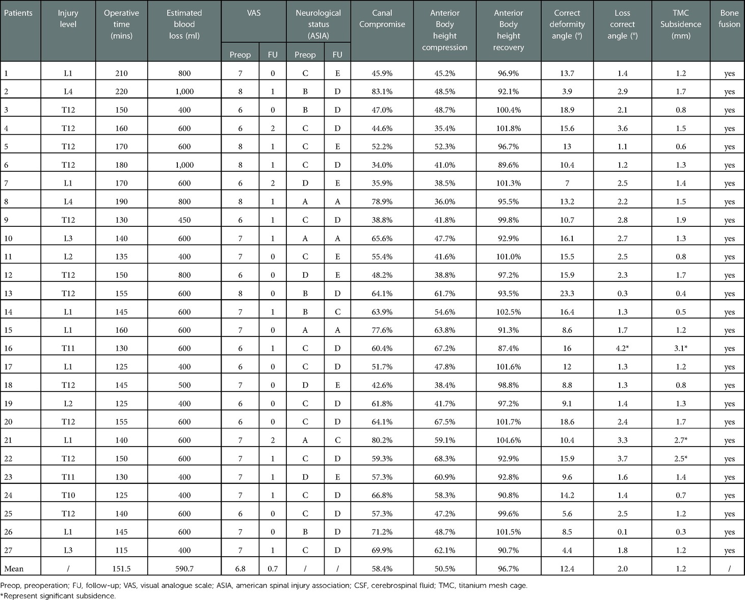
Results: The average preoperative percentages of canal compromise and anterior body height compression were 58.4% and 50.5%, respectively. All surgeries were successfully completed in one phase, the operative time was 151.5 ± 25.5 min (range: 115–220 min), the estimated blood loss was 590.7 ± 169.9 ml (range: 400–1,000 ml). Neurological function recovery was significantly improved except for 3 grade A patients. The preoperative visual analog scale (VAS) scores for back pain were significantly decreased compared with the values at the last follow-up (P = 0.000). The correct deformity angle was 12.4 ± 4.7° (range: 3.9–23.3°), and the anterior body height recovery was 96.7%. The TMC subsidence at the last follow-up was 1.3 ± 0.7 mm (range: 0.3–3.1 mm). Bony fusion was achieved in all patients.
Conclusion: The posterior unilateral approach with 270° spinal canal decompression and three-column reconstruction using double TMC is a clinically feasible, safe and alternative treatment for thoracic and lumbar burst fractures.
Introduction
Vertebral burst fracture is described as compressed fracture of the anterior and middle portions of the spinal column combined with mild to severe epidural compression by bone fragments that shifted into the spinal canal (1). They are commonly caused by severe trauma, including a fall from a height and traffic accidents (2). Vertebral burst fractures account for 10%–20% of all spine fractures. They frequently occur at the thoracic and lumbar junction and may cause neurologic complications and angular deformities (3).
The goal of surgical intervention for thoracic and lumbar burst fractures is to decompress the neural elements, stabilize the spinal column, restore vertebral body height, and correct angular deformities. Decompression and reconstruction can be achieved via anterior and posterior approaches for thoracic and lumbar burst fractures. However, the clinical results of fixation to correct kyphotic deformities via the anterior approach are obviously inferior to those of the posterior approach (3–5). Additionally, higher morbidity has been reported during the exploration of thoracic and lumbar vertebral bodies or discs via anterior transthoracic or retroperitoneal approach (6). The single-stage transpedicular or costotransversectomy approach for corpectomy and reconstruction of thoracic and lumbar burst fractures has been well described (3–5, 7). The approach allows not only circumferential decompression but also simultaneous posterior instrumentation. However, the posterior spinal structure is often completely removed for adequate decompression, thus losing the feasibility of posterior column fusion. Additionally, a large ventral supporting cage is usually required to reconstruct the anterior column, and its placement is technically demanding, carrying risk of injury to the surrounding neural elements. Especially in the lumbar spine, nerve root damage can cause lower limb dysfunction. In contrast, a small cage has insufficient bone graft contact area, which will increase the risk of non-fusion. Moreover, the pressure increase in the upper and lower endplates may lead to subsidence or endplate fracture. Therefore, a more convenient, safe, and alternative method for decompression and reconstruction of thoracic and lumbar burst fractures is needed.
Materials and methods
We received permission from the ethics committee of our hospital and written informed consent from all the patients.
Patients' characteristics and details regarding the evaluation methods
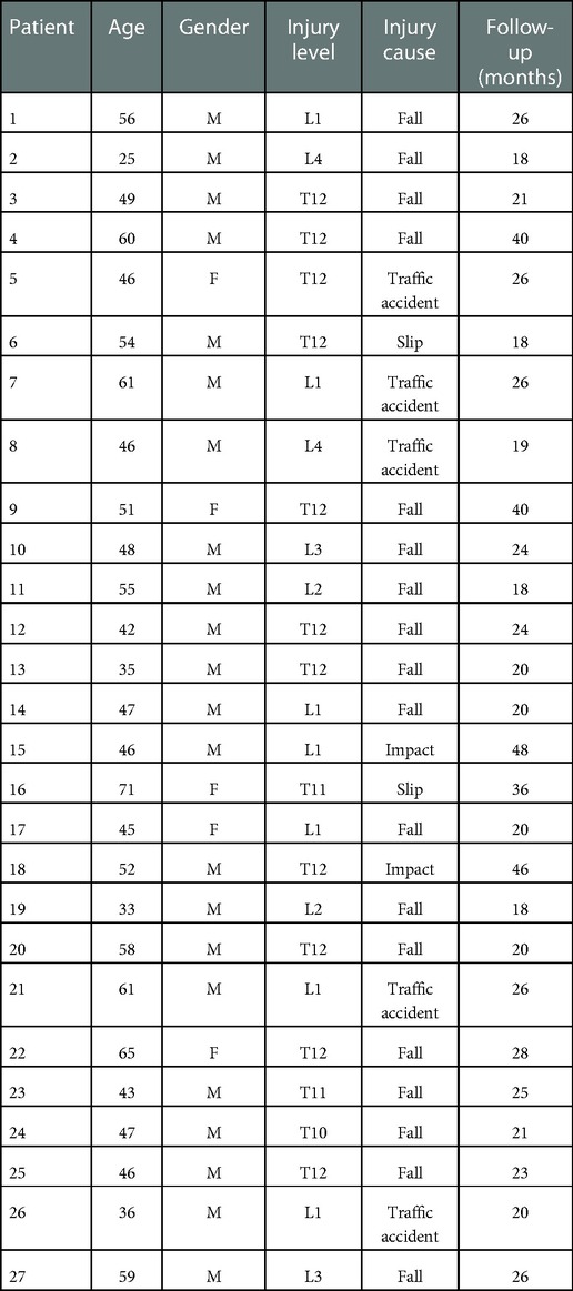
Inclusion criteria: (1) Single-level burst fractures with nerve injury located in the thoracic and lumbar and lumbar vertebrae, (2) Load Sharing Score (LSS) ≥6 points (8), (3) The thoracic and lumbar injury severity score (TLICS) ≥4 points. Exclusion criteria: (1) The fracture is located in the upper thoracic vertebra, (2) Pathological fracture due to tumor and infection. Data from 27 patients meeting the above criteria were retrospectively collected. All patients underwent a posterior unilateral approach for 270° spinal canal decompression and three-column reconstruction using double TMC between May 2013 and May 2018. The demographic data, cause, and level of the injury (Table 1), Visual Analog Scale (VAS) for back pain, neurologic status, operative time, estimated blood loss, surgical-related complications, and follow-up time were collected. Radiographs were reviewed to assess canal compromise, anterior body height compression, kyphotic/lordotic angle, anterior body height recovery, correct deformity angle, loss correct angle, bony fusion and TMC subsidence.
The clinical and radiographic indexes were described as follows
1. Neurologic status was assessed by the American Spinal Injury Association (ASIA) impairment score.
2. Canal compromise was calculated with the following formula: , where a was the percentage of canal compromise (5); D0 was the midsagittal diameter of the spinal canal of the injured level; and D1 and D2 were the midsagittal diameters of the spinal canal at the levels above and below the injured level (Figure 1).
3. Anterior body height compression was calculated with the following formula: , where b0 was the percentage of anterior body height compression (9); A0 was the anterior body height of the injured level; and Ap and Ad were the anterior body heights of the proximal and distal levels (Figure 2).
4. Anterior body height recovery was calculated with the following formula: , where b1 was the percentage of anterior body height recovery; and A1 was the anterior body height of the injured level after surgery (Figure 2).
5. The kyphotic/lordotic angle (α) was the angle of kyphotic or lordotic deformity of the injured level, measured as the angle between the superior endplate of the vertebral body above the affected level and the inferior endplate of the vertebral body below the affected level (5) (Figure 2). α0 was the preoperative angle, and α1 was the postoperative angle of deformity. The kyphotic angle was defined as the positive value, and the lordotic angle was defined as the negative value.
6. Correct deformity angle (°): kyphotic correction = α0−α1; lordotic correction = α1−α0.
7. The correct loss angle was the reductive angle between the last follow-up and immediate post-operation.
8. Definition of bone fusion: the x-ray or CT film lacked lucency at the cage-vertebra junction, or the presence of mature bony trabeculae around the cage bridging the adjacent vertebral bodies was accepted as a sign of fusion (10, 11).
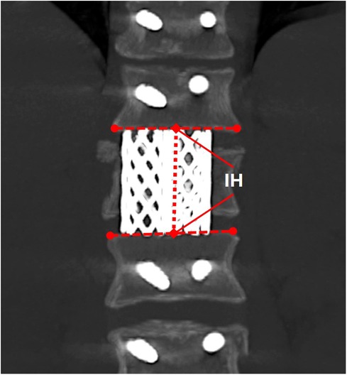
9. TMC subsidence was the total subsidence of intervertebral height (IH) from immediate post-operation to the last follow-up. IH was the distance between the midpoints of the inferior endplate of the proximal level and the superior endplate of the distal level (Figure 3). Subsidence over 2 mm or a correct loss angle over 4° was considered the presence of TMC subsidence in this study (12).
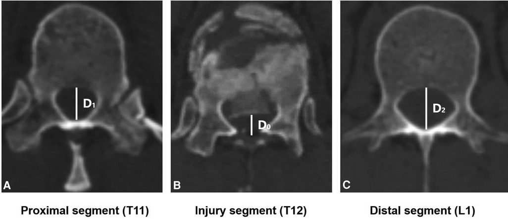
Figure 1. The midsagittal diameter of spinal canal measurement. D0 was the midsagittal diameter of the injured level's spinal canal (T12). D1 and D2 were the midsagittal diameters of the spinal canal at the levels above (T11) and below (L1) the injured level.
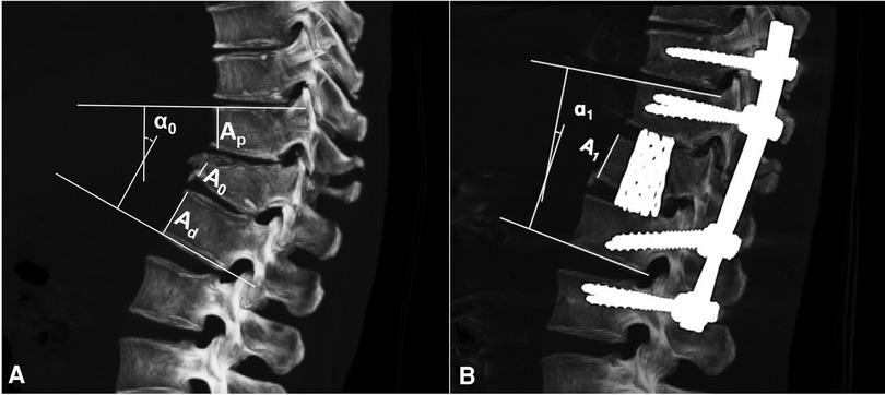
Figure 2. The measurement of angular deformity and anterior body height compression. A0 was the anterior body height of injured level; Ap and Ad was the anterior body height of the proximal and distal level; A1 was the anterior body height of injured level after surgery; α0 was pre-operative angle and α1 was post-operative angle of deformity.
Preoperative preparation
Preoperative radiologic tests included conventional radiography, thin-slice computed tomography, and magnetic resonance imaging. Neurologic status was also evaluated. The values of canal compromise (%), anterior body compression (%), and kyphotic/lordotic angle in CT reconstructed images were calculated. All radiographic parameters were measured with the DICOM Viewer System (RadiAnt, Medixant, Poland).
Surgical procedures
All surgeries were performed by the same surgeon. The patient was placed in the prone position under general anesthesia. A midline incision was made to expose the laminae and facet joints two levels above and below the injured level. Muscle dissection was performed using an electrome until the bilateral facet joints were exposed. Then, the injured vertebra was confirmed by intraoperative C-arm fluoroscopy. Pedicle screws were introduced two levels above and below the injured level if there was no affiliated injury (3). The side of decompression was determined by the severity of canal compromise at the injured level. The screws were distracted axially, first on the side of decompression and then on the opposite side. Generally, semi-laminectomy was performed by using a high-speed drill or osteotome. Under visualization of the dural sac, the facet joint and pedicle of the same side were removed conventionally to expose the nerve root. Transverse processes in the lumbar and small section rib in the thoracic vertebra should be removed for better exposure to neural elements. Epidural veins and radicular veins were cauterized with bipolar forceps to avoid massive bleeding. In some cases, the spinous process should be removed as required for repair of dural sac rupture due to severe burst fracture first. Thereafter, adjacent intervertebral discs were completely resected. Then, a subtotal corpectomy of the fractured vertebral body was performed using osteotome and bone curette, and the lateral and anterior parts of the vertebral body wall were preserved in place. During the subtotal corpectomy, the ipsilateral and ventral cancellous bones were removed firstly. Thereafter, an off-centered curette was used to carefully scrape the bone tissue from ventral to dorsal. After the fragmented vertebral body of the posterior part had collapsed, then the bone fragments were carefully dissected by the off-centered curette. After decompression was completed, a corridor was created at the lateral edge of the vertebra to insert the TMC. The medial border of corridor was the dural sac, the lateral wall was the pleura or peritoneum, the inferior border was the corresponding nerve root, and the superior border was the lower endplate of the upper level.
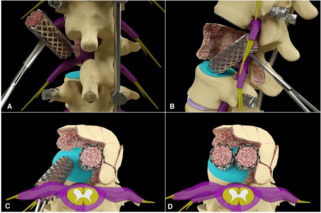
Commonly, double TMC (WEGO Co., Weihai, Shandong, China) 16 mm in diameter were stuffed with chipped autograft bone. Then, the cage with appropriate length was inserted obliquely into the corpectomy defect through the corridor without any traction of the nerve roots or the dural sac (Figure 4A). Once inside the corpectomy defect, the cage was rotated to its proper position using the bone tamp to ensure sufficient contact and a tight fit against the inferior endplate of the upper vertebra (Figure 4B). The first cage should be placed as close to the opposite side as possible to reserve enough space to insert the second cage. Additionally, two cages could be placed on the same coronal plane for better balance (Figures 4C,D). After screw-rod compression, the cortex of the contralateral lamina and upper and lower facet joints were drilled, and the chipped autograft bone was covered for posterior column fusion. Eventually, the incised wound was sutured after sufficient and careful hemostasis.
Postoperative management
Intraoperative data, including operative time, estimated blood loss, and surgery-related complications, were recorded. Thin-cut computed tomography and x-ray were performed postoperatively in all patients (Figure 5). The patients were asked to wear a thoracolumbar brace for three months after surgery. Routine follow-ups were performed at 6 weeks and 3, 6, 12, and 24 months postoperatively. When bone fusion was observed on computed tomography examination, the patient was advised to undergo internal implant removal to retain non-fusion segment movement.

Figure 5. Transverse images of pre-operation and after 270° canal decompression. The white dotted line represents the range of decompression and the yellow arrow represents the decompressed area in 270°.
Statistics
Continuous variables were expressed as mean ± standard deviation and range. The comparison of these variables were conducted using repeated measurements with a Wilcoxon signed-rank test. Categorical variables of interest were presented as percentages. All statistical analyses were performed in SPSS, version 25.0 (SPSS, Inc.). The statistical significance threshold was denoted as P values <0.05.
Results
Preoperative measurements showed that the average preoperative canal compromise and anterior body height compression were 58.4% and 50.5%, respectively. All surgeries were successfully completed in one phase. Twenty-four patients underwent corpectomy with contralateral laminae, and spinous processes remained. Three patients underwent removal of the spinous process as required to reveal the dura sac for sufficient repair of the dural sac rupture due to severe burst fracture. However, one of the patients experienced cerebrospinal fluid leakage after dura sac repair. In all patients, neurological function recovery was significantly improved except for three grade A patients. The operation lasted 151.5 ± 25.5 min (range: 115–220 min), the estimated blood loss was 590.7 ± 169.9 ml (range: 400–1,000 ml), and the follow-up time was 25.8 ± 8.6 months (range: 18–48 months). Neurological function recovery was significantly improved in all patients except 3 grade A patients. No wound infection and severe neurovascular injury occurred. Two patients experienced pleural tears intraoperatively, but no pneumothorax occurred after closing the surgical incision and chest x-ray examination. Only one patient had an aggravation of neurological damage due to accidental injury when using the off-centered curette. The VAS for back pain was significantly decreased from 6.8 to 0.7 compared with the values pre-operation and at the last follow-up (P = 0.000). The correct deformity angle was 12.4° ± 4.7, and the anterior body height recovery was 96.7% postoperatively. The correct loss angle was 2.1° ± 1.0, and TMC subsidence was 1.3 ± 0.7 mm at the last follow-up. Defined TMC subsidence occurred in 3 patients (3/27) at the last follow-up. During follow-up, CT showed efficient 270° spinal canal decompression (Figure 5), no aggravation of thoracic and lumbar pain, no screw loosening or breakage, and no displacement of TMCs. Bony fusion was achieved in all patients. All the data are displayed in Table 2.
Discussion
“Burst fractures” were first described by Holdsworth (13) in the 1960 s as fractures caused by the axial load. They frequently occur due to high-energy trauma and are most associated with falling and traffic accidents (2). These fractures can cause neurologic complications and kyphotic deformities, and they represent 10%–20% of all spine fractures at or near the thoracolumbar junction (3).
The single-stage posterior transpedicular or costotransversectomy approach for corpectomy and reconstruction has gradually become popular for thoracolumbar burst fractures (3, 5, 6, 14, 15), because spinal surgeons are familiar with the posterior approach, and studies show that the posterior approach is associated with a relatively low risk of complications compared with anterior or combined anterior and posterior approaches. However, the placement of a large fusion cage may require the sacrifice of a nerve root in the thoracic spine due to its fixed height. Despite using expandable cages, some surgeons still amputate nerve roots to accommodate a larger cage for better anterior reconstruction (16). Cages with smaller diameters are often used in the lumbar spine to preserve nerve roots and prevent nerve injury, but they carry the risk of cage subsidence when the footplate-to-vertebral body endplate ratio is less than 0.5 (17). One research (18) showed that cage subsidence was observed in the early postoperative period in as high as 35% of patients, and subsidence was demonstrated to be higher in patients with expandable cages than in patients with non-expandable cages (17). A TMC is a non-expandable cage that is a good option for supporting the anterior spinal column after a corpectomy. The anterior column supports approximately 80% of the axial load, and its efficiency for reconstruction is crucial (19). Biomechanical studies have indicated that TMC is resistant to axial compression (20). These cages are available in different diameters and heights, and the tamping of autografts inside the mesh can significantly promote osteoinduction. Furthermore, TMCs can be easily trimmed for adaptation to sagittal inclination, and interdigitation of the cage with the vertebral body endplate provides primary stability.
The posterior approach often requires the full removal of posterior spinal attachment. However, additional posterior fusion can enhance late-stage stability and reduce the occurrence of non-fusion and cage subsidence at injured segments (17). Therefore, when spinal canal decompression is required, it is necessary to preserve the posterior structure of the spinal canal for bone graft fusion and to enhance the spinal stability after reconstruction. Finally, the vertebra was subjected to subtotal corpectomy with a unilateral approach to achieve 270° of canal decompression and three-column reconstruction using double TMC.
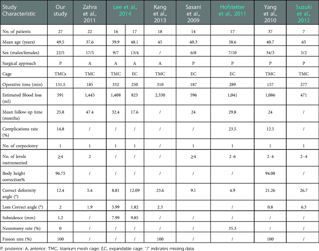
In our study, all patients suffered from thoracic and lumbar burst fractures with neurological damage. The preoperative spinal canal compromise and anterior body height compression were 58.4% and 50.5%. The indication for surgical decompression and reconstruction was necessary. We compared the results of previous studies on thoracic and lumbar burst fractures with our results (Table 3). The data showed that our technique had obvious advantages in terms of the operative time and estimated blood loss than the anterior approach. The incidence of complications was within an acceptable range, and the anterior body height was almost corrected. In terms of cage subsidence, the double cage shared the stress load from the upper and lower endplates. Theoretically, the bone contact area on one side of double TMC with a diameter of 16 mm was 402 mm2, and that of the single cage with a diameter of 22 mm was only 380 mm2. As a result, the double 16 mm cage unit area received less stress, and the larger bone contact area was better conducive to bone fusion under a theoretical condition.
Intervertebral fusion has been proven to be effective in promoting the final biomechanical stabilization of the fracture and protecting the fixation instruments from material fatigue failure (21). Rapid and firm bone fusion requires good surface contact and stability in the fusion region. It has been proven that the using of anterior fixation alone or short posterior fixation alone after corpectomy and TMC implantation provides inadequate stability, which may be associated with the loss of correction and subsidence (12). Furthermore, significant cage subsidence and angular change lead to the loss of this stability and allow progressively more flexion-extension motion (22). Therefore, posterior fixation was applied with four points of fixation superior and inferior to the level of decompression in our study. Our results showed a satisfactory fusion rate of 100% and no screw loosening or breakage at the last follow-up. However, three cases had significant cage subsidence over 2 mm, and one had a combined loss correct angle over 4°. It seemed to be more likely to occur in patients with osteoporosis. Fortunately, none of the patients required two-stage or revision surgery.
In terms of surgical techniques, TMC insertion is technically demanding, but double cage with 16 mm diameters had no difficulty in terms of insertion. As we know, the larger the diameter of the cage was, the higher the tilted height was. The tilted height was the maximal height when a cylinder rotated from a horizontal to an upright position (Figure 6). Therefore, a single cage with a large diameter should be shorten the height, or the intervertebral space should be more axially distracted. Otherwise, the endplate may be damaged when tamping the cage. As a comparison, the small cage was more convenient to insert and effectively avoided surrounding tissue damage during insertion. Additionally, the double TMC with off-center positions were relatively closer to the cortical rim of the vertebral body, which was the strongest part of the endplate, thus reducing the chances of cage subsidence (23).
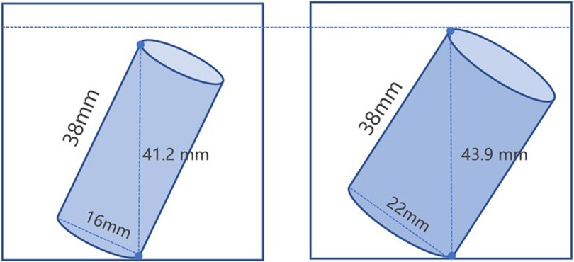
Figure 6. The schematic picture of tilted height. (A) shows the tilted height of 16 mm diameter TMC. (B) shows the tilted height of 22 mm diameter TMC.
The posterior approach allowed for simultaneous treatment of both anterior and posterior structural elements. In the case of dural sac rupture, the posterior approach might be more convenient for dural sac repair, and 3 cases were successfully repaired during the operation in our study (Figure 7). To accommodate the passage of a large supporting construct when cage insertion, the surrounding nerve elements have a risk of injury. Our technique made up for the shortcomings of the posterior approach. Double TMC were easier to place and obviously shortened the operative time with nerve root preservation. However, our study still had several shortcomings that we need to emphasize. First, decompression of the contralateral side is technical demand and the performance of this technique requires an experienced surgeon. Second, this study is retrospective, and previous cases with one TMC will be enrolled for a Case-control study. Third, the number of included samples in this study is limited, so further technical improvement and experience summary needs to be carried out continuously in a larger sample. Finally, the technique is modified from the traditional posterior approach, and it supplies an alternative method for surgeons when treated with similar fractures.
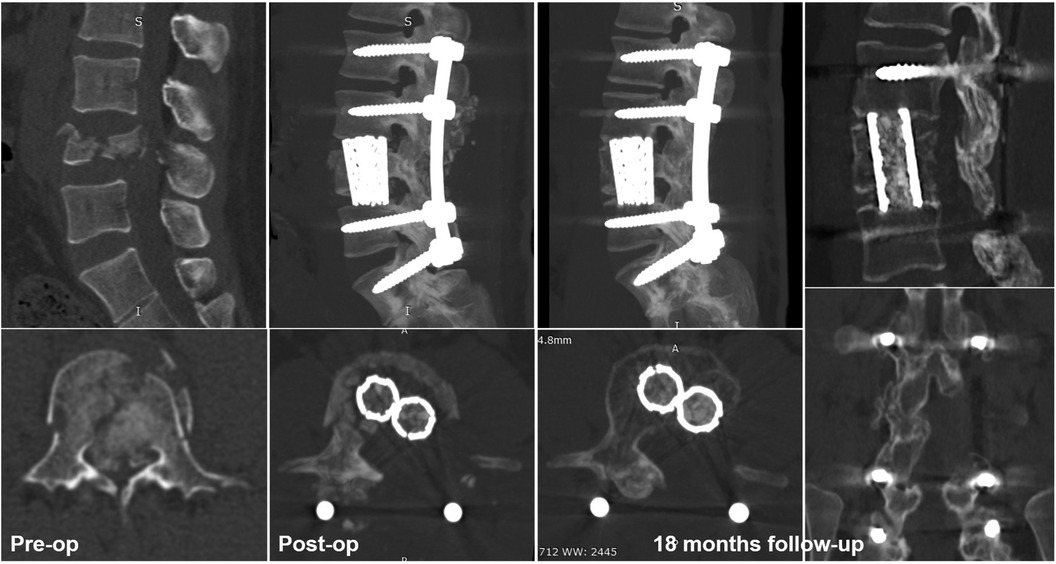
Figure 7. Typical case of intraoperative dural rupture, removal of unilateral laminae, and spinous processes for repair and completed 270° canal decompression and three-column reconstruction.
Conclusion
The posterior unilateral approach with 270° spinal canal decompression and three-column reconstruction using double titanium mesh cages is a clinically feasible, safe and alternative treatment for thoracic and lumbar burst fractures.
Data availability statement
The original contributions presented in the study are included in the article/Supplementary Material, further inquiries can be directed to the corresponding author/s.
Ethics statement
The studies involving human participants were reviewed and approved by Ethics Committee of the Second Affiliated Hospital of Chongqing Medical University. The patients/participants provided their written informed consent to participate in this study. Written informed consent was obtained from the individual(s) for the publication of any potentially identifiable images or data included in this article.
Author contributions
LS and Z-YK: provided ideas and design of the study. LL, H-QS, X-LC and SC: collected the clinical data. H-QS and Z-JY: performed the statistical analysis. Q-JG and Q-SY: contributed to investigation and the relevant literature review. LS and Q-JG: wrote this manuscript. LC, YW and Z-YK: revised the paper. The study was performed under the Supervision of LC, YW and Z-YK. All authors contributed to the article and approved the submitted version.
Funding
This study was supported by the Health Commission Scientific Research Program of Chongqing (grant no. 2012-2-056).
Acknowledgments
Thank individuals who contributed to the study or manuscript preparation but do not fulfill all the authorship criteria.
Conflict of interest
The authors declare that the research was conducted in the absence of any commercial or financial relationships that could be construed as a potential conflict of interest.
Publisher's note
All claims expressed in this article are solely those of the authors and do not necessarily represent those of their affiliated organizations, or those of the publisher, the editors and the reviewers. Any product that may be evaluated in this article, or claim that may be made by its manufacturer, is not guaranteed or endorsed by the publisher.
References
1. Denis F. The three column spine and its significance in the classification of acute thoracolumbar spinal injuries. Spine. (1983) 8:817–31. doi: 10.1097/00007632-198311000-198300003
2. Bensch FV, Koivikko MP, Kiuru MJ, Koskinen SK. The incidence and distribution of burst fractures. Emerg Radiol. (2006) 12:124–9. doi: 10.1007/s0010140-005-0457-5
3. Sasani M, Ozer AF. Single-stage posterior corpectomy and expandable cage placement for treatment of thoracic or lumbar burst fractures. Spine. (2009) 34:E33–E40. doi: 10.1097/BRS.0b013e318189fcfd
4. Sciubba DM, Gallia GL, McGirt MJ, Woodworth GF, Garonzik IM, Witham T, et al. Thoracic kyphotic deformity reduction with a distractible titanium cage via an entirely posterior approach. Neurosurgery. (2007) 60:223–30; discussion 230–221. doi: 10.1227/01.NEU.0000255385.18335.A8
5. Haiyun Y, Rui G, Shucai D, Zhanhua J, Xiaolin Z, Xin L, et al. Three-column reconstruction through single posterior approach for the treatment of unstable thoracolumbar fracture. Spine. (2010) 35:E295–302. doi: 10.1097/BRS.0b013e3181c392b9
6. Lu DC, Lau D, Lee JG, Chou D. The transpedicular approach compared with the anterior approach: an analysis of 80 thoracolumbar corpectomies. J Neurosurg Spine. (2010) 12:583–91. doi: 10.3171/2010.1.SPINE09292
7. Suzuki T, Abe E, Miyakoshi N, Murai H, Kobayashi T, Abe T, et al. Anterior decompression and shortening reconstruction with a titanium mesh cage through a posterior approach alone for the treatment of lumbar burst fractures. Asian Spine J. (2012) 6:123–30. doi: 10.4184/asj.2012.6.2.123
8. McCormack T, Karaikovic E, Gaines RW. The load sharing classification of spine fractures. Spine. (1994) 19:1741–4. doi: 10.1097/00007632-199408000-00014
9. Mumford J, Weinstein JN, Spratt KF, Goel VK. Thoracolumbar burst fractures. The clinical efficacy and outcome of nonoperative management. Spine. (1993) 18:955–70. doi: 10.1097/00007632-199306150-00003
10. Robertson PA, Rawlinson HJ, Hadlow AT. Radiologic stability of titanium mesh cages for anterior spinal reconstruction following thoracolumbar corpectomy. J Spinal Disord Tech. (2004) 17:44–52. doi: 10.1097/00024720-200402000-00010
11. Bridwell KH, Lenke LG, McEnery KW, Baldus C, Blanke K. Anterior fresh frozen structural allografts in the thoracic and lumbar spine. Do they work if combined with posterior fusion and instrumentation in adult patients with kyphosis or anterior column defects? Spine. (1995) 20:1410–8. doi: 10.1097/00007632-199506020-00014
12. Karaeminogullari O, Tezer M, Ozturk C, Bilen FE, Talu U, Hamzaoglu A. Radiological analysis of titanium mesh cages used after corpectomy in the thoracic and lumbar spine: minimum 3 years’ follow-up. Acta Orthop Belg. (2005) 71:726–31.16459866
13. Holdsworth F. Fractures, dislocations, and fracture-dislocations of the spine. J Bone Joint Surg Am. (1970) 52:1534–51. doi: 10.2106/00004623-197052080-00002
14. Lee GJ, Lee JK, Hur H, Jang JW, Kim TS, Kim SH. Comparison of clinical and radiologic results between expandable cages and titanium mesh cages for thoracolumbar burst fracture. J Korean Neurosurg Soc. (2014) 55:142–7. doi: 10.3340/jkns.2014.55.3.142
15. Nourbakhsh A, Chittiboina P, Vannemreddy P, Nanda A, Guthikonda B. Feasibility of thoracic nerve root preservation in posterior transpedicular vertebrectomy with anterior column cage insertion: a cadaveric study. J Neurosurg Spine. (2010) 13:630–5. doi: 10.3171/2010.5.SPINE09717
16. Hofstetter CP, Chou D, Newman CB, Aryan HE, Girardi FP, Härtl R. Posterior approach for thoracolumbar corpectomies with expandable cage placement and circumferential arthrodesis: a multicenter case series of 67 patients. J Neurosurg Spine. (2011) 14:388–97. doi: 10.3171/2010.11.Spine09956
17. Park P, La Marca F, Guan Z, Song Y, Lau D. Radiological outcomes of static vs expandable titanium cages after corpectomy. Neurosurgery. (2013) 72:529–39. doi: 10.1227/NEU.0b013e318282a558
18. Graillon T, Farah K, Rakotozanany P, Blondel B, Adetchessi T, Dufour H, et al. Anterior approach with expandable cage implantation in management of unstable thoracolumbar fractures: results of a series of 93 patients. Neurochirurgie. (2016) 62:78–85. doi: 10.1016/j.neuchi.2016.01.001
19. Bierry G, Buy X, Mohan PC, Cupelli J, Steib JP, Gangi A. Percutaneous vertebroplasty in a broken vertebral titanium implant (titanium mesh cage). Cardiovasc Intervent Radiol. (2006) 29:706–9. doi: 10.1007/s00270-005-5278-0
20. Chen G, Xin B, Yin M, Fan T, Wang J, Wang T, et al. Biomechanical analysis of a novel height-adjustable nano-hydroxyapatite/polyamide-66 vertebral body: a finite element study. J Orthop Surg Res. (2019) 14:368. doi: 10.1186/s13018-019-1432-2
21. Diniz JM, Botelho RV. Is fusion necessary for thoracolumbar burst fracture treated with spinal fixation? A systematic review and meta-analysis. J Neurosurg Spine. (2017) 27:584–92. doi: 10.3171/2017.1.Spine161014
22. Colman MW, Guss A, Bachus KN, Spiker WR, Lawrence BD, Brodke DS. Fixed-angle, posteriorly connected anterior cage reconstruction improves stiffness and decreases cancellous subsidence in a spondylectomy model. Spine. (2016) 41:E519–523. doi: 10.1097/BRS.0000000000001312
Keywords: spinal canal decompression, three-column reconstruction, double titanium mesh cage, posterior approach, thoracic and lumbar burst fracture
Citation: Shi L, Ge Q, Cheng Y, Lin L, Yu Q, Cheng S, Chen X, Shen H, Chen F, Yan Z, Wang Y, Chu L and Ke Z (2023) Posterior unilateral approach with 270° spinal canal decompression and three-column reconstruction using double titanium mesh cage for thoracic and lumbar burst fractures. Front. Surg. 9:1089697. doi: 10.3389/fsurg.2022.1089697
Received: 4 November 2022; Accepted: 19 December 2022;
Published: 11 January 2023.
Edited by:
Baoshan Xu, Tianjin Hospital, ChinaReviewed by:
Weibin Sheng, Xinjiang Medical University, ChinaDiego Garbossa, University of Turin, Italy
© 2023 Shi, Ge, Cheng, Lin, Yu, Cheng, Chen, Shen, Chen, Yan, Wang, Chu and Ke. This is an open-access article distributed under the terms of the Creative Commons Attribution License (CC BY). The use, distribution or reproduction in other forums is permitted, provided the original author(s) and the copyright owner(s) are credited and that the original publication in this journal is cited, in accordance with accepted academic practice. No use, distribution or reproduction is permitted which does not comply with these terms.
*Correspondence: Yang Wang MzAzNDk2QGhvc3BpdGFsLmNxbXUuZWR1LmNu Lei Chu Y2h1bGVpMjM4MEAxNjMuY29t Zhen-Yong Ke a3p5NjQxN0AxNjMuY29t
†These authors have contributed equally to this work and share first authorship
‡These authors have contributed equally to this work
Specialty Section: This article was submitted to Orthopedic Surgery, a section of the journal Frontiers in Surgery
 Lei Shi1,2,†
 Qi-jun Ge
 Qing-Shuai Yu
 Yang Wang
 Lei Chu