 Siyao Zhai
 Michael Twardowski
95% of researchers rate our articles as excellent or good
Learn more about the work of our research integrity team to safeguard the quality of each article we publish.
Find out more
ORIGINAL RESEARCH article
Front. Remote Sens. , 15 September 2021
Sec. Atmospheric Remote Sensing
Volume 2 - 2021 | https://doi.org/10.3389/frsen.2021.735512
This article is part of the Research Topic Polarization of Light as a Tool for Characterizing Different Facets of the Atmosphere-Ocean System View all 9 articles
The Degree of Linear Polarization (DoLP) for unperturbed particle fields in waters from six diverse regions around the globe was measured with the custom Multi-Angle Scattering Optical Tool (MASCOT). DoLP here is defined as the ratio of two elements of Mueller scattering matrix, i.e., -
Light scattering measurements of ocean waters have been used to infer the marine biological states and the microphysical properties of marine particulates (Brown and Gordon 1973; Gordon 1988; Zaneveld 1995; Subramaniam et al., 2001; Twardowski et al., 2001; Lee et al., 2002; Maritorena et al., 2002; Behrenfeld et al., 2005; Kostadinov et al., 2009). Elastic light scattering of a waterbody is described by its Mueller matrix (Mishchenko et al., 2002; Jonasz and Fournier 2007). The Mueller matrix (a 4-by-4 matrix) linearly transforms the incident Stokes vector (a 4-element array) to the scattered Stokes vector. The first element in the Mueller matrix is proportional to the Volume Scattering Function (VSF) in ocean optics. The VSF describes the angular distribution of unpolarized scattered radiation by a volume element of water. The VSF itself and its various derived quantities such as the scattering coefficient (
The
Accurate and meaningful simulations of the full Mueller matrix for marine particulates have also progressed slowly due to the large size and complex shape and composition of marine particulates (Zhai et al., 2013; Bi and Yang 2015; Sun et al., 2016; Xu et al., 2017; Stegmann et al., 2019; Zhai et al., 2020). An associated challenge is vector radiative transfer simulations for the entire ocean and atmospheric system (Zhai et al., 2010; Xu et al., 2016; Stamnes et al., 2018; Chowdhary et al., 2019; Ding et al., 2019). The computational speed of such simulations is relatively slower than the scalar versions. As one important element in the Mueller matrix for marine particles, DoLP affects the accuracy of vector Radiative Transfer (vRT) computations of the polarized radiance field under and above water. The term “vector” comes from the inclusion of the full Stokes vector and the entire 4-by-4 Mueller matrix in the RT code instead of just the first element of the Stokes vector. The treatment of the marine particulate Mueller matrix in vRT models generally fall into two categories: 1) based on in situ measurements (Kokhanovsky 2003; Zhai et al., 2010; You et al., 2011a, 2011b; Xu et al., 2016); and 2) numerically constructed with Lorenz-Mie or non-spherical particle single scattering models (Chowdhary et al., 2012; Ibrahim et al., 2012; Stegmann et al., 2019). As mentioned, in situ measurements are scarce and the Voss and Fry “ocean average” matrix (Voss and Fry 1984) has been typically employed (Kokhanovsky 2003; Zhai et al., 2010; Xu et al., 2016). Because of these challenges, DoLP has seen limited use in ocean optics and remote sensing applications specifically. Accuracy of the underwater Mueller matrix elements in vRT models are key to the success of future remote retrieval approaches based on either passive or active sensors (Chowdhary et al., 2019; Jamet et al., 2019).
The NASA PACE satellite mission will have two polarimeters, HARP and SPEXone (Chowdhary et al., 2019). Our understanding of particulate DoLP in the ocean is not sufficient to start formulating inversion algorithms to interpret the polarized water-leaving radiance from these polarimeters. Lidar is also an emerging tool in satellite remote sensing, with the recent CALIPSO mission demonstrating the potential of using remote lidar for particle characterizations (Hostetler et al., 2018). Jamet et al. (2019) discussed the potential applications of new remote lidar measurements for ocean science. A better understanding of polarized scattering by underwater particles is necessary to interpret and apply these emerging remote sensing techniques and the novel measurements they will provide.
In this study, we analyzed the in situ measured underwater particulate DoLP from six diverse regions around the globe. The field sites cover inland lakes, coastal oceans and open oceans. Both ocean color case I and case II waters were encountered during measurements. The angular shapes of the measured DoLPs of these waters were assessed in detail. We quantify the symmetry, peak magnitude and peak angle of the measured DoLPs. Vertical variations of DoLP at each location and for the ensemble average were analyzed. Numerical simulations of the particulate DoLP with homogeneous sphere and asymmetric hexahedral particle light scattering models were conducted with lookup tables containing particle refractive indices and size distributions. By matching the simulated and measured DoLP, we were able to retrieve particulate refractive index, which is an important particle composition parameter closely related to particle density (Aas 1996). The retrieved refractive indices were compared against values retrieved with another independent method.
The 4-by-4 Mueller matrix
where
The scattering angle
For unpolarized incident light (
The minus sign in Eq. 4 is a choice made to indicate that positive values of DoLP refer to polarization directions that are perpendicular to scattering plane, as indicated in Eq. 7. In the case of vertically polarized (perpendicular to the scattering plane) incident light (
In the case of horizontally polarized (parallel to the scattering plane) incident light (
Solving for
The particulate DoLP (
The pure seawater volume scattering functions with respect to vertical and horizontal incident light,
where
Underwater DoLP was measured with the Multi-Angle Scattering Optical Tool (MASCOT) (Twardowski et al., 2012; Sullivan et al., 2013). It has an array of 17 silicon diode detectors covering 10°–170° scattering angles (source-sample-detector angle) in 10° increments. Incident light source is a 30 mW 658 nm laser diode passing through a wedge depolarizer to provide unpolarized incident radiation. The sampling rate is 20 Hz. Full angles of the Detector field of views (FOVs) are 0.8°, 2°, 3°, and 4° for the 10°, 20°, 30°, and 40° detectors, respectively. FOV is 5° for the rest of the detectors. MASCOT covers a large scattering angle range, enabling accurate measurement of VSF at mid- and back-scatter angles. The MASCOT was designed with minimal form factors and structural elements (moving parts, sample holder, etc.) to minimize stray light contamination. It is an in situ device designed for direct measurement in nominally unperturbed waters. A filter wheel mounted in front of the source window can generate unpolarized (empty space on wheel), horizontally or vertically polarized incident light, or a dark blank (opaque location on wheel). The wheel continually spins at a rate that allows each location on the wheel to be sampled for 1 s. Particulate DoLP for unpolarized light was computed with Eq. 8. Accurate measurements of polarization elements have been verified with Lorenz-Mie theory for microspherical beads. For details on the instrument calibration and correction procedures in VSF measurement of MASCOT see (Twardowski et al., 2012). The MASCOT has been deployed extensively since 2006 (Sullivan and Twardowski 2009; You et al., 2011b; Gleason et al., 2012; Twardowski et al., 2012; Sullivan et al., 2013; Moore et al., 2017). Table 1 lists the field sites and measurements relevant in this study. Locations cover inland lakes, coastal oceans and open oceans. Relevant particulate inherent optical properties (IOPs) were also measured. Particulate absorption coefficient
where
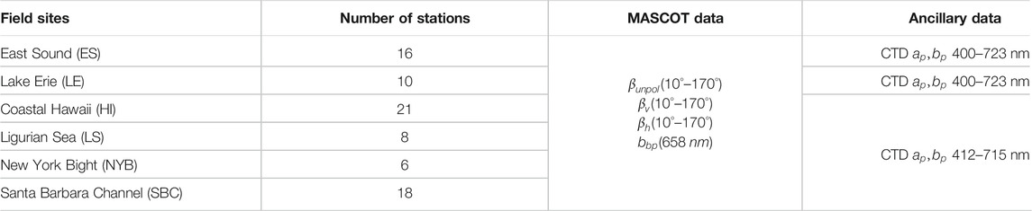
TABLE 1. Field sites and data collected with the MASCOT device and ancillary instrumentation.
To prepare the DoLP angular curve for parameterization, the MATLAB Piecewise Cubic Hermite Interpolating Polynomial (PCHIP) function was used to interpolate the measured 17-angle DoLP angular curve (10°–170° in 10° increment) to a 161-angle DoLP curve (10°–170° in 1° increment). Three parameters were then used to quantify the DoLP shape: the asymmetry parameter, the peak magnitude and the peak angle. The asymmetry parameter
where
The peak magnitude
With
The use of this method is justified by the overall small RMSE between measurements and Eq. 12, and small deviation of
Light scattering simulations with homogeneous spheres were performed to help interpret observations. Also, we attempt to retrieve the particulate refractive index by fitting simulated DoLP to measurements, i.e., through inversion. In modelling, The bulk particulate DoLP is given by,
where the bulk
where
Figure 1 shows the simulated DoLP angular functions of the homogeneous sphere model for different
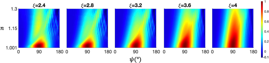
FIGURE 1. Surface plots of the DoLP angular shape modeled with the Lorenz-Mie light scattering model. X-axes are the scattering angle
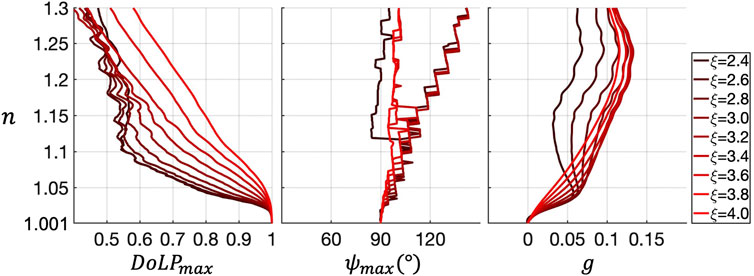
FIGURE 2. Particulate refractive index
Another light scattering model, the asymmetric hexahedral particle scattering model was examined in this study. Originally, the Volume Scattering Function (VSF) from the asymmetric hexahedral model dataset was used in another study to retrieve particle size distributions (Twardowski et al., 2012). In this study, we examine its DoLP element. In the asymmetric hexahedral model, at each particle size, the scattering properties of an ensemble of randomly distorted hexahedra were computed with the Discrete Dipole Approximation (DDA) method (Yurkin and Hoekstra 2007) and the Improved Geometric Optics Method (IGOM) (Yang and Liou 1996). After ensemble averaging at each particle size and a subsequent size averaging, the scattering properties for a polydispersion of particles were obtained. The particulate refractive index range was [1.02,1.2] in 0.02 increments, with the imaginary part fixed at 0.002. The equivalent sphere diameter increased exponentially from 0.01 to 163 μm. Incident wavelength was 658 nm in vacuum (
Figure 3 panels show the simulated DoLP angular functions of the asymmetric hexahedron model for different
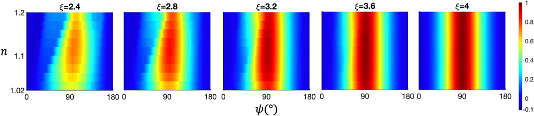
FIGURE 3. Surface plots of the DoLP angular shape modeled with the asymmetric hexahedral particle light scattering model, same as Figure 1.
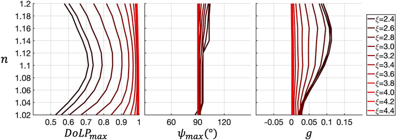
FIGURE 4. Particulate refractive index
Figure 5A panels show measured particulate DoLP at each field site. A considerable number of DoLP curves exhibited large, highly intermittent spikes at 30°, 40°, 140°, 150°, and 160°. These isolated spikes at specific angles are generated by particulates such as large organisms and aggregates drifting into the sample volume, and isolated light paths for individual detectors. At those angles, spikes with magnitudes larger than 0.2 were flagged and these DoLPs were removed from the analysis. After that screening, at each field site, outliers with magnitude falling out of the [15%, 85%] inter-percentile range at any one of the 17 scattering angles were removed. This percentile range eliminated DoLPs with potentially high uncertainty and maintained enough DoLP samples for each field sites. The HI data also showed more variability in the backward direction due to the very low measured scattering signals in this clear water; this was from a higher contribution from MASCOT instrument noise. Remaining fluctuations in the DoLP angular curves are from small scale environmental variability in particle fields; note measurements of vertical and horizontal polarized scattering in the sample volume were not made exactly simultaneously.
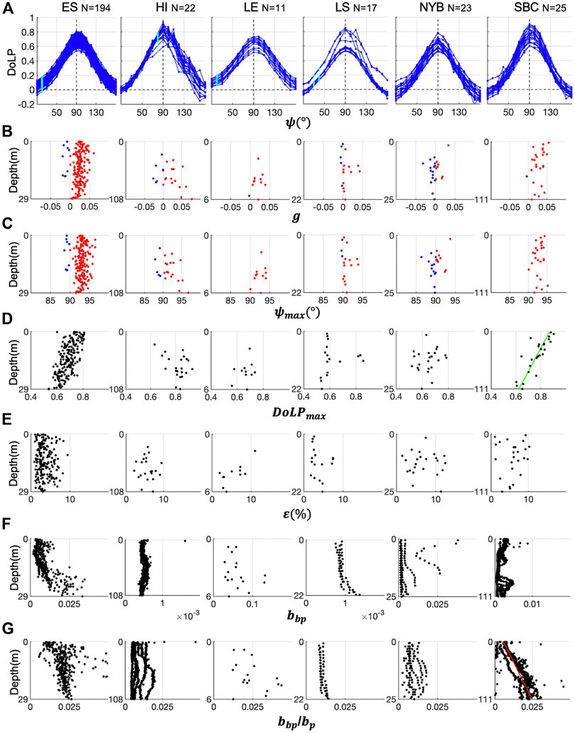
FIGURE 5. (A) Measured particulate DoLPs plotted against scattering angle
Figures 5B–G panels show vertical profiles of the three shape parameters, percent RMSE and backscattering parameters at each field site. Linear least-squares fit to the vertical profiles in Figures 5B–G panels are displayed if the coefficient of determination
In Figure 5B panels, a relatively high proportion of
Figure 6 shows the box-whisker plots of
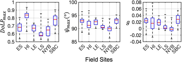
FIGURE 6. From left to right panel: Box-whisker plots of the peak magnitude
DoLPs from all field sites in Figure 5A were averaged into 1 m depth bins and sorted according to their measurement depth to obtain a ensemble averaged DoLP in Figure 7. Figure 7 shows the ensemble averaged particulate DoLP along with the vertical profiles of
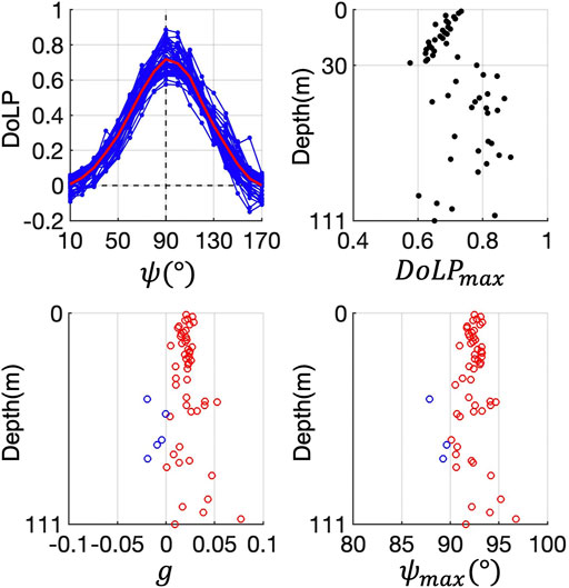
FIGURE 7. Ensemble averaged particulate DoLP. Measured DoLPs from all field sites are grouped together and then averaged into 1-m depth bins. The mean is overlaid as the red curve. Vertical profiles of the peak magnitude
Figure 8 compares the ensemble averaged DoLP in this study with the Voss and Fry global mean DoLP (Voss and Fry 1984). Figure 8 left panel shows the ensemble averaged particulate DoLP of this study and the seawater DoLP computed with Eqs. 9a–b at a temperature of 20°C and a salinity of 35 ppt. Figure 8 right panel shows the ensemble averaged total (seawater plus particulates) DoLP, Voss and Fry DoLP and seawater DoLP. The red error bars and the grey area indicate standard deviation of our DoLP and the Voss and Fry DoLP, respectively. The pure seawater DoLP has a peak of around 0.92, the total DoLP has a higher peak (0.74) than the particulate DoLP (0.72), but they are both higher than the peak of the Voss and Fry DoLP (0.66). The Voss and Fry DoLP and our total and particulate DoLPs all have highest variability around 90°. Note Voss and Fry DoLP did not converge to zero in far backward angles (beyond about 140°); this may be due to reflection errors related to the cuvette in the bench top apparatus that was used.
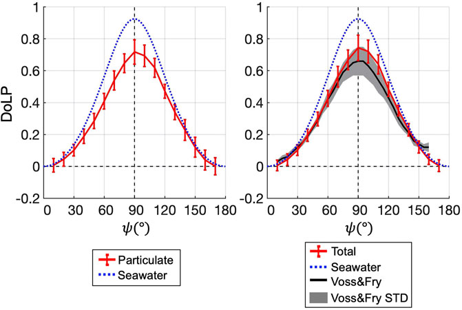
FIGURE 8. Left: The ensemble average particulate DoLP from Figure 7 is plotted as the red curve with error bars indicating standard deviation at 17 angles. Seawater DoLP at 20°C and a salinity of 35 ppt is the blue dotted curve. Right: Same as left panel, except that the particulate DoLP is replaced with total DoLP (particulate plus seawater) from this study. The global mean total DoLP from the Voss and Fry study is overlaid as the black curve. Grey area is the standard deviation of the Voss and Fry DoLP.
As mentioned in Modeling, there is potential with using a DoLP LUT to retrieve the particulate refractive index through inversion. For a measured DoLP with a corresponding PSD slope value, a bulk particulate refractive index can be retrieved by finding a best-fit to the measurement from the simulated DoLP LUT. We attempt to retrieve particulate refractive index with this approach. With the co-located backscattering ratio measurements, another bulk particulate refractive index retrieval approach (Twardowski et al., 2001) (referred to as “the Twardowski model” in texts and figures below) was used for comparison.
At each measured DoLP depth, the Junge-type PSD slope (
The PSD slope
The Twardowski et al. model relates
Figure 9 shows the results of refractive index retrieval at field site East Sound (ES). In Figure 9B, The second panel shows relative errors of the decimal part of the refractive indices retrieved with the two models,
Figures 10–14 show the same contents for the other field sites, HI (Hawaii), LE (Lake Erie), Ligurian Sea (LS), NYB (New York Bight) and SBC (Santa Barbara Channel). Overall, at ES, HI, LE and SBC, refractive indices retrieved with the DoLP model and the Twardowski model were roughly comparable. At ES (Figure 9),
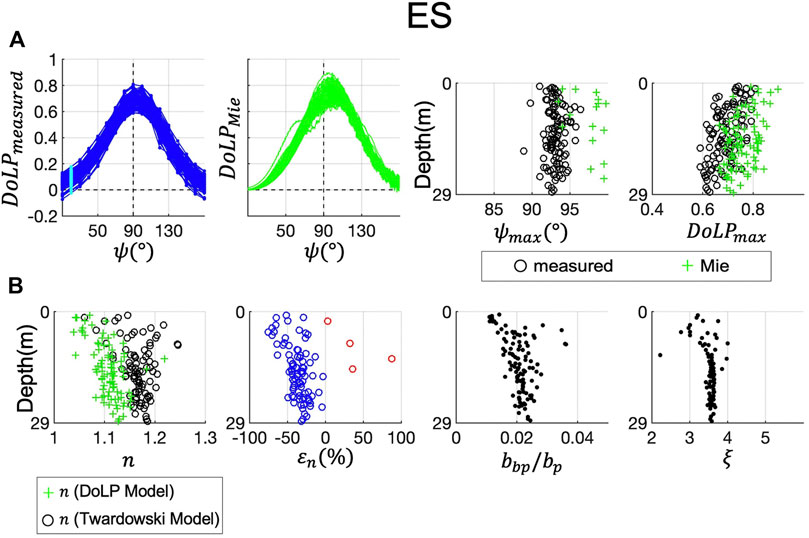
FIGURE 9. Particulate refractive index retrieval at field site ES (East Sound). (A) From left to right: Measured particulate DoLP (
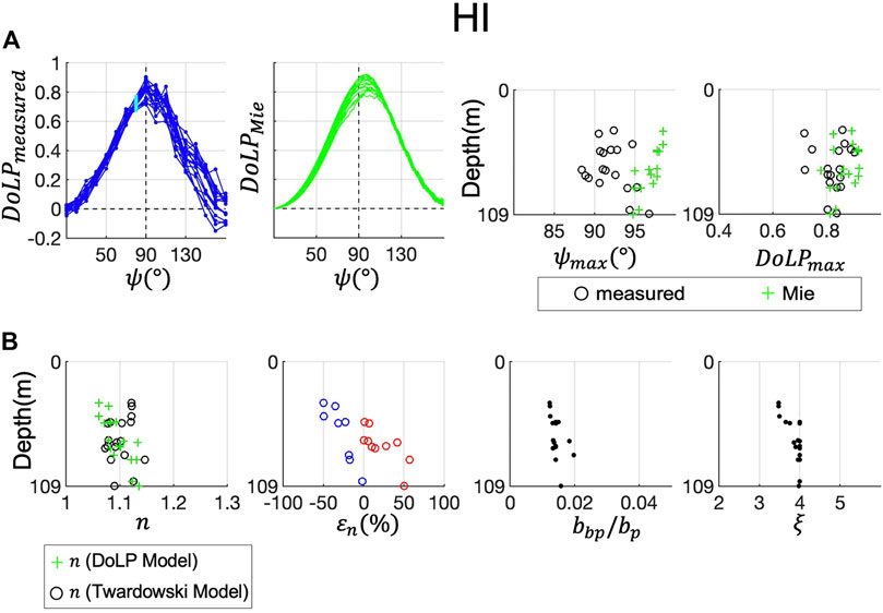
FIGURE 10. Refractive index retrieval at field site HI (Hawaii). Same as Figure 9.
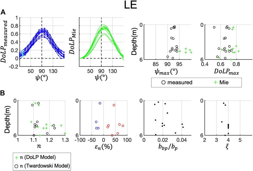
FIGURE 11. Refractive index retrieval at field site LE (Lake Erie). Same as Figure 9.
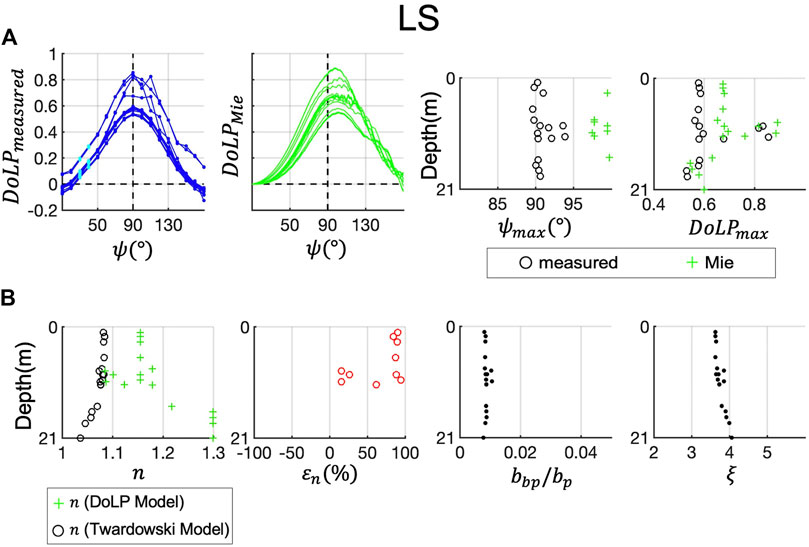
FIGURE 12. Refractive index retrieval at field site LS (Ligurian Sea). Same as Figure 9.
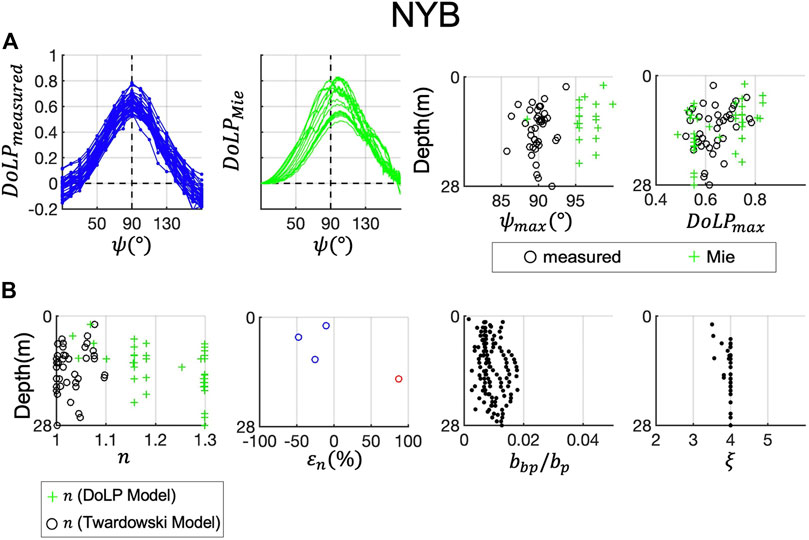
FIGURE 13. Refractive index retrieval at field site NYB (New York Bight). Same as Figure 9.
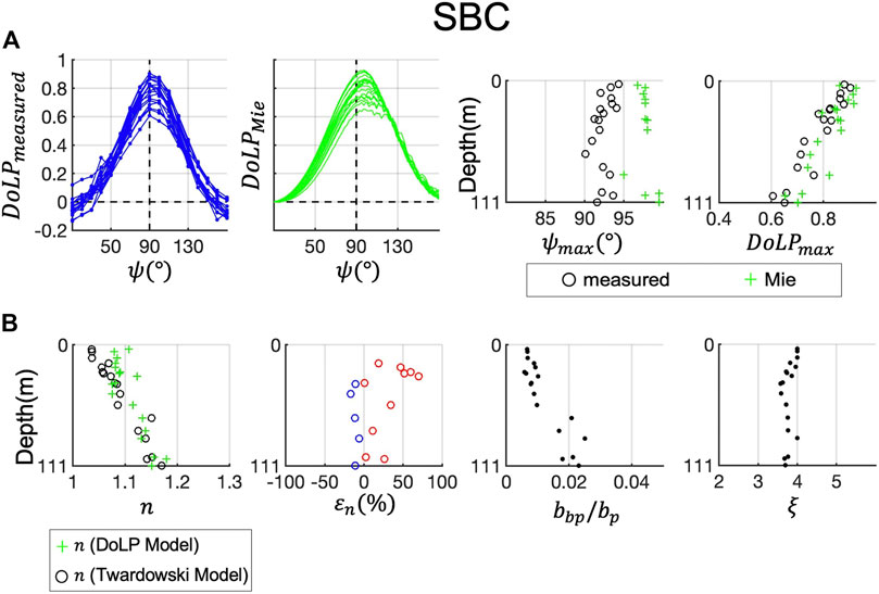
FIGURE 14. Refractive index retrieval at field site SBC (Santa Barbara Channel). Same as Figure 9.
At HI, LE and SBC, refractive indices retrieved with the DoLP model and the Twardowski model were comparable. We capped the PSD slope values at 4 for these retrievals because oceanic PSD slope values do not typically exceed 4 in natural waters (Reynolds et al., 2010; their Figure 11), thus removing some potential uncertainty in the derivation of
At LE, the DoLP peaks are low, with extreme values approaching 0.5. Thus, the DoLP model returns high refractive indices. Values of
Inconsistencies in the models for LS and NYB could be due to several factors not accounted for that may affect the two models differently, including complex particle mixtures (i.e., broad np distributions), complex particle shapes, aggregations and bubbles. Further work is needed to interpret the influence of these factors. The Twardowski model and the DoLP model here are both based on the Lorenz-Mie (homogeneous sphere) model, although the key scattering parameters of the two models, DoLP and
Although model discrepancies here require further work to elucidate impacts of these complicating factors, the DoLP model provides another tool to assess oceanic particle composition that may help constrain bulk refractive index estimates. It is also progress toward operational application of remote polarimetry data in determining bulk refractive index and closely related particle density. Particle density is essential for determinations of particle sinking rates (Briggs et al., 2020; Nayak and Twardowski 2020; Omand et al., 2020) and relationships between particulate organic carbon and chlorophyll concentrations (Loisel et al., 2007), and is currently not a parameter that can be derived remotely.
In this study, we analyzed the angular shape of the particulate Degree of Linear Polarization (DoLP) measured at six locations around the globe. The measured DoLP shapes were quantified with three parameters: the peak magnitude
An inversion model was developed to retrieve bulk particulate refractive index by finding a best fit to each measured particulate DoLP from a simulated DoLP dataset (the DoLP model). Lorenz-Mie theory for scattering by homogeneous spheres was used in the simulations. An independent retrieval model based on backscattering ratio was used (the Twardowski model) for comparison. The retrievals were performed at all six field sites. The particulate refractive index retrieved with the DoLP model and the Twardowski model were comparable at the six field sites. At ES, the DoLP model underestimated the refractive index compared to the Twardowski model due to low PSD slope values. At HI and SBC, the DoLP model and the Twardowski model produced refractive index values that are close throughtout the measurement depth. At LE, refractive indices retrieved by the two models were roughly comparable. At LS and NYB, the combination of high PSD slope values and low DoLP peaks resulted in very high refractive indices (1.25–1.3) retrieved with the DoLP model, while low backscattering ratios resulted in low refractive indices (<1.1) retrieved with the Twardowski model. Mixture of particles with a broad range of shape and composition at the two sites might be the cause for relatively large model discrepancies.
In future studies, modeling work with various particle shapes and compositions is needed to investigate the sensitivity of DoLP to these particle features. As shown in Modeling, the simulated DoLPs of Lorenz-Mie and asymmetric hexahedral model showed different variations with changing refractive index. Possible candidate particle models include coated sphere, spheroid and coated spheroid. These shapes were used in simulating marine particulate inherent optical properties (Clavano et al., 2007; Xu et al., 2017; Organelli et al., 2018; Duforêt-Gaurier et al., 2018), although the focus of those studies were not on the linear polarization element. In addition, more work is needed to better understand and validate relationships between DoLP and other IOPs such as backscattering ratio, particulate albedo, and size distributions.
The raw data supporting the conclusion of this article will be made available by the authors, without undue reservation.
MT collected the field data and carried out Lorenz-Mie modeling work. SZ analyzed the data, performed the simulations and wrote the paper. MT edited the paper.
Funding for MT and SZ was provided by the NASA PACE program (80NSSC20M0225), the NASA US Investigator program (80NSSC19K1195) and the Harbor Branch Oceanographic Institute Foundation. Additional support for MT was provided by Belgian Science Policy Office Research Programme for Earth Observation STEREO III (STEREO/00/374).
The authors declare that the research was conducted in the absence of any commercial or financial relationships that could be construed as a potential conflict of interest.
The handling editor declared a past co-authorship with one of the authors (MT).
All claims expressed in this article are solely those of the authors and do not necessarily represent those of their affiliated organizations, or those of the publisher, the editors and the reviewers. Any product that may be evaluated in this article, or claim that may be made by its manufacturer, is not guaranteed or endorsed by the publisher.
We are indebted to those that participated in the development of MASCOT, including Alex Derr, Wes Strubhar, and Dave Romanko. We are also indebted to those who helped collect and process data from MASCOT and other sensors for the various field sites, including Scott Freeman, Nicole Stockley, Heather Groundwater, and Matt Slivkoff.
Aas, E. (1996). Refractive index of Phytoplankton Derived from its Metabolite Composition. J. Plankton Res. 18 (12), 2223–2249. doi:10.1093/plankt/18.12.2223
Basterretxea, G., Font-Muñoz, J. S., and Tuval, I. (2020). Phytoplankton Orientation in a Turbulent Ocean: A Microscale Perspective. Front. Mar. Sci. 7 (March), 185. doi:10.3389/fmars.2020.00185
Behrenfeld, M. J., Boss, E., Siegel, D. A., and Shea, D. M. (2005). Carbon-Based Ocean Productivity and Phytoplankton Physiology from Space. Glob. Biogeochem. Cycles 19 (1), 1–14. doi:10.1029/2004GB002299
Bi, L., and Yang, P. (2015). Impact of Calcification State on the Inherent Optical Properties of Emiliania Huxleyi Coccoliths and Coccolithophores. J. Quantitative Spectrosc. Radiative Transfer 155 (April), 10–21. doi:10.1016/j.jqsrt.2014.12.017
Bohren, C. F., and Huffman, D. R. (1998). Absorption and Scattering of Light by Small Particles. Wiley.
Boss, E., Pegau, W. S., Lee, M., Twardowski, M., Shybanov, E., Korotaev, G., et al. (2004). Particulate Backscattering Ratio at LEO 15 and its Use to Study Particle Composition and Distribution. J. Geophys. Res. 109 (1), 1014. doi:10.1029/2002jc001514
Boss, E., Twardowski, M. S., and Herring, S. (2001). Shape of the Particulate Beam Attenuation Spectrum and its Inversion to Obtain the Shape of the Particulate Size Distribution. Appl. Opt. 40 (27), 4885. doi:10.1364/ao.40.004885
Briggs, N., Dall’Olmo, G., and Claustre, H. (2020). Major Role of Particle Fragmentation in Regulating Biological Sequestration of CO2 by the Oceans. Science 367 (6479), 791–793. doi:10.1126/science.aay1790
Brown, O. B., and Gordon, H. R. (1973). Two Component Mie Scattering Models of Sargasso Sea Particles. Appl. Opt. 12 (10), 2461. doi:10.1364/ao.12.002461
Chami, M., and McKee, D. (2007). Determination of Biogeochemical Properties of Marine Particles Using above Water Measurements of the Degree of Polarization at the Brewster Angle. Opt. Express 15 (15), 9494. doi:10.1364/oe.15.009494
Chowdhary, J., Cairns, B., Waquet, F., Knobelspiesse, K., Ottaviani, M., Redemann, J., et al. (2012). Sensitivity of Multiangle, Multispectral Polarimetric Remote Sensing over Open Oceans to Water-Leaving Radiance: Analyses of RSP Data Acquired during the MILAGRO Campaign. Remote Sensing Environ. 118 (March), 284–308. doi:10.1016/j.rse.2011.11.003
Chowdhary, J., Zhai, P.-W., Boss, E., Dierssen, H., Frouin, R., Ibrahim, A., et al. (2019). Modeling Atmosphere-Ocean Radiative Transfer: A PACE Mission Perspective. Front. Earth Sci. 7 (June), 100. doi:10.3389/feart.2019.00100
Clavano, W., Boss, E., and Karp-Boss, L. (2007). Inherent Optical Properties of Non-spherical Marine-Like Particles, Äî from Theory to Observation. Oceanography Mar. Biol. 38, 1–38. doi:10.1201/9781420050943.ch1
Ding, J., Yang, P., King, M. D., Platnick, S., Liu, X., Meyer, K. G., et al. (2019). A Fast Vector Radiative Transfer Model for the Atmosphere-Ocean Coupled System. J. Quantitative Spectrosc. Radiative Transfer 239 (December), 106667. doi:10.1016/j.jqsrt.2019.106667
Duforêt-Gaurier, L., Dessailly, D., Moutier, W., and Loisel, H. (2018). Assessing the Impact of a Two-Layered Spherical Geometry of Phytoplankton Cells on the Bulk Backscattering Ratio of Marine Particulate Matter. Appl. Sci. 8 (12), 2689. doi:10.3390/app8122689
Farinato, R. S., and Rowell, R. L. (1976). New Values of the Light Scattering Depolarization and Anisotropy of Water. J. Chem. Phys. 65 (2), 593–595. doi:10.1063/1.433115
Fry, E. S., and Voss, K. J. (1985). Measurement of the Mueller Matrix for Phytoplankton1. Limnol. Oceanogr. 30 (6), 1322–1326. doi:10.4319/lo.1985.30.6.1322
Gleason, A. C. R., Voss, K. J., Gordon, H. R., Twardowski, M., Sullivan, J., Trees, C., et al. (2012). Detailed Validation of the Bidirectional Effect in Various Case I and Case II Waters. Opt. Express 20 (7), 7630. doi:10.1364/oe.20.007630
Gordon, H. R., Brown, O. B., Evans, R. H., Brown, J. W., Smith, R. C., Baker, K. S., et al. (1988). A Semianalytic Radiance Model of Ocean Color. J. Geophys. Res. 93 (D9), 10909–10924. doi:10.1029/JD093iD09p10909
Hostetler, C. A., Behrenfeld, M. J., Hu, Y., Hair, J. W., and Schulien, J. A. (2018). Spaceborne Lidar in the Study of Marine Systems. Annu. Rev. Mar. Sci. 10 (1), 121–147. doi:10.1146/annurev-marine-121916-063335
Ibrahim, A., Gilerson, A., Harmel, T., Tonizzo, A., Chowdhary, J., and Ahmed, S. (2012). The Relationship between Upwelling Underwater Polarization and Attenuation/Absorption Ratio. Opt. Express 20 (23), 25662. doi:10.1364/oe.20.025662
Jamet, C., Ibrahim, A., Ahmad, Z., Angelini, F., Babin, M., Behrenfeld, M. J., et al. (2019). Going beyond Standard Ocean Color Observations: Lidar and Polarimetry. Front. Mar. Sci. 6 (May), 251. doi:10.3389/fmars.2019.00251
Jonasz, M., and Fournier, G. (2007). Light Scattering by Particles in Water: Theoretical and Experimental Foundations. Elsevier.
Kadyshevich, E., and Lyubovtseva, Y. S. (1976). Light-Scattering Matrices of Pacific and Atlantic Ocean Waters. Izvestiya Atmos. Oceanic Phys. 12 (2), 106–111.
Koestner, D., Stramski, D., and Reynolds, R. A. (2020). Polarized Light Scattering Measurements as a Means to Characterize Particle Size and Composition of Natural Assemblages of Marine Particles. Appl. Opt. 59 (27), 8314. doi:10.1364/ao.396709
Koestner, D., Stramski, D., and Reynolds, R. (2018). Measurements of the Volume Scattering Function and the Degree of Linear Polarization of Light Scattered by Contrasting Natural Assemblages of Marine Particles. Appl. Sci. 8 (12), 2690. doi:10.3390/app8122690
Kokhanovsky, A. A. (2003). Parameterization of the Mueller Matrix of Oceanic Waters. J. Geophys. Res. 108 (6), 3175. doi:10.1029/2001jc001222
Kostadinov, T. S., Siegel, D. A., and Maritorena, S. (2009). Retrieval of the Particle Size Distribution from Satellite Ocean Color Observations. J. Geophys. Res. 114 (9), 9015. doi:10.1029/2009JC005303
Lee, Z., Carder, K. L., and Arnone, R. A. (2002). Deriving Inherent Optical Properties from Water Color: A Multiband Quasi-Analytical Algorithm for Optically Deep Waters. Appl. Opt. 41 (27), 5755. doi:10.1364/ao.41.005755
Loisel, H., Mériaux, X., Berthon, J.-F., and Poteau, A. (2007). Investigation of the Optical Backscattering to Scattering Ratio of Marine Particles in Relation to Their Biogeochemical Composition in the Eastern English Channel and Southern North Sea. Limnol. Oceanogr. 52 (2), 739–752. doi:10.4319/lo.2007.52.2.0739
Lotsberg, J. K., and Stamnes, J. J. (2010). Impact of Particulate Oceanic Composition on the Radiance and Polarization of Underwater and Backscattered Light. Opt. Express 18 (10), 10432. doi:10.1364/oe.18.010432
Maritorena, S., Siegel, D. A., and Peterson, A. R. (2002). Optimization of a Semianalytical Ocean Color Model for Global-Scale Applications. Appl. Opt. 41 (15), 2705. doi:10.1364/ao.41.002705
McFarland, M., Nayak, A. R., Stockley, N., Twardowski, M., and Sullivan, J. (2020). Enhanced Light Absorption by Horizontally Oriented Diatom Colonies. Front. Mar. Sci. 7 (July), 494. doi:10.3389/fmars.2020.00494
Mishchenko, M. I., Travis, L. D., and Lacis, A. A. (2002). Scattering, Absorption, and Emission of Light by Small Particles. Cambridge University Press.
Mishchenko, M. I., and Yurkin, M. A. (2017). On the Concept of Random Orientation in Far-Field Electromagnetic Scattering by Nonspherical Particles. Opt. Lett. 42 (3), 494. doi:10.1364/ol.42.000494
Moore, T. S., Mouw, C. B., Sullivan, J. M., Twardowski, M. S., Burtner, A. M., Ciochetto, A. B., et al. (2017). Bio-Optical Properties of Cyanobacteria Blooms in Western Lake Erie. Front. Mar. Sci. 4 (SEP), 300. doi:10.3389/fmars.2017.00300
Nayak, A. R., McFarland, M. N., Sullivan, J. M., and Twardowski, M. S. (2018). Evidence for Ubiquitous Preferential Particle Orientation in Representative Oceanic Shear Flows. Limnol. Oceanogr. 63 (1), 122–143. doi:10.1002/lno.10618
Nayak, A. R., and Twardowski, M. S. (2020). "Breaking" News for the Ocean's Carbon Budget. Science 367 (6479), 738–739. doi:10.1126/science.aba7109
Omand, M. M., Govindarajan, R., He, J., and Mahadevan, A. (2020). Sinking Flux of Particulate Organic Matter in the Oceans: Sensitivity to Particle Characteristics. Sci. Rep. 10 (1), 1–16. doi:10.1038/s41598-020-60424-5
Organelli, E., Dall’Olmo, G., Brewin, R. J. W., Tarran, G. A., Boss, E., and Bricaud, A. (2018). The Open-Ocean Missing Backscattering Is in the Structural Complexity of Particles. Nat. Commun. 9 (1), 5439. doi:10.1038/s41467-018-07814-6
Quinby-Hunt, M. S., Hunt, A. J., Lofftus, K., and Shapiro, D. (1989). Polarized-light Scattering Studies of marine Chlorella. Limnol. Oceanogr. 34 (8), 1587–1600. doi:10.4319/lo.1989.34.8.1587
Reynolds, R. A., Stramski, D., Wright, V. M., and Woźniak, S. B. (2010). Measurements and Characterization of Particle Size Distributions in Coastal Waters. J. Geophys. Res. 115 (8), 8024. doi:10.1029/2009JC005930
Stamnes, S., Hostetler, C., Ferrare, R., Burton, S., Liu, X., Hair, J., et al. (2018). Simultaneous Polarimeter Retrievals of Microphysical Aerosol and Ocean Color Parameters from the "MAPP" Algorithm with Comparison to High-Spectral-Resolution Lidar Aerosol and Ocean Products. Appl. Opt. 57 (10), 2394. doi:10.1364/ao.57.002394
Stegmann, P. G., Sun, B., Ding, J., Yang, P., and Zhang, X. (2019). Study of the Effects of Phytoplankton Morphology and Vertical Profile on Lidar Attenuated Backscatter and Depolarization Ratio. J. Quantitative Spectrosc. Radiative Transfer 225 (March), 1–15. doi:10.1016/j.jqsrt.2018.12.009
Subramaniam, A., Christopher, W. B., Hood, R., Carpenter, E. J., and Capone, D. G. (2001). Detecting Trichodesmium Blooms in SeaWiFS Imagery. Deep-Sea Res. Part Topical Stud. Oceanography 49 (1–3), 107–121. doi:10.1016/S0967-0645(01)00096-0
Sullivan, J. M., and Twardowski, M. S. (2009). Angular Shape of the Oceanic Particulate Volume Scattering Function in the Backward Direction. Appl. Opt. 48 (35), 6811–6819. doi:10.1364/AO.48.006811
Sullivan, J. M., Twardowski, M. S., Donaghay, P. L., and Freeman, S. A. (2005). Use of Optical Scattering to Discriminate Particle Types in Coastal Waters. Appl. Opt. 44 (9), 1667–1680. doi:10.1364/AO.44.001667
Sullivan, J. M., Twardowski, M. S., Ronald, J., Zaneveld, V., and Moore, C. C. (2013). “Measuring Optical Backscattering in Water,” in Light Scattering Reviews 7: Radiative Transfer and Optical Properties of Atmosphere and Underlying Surface, 189–224. doi:10.1007/978-3-642-21907-8_6
Sun, B., Kattawar, G. W., Yang, P., Twardowski, M. S., and Sullivan, J. M. (2016). Simulation of the Scattering Properties of a Chain-Forming Triangular Prism Oceanic Diatom. J. Quantitative Spectrosc. Radiative Transfer 178 (July), 390–399. doi:10.1016/j.jqsrt.2016.02.035
Tonizzo, A., Gilerson, A., Harmel, T., Ibrahim, A., Chowdhary, J., Gross, B., et al. (2011). Estimating Particle Composition and Size Distribution from Polarized Water-Leaving Radiance. Appl. Opt. 50 (25), 5047–5058. doi:10.1364/AO.50.005047
Tonizzo, A., Zhou, J., Gilerson, A., Twardowski, M. S., Gray, D. J., Arnone, R. A., et al. (2009). Polarized Light in Coastal Waters: Hyperspectral and Multiangular Analysis. Opt. Express 17 (7), 5666. doi:10.1364/oe.17.005666
Twardowski, M. S., Boss, E., Macdonald, J. B., Pegau, W. S., Barnard, A. H., and Zaneveld, J. R. V. (2001). A Model for Estimating Bulk Refractive Index from the Optical Backscattering Ratio and the Implications for Understanding Particle Composition in Case I and Case II Waters. J. Geophys. Res. 106 (C7), 14129–14142. doi:10.1029/2000JC000404
Twardowski, M. S., Claustre, H., Freeman, S. A., Stramski, D., and Huot, Y. (2007). Optical Backscattering Properties of the "clearest" Natural Waters. Biogeosciences 4 (6), 1041–1058. doi:10.5194/bg-4-1041-2007
Twardowski, M. S., Sullivan, J. M., Donaghay, P. L., and Zaneveld, J. R. V. (1999). Microscale Quantification of the Absorption by Dissolved and Particulate Material in Coastal Waters with an Ac-9. J. Atmos. Oceanic Technol. 16 (6), 691–707. doi:10.1175/1520-0426(1999)016<0691:mqotab>2.0.co;2
Twardowski, M., and Tonizzo, A. (2018). Ocean Color Analytical Model Explicitly Dependent on the Volume Scattering Function. Appl. Sci. 8 (12), 2684. doi:10.3390/app8122684
Twardowski, M., Zhang, X., Vagle, S., Sullivan, J., Freeman, S., Czerski, H., et al. (2012). The Optical Volume Scattering Function in a Surf Zone Inverted to Derive Sediment and Bubble Particle Subpopulations. J. Geophys. Res. 117 (2), a–n. doi:10.1029/2011JC007347
Volten, H., De Haan, J. F., Hovenier, J. W., Schreurs, R., Vassen, W., Dekker, A. G., et al. (1998). Laboratory Measurements of Angular Distributions of Light Scattered by Phytoplankton and Silt. Limnol. Oceanogr. 43 (6), 1180–1197. doi:10.4319/lo.1998.43.6.1180
Voss, K. J., and Fry, E. S. (1984). Measurement of the Mueller Matrix for Ocean Water. Appl. Opt. 23 (23), 4427. doi:10.1364/ao.23.004427
Werdell, P. J., McKinna, L. I. W., Boss, E., Ackleson, S. G., Craig, S. E., Gregg, W. W., et al. (2018). An Overview of Approaches and Challenges for Retrieving Marine Inherent Optical Properties from Ocean Color Remote Sensing, Prog. Oceanography, 160, 186, 212. doi:10.1016/j.pocean.2018.01.001
Witkowski, K., Król, T., Zielirińki, A., and Kuteń, E. (1998). A Light-Scattering Matrix for Unicellular Marine Phytoplankton. Limnol. Oceanogr. 43 (5), 859–869. doi:10.4319/lo.1998.43.5.0859
Xu, F., Dubovik, O., Zhai, P.-W., Diner, D. J., Kalashnikova, O. V., Seidel, F. C., et al. (2016). Joint Retrieval of Aerosol and Water-Leaving Radiance from Multispectral, Multiangular and Polarimetric Measurements over Ocean. Atmos. Meas. Tech. 9 (7), 2877–2907. doi:10.5194/amt-9-2877-2016
Xu, G., Sun, B., Brooks, S. D., Yang, P., Kattawar, G. W., and Zhang, X. (2017). Modeling the Inherent Optical Properties of Aquatic Particles Using an Irregular Hexahedral Ensemble. J. Quantitative Spectrosc. Radiative Transfer 191 (April), 30–39. doi:10.1016/j.jqsrt.2017.01.020
Yang, P., and Liou, K. N. (1996). Geometric-optics-integral-equation Method for Light Scattering by Nonspherical Ice Crystals. Appl. Opt. 35 (33), 6568. doi:10.1364/ao.35.006568
You, Y., Kattawar, G. W., Voss, K. J., Bhandari, P., Wei, J., Lewis, M., et al. (2011a). Polarized Light Field under Dynamic Ocean Surfaces: Numerical Modeling Compared with Measurements. J. Geophys. Res. 116 (10), C00H05. doi:10.1029/2011JC007278
You, Y., Tonizzo, A., Gilerson, A. A., Cummings, M. E., Brady, P., Sullivan, J. M., et al. (2011b). Measurements and Simulations of Polarization States of Underwater Light in Clear Oceanic Waters. Appl. Opt. 50 (24), 4873–4893. doi:10.1364/AO.50.004873
Yurkin, M. A., and Hoekstra, A. G. (2007). The Discrete Dipole Approximation: An Overview and Recent Developments. J. Quantitative Spectrosc. Radiative Transfer 106 (1–3), 558–589. doi:10.1016/j.jqsrt.2007.01.034
Zaneveld, J. R. V. (1995). A Theoretical Derivation of the Dependence of the Remotely Sensed Reflectance of the Ocean on the Inherent Optical Properties. J. Geophys. Res. 100 (C7), 13135–13142. doi:10.1029/95jc00453
Zhai, P.-W., Hu, Y., Chowdhary, J., Trepte, C. R., Lucker, P. L., and Josset, D. B. (2010). A Vector Radiative Transfer Model for Coupled Atmosphere and Ocean Systems with a Rough Interface. J. Quantitative Spectrosc. Radiative Transfer 111 (7–8), 1025–1040. doi:10.1016/j.jqsrt.2009.12.005
Zhai, P.-W., Hu, Y., Trepte, C. R., Winker, D. M., Josset, D. B., Lucker, P. L., et al. (2013). Inherent Optical Properties of the Coccolithophore: Emiliania Huxleyi. Opt. Express 21 (15), 17625. doi:10.1364/oe.21.017625
Zhai, S., Twardowski, M., Hedley, J. D., McFarland, M., Nayak, A. R., and Moore, T. (2020). Optical Backscattering and Linear Polarization Properties of the Colony Forming Cyanobacterium Microcystis. Opt. Express 28 (25), 37149. doi:10.1364/oe.405871
Zhang, X., Hu, L., and He, M.-X. (2009). Scattering by Pure Seawater: Effect of Salinity. Opt. Express 17 (7), 5698. doi:10.1364/oe.17.005698
Zhang, X., Stramski, D., Reynolds, R. A., and Blocker, E. R. (2019). Light Scattering by Pure Water and Seawater: The Depolarization Ratio and its Variation with Salinity. Appl. Opt. 58 (4), 991. doi:10.1364/ao.58.000991
Keywords: ocean optics1, light scattering2, linear polarization3, optical inversion4, Lorenz-Mie model5
Citation: Zhai S and Twardowski M (2021) The Degree of Linear Polarization for Suspended Particle Fields from Diverse Natural Waters. Front. Remote Sens. 2:735512. doi: 10.3389/frsen.2021.735512
Received: 02 July 2021; Accepted: 27 August 2021;
Published: 15 September 2021.
Edited by:
Amir Ibrahim, National Aeronautics and Space Administration, United StatesReviewed by:
Jacek Chowdhary, Columbia University, United StatesCopyright © 2021 Zhai and Twardowski. This is an open-access article distributed under the terms of the Creative Commons Attribution License (CC BY). The use, distribution or reproduction in other forums is permitted, provided the original author(s) and the copyright owner(s) are credited and that the original publication in this journal is cited, in accordance with accepted academic practice. No use, distribution or reproduction is permitted which does not comply with these terms.
*Correspondence: Michael Twardowski, bXR3YXJkb3dza2lAZmF1LmVkdQ==
Disclaimer: All claims expressed in this article are solely those of the authors and do not necessarily represent those of their affiliated organizations, or those of the publisher, the editors and the reviewers. Any product that may be evaluated in this article or claim that may be made by its manufacturer is not guaranteed or endorsed by the publisher.
Research integrity at Frontiers
Learn more about the work of our research integrity team to safeguard the quality of each article we publish.