 Vanessa Valladares
 Célio Pasquini
 Silvana C. Thiengo
 Monica A. Fernandez
 Clélia C. Mello-Silva
- 1Environmental Health Monitoring and Prevention Laboratory, Instituto Oswaldo Cruz-Fiocruz, Rio de Janeiro, Brazil
- 2Chemistry Institute, Universidade Estadual de Campinas/UNICAMP, Campinas, Brazil
- 3Malacology Laboratory, Instituto Oswaldo Cruz-Fiocruz, Rio de Janeiro, Brazil
Near Infrared Spectroscopy (NIRS) is a spectroscopic technique that evaluates the vibrational energy levels of the chemical bonds of molecules within a wavelength range of 750–2,500 nm. This simple method acquires spectra that provide qualitative and quantitative data on the chemical components of the biomass of living organisms through the interaction between the electromagnetic waves and the sample. NIRS is an innovative, rapid, and non-destructive technique that can contribute to the differentiation of species based on their chemical phenotypes. Chemical profiles were obtained by NIRS from three snail species (Biomphalaria glabrata, Biomphalaria straminea, and Biomphalaria tenagophila) that are intermediate hosts of Schistosoma mansoni in Brazil. The correct identification of these species is important from an epidemiological viewpoint, given that each species has distinct biological and physiological characteristics. The present study aimed to develop a chemometric model for the interspecific and intra-specific classification of the three species, focusing on laboratory and field populations. The data were obtained from 271 live animals, including 150 snails recently collected from the field, with the remainder being raised in the laboratory. Populations were sampled at three localities in the Brazilian state of Rio de Janeiro, in the municipalities of Sumidouro (B. glabrata) and Paracambi (B. straminea), and the borough of Jacarepaguá in the Rio de Janeiro city (B. tenagophila). The chemometric analysis was run in the Unscrambler® software. The intra-specific classification of the field and laboratory populations obtained accuracy rates of 72.5% (B. tenagophila), 77.5% (B. straminea), and 85.0% (B. glabrata). The interspecific differentiation had a hit rate of 75% for the field populations and 80% for the laboratory populations. The results indicate chemical and metabolic differences between populations of the same species from the field and the laboratory. The chemical phenotype, which is closely related to the metabolic profile of the snails, varied between environments. Overall, the NIRS technique proved to be a potentially valuable tool for medical malacology, enabling the systematic discrimination of the Biomphalaria snails that are the intermediate hosts of S. mansoni in Brazil.
Introduction
The planorbid snails Biomphalaria glabrata, Biomphalaria tenagophila, and Biomphalaria straminea are intermediate hosts of Schistosoma mansoni in Brazil. However, the identification of these species in the field is currently impossible without the dissection of specimens for the examination of the diagnostic morphological traits of the three species. The correct diagnosis of the predominant Biomphalaria species found in a given area is extremely important for the adoption of adequate measures for the prevention of schistosomiasis, given the epidemiology of transmission of the parasite. B. glabrata is considered to be the principal threat to public health of the three Schistosoma mansoni transmitting species found in Brazil (1), given that it is the most susceptible of the three species. In addition, that, once infected, this snail eliminates cercariae of the parasite throughout the rest of its lifetime. The three Biomphalaria species can usually only be distinguished by specialists based on diagnostic morphological traits, although in some cases, even these criteria may be inadequate, with reliable species identification required genetic data, using destructive procedures. Given this, more modern techniques, such as spectroscopy, may provide an important complementary approach that contributes to the identification of the species without damaging the specimen (2).
Near Infrared Spectroscopy (NIRS) is considered a simple procedure for the acquisition of spectra and both qualitative and quantitative data on the chemical components of the biomass of living organisms through the interaction between the sample and electromagnetic waves within a wavelength range of 750–2,500 nm (3–5). This treatment does not require any prior processing of the samples, does not produce dangerous residues, and it is “ecofriendly.” The species are differentiated based on the chemical phenotype of the sample. Each species has a characteristic metabolic profile determined by its role in the ecosystem, which can be detected by NIRS. Mello-Silva et al. (6) applied NIRS to the analysis of these mollusk vectors, determining the potential for the identification of the species based on the shells of specimens maintained in a controlled laboratory environment.
As the correct identification of the snails intermediate hosts of S. mansoni in Brazil is essential for the application of effective measures for the control and prevention of schistosomiasis, the present study evaluated a chemometric model for the inter- and intraspecific classification of these mollusks based on the comparison of spectrophotometric data on populations of the three species, both collected in the field and raised in the laboratory.
Materials and Methods
Snail Strain
A total of 271 live mollusks were collected from three populations in the Brazilian state of Rio de Janeiro. The B. glabrata specimens were collected in the municipality of Sumidouro and the B. straminea specimens in the municipality of Paracambi, while B. tenagophila was collected in Jacarepaguá, a borough of the state capital, Rio de Janeiro. These populations were selected based on Thiengo et al. (7, 8) survey of the distribution of freshwater mollusks in the state of Rio de Janeiro.
In the laboratory, 150 of the specimens collected in the field were raised in the laboratory, using water obtained from the collecting sites in which the snails were found. These individuals were exposed to a light source once a week over 30 days to verify possible natural infection by S. mansoni, as described in Brasil (9). None of the mollusks elected for the study were considered to be positive (infected), given that they did not eliminate any kind of cercariae. Mollusks from the same populations were maintained in aquaria in the mollusk vivarium of the Brazilian National Reference Schistosomiasis Laboratory (LRNEM) at FIOCRUZ in Rio de Janeiro, Brazil. These specimens were separated by species and population, under the same temperature conditions and provisioned ad libitum with fresh washed lettuce. The laboratory analyses were conducted on specimens collected recently in the field with the local water and from the snails collected from the same populations that had been raised in the laboratory. All the specimens in this study were analyzed after sexual maturity and with a shell diameter of 8–12 mm, varying according to the species.
Near Infrared Spectrophotometry
An ABB Boomem MB 3600 near-infrared spectrophotometer with the Fourier transformation (FT-NIR) was used to collect the spectra of the live specimens of the three Biomphalaria species from the field and the laboratory. The mollusks were separated by species and dried externally using paper towels. The specimens were analyzed individually and randomly in transparent glass vial of 20 mL from Thermo Fisher Scientific. Each specimen was positioned at the bottom of the vial with the left side of the shell facing down and placed at the window of the apparatus for the collection of 50 spectra from each live animal, with a resolution of 16 cm−1. A glass vial, of the same specifications, contained spectralon was used as the spectroscopic reference for all the analyses, with the apparatus being calibrated prior to each group of samples. All the mollusks were still alive after the collection of the spectra and, in a complementary way, 20% of the animals of each group were dissected to confirm the species.
Chemometric Analyses
The chemometric analyses were run in the Unscrambler® software, in which the raw spectra were pre-processed to reduce possible noise that may have influenced the final results. It was also necessary to create a specific spectral model for each set of samples prior to their validation. A exploratory Principal Components Analysis (PCA) was used to verify the existence of distinct clusters of samples that corresponded to the different species. Following this procedure, the results were classified using a Linear Discriminant Analysis (LDA), which is a tool that uses information from the categories associated with each standard to extract the most discriminating characteristics linearly, allowing the samples to belong to a single class (10). The table of classification produced by the LDA was used to classify the percentage of hits, that is, the number of correct classifications divided by the number of samples used for the validation (multiplied by 100 to give a percentage value).
Results
Intraspecific Classification
Three models of classification were compiled based on the intraspecific relationships found in B. glabrata, B. tenagophila, and B. straminea, derived from the live specimens of the same populations collected in the field and raised in the laboratory. The Principal Components Analysis (PCA) of the B. glabrata samples (field and laboratory) is shown in Figure 1. The plot shows the clear separation of the two samples on the PC-2 axis.
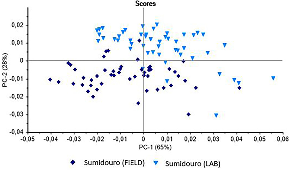
Figure 1. Principal Components Analysis (PCA) of the live specimens of Biomphalaria glabrata collected in the field in the municipality of Sumidouro (Rio de Janeiro state, Brazil) and raised in the laboratory.
In the case of the B. tenagophila population from Jacarepaguá, the analysis (Figure 2) indicated the possible separation of the two groups (field vs. laboratory). In this case, the scores separate the groups on the PC-1 axis. The PCA of the B. straminea samples (Figure 3) separated the two samples (field vs. laboratory) clearly on the PC-2 axis. The external validation of these classification models revealed different hit rates for each species (Table 1).
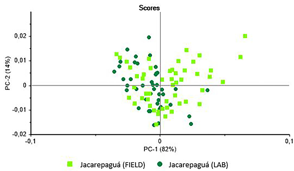
Figure 2. Principal Components Analysis (PCA) of the live specimens of Biomphalaria tenagophilacollected in the field in the borough of Jacarepaguá in the city of Rio de Janeiro (Brazil) and raised in the laboratory.
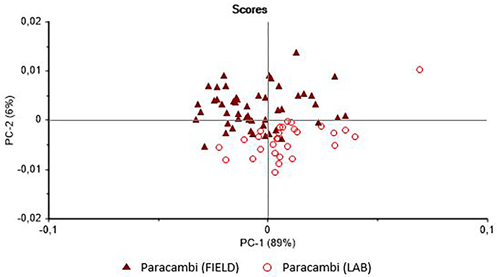
Figure 3. Principal Components Analysis (PCA) of the live specimens of Biomphalaria straminea collected in the field in the municipality of Paracambi (Rio de Janeiro state, Brazil) and raised in the laboratory.
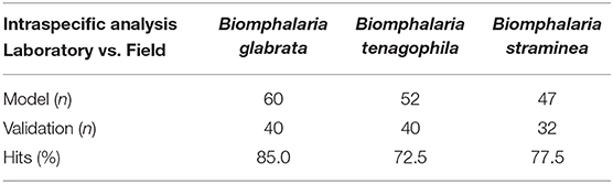
Table 1. Classification of the hits of the Biomphalaria species, based on a Linear Discriminant Analysis (LDA).
Interspecific Classification
Two models were used to classify the interspecific relationships of the three Biomphalaria species that act as intermediate hosts of S. mansoni, based on the analysis of live specimens collected in the field and raised in the laboratory. The PCA of the animals collected in the field (Figure 4) presented a clear tendency for the separation of the species, albeit with some mixed scores. The external validation of the spectra of this group had a 75% hit rate, with most errors being recorded between B. glabrata and B. tenagophila.
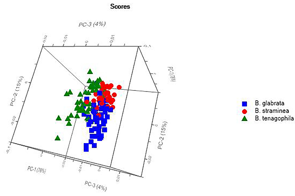
Figure 4. Principal Components Analysis (PCA) of the live specimens of Biomphalaria spp. collected in the field in Rio de Janeiro state, Brazil.
The PCA of the mollusk population raised in the laboratory (Figure 5) also revealed a tendency for the separation of species, with a hit rate of 80% for the classification of the species, as confirmed by the external validation. An interspecific analysis that included all the spectra recorded for the field and laboratory animals was also conducted to double-check the results, although in this case, there was no clear separation of the species.
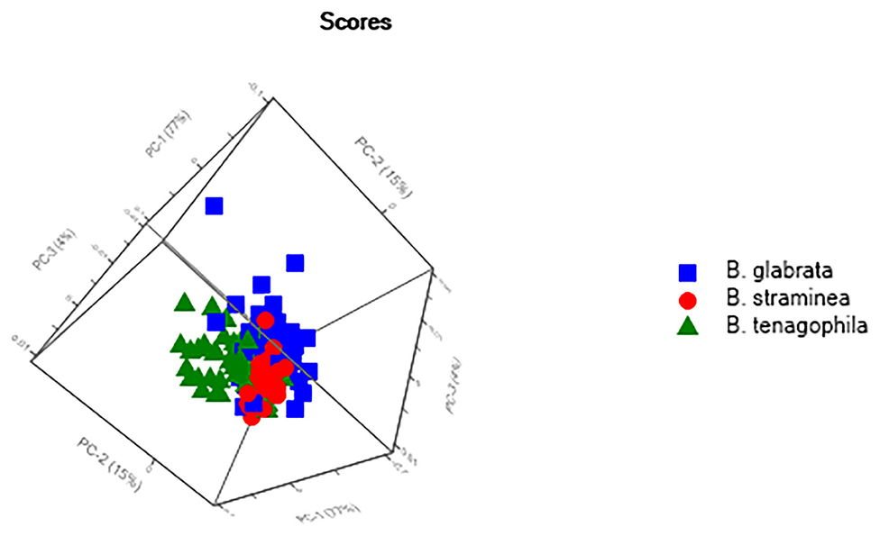
Figure 5. Principal Components Analysis (PCA) of the live specimens of ram's horn snail (Biomphalaria spp.) raised in the laboratory.
Discussion
The results of the present study indicate that the vibrational NIR spectroscopy was able to discriminate intra- and interspecific differences among the Biomphalaria species that are intermediate hosts of S. mansoni, based on the metabolic profiles presented by these snails in response to their immediate environment. The NIRS procedure provides differentiated chemical phenotypes of the specimens of the same species and different species, related to the metabolic configuration associated with the different environments, that is, the field and laboratory. As far as is known, this is the first study that has applied this approach to mollusk vectors, although Sikulu et al. (11, 12), Sikulu-Lord et al. (13), and Fernandes et al. (14) used NIRS to investigate the taxonomic, ecological, biochemical, and parasitological characteristics of mosquito vectors. Together with these analyses, the findings of the present study should contribute to the establishment of an area of research based on the application of the chemical or spectrophotometric diagnosis of the taxonomy of parasitic disease vectors. This approach should provide a valuable tool for the integrated analysis of taxonomic relationships, which complements classic taxonomic and molecular approaches.
The results of the intraspecific analyses applied in the present study emphasize the influence of the environment on the metabolism on aquatic organisms. A range of factors may have influenced the differentiation of the specimens of the same species collected in the field or raised in the laboratory.
In the wild, the snails are exposed to a wide range of contaminants and environmental impacts, which results in constant and unavoidable contact with the chemical elements dissolved in the water. Biomphalaria species are considered to be good indicators of environmental contamination, and have been analyzed to determine the bioaccumulation of heavy metals and metabolic shifts provoked by contact with herbicides and molluscicides (15–17). In particular, the herbicide oxyfluorfen can damage the reproductive and antioxidant systems of Biomphalaria species, as well as having cytotoxic and genotoxic effects (18, 19). The results of the present study demonstrated a more pronounced intraspecific pattern in B. glabrata (85%), when individuals from the same population collected in the field and raised in the laboratory were compared. This indicates clear differences in the metabolism of the two groups, which are possibly related to environmental factors. Mitta et al. (20) found that environmental factors had a direct impact on the relationship between Biomphalaria and Schistosoma, creating a mosaic of possible interactions. In the present study, although B. glabrata specimens were collected in Sumidouro, an endemic region for schistosomiasis in the state of Rio de Janeiro, none of the animals were found infected, as well as specimens of B. tenagophila and B. straminea. Pesticides are widely used on the farms in this municipality, where agriculture is a mainstay of the local economy (21), and the intoxication of local farm laborers has been reported by Oswaldo Cruz Foundation (22). Despite the evidence, complementary techniques to the NIRS, such as metabolomics and chemical analysis, are necessary to deepen the analysis in relation to environmental contamination.
The results of the present study also demonstrated the potential of NIRS for the separation and diagnosis of the three snail species without the need for the dissection of the specimens, in animals obtained from both the field and the laboratory. The identification errors found between B. glabrata and B. tenagophila indicate that the chemical phenotypes of the two species are the most similar. This may be related to the morphological and biological similarities of these two species, and their possible potential for hybridization (9, 23). However, Vidigal et al. (24) found that B. glabrata and B. tenagophila belonged to distinct clades in a phylogenetic analysis of the ITS2 molecular marker, which indicates that there is a considerable genetic distance between the two species.
Experimental metabolic studies of the three Biomphalaria species infected by S. mansoni or Angiostrongylus cantonensis have shown that the three species adopt distinct metabolic strategies to avoid infection. This may have contributed to the differentiation of the NIRS profiles of the three species (16, 25).
The potential for the use of NIRS to analyze the glycogen content of different types of tissue has already been evaluated in other mollusks, such as bivalves. This study (26) focused on the quality of the mollusk diet, showing variation in the glycogen content related to the condition of the mass of the viscera in different individuals of the same species. This study applied calibration models to predict the glycogen concentrations, analyzed using standard methods.
Clearly, then, NIRS has potential for use as a complementary approach for the analysis of the differentiation of the Biomphalaria species that are intermediate hosts of S. mansoni in Brazil, providing an important tool for an integrated taxonomic analysis. The potential for the differentiation of specimens at an intraspecific level nevertheless demonstrates the need to establish separate interspecific spectrophotometric models (distinguishing specimens from the field and the laboratory, for example) that guarantee the most effective and reliable diagnosis of the B. glabrata, B. tenagophila, and B. straminea specimens by NIRS.
Data Availability Statement
The original contributions presented in the study are included in the article/supplementary material, further inquiries can be directed to the corresponding author/s.
Author Contributions
VV collected the snails, analyzed the spectra, conceived and developed the study, and wrote and edited the manuscript. CM-S was involved in conception and development, and the writing and editing of the manuscript. CP participated in the analysis of the spectra and edited the manuscript. ST and MF contributed to the conception of the study and edited the manuscript. All authors contributed to the article and approved the submitted version.
Funding
This work was carried out with the support of the Coordination for the Improvement of Higher Education Personnel—Brazil (CAPES)—Financing Code 001.
Conflict of Interest
The authors declare that the research was conducted in the absence of any commercial or financial relationships that could be construed as a potential conflict of interest.
Acknowledgments
We are grateful to CAPES for supporting the present study. We would also like to thank the field team that helped collect the mollusks and the team at the Malacology Laboratory (Fiocruz/RJ/Brazil). We also grateful to the research group in Health and Environmental Education with emphasis on parasitic relationships.
References
1. Carvalho OC, Coelho PM, Lenzi HL. Schistosoma mansoni e Esquistossomose: uma visão multidisciplinar. Rio de Janeiro: Editora Fiocruz, (2008). 1124 p.
2. Nudelman F. Nacre biomineralisation: a review on the mechanisms of crystal nucleation. Semin Cell Dev Biol. (2015) 46:2–10. doi: 10.1016/j.semcdb.2015.07.004
3. Pasquini C. Near infrared spectroscopy: fundamentals practical aspects and analytical applications. J Braz Chem Soc. (2003) 14:138–219 doi: 10.1590/S0103-50532003000200006
4. Roggo Y, Pascal C, Lene M, Carmen L, Aurélie E, Nadine J. A review of near infrared spectroscopy and chemometrics in pharmaceutical Technologies. J Pharm Biomed Anal. (2007) 44:683–700. doi: 10.1016/j.jpba.2007.03.023
5. Silvestein RM, Webster FX, Kiemle DJ. Identificação Espectrométrica de Compostos Orgânicos. Rio de Janeiro: LTC (2013).
6. Mello-Silva CC, Thiengo S, Fernandez MA, Vidal C, Pasquini C. NIR and hyperspectral imaging in the diagnosis of the snails intermediate hosts of Schistosomiasis in Brazil. In: 17th International Conference on Near Infrared Spectroscopy. Foz do Iguaçu (2015).
7. Thiengo SC, Fernandez MA, Boaventura MF, Grault CE, Silva HFR, Mattos AC, et al. Freshwater snails and Schistosomiasis mansoni in the state of Rio de Janeiro, Brazil: I - Metropolitan Mesoregion. Mem Inst Oswaldo Cruz. (2001) 96:177–84. doi: 10.1590/S0074-02762001000900028
8. Thiengo SC, Fernandez MA, Boaventura MF, Santos SB, Mattos AC. Freshwater snails and Schistosomiasis Mansoni in the state of Rio de Janeiro, Brazil: II - Centro Fluminense Mesoregion. Mem Inst Oswaldo Cruz. (2002) 97:621–6. doi: 10.1590/S0074-02762002000500004
9. Brasil. Ministério da Saúde. Secretaria de Vigilância em Saúde. Departamento de Vigilância Epidemiológica. Vigilância e controle de moluscos de importância epidemiológica: diretrizes técnicas: Programa de Vigilância and Controle da Esquistossomose (PCE)/Ministério da Saúde, Secretaria de Vigilância em Saúde, Departamento de Vigilância Epidemiológica – 2nd edn. Brasília: Editora do Ministério da Saúde (2008). 178 p.
10. Ferreira CMM. Preparação dos dados para análise. In: Guimarães E, editor. Quimiometria: Conceitos, Métodos e Aplicações, Vol. 2. Campinas, SP: Editora Unicamp (2015) 43–90. doi: 10.7476/9788526814714
11. Sikulu MT, Majambere S, Khatib BO, Ali AS, Hugo LE, Dowell FE. Using a near-infrared spectrometer to estimate the age of Anopheles mosquitoes exposed to pyrethroids. PLoS One. (2014) 9:e0090657. doi: 10.1371/journal.pone.0090657
12. Sikulu-lord MT, Maia MF, Milali MP, Henry M, Mkandawile G, Kho A. Rapid and nondestructive detection and identification of two strains of wolbachia in Aedes aegypti by nearinfrared spectroscopy. PLoS One. (2016) 10:e0004759. doi: 10.1371/journal.pntd.0004759
13. Sikulu-lord MT, Devine GJ, Hugo LE, Dowell FE. First report on the application of near-infrared spectroscopy to predict the age of Aedes albopictus Skuse. Sci Rep. (2018) 8:9590. doi: 10.1038/s41598-018-27998-7
14. Fernandes JN, Dos Santos LMB, Chouin-carneiro T, Pavan MG, Garcia GA, David MR, et al. Rapid, non-invasive detection of Zika virus in mosquitoes by near-infrared spectroscopy. Sci Adv. (2018) 4:eaat0496. doi: 10.1126/sciadv.aat0496
15. Mahamoud KMA, Abu Taleb HMA. Fresh water snails as bioindicator for some heavy metals in the aquatic environment. Afr J Ecol. (2013) 51:193–8. doi: 10.1111/aje.12019
16. Lima MG, de Augusto RC, de Vasconcellos MC, Mello-Silva CC, Pinheiro J. Metabolic changes in Biomphalaria glabrata infected with Schistosoma mansoni exposed to latex of Euphorbia milii solution versus times of preparation. J Nat Prod. (2012) 5:222–32.
17. Mello-Silva CC, Vasconcellos MC, Bezerra JCB, Rodrigues MLA, Pinheiro J. The influence of exposure to Euphorbia splendens var. hislopii latex on the concentrations of total proteins and nitrogen products in Biomphalaria glabrata infected with Schistosoma mansoni. Acta Trop. (2011) 117:101104. doi: 10.1016/j.actatropica.2010.10.007
18. Lima MdV, de Siqueira WN, Silva HAMF, Lima Filho JM, de França EJ, de Albuquerque Melo AMM. Cytotoxic and genotoxic effect of oxyfluorfen on hemocytes of Biomphalaria glabrata. Environ Sci Pollut Res. (2018) 26: 3350–6. doi: 10.1007/s11356-018-3848-3
19. Ibrahim AM, Sayed DA. Toxicological impact of oxyfluorfen 24% herbicide on the reproductive system, antioxidant enzymes, and endocrine disruption of Biomphalaria alexandrina (Ehrenberg, 1831) snails. Environ Sci Pollut Res. (2019) 26:7960–8. doi: 10.1007/s11356-019-04251-w
20. Mitta G, Gourbal B, Grunau C, Knight M, Bridger JM, Theron A. The compatibility between Biomphalaria glabrata snails and Schistosoma mansoni: an increasingly complex puzzle. Adv Parasitol. (2017) 97:111–45. doi: 10.1016/bs.apar.2016.08.006
21. Stotz EN. Os limites da agricultura convencional and as razões de sua persistência: estudo do caso de Sumidouro, RJ. Rev Bras Saúde Ocup. (2012) 37:114–26. doi: 10.1590/S0303-76572012000100014
22. Fundação Oswaldo Cruz Instituto de Comunicação e Informação Científica and Tecnológica em Saúde. Consumo Intensivo de Agrotóxicos no Rio de Janeiro Revela Cenário de Intoxicações “Invisíveis”. (2016). Available online at: https://www.icict.fiocruz.br/content/consumo-intensivo-de-agrot%C3%B3xicos-no-rio-dejaneiro-revela-cen%C3%A1rio-de-intoxica%C3%A7%C3%B5es (accessed October 26, 2020).
23. Mello-Silva CC, Grault CE, Barbosa FS, Costa VA. Possible hibridization of Brazilian Planorbid snails and its importance in population dynamics. Mem Inst Oswaldo Cruz. (1998) 93:227232. doi: 10.1590/S0074-02761998000700040
24. Vidigal T, Kissinger JC, Caldeira RL, Pires ECR, Monteiro E, Simpson AJG. Phylogenetic relationships among Brazilian Biomphalaria species (Mollusca: Planorbidae) based upon analysis of ribossomal ITS2 sequences. Parasitology. (2000) 121:611–20. doi: 10.1017/S0031182000006831
25. Lima MG, Tunholi-Alves VM, Gaudencio FN, Martins FG, Castro RN, Thiengo SC, et al. The influence of Angiostrongylus cantonensis (Nematoda, Metastrongylidae) infection on the aerobic metabolism of Biomphalaria straminea and Biomphalaria tenagophila (Mollusca, Gastropoda). Exp Parasitol. (2016) 171:1–9. doi: 10.1016/j.exppara.2016.10.010
Keywords: near infrared spectroscopy, innovative technique, mollusk, freshwater snails, chemical phenotype, schistosomiasis
Citation: Valladares V, Pasquini C, Thiengo SC, Fernandez MA and Mello-Silva CC (2021) Field Application of NIR Spectroscopy for the Discrimination of the Biomphalaria Species That Are Intermediate Hosts of Schistosoma mansoni in Brazil. Front. Public Health 9:636206. doi: 10.3389/fpubh.2021.636206
Received: 01 December 2020; Accepted: 11 February 2021;
Published: 12 March 2021.
Edited by:
Russ Russell Stothard, Liverpool School of Tropical Medicine, United KingdomReviewed by:
Christian Albrecht, Justus Liebig Universität Giessen, GermanyMalcolm Jones, The University of Queensland, Australia
Copyright © 2021 Valladares, Pasquini, Thiengo, Fernandez and Mello-Silva. This is an open-access article distributed under the terms of the Creative Commons Attribution License (CC BY). The use, distribution or reproduction in other forums is permitted, provided the original author(s) and the copyright owner(s) are credited and that the original publication in this journal is cited, in accordance with accepted academic practice. No use, distribution or reproduction is permitted which does not comply with these terms.
*Correspondence: Silvana C. Thiengo, c2NhcnZhbGhvdGhpZW5nb0BnbWFpbC5jb20=
†These authors have contributed equally to this work and share last authorship