- 1Graduate School of Life and Environmental Sciences, Osaka Prefecture University, Sakai, Japan
- 2Graduate School of Engineering, Osaka Prefecture University, Sakai, Japan
- 3Faculty of Agriculture, Ehime University, Matsuyama, Japan
- 4Faculty of Agriculture, Ryukoku University, Otsu, Japan
- 5Center for Ecological Research, Kyoto University, Otsu, Japan
- 6Precursory Research for Embryonic Science and Technology, Japan Science and Technology Agency, Kawaguchi, Japan
The timing of measurement during plant growth is important because many genes are expressed periodically and orchestrate physiological events. Their periodicity is generated by environmental fluctuations as external factors and the circadian clock as the internal factor. The circadian clock orchestrates physiological events such as photosynthesis or flowering and it enables enhanced growth and herbivory resistance. These characteristics have possible applications for agriculture. In this study, we demonstrated the diurnal variation of the transcriptome in tomato (Solanum lycopersicum) leaves through molecular timetable method in a sunlight-type plant factory. Molecular timetable methods have been developed to detect periodic genes and estimate individual internal body time from these expression profiles in mammals. We sampled tomato leaves every 2 h for 2 days and acquired time-course transcriptome data by RNA-Seq. Many genes were expressed periodically and these expressions were stable across the 1st and 2nd days of measurement. We selected 143 time-indicating genes whose expression indicated periodically, and estimated internal time in the plant from these expression profiles. The estimated internal time was generally the same as the external environment time; however, there was a difference of more than 1 h between the two for some sampling points. Furthermore, the stress-responsive genes also showed weakly periodic expression, implying that they were usually expressed periodically, regulated by light–dark cycles as an external factor or the circadian clock as the internal factor, and could be particularly expressed when the plant experiences some specific stress under agricultural situations. This study suggests that circadian clock mediate the optimization for fluctuating environments in the field and it has possibilities to enhance resistibility to stress and floral induction by controlling circadian clock through light supplement and temperature control.
Introduction
Recently agricultural technologies have been rapidly developing, for example, application of information and communication technology, automation, and cultivation in closed systems with controlled temperature, humidity, and light conditions. In particular, plant factories are the more recent and highly regarded. These are of two types, closed-type, and sunlight-type. Both types have abilities of mass production, stable supply, and safe products. Furthermore, both types can control the cultivation environment artificially compared with open culture. Accordingly, there has been increased expectation of the ability to create cultivation control technologies for several cultivars according to various internal factors, which could be a key for high productivity or high quality. There is a real need for an exhaustive understanding of gene expression or metabolism to determine what these internal factors are, for example stress responses (Mittler and Blumwald, 2010). With this background, there have been great advances in technologies to analyze the genome, transcriptome, proteome, and metabolome. These analyses are increasingly user-friendly and their use in studies has increased rapidly. These molecular biological approaches have potential to solve several agricultural problems. In the case of fruit vegetables, there is a need to clarify some of the internal factors responsible for such physiological disorders as fruit cracking, blossom-end rot (Cuartero and Fernández-Muñoz, 1999; Guichard et al., 2001), and floral induction (Lifschitz et al., 2006; Corbesier et al., 2007). These are also critical issues for yield improvements.
Searching of these internal factors through biological statistical analysis has shown the usefulness of biological models (Takahashi et al., 2012; Guanter et al., 2014). However, because many genes are expressed periodically, diurnal variation must be considered when choosing a time for measurement (Harmer et al., 2000). These periodic expressions are generated by the day–night environment as the external factor and the circadian clock as the internal factor. Most living organisms have a circadian clock system and that of plants orchestrates physiological events such as gene expression, protein phosphorylation, chloroplast movement, stomatal opening, and flowering (Barak et al., 2000). The circadian clock system has three components (Farré and Weise, 2012): input pathways receive the external stimuli such as light and temperature fluctuations; the oscillator generates the endogenous circadian rhythms; and output pathways regulate general metabolism (e.g., of nitrogen and sugars). Endogenous circadian rhythms are generated by a number of genes; for example, CCA1 (CIRCADIAN CLOCK ASSOCIATED 1), TOC1 (TIMING OF CAB EXPRESSION 1), LHY (LATE ELONGATED HYPOCOTYL), and PRRs (PSEUDO-RESPONSE REGULATORs), which create feedback loops in each cell (Alabadí et al., 2001; Nakamichi et al., 2010, 2012; Haydon et al., 2011). These genes—including PHYs, which are the far-red light receptors; and CRYs, which are the blue light receptors (Pruneda-Paz and Kay, 2010)—receive signals from the input pathway and are expressed periodically with control of their own amplitude, period, and phase. It is well known that controlling the circadian rhythm enhances growth and herbivory resistance (Dodd et al., 2005; Goodspeed et al., 2012, 2013; Higashi et al., 2015), and the circadian clock can be controlled by external stimuli that change the light–dark cycle and by temperature fluctuation, because the circadian clock has characteristics of entrainment to the environment (Rensing and Ruoff, 2002; James et al., 2012; Fukuda et al., 2013). The circadian clock regulates hundreds of genes in Arabidopsis (Arabidopsis thaliana). Photosynthesis or phenylpropanoid biosynthesis genes are expressed periodically, with these genes peaking at different times such as subjective day and dawn under constant light (Harmer et al., 2000). These studies have shown that many genes generate circadian fluctuations without external stimuli. One recent study indicated that the day–night environment as the external oscillator and the circadian clock as the internal molecular oscillator activated transcription factors and mediated the circadian outputs (Haydon et al., 2011). The output adapts the circadian gene expression and entrains it to the external environment. This study suggests that the day–night environment as the external oscillator is a fast pathway and that the circadian clock as the internal molecular oscillator is a slow pathway for activation of transcriptome factors. Furthermore, hundreds of genes oscillate with circadian and circannual rhythms in rice (Oryza sativa) under field conditions (Nagano et al., 2012). They can generate comparatively stable rhythms under greatly variable conditions through the internal molecular oscillator. This indicates that in many cases we should consider the plant's internal rhythm and take care choosing a measurement time. Thus, very accurate analysis of diurnal variation will have application in development of biological models for yield improvements.
In this study, we tried to develop a highly accurate analysis of diurnal variation in tomato leaves in a sunlight-type plant factory as a basis for exhaustive study of the transcriptome and metabolome. We obtained time-course transcriptome data and used the molecular timetable method (Ueda et al., 2004) to analyze diurnal variation. Using these methods, we selected some periodic genes and estimated the phase of the circadian clock in tomato (internal time) from these periodic gene expression profiles.
Materials and Methods
Plant Materials and Growing Systems
Experiments were carried out using tomato (Solanum lycopersicum cv. Taiankichijitsu, Nanto Seed Co. Ltd., Nara, Japan) cultivated in a sunlight-type plant factory (4480 cm [W] × 2300 cm [D] × 500 cm [H]) in the Faculty of Agriculture, Ehime University, Japan. Individual plants are usually cultivated for a year; in this experiment, tomato seedlings were grown by Berg Earth Co. Ltd. (Ehime, Japan) and transplanted into rockwool cubes (10 cm [W] × 10 cm [D] × 6.5 cm [H], Grodan Delta, GRODAN, Roermond, The Netherlands) in August 2013. Rockwool cubes were placed on rockwool slabs (100 cm [W] × 20 cm [D] × 7.5 cm [H], Grotop Expert, GRODAN) at four per slab. The four rockwool cubes were placed at 25 cm intervals and watered using nutrient solution (Sonneveld, 1985). There were 20 slabs set in a line, with 28 lines per greenhouse. We sampled their leaves in January 2014. The light condition, relative humidity, and carbon dioxide concentration were ambient. Air temperature was maintained at 14°C during 18:00–8:00.
We sampled the fifth leaves every 2 h for 2 days, starting at 14:00 on 6 January 2014 and ending at 14:00 on 8 January 2014. We sliced leaf segments and stored them at 0°C with RNA-later solution (Qiagen, Valencia, CA, USA), an aqueous nontoxic tissue storage reagent that rapidly permeates tissue to stabilize and protect the integrity of RNA.
RNA-Seq Assay and Data Analysis
We isolated total RNA using an RNeasy Plant Mini Kit (Qiagen). RNA quality was checked using an Agilent 2100 Bioanalyzer (Agilent Technologies, Palo Alto, CA, USA) and RNA quantity control was performed using a Qubit® 2.0 Fluorometer (Life Technologies, Carlsbad, CA, USA). We prepared a RNA-Seq library (Wang et al., 2011; Nagano et al., 2015) and sequencing was performed by BGI (Yantian District, Shenzhen, China). Then we obtained the sequence read files using a HiSeq 2000 sequencer (single end, 50 bp; Illumina, San Diego, CA, USA). These sequence data are available in the DDBJ Sequenced Read Archive (http://trace.ddbj.nig.ac.jp/DRASearch) under the accession number DRA003529 and DRA003530.
All reads of each sample were quality checked by FastQC and mapped using RSEM (RNA-Seq by Expectation Maximization) (Li and Dewey, 2011) with Bowtie2 software (Langmead and Salzberg, 2012) to the reference sequence. RSEM is a user-friendly software package able to accurately quantify transcript abundances. By these processes, we obtained the data of gene expression levels for each sample. Finally, we used the reads per kilobase per million mapped reads measure to normalize the gene expression for total read length and the number of sequencing reads. In this study, we calculated the average expression level of each sampling time for each genes. Then we cut off some genes whose average expression level is less than 5% of average expression level of whole genes and we identified 18,332 genes were significantly expressed.
Molecular Timetable Method
Molecular timetable methods have been developed for administration of medicine to animals, with the objective to detect internal body time from periodic gene expression profiles (Ueda et al., 2004). As an outline of this methods, they selected “time-indicating genes,” which expressed periodically and represented internal body time from every 2 h for 2 days microarray data in the mouse liver. Then they developed gene expression profiles from time-indicating genes and estimated internal body time at each sampling time. To verify the performance of this methods, they calculated the sensitivity and specificity in the presence of different measurement noise and different number of time-indicating genes. We adapted this methods to use RNA-Seq data processing with cultivated plants.
First, we selected genes whose expression indicated periodicity and high amplitude from the time-course transcriptome data—these were time-indicating genes. To analyze periodicity, we prepared 1440 test cosine curves. These curves had different peaks (0–24 h) measured at increments of 1 min. We fitted test cosine curves to data from each time-course gene expression generated via RNA-Seq and calculated the correlation value (r) to identify the best-fitting cosine curve. The peak time of the best-fitting curve was estimated as the peak time for each gene. This estimated peak time defined as the molecular peak time. Thus, molecular peak time was estimated from a single gene, and all genes were estimated it individually. Then, to analyze amplitude, we calculated the average and standard deviation for every gene expression level. The amplitude value (a) was calculated as the standard deviation divided by the average of gene expression level.
Second, we plotted expression profiles of time-indicating genes. The number of time-indicating genes was determined by the cut-off values of r and a. We normalized the expression level of each time-indicating gene using its average and standard deviation. Normalized expression level was defined as the value of expression level minus average expression level, divided by the standard deviation. We then plotted expression profiles composed of the molecular peak time and normalized expression level for every sampling time. The horizontal axis indicated the molecular peak time and the vertical axis indicated normalized expression level.
Finally, the internal time was estimated by the plotted expression profile. We prepared 1440 test cosine curves with 1-min differences from each other and fitted them to the expression profiles. We identified the best-fitting cosine curve, and its peak time indicated the estimated internal time. Thus, the internal time was estimated from a number of periodic genes.
In this study, we selected 143 time-indicating genes by setting the cut-off values of r = 0.915 and a = 0.15 in whole genes and 150 time-indicating genes by setting the cut-off values of r = 0.635 and a = 0.15 in stress-responsive genes. In the previous study, they indicated that 150 time-indicating genes with 100% measurement noise could estimate internal time with high accuracy. Table S1 shows the measurement noise with 143 time-indicating genes in whole genes and 150 time-indicating genes in stress-responsive genes for each sampling time (see Supplementary Material). We calculated the standard deviation of the difference between a real and an estimated expression of all time-indicating genes as the measurement noise. Each measurement noise represent less than 100%. Thus, 143 and 150 time-indicating genes are enough to estimate the internal time with high accuracy.
Mapman Analysis
We used MapMan to categorize stress-responsive genes in tomato (Thimm et al., 2004). MapMan BINs were generated by Mercator (http://mapman.gabipd.org/web/guest/app/mercator) based on TAIR10 in Arabidopsis. MapMan software can be downloaded from http://mapman.gabipd.org/web/guest/mapman.
Results
Environmental Conditions in a Sunlight-Type Plant Factory
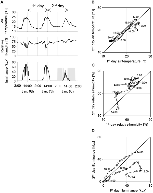
We sampled tomato leaves every 2 h during 6–8 January 2014 in a sunlight-type plant factory in the Faculty of Agriculture, Ehime University, Japan (Takayama et al., 2012). Because it was winter during the experiments, the air temperature, relative humidity, and illuminance were comparatively low and day–night cycle had short days (day of 10 h and night of 14 h) (Figure 1A). As it rained on 8 January, this caused decreases in air temperature and illuminance, and increased relative humidity. Air temperature was relatively stable over the 1st and 2nd days of the experiment (Figure 1B); in contrast, relative humidity, and illuminance were relatively unstable on each day (Figures 1C,D). These results show that relative humidity and illuminance were strongly influenced by the rainy day.

Figure 1. Environmental conditions in a sunlight-type plant factory in Ehime University (A) and comparison of the 1st with the 2nd day for each environmental condition (B–D). (A) Air temperature, relative humidity, and illuminance in a sunlight-type plant factory. Gray area of illuminance indicates precipitation. (B–D) Open circles indicate each values for every 10 min and filled circles indicate the sampling point. Sampling started at 14:00.
Selection of Time-Indicating Genes and Development of These Expression Profiles
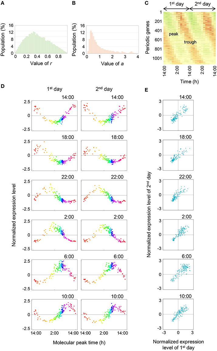
Then we analyzed the diurnal oscillation in gene expressions using the molecular timetable method. To select time-indicating genes that were definitely expressed periodically, we calculated the correlation value (r) which indicates periodicity, and amplitude value (a) for 18,332 genes (see Materials and Methods). The histograms showed that neither r nor a values were normally distributed (Figures 2A,B) and more than half of all genes had r < 0.5. About 5% of all genes exhibited clear periodic expression. In contrast, the histogram of a values showed that a values were distributed around 0.5 and spread widely around high value. About 70% of all genes had a values within 0–1. The heat map of 1132 periodic genes, which indicated periodicity and high amplitude through setting of cut-off values of r = 0.80 and a = 0.15, clearly showed a diagonal striped pattern, indicating that we could correctly select the periodic genes (Figure 2C). We showed the expression profiles of 143 time-indicating genes through setting the cut-off values of r = 0.915 and a = 0.15 for each time point on the 1st and 2nd days (Figure 2D). The vertical axis represents the normalized expression level and the horizontal axis represents the molecular peak time. Thus, Figure 2D shows the overall behavior of 143 time-indicating genes at each sampling time. Genes related to photosynthesis, translocation of sugar and photoreceptor were pooled in 143 time-indicating genes and the timing of their expression was consistent with their physiological function (Table S2). As time proceeded, the peak position shifted at regular intervals. Furthermore, the expression profiles on the 1st and 2nd days were similar in most respects. This indicates that these 143 genes showed stable periodic expression at daily intervals under fluctuating field conditions. We plotted normalized expression levels for the 1st and 2nd days to verify stability (Figure 2E). All genes were plotted in a linear fashion despite the 2nd day being rainy. The determination coefficient (R2) between them was high (R2 ≈ 0.7; Figure 3B). Thus, this supports the claim that time-indicating genes were stably expressed under fluctuating field conditions.

Figure 2. Comprehensive analysis of diurnal oscillation in gene expressions. (A,B) Histograms of the r and a values, respectively. (C) Oscillation behavior of time-indicating genes. Green area indicates the peak and red area indicates the trough. (D) Expression profiles of 143 time-indicating genes for the 1st and 2nd days. The time at the top right of the plot area indicates the sampling time. The range from orange to purple represents night-time and other colored areas represent day-time. (E) Correlations between normalized expression levels for the 1st and 2nd days. The time at the top right of the plot area indicates the sampling time.

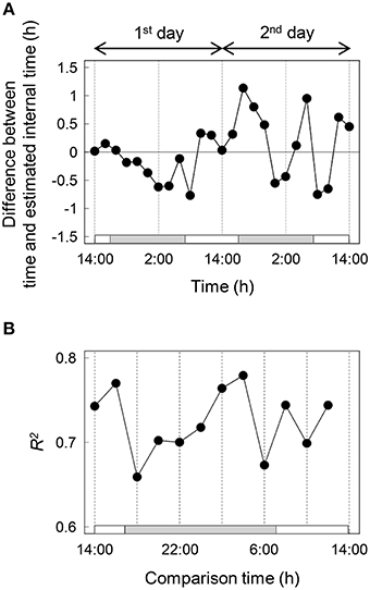
Figure 3. Validation of the internal time estimation (A) and transition of the determination coefficient between normalized expression levels for the 1st and 2nd days (B). White and gray bars represent light and dark, respectively.
Validation of the Internal Time Estimation
We estimated the internal time from the expression profiles of 143 time-indicating genes for each time and calculated the difference between time and estimated internal time (Figure 3A). Internal time showed much the same time as the sampling time; however, there was a difference of more than 1 h between the two for some sampling points. As an additional indication of stability, we showed the transition of R2 between normalized expression levels of the 1st and 2nd days (Figure 3B). There was a high correlation indicated by high R2 values (>0.65) at all times.
Diurnal Gene Expression Profiles of Stress-Responsive Genes
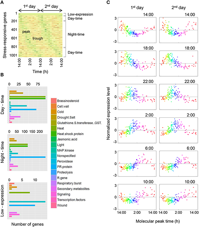
We focused on the stress-responsive genes, which are important to increase sweetness in tomato fruit and to defend against disease. Furthermore, stress-responsive genes are possible markers of the internal factors because these genes show sensitivity to external stimuli and affect several physiological events (Wang et al., 2003; Chinnusamy et al., 2005; Umezawa et al., 2006; Cattivelli et al., 2008). We extracted 1048 stress-responsive genes from all genes using the MapMan and analyzed them by the molecular timetable method. The heat map of the 1048 stress-responsive genes indicated that they were expressed periodically under fluctuating field conditions without the addition of external stress such as heat or drought during the experiment (Figure 4A). Then we separated the stress-responsive genes into three types—day-time (434 genes), night-time (786 genes), and low-expression (47 genes)—according to the molecular peak times, and categorized them using the MapMan (Figure 4B). Day-time was 7:00–17:00 (10 h) and night-time was 17:00–7:00 (14 h). More than half of the stress-responsive genes had expression peaks at night in tomato leaves. Genes related to cold, heat, and drought stress had expression peaks equally during day and night. Then we showed the expression profiles of 150 time-indicating genes, which indicated periodicity and high amplitude, through setting the cut-off values of r = 0.635 and a = 0.15 in the stress-responsive genes (Figure 4C). These expression profiles were clearly noisy compared with expression profiles of the 143 most-periodic time-indicating genes (Figure 2D); however, there was certainly periodicity without stress being applied.

Figure 4. Diurnal gene expression profiles of stress-responsive genes. (A) Oscillation behavior of stress-responsive genes. Green areas indicate the peak and red areas indicate the trough. (B) Categorization of stress-responsive genes using MapMan. Day-time was 7:00–17:00 (10 h) and night-time was 17:00–7:00 (14 h). (C) Expression profiles of 150 time-indicating genes for the 1st and 2nd days in stress-responsive genes. The time at the top right of the plot area indicates the sampling time. The range from orange to purple represents night-time and other colored areas represent day-time.
Discussion
At least 1000 genes, included some clock-related genes (Presentation S1), indicated periodic expression clearly in tomato leaves under agricultural situations in winter. In addition, 143 time-indicating genes indicated stable periodic expression even though the alterations in the environment had a possibility to affect such aspects of plant physiology as photosynthesis and sucrose metabolism (Feugier and Satake, 2013; McCormick and Kruger, 2015), and so the expression of relevant genes would likely be influenced by external stimuli (Nagano et al., 2012). This mediation system seemed to be generated by the internal oscillator of the circadian clock. Although 143 time-indicating genes indicated stable periodic expression, some estimated internal time showed little difference between the sampling times. It suggested that the phase of periodic genes, or of genes downstream, had their circadian clock precisely regulated to adjust to a non-constant environment and so kept their periodicity (Presentation S1). Thus, the periodicity of these genes was sufficiently stable, although there were some differences in expression level. Circadian stability was reported in rice leaves under fluctuating field conditions (Matsuzaki et al., 2015). In the case of rice, the difference between sampling times and estimated internal times was ≤ 22 min and gene expression was not affected by external factors. These differences between tomato and rice may be due to species differences. Circadian clocks exist in almost all living organisms and characteristics of plant circadian rhythms have been studied in Arabidopsis (Nakamichi et al., 2004); however, characteristics of circadian rhythm in Lactuca sativa differed from Arabidopsis (Higashi et al., 2014). Furthermore, tomato leaves may have an unusual circadian clock system because they develop a detrimental leaf injury when grown under constant light (Cushman and Tibbitts, 1998; Velez-Ramirez et al., 2011). One characteristic of circadian clocks is that they generate circadian rhythm under continuous conditions. This suggest that an unusual circadian clock system in tomato affected expression of the clock genes or downstream genes and resulted in the difference of estimated internal time. Recently, the inclusive CCA1 target genes in Arabidopsis were reported (Nagel et al., 2015). Then we located orthologs of the time-indicating genes using the KEGG database and identified how many orthologs of the CCA1 target genes were present in the time-indicating gene pool of tomato. We identified 23 CCA1 target genes among the time-indicating genes, accounting for about 16% of 143 time-indicating genes. This suggests that a number of time-indicating genes were expressed periodically depending on the day–night environment or influence of other clock genes, at least in this experiment. This low proportion of CCA1 target genes to time-indicating genes in tomato might affect the difference in estimated internal time. On the other hand, to detect the accurate internal time is important because plant circadian rhythms can be controlled by external stimuli (Barak et al., 2000; Covington et al., 2008; Fukuda et al., 2013). Thus, the timing of environmental control such as light supplement and temperature control affect the circadian rhythms and therefore the dimension of physiological events. It has possibilities to enhance growth and floral induction.
Stress-responsive genes were also indicated for periodic expression. Thus the stress-responsive genes were normally expressed periodically as regulated by light–dark cycle as the external factor or the circadian clock as the internal factor, and they could be specifically expressed when some stresses were experienced by the plant. Similar events were also reported for soybean, barley, and Arabidopsis (Covington et al., 2008; Habte et al., 2014; Marcolino-Gomes et al., 2014). It suggests that circadian regulation of stress-responsive genes expression may be widely-conserved systems and it provides some advantage that plant survive under fluctuating field conditions (Goodspeed et al., 2012, 2013; Grundy et al., 2015). Furthermore, this result suggests that stress-responsive genes may be competent markers for agriculture because they are simple to apply although normally expressed periodically and especially transiently.
In conclusion, we demonstrated that many genes were expressed periodically and that gene expression was stable in a sunlight-type plant factory. Furthermore, internal time could be estimated from time-course gene expression data in tomato leaves through molecular timetable method. The results also showed that stress-responsive genes were expressed periodically under non-stressed conditions. This study suggests that circadian clock mediate the optimization for fluctuating environments and environmental control tailored to internal time may enhance resistibility to stress and floral induction, eventually the yieldability.
Author Contributions
HF and TH designed the experiments. YT did the MapMan analysis and constructed the heat map. KT supported the sampling in a sunlight-type plant factory. AN and MH prepared a RNA-Seq library. TH performed RNA-Seq data analysis and the molecular timetable method. TH and HF wrote the manuscript. All authors discussed the results and implications and commented on the manuscript.
Conflict of Interest Statement
The authors declare that the research was conducted in the absence of any commercial or financial relationships that could be construed as a potential conflict of interest.
Acknowledgments
We are grateful to Dr. Noriko Takahashi for the plant cultivation, Professor Hiroshi Kudoh for RNA-Seq and Professor Koh Aoki for discussion. The present study was supported by the Joint Usage/Research Program of the Center for Ecological Research, Kyoto University. This study was partially supported by Grants-in-Aid for JSPS Fellows (No. 15J12133 to TH), Grants-in-Aid for Scientific Research (No. 25712029 and 25119721 to HF), PRESTO JST, and the Canon Foundation.
Supplementary Material
The Supplementary Material for this article can be found online at: http://journal.frontiersin.org/article/10.3389/fpls.2016.00087
References
Alabadí, D., Oyama, T., Yanovsky, M. J., Harmon, F. G., Más, P., and Kay, S. A. (2001). Reciprocal regulation between TOC1 and LHY/CCA1 within the Arabidopsis circadian clock. Science 293, 880–883. doi: 10.1126/science.1061320
Barak, S., Tobin, E. M., Andronis, C., Sugano, S., and Green, R. M. (2000). All in good time: the Arabidopsis circadian clock. Trends Plant Sci. 5, 517–522. doi: 10.1016/S1360-1385(00)01785-4
Cattivelli, L., Rizza, F., Badeck, F. W., Mazzucotelli, E., Mastrangelo, A. M., Francia, E., et al. (2008). Drought tolerance improvement in crop plants: an integrated view from breeding to genomics. Field Crops Res. 105, 1–14. doi: 10.1016/j.fcr.2007.07.004
Chinnusamy, V., Jagendorf, A., and Zhu, J. K. (2005). Understanding and improving salt tolerance in plants. Crop Sci. 45, 437–448. doi: 10.2135/cropsci2005.0437
Corbesier, L., Vincent, C., Jang, S., Fornara, F., Fan, Q., Searle, I., et al. (2007). FT protein movement contributes to long-distance signaling in floral induction of Arabidopsis. Science 316, 1030–1033. doi: 10.1126/science.1141752
Covington, M. F., Maloof, J. N., Straume, M., Kay, S. A., and Harmer, S. L. (2008). Global transcriptome analysis reveals circadian regulation of key pathways in plant growth and development. Genome Biol. 9:R130. doi: 10.1186/gb-2008-9-8-r130
Cushman, K. E., and Tibbitts, T. W. (1998). The role of ethylene in the development of constant-light injury of potato and tomato. J. Am. Soc. Hort. Sci. 123, 239–245.
Dodd, A. N., Salathia, N., Hall, A., Kévei, E., Tóth, R., Nagy, F., et al. (2005). Plant circadian clocks increase photosynthesis, growth, survival, and competitive advantage. Science 309, 630–633. doi: 10.1126/science.1115581
Farré, E. M., and Weise, S. E. (2012). The interactions between the circadian clock and primary metabolism. Plant Biol. 15, 293–300. doi: 10.1016/j.pbi.2012.01.013
Feugier, F. G., and Satake, A. (2013). Dynamical feedback between circadian clock and sucrose availability explains adaptive response of starch metabolism to various photoperiods. Front. Plant Sci. 3:305. doi: 10.3389/fpls.2012.00305
Fukuda, H., Murase, H., and Tokuda, I. T. (2013). Controlling circadian rhythms by dark-pulse perturbations in Arabidopsis thaliana. Sci. Rep. 3:1533. doi: 10.1038/srep01533
Goodspeed, D., Chehab, E. W., Min-Venditti, A., Braam, J., and Covington, M. F. (2012). Arabidopsis synchronizes jasmonate-mediated defense with insect circadian behavior. Proc. Natl. Acad. Sci. U.S.A. 109, 4674–4677. doi: 10.1073/pnas.1116368109
Goodspeed, D., Liu, J. D., Chehab, E. W., Sheng, Z., Francisco, M., Kliebenstein, D. J., et al. (2013). Postharvest circadian entrainment enhances crop pest resistance and phytochemical cycling. Curr. Biol. 23, 1235–1241. doi: 10.1016/j.cub.2013.05.034
Grundy, J., Stoker, C., and Carré, I. A. (2015). Circadian regulation of abiotic stress tolerance in plants. Front. Plant Sci. 6:648. doi: 10.3389/fpls.2015.00648
Guanter, L., Zhang, Y., Jung, M., Joiner, J., Voigt, M., Berry, J. A., et al. (2014). Global and time-resolved monitoring of crop photosynthesis with chlorophyll fluorescence. Proc. Natl. Acad. Sci. U.S.A. 111, E1327–E1333. doi: 10.1073/pnas.1320008111
Guichard, S., Bertin, N., Leonardi, C., and Gary, C. (2001). Tomato fruit quality in relation to water and carbon fluxes. Agronomie 21, 385–392. doi: 10.1051/agro:2001131
Habte, E., Müller, L. M., Shtaya, M., Davis, S. J., and VonKorff, M. (2014). Osmotic stress at the barley root affects expression of circadian clock genes in the shoot. Plant Cell Environ. 37, 1321–1327. doi: 10.1111/pce.12242
Harmer, S. L., Hogenesch, J. B., Strume, M., Chang, H. S., Han, B., Zhu, T., et al. (2000). Orchestrated transcription of key pathways in Arabidopsis by the circadian clock. Science 290, 2110–2113. doi: 10.1126/science.290.5499.2110
Haydon, M. J., Bell, L. J., and Webb, A. A. R. (2011). Interactions between plant circadian clocks and solute transport. J. Exp. Bot. 62, 2333–2348. doi: 10.1093/jxb/err040
Higashi, T., Kamitamari, A., Okamura, N., Ukai, K., Okamura, K., Tezuka, T., et al. (2014). Characterization of circadian rhythms through a bioluminescence reporter assay in Lactuca sativa L. Environ. Control Biol. 52, 21–27. doi: 10.2525/ecb.52.21
Higashi, T., Nishikawa, S., Okamura, N., and Fukuda, H. (2015). Evaluation of growth under Non-24 h period lighting conditions in Lactuca sativa L. Environ. Control Biol. 53, 7–12. doi: 10.2525/ecb.53.7
James, A. B., Syed, N. H., Bordage, S., Marshall, J., Nimmo, G. A., Jenkins, G. I., et al. (2012). Alternative splicing mediates responses of the Arabidopsis circadian clock to temperature changes. Plant Cell 24, 961–981. doi: 10.1105/tpc.111.093948
Langmead, B., and Salzberg, S. L. (2012). Fast gapped-read alignment with Bowtie2. Nat. Methods 9, 357–359. doi: 10.1038/nmeth.1923
Li, B., and Dewey, C. N. (2011). RSEM: accurate transcript quantification from RNA-Seq data with or without a reference genome. BMC Bioinformatics 12:323. doi: 10.1186/1471-2105-12-323
Lifschitz, E., Eviatar, T., Rozman, A., Shalit, A., Goldshmidt, A., Amsellem, Z., et al. (2006). The tomato FT ortholog triggers systemic signals that regulate growth and flowering and substitute for diverse environmental stimuli. Proc. Natl. Acad. Sci. U.S.A. 103, 6398–6403. doi: 10.1073/pnas.0601620103
Marcolino-Gomes, J., Rodrigues, F. A., Fuganti-Pagliarini, R., Bendix, C., Nakayama, T. J., Celaya, B., et al. (2014). Diurnal oscillations of soybean circadian clock and drought responsive genes. PLoS ONE 9:e86402. doi: 10.1371/journal.pone.0086402
Matsuzaki, J., Kawahara, Y., and Izawa, T. (2015). Punctual transcriptional regulation by the rice circadian clock under fluctuating conditions. Plant Cell 27, 633–648. doi: 10.1105/tpc.114.135582
McCormick, A. J., and Kruger, N. J. (2015). Lack of fructose 2, 6-bisphosphate compromises photosynthesis and growth in Arabidopsis in fluctuating environments. Plant J. 81, 670–683. doi: 10.1111/tpj.12765
Mittler, R., and Blumwald, E. (2010). Genetic engineering for modern agriculture: challenges and perspectives. Annu. Rev. Plant Biol. 61, 443–462. doi: 10.1146/annurev-arplant-042809-112116
Nagano, A. J., Honjo, M. N., Mihara, M., Sato, M., and Kudoh, H. (2015). Detection of plant viruses in natural environments by using RNA-Seq. Methods Mol. Biol. 1236, 89–98. doi: 10.1007/978-1-4939-1743-3_8
Nagano, A. J., Sato, Y., Mihara, M., Antonio, B. A., Motoyama, R., Itoh, H., et al. (2012). Deciphering and prediction of transcriptome dynamics under fluctuating field conditions. Cell 151, 1358–1369. doi: 10.1016/j.cell.2012.10.048
Nagel, D. H., Doherty, C. J., Pruneda-Paz, J. L., Schmitz, R. J., Ecker, J. R., and Kay, S. A. (2015). Genome-wide identification of CCA1 targets uncovers an expanded clock network in Arabidopsis. Proc. Natl. Acad. Sci. U.S.A. 112, E4802–E4810. doi: 10.1073/pnas.1513609112
Nakamichi, N., Ito, S., Oyama, T., Yamashino, T., Kondo, T., and Mizuno, T. (2004). Characterization of plant circadian rhythms by employing Arabidopsis cultured cells with bioluminescence reporters. Plant Cell Physiol. 45, 57–67. doi: 10.1093/pcp/pch003
Nakamichi, N., Kiba, T., Henriques, R., Mizuno, T., Chua, N. H., and Sakakibara, H. (2010). PSEUDO-RESPONSE REGULATORS 9, 7, and 5 are transcriptional repressors in the Arabidopsis circadian clock. Plant Cell 22, 594–605. doi: 10.1105/tpc.109.072892
Nakamichi, N., Kiba, T., Kamioka, M., Suzuki, T., Yamashino, T., Higashiyama, T., et al. (2012). Transcriptional repressor PRR5 directly regulates clock-output pathways. Proc. Natl. Acad. Sci. U.S.A. 109, 17123–17128. doi: 10.1073/pnas.1205156109
Pruneda-Paz, J. L., and Kay, S. A. (2010). An expanding universe of circadian networks in higher plants. Trends Plant Sci. 15, 259–265. doi: 10.1016/j.tplants.2010.03.003
Rensing, L., and Ruoff, P. (2002). Temperature effect on entrainment, phase shifting, and amplitude of circadian clocks and its molecular bases. Chronobiol. Int. 19, 807–864. doi: 10.1081/CBI-120014569
Sonneveld, C. (1985). A Method for Calculating the Composition of Nutrient Solutions for Soilless Cultures, 2nd Edn. Naaldwijk: Glasshouse Crops.
Takahashi, T., Ishigami, Y., Goto, E., Niibori, K., and Goto, K. (2012). Modeling the growth and yield of tomatoes cultivated with a low node-order pinching system at high plant density. Environ. Control Biol. 50, 53–61. doi: 10.2525/ecb.50.53
Takayama, K., Miguchi, Y., Manabe, Y., Takahashi, N., and Nishina, H. (2012). Analysis of ΦPS II and NPQ during the slow phase of the chlorophyll fluorescence induction phenomenon in tomato leaves. Environ. Control Biol. 50, 181–187. doi: 10.2525/ecb.50.181
Thimm, O., Bläsing, O., Gibon, Y., Nagel, A., Meyer, S., Krüger, P., et al. (2004). MAPMAN: a user-driven tool to display genomics data sets onto diagrams of metabolic pathways and other biological processes. Plant J. 37, 914–939. doi: 10.1111/j.1365-313X.2004.02016.x
Ueda, H. R., Chen, W., Minami, Y., Honma, S., Honma, K., Iino, M., et al. (2004). Molecular-timetable methods for detection of body time and rhythm disorders from single-time-point genome-wide expression profiles. Proc. Natl. Acad. Sci. U.S.A. 101, 11227–11232. doi: 10.1073/pnas.0401882101
Umezawa, T., Fujita, M., Fujita, Y., Yamaguchi-Shinozaki, K., and Shinozaki, K. (2006). Engineering drought tolerance in plants: discovering and tailoring genes to unlock the future. Curr. Opin. Biotechnol. 17, 113–122. doi: 10.1016/j.copbio.2006.02.002
Velez-Ramirez, A. I., Ieperen, W. V., Vreugdenhil, D., and Millenaar, F. F. (2011). Plants under continuous light. Trends Plant Sci. 16, 310–318. doi: 10.1016/j.tplants.2011.02.003
Wang, L., Si, Y., Dedow, L. K., Shao, Y., Liu, P., and Brutnell, T. P. (2011). A low-cost library construction protocol and data analysis pipeline for illumina-based strand-specific multiplex RNA-Seq. PLoS ONE 6:e26426. doi: 10.1371/journal.pone.0026426
Keywords: circadian clocks, molecular timetable method, stress-responsive genes, plant factory, tomato, transcriptome
Citation: Higashi T, Tanigaki Y, Takayama K, Nagano AJ, Honjo MN and Fukuda H (2016) Detection of Diurnal Variation of Tomato Transcriptome through the Molecular Timetable Method in a Sunlight-Type Plant Factory. Front. Plant Sci. 7:87. doi: 10.3389/fpls.2016.00087
Received: 06 November 2015; Accepted: 17 January 2016;
Published: 08 February 2016.
Edited by:
Alejandro Isabel Luna Maldonado, Universidad Autónoma de Nuevo León, MexicoReviewed by:
Hao Peng, Washington State University, USAMichael H. Walter, Leibniz Institute of Plant Biochemistry, Germany
Copyright © 2016 Higashi, Tanigaki, Takayama, Nagano, Honjo and Fukuda. This is an open-access article distributed under the terms of the Creative Commons Attribution License (CC BY). The use, distribution or reproduction in other forums is permitted, provided the original author(s) or licensor are credited and that the original publication in this journal is cited, in accordance with accepted academic practice. No use, distribution or reproduction is permitted which does not comply with these terms.
*Correspondence: Hirokazu Fukuda, ZnVrdWRhQG1lLm9zYWthZnUtdS5hYy5qcA==
 Takanobu Higashi
 Yusuke Tanigaki
 Kotaro Takayama3
 Atsushi J. Nagano
 Mie N. Honjo
 Hirokazu Fukuda