 Débora S. A. Colombari
 Colin Sumners
 Khalid Elsaafien
- 1Department of Physiology and Pathology, School of Dentistry, São Paulo State University, Araraquara, São Paulo, Brazil
- 2Department of Physiology and Aging, College of Medicine, University of Florida, Gainesville, FL, United States
- 3Centre for Integrative Cardiovascular and Metabolic Diseases, University of Florida, Gainesville, FL, United States
- 4Evelyn F. and William L. McKnight Brain Institute, University of Florida, Gainesville, FL, United States
- 5The Neuroscience Institute, Georgia State University, Atlanta, GA, United States
- 6Centre for Neuroinflammation and Cardiometabolic Diseases, Georgia State University, Atlanta, GA, United States
Editorial on the Research Topic
The neuroendocrine, autonomic and neuroinflammatory stress axes in cardiometabolic disease
1 Introduction
The survival of organisms requires homeostatic reflexes that stabilize the internal body environment in response to changes elicited by external and internal physiological stressors. The brain contains extensive neural networks spanning several specialized regions that control and regulate neuroendocrine, autonomic and neuroinflammatory output. These outputs dysfunction in cardiometabolic disease, and recent studies suggest that the interplay between all three outputs is integral in cardiometabolic physiology (Figure 1). The Paraventricular Nucleus of the Hypothalamus (PVN) contains neuroendocrine neurons that synthesize corticotrophin releasing hormone (CRH) to regulate the systemic release of corticosterone in response to stressors (Szafarczyk et al., 1986). The PVN also contains pre-autonomic neurons that influence sympathetic nerve activity to the cardiovascular organs to modulate blood pressure (BP) and heart rate (HR) (Strack et al., 1989). Furthermore, sympathetic nerve activity to the spleen influences the levels of circulating inflammatory cytokines (Katayama et al., 2022). Inflammatory cytokines can act on receptors expressed on neurons and glia within the PVN to regulate neuronal excitability and plasticity (Song et al., 2014).
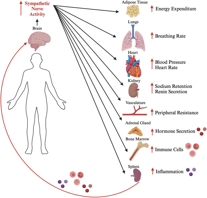
FIGURE 1. The neuroendocrine, autonomic and neuroinflammatory interplay. A schematic diagram depicting the role sympathetic nerves play in regulating different physiological outputs, which in turn can act on the brain in a feedback loop to modulate sympathetic nerve activity. This coordination of the different physiological functions through neuroendocrine, autonomic, and inflammatory mechanisms can become dysregulated resulting in a vicious positive feedback loop that leads to cardiometabolic disease. Figure generated by the authors using Biorender.com.
The emergence of new technologies, such as CRISPR/Cas9 and Cre-LoxP recombination have enabled physiologists to selectively target neuronal populations of interest. A recent study investigated the neuroendocrine and autonomic populations of the PVN using such technologies (Elsaafien et al., 2021). It was demonstrated that the different neuronal populations of the PVN coordinate their activity to regulate cardiovascular function. CRH-synthesizing neurons signal inter-neuronally within the PVN to activate pre-autonomic neurons. This couples the increase in sympathetic nerve activity and systemic corticosterone secretion to BP elevations. The study demonstrated the importance of the interplay between the different neuronal populations to induce coordinated physiological responses.
This Research Topic provides an updated understanding of the interplay between neuroendocrine, autonomic and neuroinflammatory outputs in cardiometabolic disease. We have five excellent papers that include a brief research report, a mini review, a review, and two original research papers. These contributions span studies utilizing rodent models (in vitro and in vivo) and human studies. Of these articles, four were published in Frontiers in Physiology, while one was published in Frontiers in Neuroscience and have been viewed over 9,000 times so far. Physiologists are increasingly working towards providing a better understanding of neural circuits that regulate cardiometabolic function. This understanding is crucial for unraveling dysregulations that occur in cardiometabolic disease, allowing for the development of novel therapeutic targets.
2 The autonomic nervous system in cardiometabolic disease
In 1989, Arthur Loewy and others asked the question of what influences the releases of epinephrine and norepinephrine from the adrenal glands into systemic circulation (Strack et al., 1989). By applying pseudorabies virus into the left adrenal gland, they unraveled the sympathetic innervation of adrenal glands, which arises from five brain regions that include the PVN. Elia and Fossati provide an extensive review describing the autonomic innervation of the cardiovascular organs and their implication in cardiovascular disease. This understanding has led the authors to draw a link between autonomic dysregulation in cardiovascular disease and Alzheimer’s disease. It is well established that hypoperfusion of the brain leads to neuronal deterioration and accumulation of β-amyloid plaques that cause cognitive decline (Cermakova et al., 2015). The sustained adrenergic hyperactivity and robust sympathetic responses that lead to heart failure, result in reducing BP and causing hypoperfusion of the brain. This sustained adrenergic hyperactivity is accompanied by strong neuroendocrine stimulation that includes norepinephrine, renin, angiotensin, and aldosterone release (Leenen, 2007). Thus, the authors propose several interventions to the autonomic and neuroendocrine systems as therapeutic targets to mitigate Alzheimer’s risk in heart failure. Another link between cognitive function and autonomic regulation comes from Grosprêtre et al. where human participants performed motor imagery as a form of motor rehabilitation. The study demonstrated that motor imagery and posture can modulate spinal excitability, autonomic and cardiometabolic responses. Overall, these contributions demonstrate the importance of the interplay between cortical, autonomic, and neuroendocrine systems in cardiovascular disease.
3 The neuroendocrine system in cardiometabolic disease
Within the brain reside populations of neurons that influence the systemic release of endocrine signals. These hormones regulate physiological systems that are implicated in cardiometabolic disease. The study by Queathem et al. uses a novel transgenic mouse model to demonstrate the importance of the hormone estrogen in energy expenditure. They demonstrate a novel role of estrogen receptor β in white adipose tissue browning, via activation of protective adipocyte mitochondrial responses involving UCP1. This discovery highlights a novel pharmacological target for the treatment of metabolic disease. Similarly, the review by Pereira et al. proposes an important role that estrogen plays in sodium appetite. Excessive salt intake is well documented to contribute to hypertension. This review proposes that estrogen can act through the brain to alter salt palatability and inhibit sodium appetite, and proposes that neuroendocrine mechanisms that can be targeted to alleviate excessive sodium intake in hypertension.
4 Neuroinflammation in cardiometabolic disease
Several emerging studies have demonstrated an important role inflammatory cytokines and immune cells play in influencing the activity of neurons that regulate cardiometabolic function (Korim et al., 2018; Elsaafien et al., 2019; Elsaafien et al., 2020). These insights are implicated in cardiometabolic disease, where high grade inflammation is a hall-marker of the disease. The study by Oliveira et al. utilizes a transgenic mouse model to demonstrate that knock-in of angiotensin converting enzyme two in PVN CRH-synthesizing neurons rescues chronic hypoxia-induced pulmonary hypertension. This protection against pulmonary hypertension was mediated through autonomic modulation, counteracting vascular and lung inflammation, and reducing microglia activation in the PVN. The authors propose a vicious positive feedback cycle involving the neuroendocrine, autonomic and neuroinflammatory systems in pulmonary hypertension. This involves microglia activation that contributes to neuronal plasticity in CRH neurons of the PVN, leading to chronic sympathetic activation and immune cell recruitment which exacerbates this chronic pro-inflammatory and pro-sympathetic state in pulmonary hypertension.
5 Conclusion
This Research Topic of papers and reviews provides the reader with an overview of the interplay between neuroendocrine, autonomic and neuroinflammatory systems in cardiometabolic disease. We have highlighted some of the findings that help to unravel the complexity of this coordinated interplay. Investigating disease states requires an integrative approach, whereby the different systems at interplay are examined collectively. Such investigations are crucial for the development of novel effective therapeutics.
Author contributions
DC: Conceptualization, Writing–original draft, Writing–review and editing. CS: Writing–original draft, Writing–review and editing, Conceptualization. KE: Conceptualization, Writing–original draft, Writing–review and editing.
Funding
This work was supported by American Heart Association Postdoctoral Fellowship (23POST1020034) to KE.
Acknowledgments
We would like to acknowledge the excellent submissions that have made this Research Topic successful.
Conflict of interest
The authors declare that the research was conducted in the absence of any commercial or financial relationships that could be construed as a potential conflict of interest.
Publisher’s note
All claims expressed in this article are solely those of the authors and do not necessarily represent those of their affiliated organizations, or those of the publisher, the editors and the reviewers. Any product that may be evaluated in this article, or claim that may be made by its manufacturer, is not guaranteed or endorsed by the publisher.
References
Cermakova P., Eriksdotter M., Lund L., Winblad B., Religa P., Religa D. (2015). Heart failure and Alzheimer′ s disease. J. Intern. Med. 277 (4), 406–425. doi:10.1111/joim.12287
Elsaafien K., de Kloet A. D., Krause E. G., Sumners C. (2020). Brain angiotensin type-1 and type-2 receptors in physiological and hypertensive conditions: focus on neuroinflammation. Curr. Hypertens. Rep. 22, 48–13. doi:10.1007/s11906-020-01062-0
Elsaafien K., Kirchner M. K., Mohammed M., Eikenberry S. A., West C., Scott K. A., et al. (2021). Identification of novel cross-talk between the neuroendocrine and autonomic stress axes controlling blood pressure. J. Neurosci. 41 (21), 4641–4657. doi:10.1523/JNEUROSCI.0251-21.2021
Elsaafien K., Korim W. S., Setiadi A., May C. N., Yao S. T. (2019). Chemoattraction and recruitment of activated immune cells, central autonomic control and blood pressure regulation. Front. Physiology 10, 984. doi:10.3389/fphys.2019.00984
Katayama P. L., Leirão I. P., Kanashiro A., Luiz J. P., Cunha F. Q., Navegantes L. C., et al. (2022). The carotid body detects circulating tumor necrosis factor-alpha to activate a sympathetic anti-inflammatory reflex. Brain, Behav. Immun. 102, 370–386. doi:10.1016/j.bbi.2022.03.014
Korim W. S., Elsaafien K., Basser J. R., Setiadi A., May C. N., Yao S. T. (2018). In renovascular hypertension, TNF-α type-1 receptors in the area postrema mediate increases in cardiac and renal sympathetic nerve activity and blood pressure. Cardiovasc. Res. 115 (6), 1092–1101. doi:10.1093/cvr/cvy268
Leenen F. H. (2007). Brain mechanisms contributing to sympathetic hyperactivity and heart failure. Am. Heart Assoc. 101, 221–223. doi:10.1161/CIRCRESAHA.107.158261
Song X.-A., Jia L.-L., Cui W., Zhang M., Chen W., Yuan Z.-Y., et al. (2014). Inhibition of TNF-α in hypothalamic paraventricular nucleus attenuates hypertension and cardiac hypertrophy by inhibiting neurohormonal excitation in spontaneously hypertensive rats. Toxicol. Appl. Pharmacol. 281 (1), 101–108. doi:10.1016/j.taap.2014.09.004
Strack A., Sawyer W., Hughes J., Platt K., Loewy A. (1989). A general pattern of CNS innervation of the sympathetic outflow demonstrated by transneuronal pseudorabies viral infections. Brain Res. 491 (1), 156–162. doi:10.1016/0006-8993(89)90098-x
Szafarczyk A., Balmefre M., Assenmacher I. (1986). Immunocytochemical evidence for stimulatory control by the ventral noradrenergic bundle of parvocellular neurons of the paraventricular nucleus secreting corticotropin releasing hormone and vasopressin in rats. Brain Res. 397 (2), 297–307. doi:10.1016/0006-8993(86)90631-1
Keywords: neuroendocrine signaling, autonomic nervous system, neuroinflammation, cardiometabolic disease, stress
Citation: Colombari DSA, Sumners C and Elsaafien K (2023) Editorial: The neuroendocrine, autonomic and neuroinflammatory stress axes in cardiometabolic disease. Front. Physiol. 14:1293219. doi: 10.3389/fphys.2023.1293219
Received: 12 September 2023; Accepted: 18 September 2023;
Published: 25 September 2023.
Edited and reviewed by:
Geoffrey A. Head, Baker Heart and Diabetes Institute, AustraliaCopyright © 2023 Colombari, Sumners and Elsaafien. This is an open-access article distributed under the terms of the Creative Commons Attribution License (CC BY). The use, distribution or reproduction in other forums is permitted, provided the original author(s) and the copyright owner(s) are credited and that the original publication in this journal is cited, in accordance with accepted academic practice. No use, distribution or reproduction is permitted which does not comply with these terms.
*Correspondence: Khalid Elsaafien, a2Vsc2FhZmllbkBnc3UuZWR1