94% of researchers rate our articles as excellent or good
Learn more about the work of our research integrity team to safeguard the quality of each article we publish.
Find out more
CORRECTION article
Front. Physiol. , 30 October 2017
Sec. Striated Muscle Physiology
Volume 8 - 2017 | https://doi.org/10.3389/fphys.2017.00886
This article is a correction to:
G-CSF does not influence C2C12 myogenesis despite receptor expression in healthy and dystrophic skeletal muscle
 Craig R. Wright1
 Erin L. Brown1
 Paul A. Della-Gatta1
 Alister C. Ward2
 Gordon S. Lynch3
 Aaron P. Russell1*
A corrigendum on
G-CSF does not influence C2C12 myogenesis despite receptor expression in healthy and dystrophic skeletal muscle
by Wright, C. R., Brown, E. L., Della-Gatta, P. A., Ward, A. C., Lynch, G. S., and Russell, A. P. (2014). Front. Physiol. 5:170. doi: 10.3389/fphys.2014.00170
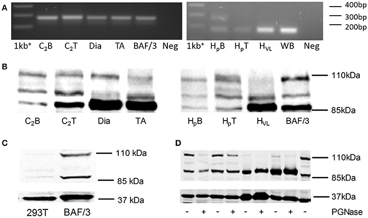
In the original article, there was a mistake in Figure 1. Identification of G-CSFR in human and rodent skeletal muscle, as published. The mistake is found in part 1A of the figure which should show that PCR product from mouse tissues (left hand image) and human tissues (right hand image). Unfortunately, when preparing the image the right hand agarose gel image that was embedded is the same as the left hand image. The corrected Figure 1 and figure legend appears below. The authors apologize for this error and state that this does not change the scientific conclusions of the article in any way.

Figure 1. Identification of G-CSFR in rodent and human skeletal muscle. (A) cDNA fragment amplified during Real Time-PCR using the primers described in Table 1, separated on a 1.8% Sybr safe (Invitrogen) agarose gel and exposed to UV light. (B) Western blot image identifying G-CSFR in rodent and human skeletal muscle in vitro and ex vivo. (C) Western blot for G-CSFR in positive (BAF/3[G]) and negative (293T) control cells. (D) G-CSFR after deglycosylation with PGNase. C2B (C2C12 myoblasts), C2T (C2C12 myotubes), Dia (diaphragm muscle from C57BLK mice), TA (tibialis anterior muscle from C57BLK mice), HpB (human primary myoblasts), HpT (human primary myotubes) HVL (human vastus lateralis muscle), BAF/3[G] (murine pro B cell line overexpressing G-CSFR), 293T (human embryonic kidney 293T cell line), 1kb+ [1 kb plus ladder (Invitrogen) and WB (Whole blood (human)].
The authors declare that the research was conducted in the absence of any commercial or financial relationships that could be construed as a potential conflict of interest.
The reviewer BGH and handling Editor declared their shared affiliation.
Keywords: G-CSF, cytokine receptor, skeletal muscle, duchenne muscular dystrophy, mdx, C2C12, proliferation, differentiation
Citation: Wright CR, Brown EL, Della-Gatta PA, Ward AC, Lynch GS and Russell AP (2017) Corrigendum: G-CSF does not influence C2C12 myogenesis despite receptor expression in healthy and dystrophic skeletal muscle. Front. Physiol. 8:886. doi: 10.3389/fphys.2017.00886
Received: 24 September 2017; Accepted: 19 October 2017;
Published: 30 October 2017.
Edited by:
Steven P. Jones, University of Louisville, United StatesReviewed by:
Frontiers Physiology Editorial Office, Frontiers Media SA, SwitzerlandCopyright © 2017 Wright, Brown, Della-Gatta, Ward, Lynch and Russell. This is an open-access article distributed under the terms of the Creative Commons Attribution License (CC BY). The use, distribution or reproduction in other forums is permitted, provided the original author(s) or licensor are credited and that the original publication in this journal is cited, in accordance with accepted academic practice. No use, distribution or reproduction is permitted which does not comply with these terms.
*Correspondence: Aaron P. Russell, YWFyb24ucnVzc2VsbEBkZWFraW4uZWR1LmF1
Disclaimer: All claims expressed in this article are solely those of the authors and do not necessarily represent those of their affiliated organizations, or those of the publisher, the editors and the reviewers. Any product that may be evaluated in this article or claim that may be made by its manufacturer is not guaranteed or endorsed by the publisher.
Research integrity at Frontiers

Learn more about the work of our research integrity team to safeguard the quality of each article we publish.