- 1State Key Laboratory of Agricultural Microbiology, Huazhong Agricultural University, Wuhan, China
- 2The Provincial Key Lab of Plant Pathology of Hubei Province, College of Plant Science and Technology, Huazhong Agricultural University, Wuhan, China
Botrytis-induced kinase1 (BIK1), a receptor-like cytoplasmic kinase, plays an important role in resistance against pathogens and insects in Arabidopsis thaliana. However, it remains unknown whether BIK1 functions against Plasmodiophora brassicae, an obligate biotrophic protist that attacks cruciferous plants and induces gall formation on roots. Here, we investigated the potential roles of receptors FLS2, BAK1, and BIK1 in the infection of P. brassicae cruciferous plants. Wild-type plants, fls2, and bak1 mutants showed typical symptom on roots, and the galls were filled with large quantities of resting spores, while bik1 mutant plants exhibited strong resistance to P. brassicae. Compared with that of the wild-type plants, the root hair and cortical infection rate of bik1 mutant were significantly reduced by about 40–50%. A considerable portion of bik1 roots failed to form typical galls. Even if some small galls were formed, they were filled with multinucleate secondary plasmodia. The bik1 plants accumulated less reactive oxygen species (ROS) at infected roots than other mutants and wild-type plants. Exogenous salicylic acid (SA) treatment alleviated the clubroot symptoms in wild-type plants, and the expression of the SA signaling marker gene PR1 was significantly increased in bik1. Both sid2 (salicylic acid induction-deficient 2) and npr1-1 [non-expresser of PR genes that regulate systemic acquired resistance (SAR)] mutants showed increased susceptibility to P. brassicae compared with wild-type plants. These results suggest that the resistance of bik1 to P. brassicae is possibly mediated by SA inducible mechanisms.
Introduction
The soil-borne obligate pathogen Plasmodiophora brassicae causes clubroot disease in species of Brassicaceae, including Arabidopsis. P. brassicae plunders nutrients from host roots to complete their life cycle, and causes the formation of root galls. Clubroot occurs in more than 60 countries and results in a 10–15% reduction in the yields of Cruciferae crops on a global scale (Dixon, 2009). P. brassicae has a complex but not completely understood infection process. A primary zoospore is released from each resting spore, which reaches the surface of a root hair and penetrates the cell wall, forming primary plasmodia in the root hair. After a number of nuclear divisions, the plasmodia cleave into zoosporangia, and the zoosporangia form clusters in the root hair or penetrate root cortex cells, in which the pathogen develops into secondary plasmodia (Kageyama and Asano, 2009). Plasmodia provoke abnormal cell enlargement and uncontrolled cell division, leading to the development of club-shaped galls on the roots and the above-ground symptoms, such as wilting, stunting, yellowing, and premature senescence compared with healthy plants (Hwang et al., 2012). Each gall contains millions of resting spores that persist in the soil for up to 15 years even in the absence of a suitable host, making it difficult to manage clubroot diseases (Donald and Porter, 2009). The resting spores of P. brassicae are easily transmitted to elsewhere with contaminated soil, including farm machinery, boots, grazing animal hooves, infected transplants, and surface floodwater (Donald and Porter, 2009). Crop rotation, increased soil pH, improved drainage conditions, and fungicide application provide certain protection against the disease; however, under high clubroot pressure, these measures are generally not effective (Abbasi and Lazarovits, 2006). Besides, government policies concerning human health and environmental safety have led to the restriction or deregistration of a large number of previously useful active ingredients. Genetic resistance is the most effective and economical approach to clubroot management, and several resistant cultivars against clubroot have been previously reported (Hirai et al., 2004; Rocherieux et al., 2004; Chu et al., 2014). However, new races of the pathogen can rapidly appear (Fähling et al., 2003). Using Arabidopsis thaliana as a model system has facilitated the application of available genetic and molecular tools and has thus advanced our understanding of this economically important plant disease (Siemens et al., 2002). In clubroot-infected Arabidopsis, it is thought that perturbation in phytohormone content plays important roles in disease development (Malinowski et al., 2012), but little has been known about which genes are involved in the plant defense response.
Salicylic acid (SA) is an important secondary phenolic metabolite in a wide range of prokaryotic and eukaryotic organisms, including plants. SA regulates a multitude of developmental processes, such as plant cell growth, seed germination and development, thermo-tolerance, respiration, stomatal aperture, fruit yield, nodulation in legumes, and leaf senescence. More importantly, SA serves as a key signaling and regulatory molecule in plant defense responses, and it is regarded as the key plant immune hormone (Spoel and Dong, 2012; Liu et al., 2015). The biosynthesis of SA on pathogen detection is essential for local and systemic acquired resistance (SAR) and the accumulation of pathogenesis-related (PR) proteins (Boatwright and Pajerowska-Mukhtar, 2013). SA has long been recognized as a central component of defense in plants against a number of biotrophic pathogens and viruses (Vlot et al., 2009). The development of disease in a susceptible host and its molecular basis has been recently examined using an Arabidopsis model (Agarwal et al., 2011). In two Arabidopsis genotypes Col-0 (susceptible) and Bur-0 (partially resistant) which were infected with the virulent P. brassicae, clubroot development was partially inhibited by camalexin, SA and jasmonic acid (JA) signaling pathway (Lemarié et al., 2015a,b). Exogenous SA in B. oleracea enhances resistance to clubroot (Lovelock et al., 2013, 2016). SA methyltransferase gene PbBSMT was identified from P. brassicae, which can methylate SA in host cells (Ludwig-Muller et al., 2015).
Basal resistance, the first step in plant defense response, involves perception through surface-localized pattern recognition receptors (PRRs) of conserved molecules characterized by pathogen-associated molecular patterns (PAMPs) or microbe-associated molecular patterns (MAMPs) (Monaghan and Zipfel, 2012). A well-known PRR is Arabidopsis receptor kinase FLS2, which recognizes a conserved 22 amino acid N-terminal sequence of the bacterial flagellin protein (flg22) (Gomez-Gomez and Boller, 2000). FLS2 serves as an excellent model to understand plant innate immune signaling, and heterotrimeric G proteins are directly coupled to the FLS2 receptor complex to regulate immunity (Macho and Zipfel, 2014; Liang et al., 2016). The extracellular leucine-rich repeat domain of FLS2 perceives flg22 and rapidly recruits another LRR receptor-like kinase called BAK1, which plays a role in brassinolide signaling (Chinchilla et al., 2007; Schulze et al., 2010). BIK1, a receptor-like cytoplasmic kinase, is directly phosphorylated through BAK1 and is associated with the FLS2/BAK1 complex in modulating PAMP-mediated signaling (Lu et al., 2010; Zhang et al., 2010). The inactivation of BIK1 causes severe susceptibility to necrotrophic fungal pathogens but enhances the resistance against a virulent strain of the bacterial pathogen Pseudomonas syringae pv tomato (Veronese et al., 2006), and bik1 plants displayed enhanced antibiosis and antixenosis toward aphids through inducing the up-regulation of PAD4 expression (Lei et al., 2014).
In the present study, we investigated the potential roles of the receptors FLS2, BAK1 and BIK1, which have different levels of basal resistance to the compatible strain ZJ-1 of the clubroot pathogen P. brassicae. We challenged these loss-of-function mutants with P. brassicae. bik1 exhibited strong resistance to P. brassicae, and the root hair and cortical infections were significantly decreased. Besides, the development of P. brassicae was inhibited in the infected bik1 plants. We attempted to explain the mechanism underlying the resistance of bik1 mutants against P. brassicae. bik1 plants were reported to have a higher basal SA level than wild type plants (Veronese et al., 2006; Lei et al., 2014), and SA suppresses the formation of clubroots in broccoli (Lovelock et al., 2013, 2016). The obtained results showed that exogenous SA treatment could alleviate the symptoms of clubroot. In the mutant line sid2 and npr1-1 mutants, which had blocked SA biosynthesis and were SAR-deficient respectively, clubroot symptoms were found to be clearly more severe compared with in Col-0. These findings indicate that the Arabidopsis mutant bik1 exhibits strong resistance to P. brassicae possibly because SA inducible mechanisms enhance the resistance to clubroot disease. However, the bik1 sid2 double mutant showed strong resistance to P. brassicae, suggesting that this resistance is possibly attributable to SA pathway and other unknown pathways.
Materials and Methods
Plant Materials, P. brassicae Inoculation and Growth Conditions
Arabidopsis thaliana ecotype Columbia (Col-0), which was used as the wild-type control in the present study, was kindly provided by Dr. Yangdou Wei at the University of Saskatchewan, and the mutants fls2, bik1, bak1-4, bik1 sid2 were kindly donated by Dr. Libo Shan (Texas A & M university). sid2 was bought from the Arabidopsis Biological Resource Center. The bik1 mutant line was further identified as a homozygous mutant. The RT-PCR results showed that the bik1 mRNA could not be detected in homozygous mutant plants (Figure S1), suggesting that these plants represent knockout mutants at the BIK1 locus. The bik1 sid2 double mutant line was identified as a homozygous mutant. The RT-PCR results showed that the bik1 and sid2 mRNA could not be detected in homozygous mutant plants (Figure S2).
P. brassicae strain ZJ-1 was originally isolated from a diseased plant in a rapeseed field in Zhijiang County, Hubei Province, P R China. The virulence of single-spore isolated from P. brassicae strain ZJ-1 was tested on the differential hosts of Williams, showing the single-spore isolate derived from race 1 (Williams, 1966; Fähling et al., 2004). Resting spores of P. brassicae were extracted from clubroot galls (Asano et al., 1999), surface disinfested by freshly prepared 2% chloramine-T solution at room temperature for 20 min, washed twice with sterile water, adjusted to a concentration of 1.0 × 10⁷ spores per mL, and were then stored at 4°C. P. brassicae was proliferated using the stored resting spores in greenhouse with rapeseed. Arabidopsis Col-0 and mutant seeds were germinated on the surface of vermiculite in small pots. The seedlings were transplanted to the soil at 14 days post-germination. After growing of 7 days in a growth chamber, plants were inoculated with 1 mL of the resting spore suspension (1 × 10⁷ spores per mL) by injecting the soil around each plant. Arabidopsis and mutant plants grown in the 50 holes plate (54 cm × 28 cm × 5 cm), each seeding planted in one hole, the wild type and mutants Arabidopsis were planted in the same holes plate to reduce errors. The plants were grown in a plant growth chamber maintained at 70% humidity and 23°C with a 16/8-h day/night cycle. For all the mutants analyzed, gene expression was verified at 21 or 28 days post inoculation. Disease severity was assessed using a scoring system of 0–4 modified from Siemens reported (Siemens et al., 2002). A score of 0 indicated no disease; 1, very small galls mainly on lateral roots that did not impair the main root; 2, small galls covering the main root and few lateral roots; 3, medium to large galls, also on the main root; and 4, severe galls on lateral root, main root or rosette, with fine roots completely destroyed. Disease index (DI) was calculated using the five-grade scale according to the formula: DI = (1n1 + 2n2 + 3n3 + 4n4) × 100/4Nt, where n1–n4 is the number of plants in the indicated class and Nt is the total number of plants tested.
ROS Determination
Reactive oxygen species (ROS) production was detected using the nitroblue tetrazolium (NBT) staining method (Montiel et al., 2012; Arthikala et al., 2014). The plants grown in the soil pot for 21 days after infection were used to determine concentrations. The healthy roots and the galled roots were incubated for 5 h in dark at room temperature, and the roots were cleared in 90% ethanol.
DAB Staining
To estimate the produced H2O2 in situ, the roots were hand-sectioned using a double-edged razorblade. The sections were subsequently rapidly immersed in 1 mg/ml of 3′, 3′-diaminobenzidine (DAB) solution, vacuum infiltrated for 2 min, and incubated for 2–3 h at 25°C in darkness (Gorska-Czekaj and Borucki, 2013).
SA and MeSA Exogenous Treatment
A stock solution of SA (99.5%, Sigma Aldrich, dried substance) was prepared in ethanol/ddH2O (v/v1:1), and diluted with sterile water at 2.5 × 10⁻⁵mol/L concentration for spraying (Lovelock et al., 2013). MeSA (99%, VETEC) was diluted in 10% ethanol and was sprayed at the 2.5 × 10⁻⁵mol/L concentration. SA and MeSA were used for three times of exogenous treatments. The first treatment was conducted 2 days prior to the inoculation, while the second treatment was carried out 2 days after inoculation, and the last was performed 10 days after inoculation. The total volume of each spraying was 2 L, and the remaining water was gently poured into the holding tray for slow absorption through the roots. Approximately 30 plants were treated for each sample (Lovelock et al., 2013).
Quantification of P. brassicae DNA Content in Infected Roots
DNA was extracted from root samples using the cetyl trimethyl ammonium bromide (CTAB) method (Allen et al., 2006). Quantitative PCR was performed on a CFX96 real-time PCR system (BioRad) using iTaq Universal SYBR Green supermix (BioRad) to quantify the P. brassicae target actin gene AY452179.1. Each reaction was performed with 2.5 ng of total DNA as template, and Arabidopsis actin gene AT3G18780 was used as an internal control for data normalization. Standard curves were constructed using serial dilutions of DNA extracted from the roots of Col-0 at 21 days after inoculation with P. brassicae, which was defined as a reference condition. Quantitative results were then expressed as the % of the P. brassicae mean DNA content in this reference condition (Lemarié et al., 2015a,b).
RNA Isolation and Quantitative Real-Time PCR
Total RNA was isolated from control roots, and the roots were inoculated with P. brassicae using TRIZOL reagent (Invitrogen). RNA samples were treated with DNase I to remove potential contaminating genomic DNA, followed by extraction with phenol:chloroform. First-strand cDNA was prepared using oligo (dT) primer. Quantitative PCR was performed on a CFX96 real-time PCR system (BioRad), using iTaq Universal SYBR Green supermix (BioRad). The following cycling conditions were used: 95°C for 30 s, 95°C for 5 s, 60°C for 15 s, and 72°C for 12 s. The reaction was performed for 40 cycles, followed by a step at 72°C for 5 s. Each amplification used 3 technical replicates the results of which were averaged to give the value for a single biological replicate. The primer sequences are provided in Table S1. We tried using the two Arabiodopsis genes (actin and ubiquitin) for qRTPCR, and the results were similar. The data shown in this paper selected Arabidopsis ubiquitin10 (AT4G05320) served as an internal control for normalization.
Microscopic Analysis
Fluorescent and transmission electron microscopy (TEM) were performed using the following protocol. For fluorescent microscopy, the healthy roots or galled roots were treated with Nile red solution (10 μg/ml Nile red in acetone) for 3–5 s, and excess dye was removed after brief rinsing in H2O. The root samples were subsequently observed using Nikon fluorescence microscopy. Nile red emits fluorescence over a broad range of wavelengths, but observation using a filter set for B excitation (used for FITC, Cy2, Alexa488, or GFP) generates the best images (Suzuki et al., 2013). For electron microscopy analysis, the roots were fixed in 2.5% glutaraldehyde for 4 h, and were subsequently postfixed in 1% osmium tetroxide for 3 h, washed, dehydrated through an ethanol series, and embedded in London resin white. Ultrathin sections were examined through TEM (HITACHI, H-7000).
Transverse Root Sectioning
Wild-type and mutant Arabidopsis were infected with P. brassicae for 21 days, and uninfected roots were used as the control. The healthy root tissues and galls were carefully washed with tap water and embedded in a frozen embedding medium (Sakura Finetek USA, Inc., Torrance, CA) at −23°C overnight. Transverse root sections (50 μm thick) were sliced using a freezing microtome (Leica CM1950, Germany) and attached to the slides. A drop of water was added, and the specimens were covered with cover glass. The plant cells and spores were observed using a Nikon light microscope.
Statistical Analysis
Statistical analysis was performed using SPSS (13.0) and a statistics package (Microsoft Excel 2010). In all experiments, One-way ANOVA, specifically Tukey's test with a P = 0.05, was used to analyze significant differences between treatment groups.
Results
bik1 Exhibited Strong Resistance to P. brassicae
The symptoms of P. brassicae-infected plants included the formation of club-shaped galls on the roots, and the wilting, yellowing and premature senescence of the shoots. The plant defense response upon P. brassicae infection is often reflected by reduced gall size, root condition, and reduced resting spore production. To determine whether several known receptor-like kinases have function in clubroot-associated defense responses, we observed the infection and colonization of P. brassicae on the roots of the loss-of-function mutants (Figure 1). The roots of wild-type Arabidopsis infected by P. brassicae were formed of a typical galled, resulting in few rootlets; besides, the infected plants were yellowing (Figure 1A and Figure S3), and the ratio of gall formation was 100% (Figure 1B). The symptoms of fls2 and bak1 infected by clubroot-pathogen were similar to those of the wild type (Figure 1A and Figure S3), with a gall formation ratio of nearly 100% (Figure 1B). Interestingly, in bik1, the gall formation was inhibited as 79% of the plants that did not form a gall, and the root systems of P. brassicae-inoculated bik1 plants still developed with plentiful lateral roots (Figures 1A,B). The bik1 mutant line was identified as a homozygous mutant, and the RT-PCR results showed that the bik1 mRNA could not be detected in homozygous mutant plants (Figure S1), suggesting that these plants were the knockout mutants at the BIK1 locus. To evaluate P. brassicae production in the galls, the actin gene expression levels of P. brassicae were measured using quantitative real-time PCR. For the phenotype of no gall formation on bik1-infected roots due to the largely reduced P. brassicae production (by 99.78%), and for the phenotype of mid gall formation, root spore production was reduced by approximately 92% (Figure 1C). The relative amount of P. brassicae DNA in total root-extracted DNA was evaluated through quantitative PCR (Figure 1D). The results indicated that root pathogen density was not reduced within the infected roots of fls2 and bak1 mutants compared with Col-0, suggesting that fls2 and bak1 mutants had no significant change of pathogen density within the root samples. However, the density of P. brassicae in the infected roots with and without the formation of small galls in bik1 mutant was approximately 40 and 90% less than that in Col-0, respectively (Figure 1D). Taken together, the results showed that bik1 plants exhibited strong resistance to P. brassicae.
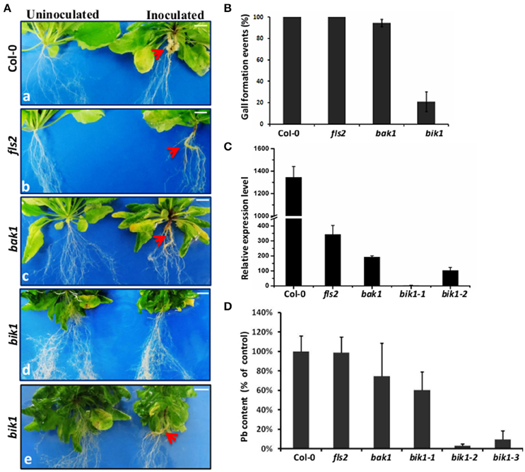
Figure 1. Phenotypes and P. brassicae DNA contents in Arabidopsis control and several loss-of-function mutant roots. (A) Arabidopsis Col-0 roots (a) and several loss-of-function mutant RLKs, including FLS2 (b), BAK1 (c), and BIK1 (d,e) infected with P. brassicae; the root images were captured at 21 days after P. brassicae infection. The red arrows show the galls, and bik1 showed two phenotypes: formation of small galls and no gall formation on the roots. Bar = 0.5 cm. (B) Disease symptoms of Arabidopsis control roots and several loss-of-function mutants for gall comparison. Clubroot symptoms were quantified for three biological replicates, each containing 30 plants per genotype. However, for some mutants especially the bik1 mutants several plants were excluded before statistics due to a poor growth (with very small shoots and roots). The error bars represent the SD of the experimental values obtained from three biological replicates. (C) Real-time PCR analysis was performed to assess the expression levels of P. brassicae at 21 days after inoculation. bik1-1 showed no root gall formation, and bik1-2 showed root gall formation. Root sample is a mixture of roots from different plants of the same mutant (over 3 plants). The error bars represent the SD of the experimental values obtained from three technical replicates. (D) Pathogen DNA quantification (Pb) by quantitative PCR, expressed the percentage of the mean Pb content in inoculated roots of Col-0, fls2, bak1, and bik1 at 21 days after inoculation with P. brassicae. At least 3 plant roots were taken as mixed sample. bik1-1 showed the formation of small galls on the roots, and bik1-2 and bik1-3 showed no formation of galls on the roots. Root sample is a mixture of roots from different plants of the same mutant (over 3 plants). The error bars represent the SD of the experimental values obtained from three technical replicates.
To observe the P. brassicae development state in the galls of the infected roots, longitudinal sections of Arabidopsis control and loss-of-function mutant roots were observed. There was no zoospores in the negative control (uninfected wild type Arabidopsis root cells), and many resting spores were found in infected wild type, fls2 and bak1 root cells. No obvious zoospores were observed on the infected roots of bik1, which showed no gall formation phenotype (Figure 2A). TEM was used to clearly demonstrate the zoospore developmental stages in the very small galls on bik1 root. The development of P. brassicae was in the state of multinucleate secondary plasmodium during cell division on bik1 root (Figure 2Bb), while the wild-type root cells were filled with resting spores and the cell division was completed (Figure 2Ba). These observations suggested that bik1 had inhibited development of P. brassicae, which explains the results that the wild-type roots showed severe galls with a high production of resting spores, while the bik1 roots had fewer and smaller galls with less P. brassicae.
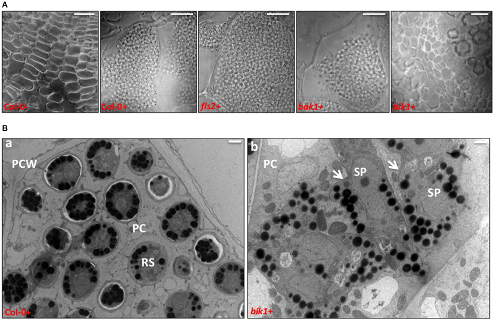
Figure 2. Longitudinal sections and TEM of Arabidopsis control and loss-of-function mutant roots infected with P. brassicae. (A) Longitudinal sections of Arabidopsis control and loss-of-function mutant roots infected with P. brassicae after 21 days. Bar = 20 μm. (B) Transmission electron microscopy of Col-0 (a) and bik1 (b) infected with P. brassicae for 21 days. Selected clearly visible clubs to section (− = uninoculated roots, + = inoculated roots). RS = resting spores; PC = plant cell; PCW = plant cell wall; SP = secondary plasmodium, white arrow. Bar = 1 μm.
Suppression of Root Hair and Cortical Infections by the Loss of BIK1 Function
The life cycle of P. brassicae consists of three stages: survival in soil, root hair infection, and cortical infection (Schwelm et al., 2015). To investigate the potential involvement of BIK1 in the infection process, we compared the root hair and cortical infections between the wild-type and bik1 mutant plants, including the colonization rate of primary plasmodia, zoosporangia, and secondary plasmodia (Figures 3A,B). Nile red was used to label P. brassicae for convenient and rapid detection of the stages of infection. The roots of more than 15 plants were selected for the control and bik1 mutants and sliced into 1–2 cm segments. A total of approximately 100 root segments per sample were observed and counted to determine the presence of infection. Compared with root hair infection of the control (100% primary plasmodia and 87.17% zoosporangia, respectively), the root hair infection ratio of bik1 mutant roots was reduced, showing 40.40% primary plasmodia and 54.47% zoosporangia, respectively (Figure 3B). Compared with the control roots (85.70% secondary plasmodia), the bik1 mutant roots showed a reduced cortical infections ratio (43.17% secondary plasmodia) (Figure 3B). Thus, root hair and cortical infections appeared to be impaired by the knockout of BIK1 expression in the mutant.
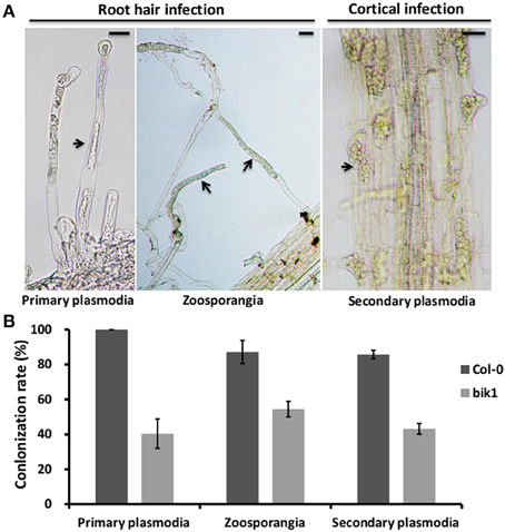
Figure 3. Root hair and cortical infection stages in the roots of Arabidopsis control and bik1 mutant plants. (A) The images show the key steps of P. brassicae infection in Arabidopsis control and bik1 mutant plants, including root hair and cortical infections stages. Primary plasmodia of root hair infection were observed in the roots stained with Nile red for 3 days post inoculation. Zoosporangia of root hair infection and secondary plasmodia of cortical infection were observed in the roots stained with Nile red for 12 days post inoculation. The pictures were under light microscopy. Arrows indicate primary plasmodia, zoosporangia or secondary plasmodia. (B) Mean rate (100%) of primary plasmodia, zoosporangia and secondary plasmodia per root of the control and bik1 mutant. The roots of more than 15 plants were selected for the control and bik1 mutants and sliced into 1–2 cm segments. A total of approximately 100 root segments per sample were observed and counted to determine the presence of infection. The error bars represent the SD of the experimental values obtained from three biological replicates. Bar = 10 μm.
High Level of ROS Accumulated in the Infected Roots of Arabidopsis
ROS plays dual roles in plants, both as toxic compounds and as the key regulators of many biological processes, such as growth, cell cycle, programmed cell death, hormone signaling, development, stress, and defense responses (Mittler et al., 2004). Pathogen infection may elicit ROS production. We used nitroblue tetrazolium (NBT), a reagent that allows the visualization of superoxide accumulation, to evaluate the ROS production in P. brassicae-infected roots of wild type Arabidopsis, fls2, bak1, and bik1 mutants. NBT-formazan precipitates within the roots of all the mutants and control wild type were localized to the places of gall formation (Figure 4), and the NBT-formazan precipitates were much less abundant in the infected roots of bik1 than in the roots of wild type or fls2 and bak1 mutant plants (Figure 4). We further observed the galls of P. brassicae-infected Brassica rapa through 3-3′-diaminobenzidine (DAB) staining. The results showed that the galled roots had much higher H2O2 accumulation than healthy roots in the cortex, which was filled with millions of P. brassicae resting spores (Figure S4). These results indicated that ROS might be an indicator of defense responses.
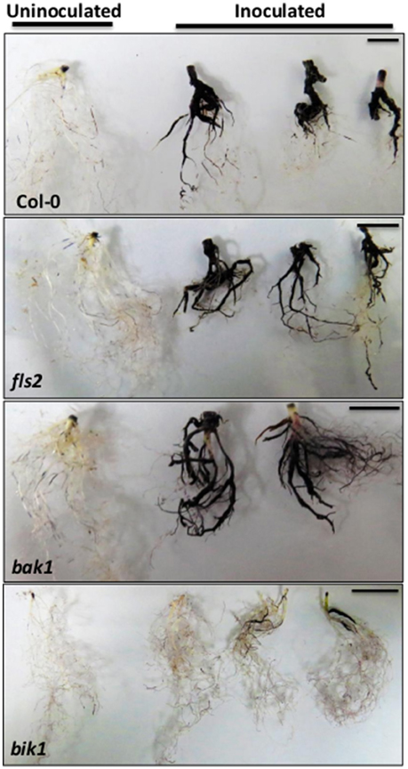
Figure 4. Analysis of ROS production. Visualization of production in representative NBT-stained control, and fls2, bak1 and bik1 mutant roots at 21 days post inoculation with P. brassicae. Dense blue forazan precipitates were observed at the site of H2O2 production. Bar = 1 cm.
Treatment of Roots with SA Alleviated the Symptoms of Clubroot
SA is a central component of defense in plants against a number of biotrophic pathogens (Catinot et al., 2008; Vlot et al., 2009). Thus, we investigated the potential of SA to stimulate defense in Arabidopsis against P. brassicae, and methyl salicylate (MeSA) was used as the control. The formation of root galls was assessed at 3 weeks after three repeated treatments with SA or MeSA. SA treatment significantly reduced gall formation, and the proportion of galling was decreased by 90% (from 100 to 10%). MeSA treatment resulted in no reduction of gall formation (from 100 to 100%) (Figures 5A–C). The disease symptoms associated with P. brassicae were observed to be decreased in the roots treated with SA, but no observable change was found in shoot morphology (Figure 5D). Nile red staining clearly showed that a very small amount of spores were observed in SA-treated roots (Figures 5E–G). These results indicate that SA enhances the resistance against P. brassicae.
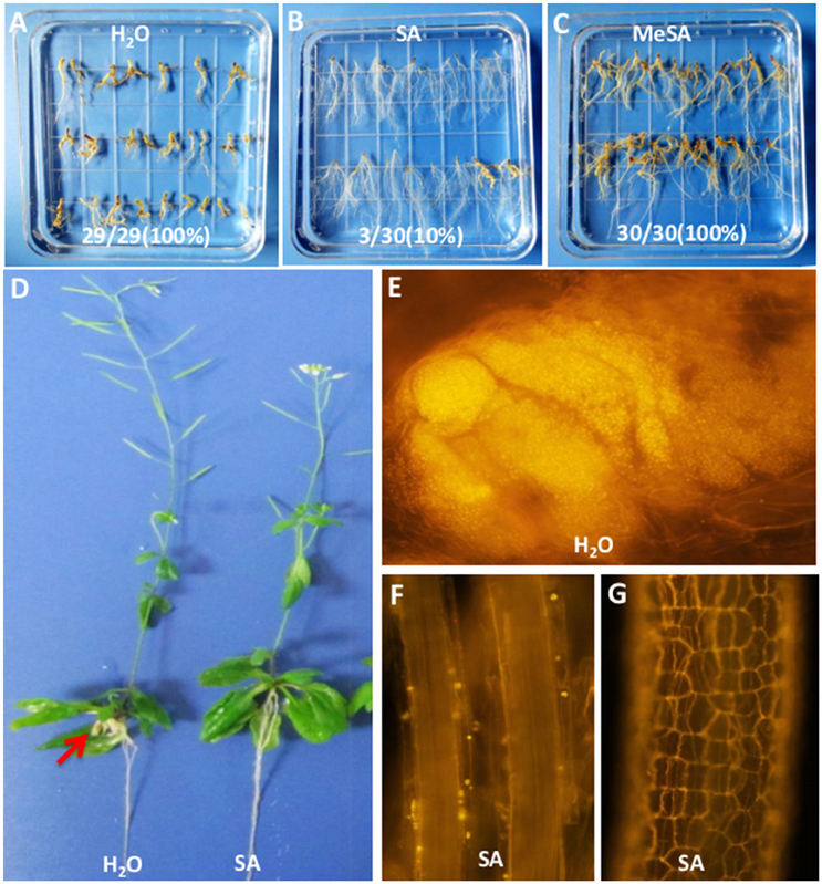
Figure 5. Effect of SA treatment in the pot on gall reduction in 3-week-old infected plants. (A–C) Gall phenotype of Arabidopsis Col-0 infected with P. brassicae for 21 days in 3 different treatments, including H2O (left), SA 2.5 × 10⁻⁵mol/L for 3 times (middle), and 2.5 × 10⁻⁵mol/L MeSA for 3 times (right). The number on the picture showed the gall formation events. (D) Images of whole plants of Arabidopsis Col-0 infected with P. brassicae for 21 days and treated with SA 2.5 × 10⁻⁵mol/L for 3 times (right) or not (left). (E) Nile red staining for infected Col-0 clubroots. (F,G) Nile red staining for infected Col-0 clubroots treated with SA.
P. brassicae Altered the Expression of Defense Genes in bik1, Particularly PR1
P. brassicae-induced plant defense pathways are often regulated through certain plant hormones, JA pathway, and SA pathway (Lemarié et al., 2015b). The loss of BIK1 function resulted in higher basal SA levels and PR1 gene expression than wild-type Col-0 Arabidopsis (Veronese et al., 2006; Lei et al., 2014). To determine whether the resistance to P. brassicae was conferred through the loss of BIK1 function involving defense-related plant hormones, we detected the expression levels of the SA-signaling marker gene PR1 and the ET/JA marker genes ERF1 and PDF1.2 at 21 days after infection of P. brassicae (Figure 6). In wild-type roots, the expression levels of PR1, PDF1.2, and ERF1 were 6.5, 9.4, and 2.2-folds up-regulated after infection, respectively. Similar results were obtained for fls2 mutant plants. However, the transcript level of the SA-responsive gene PR1 in bik1 mutant roots was 48.4-folds higher before P. brassicae infection and 11.5-folds higher after the infection compared with that in the wild type roots. These data implied that BIK1 might function as a negative regulator of SA accumulation both in the presence and absence of P. brassicae infection.
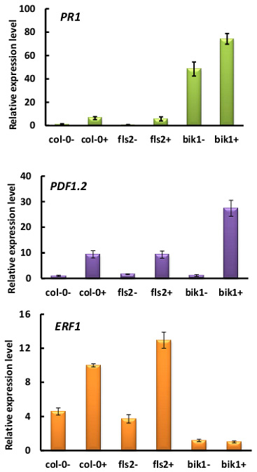
Figure 6. Relative expression of SA, JA, and ET marker genes PR1, ERF1, and PDF1.2 in response to infection with P. brassicae for 21 days (− = uninfection, + = infection). P. brassicae infection up-regulated the expression of these genes in both wild-type and mutant plants.
bik1 sid2 Double Mutant Failed to Restore the Susceptibility to P. brassicae
The loss of SID2 function blocks SA biosynthesis (Wildermuth et al., 2001). To assess the role of SA in bik1 resistance to P. brassicae, bik1, sid2, and bik1 sid2 double mutant plants were used for infection (Figure 7). The non-inoculated sid2 mutant did not show any observable change in morphology compared with the wild type (Figures 7A,D, Figure S5). Besides, the severity of clubroot symptoms induced by P. brassicae was similar to that of wild-type plants: both of them showed yellowing leaves, severely galled and rotten taproots, and similar density of P. brassicae were detected (Figures 7A–C,E). These results are consistent with those reported previously (Lovelock et al., 2016). Apparently, bik1 reduced the clubroot symptom and density of P. brassicae (Figures 7A–E). These results indicated that the elevation of SA accumulation was required for bik1 resistance. Surprisingly, in the bik1 sid2 double mutant with reduced SA accumulation (Laluk et al., 2011; Lei et al., 2014), the phenotypes were similar to those of bik1 plants. These plants showed leaves with serrated margins and wrinkled surfaces, occasional curling, partial recovery to wild type, and strong resistance to P. brassicae similarly to bik1 (Figure 7 and Figure S5). The bik1 sid2 double mutant line was identified as a homozygous mutant (Figure S2), which showed partial resistance to clubroot possibly due to an epistatic effect or some other unknown signal pathways that contribute to the resistance of bik1 against P. brassicae.
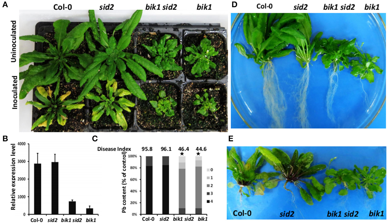
Figure 7. Effect of sid2 mutation on bik1-mediated resistance against P. brassicae. (A) Mature shoot phenotypes of wild type and mutant plants. The top of the picture shows uninoculated plants, while the bottom shows plants inoculated with P. brassicae. (B) Real-time PCR analysis was performed to assess the expression levels of P. brassicae at 28 days after inoculation with P. brassicae. (C) Phytopathological analysis of wild type and mutant plants. The percentages of plants in the individual disease classes are shown. 0, no symptoms; 1, very small galls mainly on lateral roots and that do not impair the main root; 2, small galls covering the main root and few lateral roots; 3, medium to large galls, also including the main root; and 4, severe galls on lateral root, main root or rosette; fine roots completely destroyed. For each treatment, 26–30 Arabidopsis plants were analyzed. The qualitative disease assessment data were initially analyzed using spss and subsequently further analyzed after comparing the mean rank differences. The asterisk indicates a significant difference at P < 0.01. The disease index for each sample is shown as a number above the respective histograms. (D) Images of the shoots without inoculation are shown in (A). (E) Clubroot symptoms in plants. Images of the shoots with inoculation are shown in (A).
SAR-Deficient Mutant npr1-1 Was More Susceptible to P. brassicae than Col-0
SA and the SA-dependent signaling pathways play a major role in modulating SAR. In Arabidopsis plants defective in SA accumulation, SAR is significantly impaired, and in plants where SA is either over-expressed or externally supplied, enhanced SAR is typically observed (Fu and Dong, 2013). The bik1 mutant plants showed higher SA level and up-regulated PR1 gene expression compared with Col-0, indicating that the bik1 mutant plants had enhanced SAR. To examine whether SAR is important for Arabidopsis to resist P. brassicae, the npr1-1 mutant was inoculated with P. brassicae. The results showed that at 21 days post inoculation, the symptoms of npr1-1 were more severe than those of Col-0 (Figure 8). The infected npr1-1 leaves turned yellow and showed root rot, while the control plants showed galls on the roots (Figures 8A–C). We then evaluated the disease index in infected Col-0 and npr1-1 plants (Figure 8D), and the index for Col-0 was 69.4, while that for npr1-1 was 88.1. These results indicated that npr1-1 was more susceptible to P. brassicae than Col-0. Thus, the SAR of Arabidopsis also contributed to the resistance against P. brassicae.
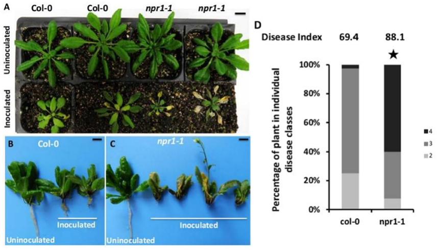
Figure 8. Gall phenotype and phytopathological analysis of the wild type compared with npr1-1 mutant plants at 21 days post inoculation with P. brassicae. (A) Mature shoot phenotypes of wild-type and mutant plants. The top of the picture shows uninoculated plants, and the bottom of the picture shows plants inoculated with P. brassicae. The left two panels show Arabidopsis Col-0 plants, while the right two panels show npr1-1 mutant plants. (B) Gall phenotype of Arabidopsis Col-0. (C) Gall phenotype of npr1-1 mutants. (D) Phytopathological analysis of wild type and mutant plants. The percentages of plants in the individual disease classes are shown. For each treatment, 40 Arabidopsis plants were analyzed. The qualitative disease assessment data were initially analyzed using spss and subsequently further analyzed after comparing the mean rank differences. The asterisk indicates a significant difference at P < 0.01. The disease index for each sample is shown as a number above the respective histogram.
Discussion
In the present study, we investigated the role of PAMP/MAMP signal receptors in the resistance of Arabidopsis against the infection of clubroot pathogen, and we found that Arabidposis mutant fls2 and bak1 were as susceptible to P. brassicae as wild-type plants; however, mutant bik1 displayed strong resistance to P. brassicae, and exhibited suppressed root hair and cortical infections. The bik1 mutant plants were reported to have a higher level of SA than wild-type plants (Veronese et al., 2006; Lei et al., 2014), and real-time PCR proved that the infection of P. brassicae induced PR1 gene expression. bik1 plants showed higher PR1 gene expression than Col-0 both in the presence and absence of P. brassicae infection. Unsurprisingly, exogenous treatment with SA could enhance the resistance of wild type plants since similar findings were reported by other groups recently (Lovelock et al., 2013, 2016; Lemarié et al., 2015b). However, we found that bik1 sid2 double mutant with reduced SA accumulation also had strong resistance to P. brassica. If it was not due to epistatic effect, the resistance of bik1 could be attributed to both SA-mediated signal pathway and other unknown factors.
BIK1 is an immediate convergent substrate of several different pattern recognition receptors, including FLS2 (which binds bacterial flagellin), EFR (which binds bacterial elongation factor Tu), PEPR1 (which binds endogenous AtPep peptides), CERK1 (which binds fungal chitin), and BAK1 (which phosphorylates several immune receptors), and is a key component of the plant immune system (Monaghan et al., 2015). Plants lacking functional BIK1 and related proteins such as PBL1 (bik1 pbl1 mutants) are strongly impaired in PTI signaling and are more susceptible to bacterial and fungal pathogens (Veronese et al., 2006; Lu et al., 2010; Feng et al., 2012). Inactivation of BIK1 causes severe susceptibility to necrotrophic fungal pathogens B. cinerea and Alternaria brassicicola, but enhances resistance to biotrophic bacterial pathogen P. syringae pv tomato. BIK1 acts as a negative regulator of basal resistance to virulent bacterial pathogens (Veronese et al., 2006). BIK1 is highly induced by B. cinerea and P. syringae (Veronese et al., 2006). Here, we did not detect any significant change in BIK1 (Figure S6A). The strong resistance mediated by bik1 to aphids depends on the suppression of PAD4 expression (Lei et al., 2014). PAD4 was induced in response to aphid feeding, but was significantly reduced in response to clubroot infection (Figure S6B), Thus, BIK1 acts as a negative regulator of the defense response against the infection of P. brassicae, and the mechanism may be different from that of the resistance of BIK1 against biotrophic bacterial pathogens and phloem sap-feeding aphids. P. brassicae infection alters the phytohormone contents in Arabidopsis, and the plant hormone brassinosteroids (BR) plays a role during gall formation; besides, the BR synthesis inhibitor propiconazole and the BR receptor mutant bri1-6 reduce gall formation (Schuller et al., 2014). However, phenotypic, molecular, and biochemical data suggested that BIK1 negatively regulates BR-mediated responses and signaling via the phosphorylation through BRI1, and the bik1 mutant possesses enhanced BR signaling (Lin et al., 2013). These results suggest that resistance to P. brassicae in bik1 mutant is unlikely to be caused by the plant defense-associated hormone BR; on the contrary, the mutant bik1 needs partial compensation for the negative effect caused by the enhanced BR signaling.
SAR is one mechanism of induced defense that confers long-lasting protection against a broad spectrum of microorganisms (Durrant and Dong, 2004). SA and the SA-dependent signaling pathways play a major role in modulating SAR. In Arabidopsis plants defective in SA accumulation, SAR is significantly impaired, and in plants where SA is either over-expressed or externally supplied, enhanced SAR is typically observed (Fu and Dong, 2013). The Arabidopsis NPR1 is a key regulator in the signal transduction pathway that leads to SAR (Kinkema et al., 2000), and npr1 mutant fails to respond to various SAR-inducing agents (SA, INA, and avirulent pathogens), displaying little expression of PR genes (PR5 was 5-fold lower and PR1 was 20-fold lower than the wild type), and exhibiting increased susceptibility to bacterial and fungal infections (Cao et al., 1994). We found that npr1-1 mutant was more susceptible to P. brassicae than wild-type plants, suggesting that SAR might be involved in the defense of plants to deal with the infection and colonization of P. brassicae. In dicotyledonous plants, SA is necessary and sufficient SA induces SAR (Vernooij et al., 1994). The bik1 mutant plants showed higher SA levels and up-regulated PR1 gene expression compared with Col-0, indicating that SAR was activated in bik1 mutant plants, which is consistent with the results of Veronese et al. (2006). The bik1 mutation affects the expression of defense-related genes ERF1 and PDF1.2 (Figure 6). These results indicate that bik1 mutants had some changes in certain plant hormones, including SA, JA, and ET, which is consistent with the results of Veronese et al. (2006). Both the JA and the SA pathways contribute to the resistance against the biotrophic clubroot agent P. brassicae in Arabidopsis (Lemarié et al., 2015b). Further studies are needed to examine whether elevated JA and ET signaling in bik1 plays a role in the resistance against P. brassicae.
Root hair infection is the first step for the colonization of P. brassicae on host roots. When we examined the infection of P. brassicae on bik1 mutant, the root hair infection and cortical infection were reduced by nearly 50% compared with wild-type plants. This result suggests that the resistance of bik1 mutant also occurred as early as at primary infection stage. The possible explanation is that BIK1 produces some unknown active compounds to inhibit the infection, or the structure of root hairs is likely to be slightly changed compared with that of the wild-type plants. A previous study showed that the root system of bik1 was different from that of wild-type plants: bik1 mutant was rich in root hairs, and its root hairs were longer than those of wild-type plants (Veronese et al., 2006). We also found that the developmental process of P. brassicae in cortical cells of bik1 mutant was significantly suppressed. In the galls of wild-type plants, numerous resting spores were observed, while in the infected-roots of bik1 mutant, only a few resting spores could be observed, and most of them were plasmodia. Furthermore, based on the quantity analysis of the expression of P. brassicae actin gene, the density of P. brassicae in bik1 mutant was significantly reduced. Whether it is SA-meditated signal pathway or other factors that are involved in this suppression of primary infection and the development of P. brassicae in cortical cells needs further clarification.
ROS is important for counteracting against the invasion of other organisms. ROS is used to trigger cell death to prevent pathogens from establishing parasitic relationships with their hosts; besides, ROS also could kill pathogens directly. As a biotrophic pathogen, P. brassicae depends on host living cells for nutrients and proliferation. In this study, we found high level ROS accumulation at the place where P. brassicae colonized in the roots of wild-type plants, fls2 and bak1, while relatively low level of ROS accumulation was observed in the roots of P. brassicae-inoculated bik1. These results suggest that oxidative stress induced by clubroots is an important immune response against invasion of pathogens. However, it is still largely unknown whether these ROS are generated by the host or by P. brassicae. NADPH oxidase (respiratory burst oxidase homologs) genes can be found in the P. brassicae genome; by analyzing the transcripts within specific life stages, NADPH oxidase genes were found to be expressed at every stage of development. We may use Arabidopsis knockout mutants without ROS production or with ROS overproduction to investigate the role of ROS during P. brassicae infection in th future.
Author Contributions
TC and DJ designed research; TC, KB, ZH, ZG, and YZ performed research; TC, YF, JC, JX, and DJ analyzed data; TC and DJ wrote the paper. All authors read and approved the final manuscript.
Funding
This work was supported by the earmarked fund for China Agriculture Research System (CARS-13), the Fundamental Research Funds for the Central Universities (2013PY115), Natural Science Foundation of Hubei Province of China (2013CFB202) and the Specialized Research Fund for the Doctoral Program of Higher Education of China (20130146120032).
Conflict of Interest Statement
The authors declare that the research was conducted in the absence of any commercial or financial relationships that could be construed as a potential conflict of interest.
Acknowledgments
We thank Dr. Libo Shan for fls2, bik1, bak1-4, nahG, bik1 sid2 Arabidopsis mutant seeds and Dr. Yangdou Wei for Arabidopsis ecotype Columbia (Col-0) seeds.
Supplementary Material
The Supplementary Material for this article can be found online at: http://journal.frontiersin.org/article/10.3389/fphys.2016.00402
Figure S1. Phenotype and identification of the homozygous bik1 mutant. (A) Phenotype of 5- (a,b) and 10-week-old (c) Arabidopsis plants and bik1 mutants. (B) Identification of the homozygous bik1 mutant. (a) Diagram showing primer and T-DNA insertion sites. Genomic PCR (b) and RT-PCR (c) of homozygous bik1 lines. Ubiquitin PCR product was used as an internal control.
Figure S2. Identification of the homozygous bik1 sid2 double mutant. Genomic PCR (A) and RT-PCR (B) of homozygous bik1 sid2 lines. Ubiquitin PCR product was used as an internal control. Primer bik1(3) indicates the 3 primers used once, BIK1-LB, BIK1-LP, and BIK1-RP. Primer sid2 (3) indicates the 3 primers used once, SID2-LB, SID2-LP, and SID2-RP.
Figure S3. Mature shoot phenotypes of various Arabidopsis genotypes used in the present study. Shoot images were acquired at 21 days after P. brassicae infection. Bar = 2 cm.
Figure S4. DAB staining for production of H2O2 in P. brassicae-inoculated plants. Production of H2O2 in mock-inoculated (A–C) or P. brassicae-inoculated (D–E) plants at 21 days after infection. The roots were stained with DAB as described in the Methods section. (A,D) Were root phenotype, bar = 5 mm. (B,E) Were the root slices, the red arrows showed the resting spores, bar = 20 μm. (C,F) were the roots stained with DAB. The brown precipitate shows DAB polymerization at the site of H2O2 production, Bar = 20 μm. The experiments were repeated three times with similar results.
Figure S5. Phenotypes of various Arabidopsis genotype mutants. Growth morphology of Arabidopsis wild type and mutant plants in the present study without infection with P. brassicae.
Figure S6. Relative expression of BIK1 (A) and PAD4 (B) in wild type with or without P. brassicae inoculation for 21 days. (− = uninoculated roots, + = inoculated roots).
Table S1. Primers used in this study.
References
Abbasi, P. A., and Lazarovits, G. (2006). Effect of soil application of AG3 phosphonate on the severity of clubroot of bok choy and cabbage caused by Plasmodiophora brassicae. Plant Dis. 90, 1517–1522. doi: 10.1094/PD-90-1517
Agarwal, A., Kaul, V., Faggian, R., Rookes, J. E., Ludwig-Muller, J., and Cahill, D. M. (2011). Analysis of global host gene expression during the primary phase of the Arabidopsis thaliana-Plasmodiophora brassicae interaction. Funct. Plant Biol. 38, 462–478. doi: 10.1071/FP11026
Allen, G. C., Flores-Vergara, M. A., Krasnyanski, S., Kumar, S., and Thompson, W. F. (2006). A modified protocol for rapid DNA isolation from plant tissues using cetyltrimethylammonium bromide. Nat. Protoc. 1, 2320–2325. doi: 10.1038/nprot.2006.384
Arthikala, M. K., Sánchez-López, R., Nava, N., Santana, O., Cárdenas, L., and Quinto, C. (2014). RbohB, a Phaseolus vulgaris NADPH oxidase gene, enhances symbiosome number, bacteroid size, and nitrogen fixation in nodules and impairs mycorrhizal colonization. New Phytol. 202, 886–900. doi: 10.1111/nph.12714
Asano, T., Kageyama, K., and Hyakumachi, M. (1999). Surface disinfestation of resting spores of Plasmodiophora brassicae used to infect hairy roots of Brassica spp. Phytopathology 89, 314–319. doi: 10.1094/PHYTO.1999.89.4.314
Boatwright, J. L., and Pajerowska-Mukhtar, K. (2013). Salicylic acid: an old hormone up to new tricks. Mol. Plant Pathol. 14, 623–634. doi: 10.1111/mpp.12035
Cao, H., Bowling, S. A., Gordon, A. S., and Dong, X. (1994). Characterization of an arabidopsis mutant that is nonresponsive to inducers of systemic acquired resistance. Plant Cell 6, 1583–1592. doi: 10.1105/tpc.6.11.1583
Catinot, J., Buchala, A., Abou-Mansour, E., and Métraux, J. P. (2008). Salicylic acid production in response to biotic and abiotic stress depends on isochorismate in Nicotiana benthamiana. FEBS Lett. 582, 473–478. doi: 10.1016/j.febslet.2007.12.039
Chinchilla, D., Zipfel, C., Robatzek, S., Kemmerling, B., Nürnberger, T., Jones, J. D., et al. (2007). A flagellin-induced complex of the receptor FLS2 and BAK1 initiates plant defence. Nature 448, 497–500. doi: 10.1038/nature05999
Chu, M. G., Song, T., Falk, K. C., Zhang, X. G., Liu, X. J., Chang, A., et al. (2014). Fine mapping of Rcr1 and analyses of its effect on transcriptome patterns during infection by Plasmodiophora brassicae. BMC Genomics 15:1166. doi: 10.1186/1471-2164-15-1166
Dixon, G. R. (2009). The occurrence and economic impact of Plasmodiophora brassicae and clubroot disease. J. Plant Growth Regul. 28, 194–202. doi: 10.1007/s00344-009-9090-y
Donald, C., and Porter, I. (2009). Integrated control of clubroot. J. Plant Growth Regul. 28, 289–303. doi: 10.1007/s00344-009-9094-7
Durrant, W. E., and Dong, X. (2004). Systemic acquired resistance. Annu. Rev. Phytopathol. 42, 185–209. doi: 10.1146/annurev.phyto.42.040803.140421
Fähling, M., Graf, H., and Siemens, J. (2003). Pathotype separation of Plasmodiophora brassicae by the host plant. J. Phytopathol. 151, 425–430. doi: 10.1046/j.1439-0434.2003.00744.x
Fähling, M., Graf, H., and Siemens, J. (2004). Characterization of a single-spore isolate population of Plasmodiophora brassicae resulting from a single club. J. Phytopathol. 152, 438–444. doi: 10.1111/j.1439-0434.2004.00868.x
Feng, F., Yang, F., Rong, W., Wu, X. G., Zhang, J., Chen, S., et al. (2012). A Xanthomonas uridine 5 ′-monophosphate transferase inhibits plant immune kinases. Nature 485, U114–U149. doi: 10.1038/nature10962
Fu, Z. Q., and Dong, X. N. (2013). Systemic acquired resistance: turning local infection into global defense. Annu. Rev. Plant Biol. 64, 839–863. doi: 10.1146/annurev-arplant-042811-105606
Gómez-Gómez, L., and Boller, T. (2000). FLS2: an LRR receptor-like kinase involved in the perception of the bacterial elicitor flagellin in Arabidopsis. Mol. Cell 5, 1003–1011. doi: 10.1016/S1097-2765(00)80265-8
Górska-Czekaj, M., and Borucki, W. (2013). A correlative study of hydrogen peroxide accumulation after mercury or copper treatment observed in root nodules of Medicago truncatula under light, confocal and electron microscopy. Micron 52–53, 24–32. doi: 10.1016/j.micron.2013.07.007
Hirai, M., Harada, T., Kubo, N., Tsukada, M., Suwabe, K., and Matsumoto, S. (2004). A novel locus for clubroot resistance in Brassica rapa and its linkage markers. Theor. Appl. Genet. 108, 639–643. doi: 10.1007/s00122-003-1475-x
Hwang, S. F., Strelkov, S. E., Feng, J., Gossen, B. D., and Howard, R. J. (2012). Plasmodiophora brassicae: a review of an emerging pathogen of the Canadian canola (Brassica napus) crop. Mol. Plant Pathol. 13, 105–113. doi: 10.1111/j.1364-3703.2011.00729.x
Kageyama, K., and Asano, T. (2009). Life cycle of Plasmodiophora brassicae. J. Plant Growth Regul. 28, 203–211. doi: 10.1007/s00344-009-9101-z
Kinkema, M., Fan, W., and Dong, X. (2000). Nuclear localization of NPR1 is required for activation of PR gene expression. Plant Cell 12, 2339–2350. doi: 10.1105/tpc.12.12.2339
Laluk, K., Luo, H., Chai, M., Dhawan, R., Lai, Z., and Mengiste, T. (2011). Biochemical and genetic requirements for function of the immune response regulator BOTRYTIS-INDUCED KINASE1 in plant growth, ethylene signaling, and PAMP-triggered immunity in Arabidopsis. Plant Cell 23, 2831–2849. doi: 10.1105/tpc.111.087122
Lei, J., Finlayson, S. A., Salzman, R. A., Shan, L., and Zhu-Salzman, K. (2014). BOTRYTIS-INDUCED KINASE1 Modulates Arabidopsis Resistance to Green Peach Aphids via PHYTOALEXIN DEFICIENT4. Plant Physiol. 165, 1657–1670. doi: 10.1104/pp.114.242206
Lemarié, S., Robert-Seilaniantz, A., Lariagon, C., Lemoine, J., Marnet, N., Jubault, M., et al. (2015b). Both the jasmonic acid and the salicylic acid pathways contribute to resistance to the biotrophic clubroot agent Plasmodiophora brassicae in Arabidopsis. Plant Cell Physiol. 56, 2158–2168. doi: 10.1093/pcp/pcv127
Lemarié, S., Robert-Seilaniantz, A., Lariagon, C., Lemoine, J., Marnet, N., Levrel, A., et al. (2015a). Camalexin contributes to the partial resistance of Arabidopsis thaliana to the biotrophic soilborne protist Plasmodiophora brassicae. Front. Plant Sci. 6:539. doi: 10.3389/fpls.2015.00539
Liang, X., Ding, P., Lian, K., Wang, J., Ma, M., Li, L., et al. (2016). Arabidopsis heterotrimeric G proteins regulate immunity by directly coupling to the FLS2 receptor. Elife 5:e13568. doi: 10.7554/eLife.13568
Lin, W., Lu, D., Gao, X., Jiang, S., Ma, X., Wang, Z., et al. (2013). Inverse modulation of plant immune and brassinosteroid signaling pathways by the receptor-like cytoplasmic kinase BIK1. Proc. Natl. Acad. Sci. U.S.A. 110, 12114–12119. doi: 10.1073/pnas.1302154110
Liu, X., Rockett, K. S., Kørner, C. J., and Pajerowska-Mukhtar, K. M. (2015). Salicylic acid signalling: new insights and prospects at a quarter-century milestone. Essays Biochem. 58, 101–113. doi: 10.1042/bse0580101
Lovelock, D. A., Donald, C. E., Conlan, X. A., and Cahill, D. M. (2013). Salicylic acid suppression of clubroot in broccoli (Brassicae oleracea var. italica) caused by the obligate biotroph Plasmodiophora brassicae. Australas. Plant Pathol. 42, 141–153. doi: 10.1007/s13313-012-0167-x
Lovelock, D. A., Šola, I., Marschollek, S., Donald, C. E., Rusak, G., van Pée, K. H., et al. (2016). Analysis of salicylic acid-dependent pathways in Arabidopsis thaliana following infection with Plasmodiophora brassicae and the influence of salicylic acid on disease. Mol. Plant Pathol. 17, 1237–1251. doi: 10.1111/mpp.12361
Ludwig-Müller, J., Jülke, S., Geiss, K., Richter, F., Mithöfer, A., Šola, I., et al. (2015). A novel methyltransferase from the intracellular pathogen Plasmodiophora brassicae methylates salicylic acid. Mol. Plant Pathol. 16, 349–364. doi: 10.1111/mpp.12185
Lu, D., Wu, S., Gao, X., Zhang, Y., Shan, L., and He, P. (2010). A receptor-like cytoplasmic kinase, BIK1, associates with a flagellin receptor complex to initiate plant innate immunity. Proc. Natl. Acad. Sci. U.S.A. 107, 496–501. doi: 10.1073/pnas.0909705107
Macho, A. P., and Zipfel, C. (2014). Plant PRRs and the activation of innate immune signaling. Mol. Cell 54, 263–272. doi: 10.1016/j.molcel.2014.03.028
Malinowski, R., Smith, J. A., Fleming, A. J., Scholes, J. D., and Rolfe, S. A. (2012). Gall formation in clubroot-infected Arabidopsis results from an increase in existing meristematic activities of the host but is not essential for the completion of the pathogen life cycle. Plant J. 71, 226–238. doi: 10.1111/j.1365-313X.2012.04983.x
Mittler, R., Vanderauwera, S., Gollery, M., and Van Breusegem, F. (2004). Reactive oxygen gene network of plants. Trends Plant Sci. 9, 490–498. doi: 10.1016/j.tplants.2004.08.009
Monaghan, J., Matschi, S., Romeis, T., and Zipfel, C. (2015). The calcium-dependent protein kinase CPK28 negatively regulates the BIK1-mediated PAMP-induced calcium burst. Plant Signal. Behav. 10:e1018497. doi: 10.1080/15592324.2015.1018497
Monaghan, J., and Zipfel, C. (2012). Plant pattern recognition receptor complexes at the plasma membrane. Curr. Opin. Plant Biol. 15, 349–357. doi: 10.1016/j.pbi.2012.05.006
Montiel, J., Nava, N., Cárdenas, L., Sánchez-López, R., Arthikala, M. K., Sántana, O., et al. (2012). A Phaseolus vulgaris NADPH oxidase gene is required for root infection by Rhizobia. Plant Cell Physiol. 53, 1751–1767. doi: 10.1093/pcp/pcs120
Rocherieux, J., Glory, P., Giboulot, A., Boury, S., Barbeyron, G., Thomas, G., et al. (2004). Isolate-specific and broad-spectrum QTLs are involved in the control of clubroot in Brassica oleracea. Theor. and Appl. Genet. 108, 1555–1563. doi: 10.1007/s00122-003-1580-x
Schuller, A., Kehr, J., and Ludwig-Müller, J. (2014). Laser microdissection coupled to transcriptional profiling of Arabidopsis roots inoculated by Plasmodiophora brassicae indicates a role for brassinosteroids in clubroot formation. Plant Cell Physiol. 55, 392–411. doi: 10.1093/pcp/pct174
Schulze, B., Mentzel, T., Jehle, A. K., Mueller, K., Beeler, S., Boller, T., et al. (2010). Rapid heteromerization and phosphorylation of ligand-activated plant transmembrane receptors and their associated kinase BAK1. J. Biol. Chem. 285, 9444–9451. doi: 10.1074/jbc.M109.096842
Schwelm, A., Fogelqvist, J., Knaust, A., Jülke, S., Lilja, T., Bonilla-Rosso, G., et al. (2015). The Plasmodiophora brassicae genome reveals insights in its life cycle and ancestry of chitin synthases. Sci. Rep. 5:11153. doi: 10.1038/srep11153
Siemens, J., Nagel, M., Ludwig-Muller, J., and Sacristan, M. D. (2002). The interaction of Plasmodiophora brassicae and Arabidopsis thaliana: parameters for disease quantification and screening of mutant lines. J. Phytopathol. 150, 592–605. doi: 10.1046/j.1439-0434.2002.00818.x
Spoel, S. H., and Dong, X. N. (2012). How do plants achieve immunity? Defence without specialized immune cells. Nat. Rev. Immunol. 12, 89–100. doi: 10.1038/nri3141
Suzuki, M., Shinohara, Y., and Fujimoto, T. (2013). Histochemical detection of lipid droplets in cultured cells. Methods Mol. Biol. 931, 483–491. doi: 10.1007/978-1-62703-056-4_25
Vernooij, B., Friedrich, L., Morse, A., Reist, R., Kolditz-Jawhar, R., Ward, E., et al. (1994). Salicylic acid is not the translocated signal responsible for inducing systemic acquired resistance but is required in signal transduction. Plant Cell 6, 959–965. doi: 10.1105/tpc.6.7.959
Veronese, P., Nakagami, H., Bluhm, B., Abuqamar, S., Chen, X., Salmeron, J., et al. (2006). The membrane-anchored BOTRYTIS-INDUCED KINASE1 plays distinct roles in Arabidopsis resistance to necrotrophic and biotrophic pathogens. Plant Cell 18, 257–273. doi: 10.1105/tpc.105.035576
Vlot, A. C., Dempsey, D. A., and Klessig, D. F. (2009). Salicylic Acid, a multifaceted hormone to combat disease. Annu. Rev. Phytopathol. 47, 177–206. doi: 10.1146/annurev.phyto.050908.135202
Wildermuth, M. C., Dewdney, J., Wu, G., and Ausubel, F. M. (2001). Isochorismate synthase is required to synthesize salicylic acid for plant defence. Nature 414, 562–565. doi: 10.1038/35107108
Williams, P. H. (1966). A system for the determination of races of Plasmodiophora brassicae that infect Cabbage and Rutabaga. Phytopathology 56, 624–626.
Keywords: Arabidopsis thaliana, Plasmodiophora brassicae, BIK1, SA, ROS, SAR, Clubroot
Citation: Chen T, Bi K, He Z, Gao Z, Zhao Y, Fu Y, Cheng J, Xie J and Jiang D (2016) Arabidopsis Mutant bik1 Exhibits Strong Resistance to Plasmodiophora brassicae. Front. Physiol. 7:402. doi: 10.3389/fphys.2016.00402
Received: 26 May 2016; Accepted: 29 August 2016;
Published: 13 September 2016.
Edited by:
Janin Riedelsberger, University of Talca, ChileReviewed by:
Jutta Ludwig-Müller, Dresden University of Technology, GermanyMaria J. Manzanares-Dauleux, Agrocampus Ouest, France
Copyright © 2016 Chen, Bi, He, Gao, Zhao, Fu, Cheng, Xie and Jiang. This is an open-access article distributed under the terms of the Creative Commons Attribution License (CC BY). The use, distribution or reproduction in other forums is permitted, provided the original author(s) or licensor are credited and that the original publication in this journal is cited, in accordance with accepted academic practice. No use, distribution or reproduction is permitted which does not comply with these terms.
*Correspondence: Daohong Jiang, ZGFvaG9uZ2ppYW5nQG1haWwuaHphdS5lZHUuY24=
 Tao Chen
 Kai Bi1,2
 Ying Zhao
 Yanping Fu
 Jiasen Cheng
 Jiatao Xie
 Daohong Jiang